! "
# # "$ % %#
& ''' (
Effective Border Detection of Noisy Real Skin Lesions for Skin Lesion Diagnosis by
Robust Segmentation Algorithm
J.H.Jaseema Yasmin*
Associate Professor, Department of Computer Science and Engineering, National College of Engineering,
Maruthakulam, Tirunelveli, India. [email protected]
M.Mohamed Sathik
Associate Professor in Computer Science, Sadakathullah Appa College,
Tirunelveli – India [email protected]
S. Zulaikha Beevi
Associate Professor, Department of Information Technology, National College of Engineering,
Maruthakulam, Tirunelveli, India. [email protected]
Abstract: One of the most important steps in image analysis is the automated detection of lesion borders. Early detection of melanoma is one of the greatest challenges of dermatological practice today. Accurate image segmentation of skin lesions is one of the key steps for useful, early, and non-invasive diagnosis of coetaneous melanomas. The medical images generally are bound to contain noise while acquisition. This paper proposes a robust and efficient image segmentation algorithm to extract the true border of noisy clinical skin images containing lesions, that reveals the global structure irregularity, which may suggest excessive cell growth or regression of a melanoma. The proposed segmentation algorithm is applied to a RGB image containing the lesion, where the RGB image is converted to grayscale intensity image by eliminating the hue and saturation information while retaining the luminance and adds salt and pepper noise to the image and uses background noise reduction techniques to filter noise. The proposed algorithm converts an image to a binary image, based on threshold, and finds edges in the image using canny method and traces the object in the image. To verify the capability of the segmentation algorithm in detecting the borders of the lesions for skin lesion diagnosis, the algorithms was applied on diversity of clinical skin images containing lesions. The results demonstrate the successful border detection of real skin lesions by the proposed segmentation algorithm for noisy clinical skin images and make them accessible for further analysis and research.
Keywords: Image Segmentation, Skin Lesion, Canny detector, Border detection, Melenoma
I. INTRODUCTION
Image segmentation plays a very important role in medical imaging application by facilitating the delineation of the anatomical structures and other regions of interest [14]. The goal of segmentation is to change the representation of an image into something that is meaningful and easier to analyze. Image segmentation is used to locate objects and boundaries in images [14]. The segmentation stage is not a straightforward task due to the great variety of lesions, skin types, presence of hair and so forth.
Once a dermoscopic image is selected, the system should provide an automatic identification (or segmentation) of the lesion, which aims at identifying the lesion and separate it from the background. The algorithm will have to be able to remove noise and other undesired features in the image, and to correctly segment the lesion [1]. Most of the time, visual segmentation of tumor (i.e. localization of tumor boundary) by dermatologist is easy. However, irreducible fuzziness remains occasionally, when transition between lesion and surrounding skin is too smooth. The numerous papers devoted to boundary detection of skin tumors demonstrate that it is still an open problem for computers. As a matter of fact, lesions have a large range of size, color, texture and are more or less contrasted. In addition, contrast between skin and tumor is highly variable and it may even change along the border [2].
Edge detection is the process of contour extraction of different objects from background, and it is very important to image understanding and computer vision. Edge location errors, false edges, and broken or missing edge fragments are often problems with edge detection [3].
Because advanced skin cancers remain incurable, early detection and surgical excision is currently the only approach to reduce mortality. The traditional screening tests require a skin naked-eye examination by an experienced clinician. One of the most widely used methods for evaluating pigmented skin lesions ( PSLs ) with the naked-eye is the ABCD rule [7]. However, this system may fail to detect many difficult or borderline PSLs that are small or/and regular in shape or color [4]. Invasive and in-situ malignant melanoma together comprise one of the most rapidly increasing cancers in the world. Invasive melanoma alone has an estimated incidence of 62,480 and an estimated total of 8420 deaths in the United States in 2008. Early diagnosis is particularly important since melanoma can be cured with a simple excision if detected early [5].
Automated border detection is a challenging task due to several reasons: (i) low contrast between the lesion and the surrounding skin, (ii) irregular and fuzzy lesion borders, (iii) artifacts and intrinsic cutaneous features such as black frames, skin lines, blood vessels, hairs, and air bubbles, (iv) variegated coloring inside the lesion, and (v) fragmentation due to various reasons such as scar-like depigmentation [5].
Skin cancers are the most common form of cancers in humans. Skin cancers can be classified into melanoma and non-melanoma. Although melanomas are much less common than non-melanomas, they account for most of the mortality from skin cancers. Detection of malignant melanoma in its early stages considerably reduces morbidity and mortality. Early detection also saves hundreds of millions of dollars that otherwise would be spent on the treatment of advanced diseases. If cutaneous melanoma is detected in its early stages and removed, there is a very high likelihood that the patient will survive. Clinical features of pigmented lesions suggestive of melanoma are what are known as the ABCDs of melanoma : asymmetry, border irregularity, color variegation, and diameter greater than 6 mm. Image analysis techniques for measuring these features have been developed Measurement of image features for diagnosis of melanoma requires that first the lesions be detected and localized in an image. It is essential that lesion boundaries are determined accurately so that measurements, e.g. maximum diameter, asymmetry, irregularity of the boundary, and color characteristics can be accurately computed. For delineating lesion boundaries, various image segmentation methods have been developed [6]. Our proposed algorithm when applied to the images containing the lesion, and even in the presence of noise, does effective border detection for skin lesion diagnosis. The rest of the paper is organized as follows: section 2 states about the related work regarding this topic, section 3 states the proposed methodology, section 4 demonstrates the results to show the effectiveness of new method and the conclusion is drawn in section 5.
II. REVIEWOF RELATEDWORK
The segmentation stage is not a straightforward task due to the great variety of lesions, skin types, presence of hair and so forth. A variety of image segmentation methods have been proposed for this purpose. L. Xu et al. [6] developed a three-step segmentation method using the properties of skin cancer images. The steps of their method are as follows: 1. Preprocessing: a color image is first transformed into an intensity image in such a way that the intensity at a pixel shows the color distance of that pixel with the color of the background. The color of the background is taken to be the median color of pixels in small windows in the four corners of the image. 2. Initial segmentation: a threshold value is determined from the average intensity of high gradient pixels in the obtained intensity image. This threshold value is used to find approximate lesion boundaries. 3. Region refinement: a region boundary is refined using edge information in the image. This involves initializing a closed elastic curve at the approximate boundary, and shrinking and expanding it to fit to the edges in its neighborhood [6].
Roberto Rodriguez et al. [8] developed a segmentation algorithm where the entropy is used as stopping criterion in the segmentation process by using recursively the mean shift filtering [8]. Teresa Mendonca et al. [1] performed the segmentation by Manual Segmentation by a non-specialist (M) and by three automatic methods: Robust adaptive contour - Robust Snakes (RS), Vector valued active contours - Level Sets (LS) and Adaptive Thresholding (AT) [1].
Active contours are a popular approach to estimate the organs boundaries in medical applications. Two types of algorithms have been proposed: parametric active contours which adapt a deformable curve until it fits the object boundary and geometric active contours based on level set theory. The geometric models are able to perform topological changes e.g., curve splitting. Despite all the research efforts in this area, most of the algorithms require an initialization of the contour model close to the object boundary since the contour is attracted towards spurious features (outliers) belonging to other objects or produced by the image texture. Recent approaches to overcome this difficulty are the gradient vector flow algorithm based on anisotropic diffusion and the robust algorithms (adaptive snakes and shape-probability data association model) [1].
Chunming Li et al. [13] presented a new variational formulation for geometric active contours that forces the level set function to be close to a signed distance function, and therefore completely eliminates the need of the costly re-initialization procedure. Their variational formulation consists of an internal energy term that penalizes the deviation of the level set function from a signed distance function, and an external energy term that drives the motion of the zero level set toward the desired image features, such as object boundaries [13]. Image segmentation is perhaps the most studied area in computer vision, with numerous methods reported. A segmentation method is usually designed taking into consideration the properties of a particular class of images. The algorithm will have to be able to remove noise and other undesired features in the image, and to correctly segment the lesion. Over the last decade, developing robust and efficient algorithm for medical image segmentation has been a challenging area of interesting research interest [15].
The medical images generally are bound to contain noise while acquisition. Medical image segmentation demands an efficient and robust segmentation algorithm against noise. Therefore, accurate segmentation of medical images is highly challenging; however, accurate segmentation of these images is very important in correct diagnosis by clinical tools [16].
This paper demonstrates the use of a robust segmentation algorithm as a tool for border detection of real skin lesions of noisy skin lesion images as an aid to skin lesion diagnosis. The algorithms has been developed and compared with variational level set formulation for geometric active contours for detecting, the desired image features, such as object boundaries by Chunming Li et al. [13].
III. PROPOSEDMETHODOLOGY
This paper proposes an image segmentation algorithm to extract the true border that reveals the global structure irregularity, which may suggest excessive cell growth or regression of a melanoma. This algorithm is applied to the image containing the lesion, where the RGB image is converted to grayscale intensity image by eliminating the hue and saturation information while retaining the luminance and adds salt and pepper noise to the image and uses background noise reduction techniques to filter noise. The algorithm converts an image to a binary image, based on threshold, and finds edges in the image using canny method and traces the object in the image.
A. Median filtering
B. Edge detection
Understandably, an edge is a set of connected pixels that lie on the boundary between two regions [10].An image can be segmented by detecting those discontinuities.
The key to a satisfactory segmentation result lies in keeping a balance between detecting accuracy and noise immunity. If the level of detecting accuracy is too high, noise may bring in fake edges making the outline of images unreasonable .Otherwise, some parts of the image outline may get undetected and the position of objects may be mistaken if the degree of noise immunity is excessive [12].
Edge detection is a most common approach for detecting meaningful discontinuities in grey level .Such discontinuities are detected using first order and second order derivatives [5]. The first order derivative of choice is the gradient. The gradient of the 2D function f(x, y), is defined as a vector. The magnitude of this vector is given by
g = [Gx2+Gy2]1/2 (1) Where Gx = ƒ/ x and Gy= ƒ/ y
The second derivative in image processing is computed using the laplacian. The laplacian is soldem used by itself for edge detection because as a second order derivative it is unacceptably sensitive to noise, its magnitude produces double edges and it is unable to detect edge direction. However Laplacian can be a powerful complement when used in combination of other edge detection techniques. The basic idea behind edge detection is to find places in an image where the intensity changes very rapidly using one of the two general criteria:
1. Find places where the first derivative of the intensity is greater in magnitude than a specified threshold.
2. Find places where the second derivative of the intensity has zero crossing.
1) Canny Detector
Finds the edges by looking for local maxima of the gradient of f(x, y). The gradient is calculated using the derivative of the Gaussian filter The method uses two threshold to detect strong and weak edges and include the weak edges in the output only if they are connected to the strong edges. Therefore this method is likely to detect true weak edges [11].
IV. RESULTS AND DISCUSSION
An image segmentation algorithm to extract the true border that reveals the global structure irregularity, which may suggest excessive cell growth or regression of a melanoma, has been implemented using Matlab. Our aim is to select an image and the system should provide an automatic identification (or segmentation) of the lesion, which aims at identifying the lesion and separate it from the background. The algorithm will have to be able to remove noise and other undesired features in the image, and to correctly segment the lesion. The algorithm should work well even when the transition between lesion and surrounding skin is too smooth. The segmentation stage is not a straightforward task due to the great variety of lesions, skin types, presence of hair and so forth. The proposed segmentation algorithm works well even in the presence of noise and hair, to detect the border of the lesion, which helps the medical practitioners in diagnosis.
(a)
(b) (c)
Border detection for Skin Lesion 1 by Variational level set formulation for geometric active contours for detecting object boundaries[13] (a) Skin lesion 1. (b) Variational level set formulation for geometric active contours for detecting object boundaries[13],without noise. (c) Variational level set formulation for geometric active contours for detecting object boundaries[13], for a noisy image.
(a) (b)
(c) (d)
(e) (f)
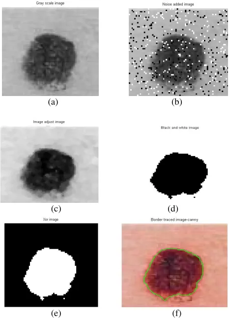
Figure 2. Demonstration of border detection for Skin Lesion 1 by the proposed robust segmentation algorithm
(a) Gray scale image. (b) .Noisy image. (c) Noise removed and enhanced image. (d) Black and white image. (e) xor image. (f) ) Border detection of noisy image by the proposed robust segmentation algorithm
[image:3.612.324.546.56.267.2] [image:3.612.315.548.327.654.2]level set formulation for geometric active contours for detecting object boundaries by Chunming Li et al. [13], when they are applied to skin lesion 1,without noise. Figure1(c) shows the final output results of the variational level set formulation for geometric active contours for detecting object boundaries by Chunming Li et al. [13],when they are applied to noisy skin lesion 1.For Skin Lesion 1, the Chunming Li et al. method poorly delineates the boundary of the lesion. The figure demonstrates the failure of this method to delineate the boundary of the lesion.
The proposed algorithm converts the original skin lesion image (skin lesion 1) in Figure 1(a) into a gray scale image as shown in Figure 2(a). 10% salt and pepper noise was added to the original image and that is illustrated in Figure 2(b).The noisy image is the input image to the proposed algorithm. The median filter is applied and the noise is removed. After noise removal the image is enhanced and that is illustrated in figure 2(c). Based on a threshold value the enhanced image is converted to black and white image this is illustrated in Figure 2(d). The proposed algorithm converts the black and white image into xor image which is illustrated in Figure 2(e).The edges are detected using canny edge detector and the border of the lesion is traced using the proposed robust segmentation algorithm successfully is shown in Figure 2(f). The proposed algorithm segments the lesion from the image even in the presence of noise and presence of hair for a variety of lesions, and skin types.
(a)
(b) (c)
Figure 3. Border detection for Skin Lesion 2 by Variational level set formulation for geometric active contours for detecting object boundaries[13] (a) Skin lesion 2. (b) Variational level set formulation for geometric active contours for detecting object boundaries[13],without noise. (c) Variational level set formulation for geometric active contours for detecting object boundaries[13], for a noisy image.
(a) (b)
(c) (d)
(e) (f)
Figure 4. Demonstration of border detection for Skin Lesion 2 by the proposed robust segmentation algorithm
(a) Gray scale image. (b) .Noisy image. (c) Noise removed and enhanced image. (d) Black and white image. (e) xor image. (f) ) Border detection of noisy image by the proposed robust segmentation algorithm
Figure 3(a). illustrates the original skin lesion 2. Figure 3(b) shows the final output results of the variational level set formulation for geometric active contours for detecting object boundaries by Chunming Li et al. [13], when they are applied to skin lesion 2, without noise. Figure 3(c) shows the final output results of the variational level set formulation for geometric active contours for detecting object boundaries by Chunming Li et al. [13], when they are applied to noisy skin lesion 2. For Skin Lesion 2, the Chunming Li et al. method poorly delineates the boundary of the lesion. The figure demonstrates the failure of this method to delineate the boundary of the lesion.
[image:4.612.337.535.54.377.2] [image:4.612.69.274.355.584.2]
(a)
(b) (c)
Figure 5. Border detection for Skin Lesion 3 by Variational level set formulation for geometric active contours for detecting object boundaries[13] (a) Skin lesion 3. (b) Variational level set formulation for geometric active contours for detecting object boundaries[13],without noise. (c) Variational level set formulation for geometric active contours for detecting object boundaries[13], for a noisy image
(a) (b)
(c) (d)
(e) (f)
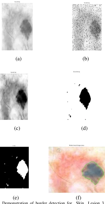
Figure 6. Demonstration of border detection for Skin Lesion 3 by the proposed robust segmentation algorithm
(a) Gray scale image. (b) .Noisy image. (c) Noise removed and enhanced image. (d) Black and white image. (e) xor image. (f) ) Border detection of noisy image by the proposed robust segmentation algorithm
Figure 5(a). illustrates the original skin lesion 3. Figure 5(b) shows the final output results of the variational level set formulation for geometric active contours for detecting object boundaries by Chunming Li et al. [13], when they are applied to skin lesion 3, without noise. Figure 5(c) shows the final output results of the variational level set formulation for geometric active contours for detecting object boundaries by Chunming Li et al. [13], when they are applied to noisy skin lesion 3.For Skin Lesion 3, the Chunming Li et al. method poorly delineates the boundary of the lesion. The figure demonstrates the failure of this method to delineate the boundary of the lesion.
The proposed algorithm converts the original lesion image (skin lesion 3) in Figure 5(a) into a gray scale image as shown in Figure 6(a). 10% salt and pepper noise was added to the original image and that is illustrated in Figure 6(b).The noisy image is the input image to the proposed algorithm. The median filter is applied and the noise is removed. After noise removal the image is enhanced and that is illustrated in figure 6(c). Based on a threshold value the enhanced image is converted to black and white image this is illustrated in Figure 6(d). The proposed algorithm converts the black and white image into xor image which is illustrated in Figure 6(e).The edges are detected using canny edge detector and the border of the lesion is traced using the proposed robust segmentation algorithm successfully is shown in Figure 6(f). The proposed algorithm segments the lesion from the image even in the presence of noise and presence of hair for a variety of lesions, and skin types.
(a)
(b) (c)
Figure 7. Border detection for Skin Lesion 4 by Variational level set formulation for geometric active contours for detecting object boundaries[13] (a) Skin lesion 4. (b) Variational level set formulation for geometric active contours for detecting object boundaries[13],without noise. (c) Variational level set formulation for geometric active contours for detecting object boundaries[13], for a noisy image
[image:5.612.70.290.68.272.2] [image:5.612.90.272.346.700.2] [image:5.612.320.540.378.587.2](a) (b)
(c) (d)
(e) (f)
Figure 8. Demonstration of border detection for Skin Lesion 4 by the proposed robust segmentation algorithm
(a) Gray scale image. (b) .Noisy image. (c) Noise removed and enhanced image. (d) Black and white image. (e) xor image. (f) ) Border detection of noisy image by the proposed robust segmentation algorithm
Figure 7(a). illustrates the original skin lesion 4. Figure 7(b) shows the final output results of the variational level set formulation for geometric active contours for detecting object boundaries by Chunming Li et al. [13], when they are applied to skin lesion 4, without noise. Figure 7(c) shows the final output results of the variational level set formulation for geometric active contours for detecting object boundaries by Chunming Li et al. [13], when they are applied to noisy skin lesion 4.For Skin Lesion 4, the Chunming Li et al. method poorly delineates the boundary of the lesion. The figure demonstrates the failure of this method to delineate the boundary of the lesion.
The proposed algorithm converts the original lesion image (skin lesion 4) in Figure 7(a) into a gray scale image as shown in Figure 8(a). 10% salt and pepper noise was added to the original image and that is illustrated in Figure 8(b).The noisy image is the input image to the proposed algorithm. The median filter is applied and the noise is removed. After noise removal the image is enhanced and that is illustrated in figure 8(c). Based on a threshold value the enhanced image is converted to black and white image this is illustrated in Figure 8(d). The proposed algorithm converts the black and white image into xor image which is illustrated in Figure 8(e).The edges are detected using canny edge detector and the border of the lesion is traced using the proposed robust segmentation algorithm successfully is shown in Figure 8(f). The proposed algorithm segments the lesion from the image even in the presence of noise and presence of hair for a variety of lesions, and skin types.
V. CONCLUSION
In this paper, we have proposed a new robust and effective segmentation algorithm, for border detection of real skin lesions for noisy skin lesion diagnosis. To verify the capability of the segmentation algorithm in detecting the borders of the lesions for skin lesion diagnosis, the algorithm was applied on diversity of clinical skin image containing lesions with and without noise. The results demonstrated the successful border detection of real skin lesions by the proposed
segmentation algorithm for clinical skin images even with noise and make them accessible for further analysis and research. We conclude that, the proposed robust segmentation algorithm effectively detects the border of the skin lesion, even in the presence of noise and presence of hair for a variety of lesions, and skin types.
As the basic technique of image processing and computer vision, image segmentation has a promising future and a universal segmentation algorithm has become the focus of contemporary research [12]. We can foresee the trend of image segmentation as a combination of multialgorithms and introduction of data mining techniques to image segmentation. For a concrete medical image segmentation task, we should combine the application background and practical requirements to design proper segmentation algorithms.
VI. REFERENCES
[1] Teresa Mendonc¸a, Andr´e R. S. Marc¸al, Angela Vieira Jacinto C. Nascimento, Margarida Silveira, Jorge S. Marques, Jorge Rozeira, “Comparison of Segmentation Methods for Automatic Diagnosis of Dermoscopy Images”,Proc. of the 29th Annual International Conference of the IEEE EMBS Cité Internationale, Lyon, France,August 23-26, 2007.pp. 6572-6575
[2] Arthur Tenenhaus1, Alex Nkengne2, Jean-François Horn3,4, Camille Serruys3,4, Alain Giron3,4 and Bernard Fertil5 “Detection of melanoma from dermoscopic images of naevi acquired under uncontrolled conditions”, Author manuscript, published in Skin Research and Technology Vol. 16, N° 1 (2009) pp. 85-97
[3] LI Xue-wei, ZHANG Xin-rong, “ A Perceptual Color Edge Detection Algorithm”, 2008 International Conference on Computer Science and Software Engineering,pp.297-300
[4] Alfonso Baldi 1,2,*, Marco Quartulli 3, Raffaele Murace 2, Emanuele Dragonetti 2, Mario Manganaro 3, Oscar Guerra 3 and Stefano Bizzi 3, “Automated Dermoscopy Image Analysis of Pigmented Skin Lesions” , Cancers 2010, 2, pp.262-273
[5] M.Emre Celebia, , Hitoshi Iyatomib, Gerald Schaeferc,William V.Stoecker d, “Lesion border detection in dermoscopy images”, Computerized Medical Imaging and Graphics 33 (2009),pp. 148–153
[6] L. Xua, M. Jackowskia, A. Goshtasbya,*, D. Rosemanb, S. Binesb, C.Yuc, A. Dhawand, A. Huntleye , “Segmentation of skin cancer images”, Image and Vision Computing 17 (1999) ,pp. 65–74
[7] Harald Ganster*, Axel Pinz, Reinhard Röhrer, Ernst Wildling, Michael Binder, and Harald Kittler, “Automated Melanoma Recognition ”,IEEE Transactions on Medical Imaging, vol. 20, no. 3, March 2001
[8] Roberto Rodríguez* and Ana G. Suarez, “ A new algorithm for image segmentation by using iteratively the mean shift filtering”, Scientific Research and Essay Vol. 1 (2), pp. 043-048, November 2006
[9] Wen-Xiong Kang, Qing-Qiang Yang, Run-peng Liang, “The Comparative Research on Image Segmentation Algorithms” ,2009 First International Workshop on Education Technology and Computer Science.
[10] Rafael C.Gonzalez, Richard E.Woods, “Digital Image Processing”, second edition, Prentice-Hall, India.
[11] Rafael C.Gonzalez, Richard E.Woods, Stevan L.Eddins “Digital Image Processing using Matlab”
[image:6.612.59.297.54.279.2][13] Chunming Li, Chenyang Xu , Changfeng Gui, and Martin D. Fox , “Level Set Evolution Without Re-initialization: A New Variational Formulation”,Proceedings of the 2005 IEEE Computer Society Conference on Computer Vision and Pattern Recognition (CVPR’05)
[14] J.H.Jaseema Yasmin, M.Mohamed Sathik, S. Zulaikha Beevi ,“Edge Detection Algorithms for Medical Image Segmentation”, Proceedings of the International Conference on Intelligent Design and Analysis of Engineering Products Systems and Computation 9-10 July 2010 (IDAPSC-10),Coimbatore, Pg 63.
[15] S.Zulaikha Beevi,M.Mohamed Sathik, “A Robust segmentation Approach for Noisy Medical Images Using
Fuzzy Clustering with Spatial Probability” ,European Journal of Scientific Research,Vol.41,No.3 (2010),pp .437-451.
[16] S.Zulaikha Beevi,M.Mohamed Sathik, K.Senthamarai Kannan ,J.H.Jaseema Yasmin, “Hybrid Segmentation Approach using FCM and Dominant Intensity Grouping
with Region Growing on Medical Image”,