Page 1 of 7 Original Article
Rosiglitazone inhibits PM2.5-induced cytotoxicity in human lung
epithelial A549 cells
Xian-Juan Pu1*, Jin Li1,2*, Qiu-Lian Zhou1,2, Wen Pan2, Yong-Qin Li2, Yuhui Zhang3, Jinhua Wang1, Zheng Jiao1
1Shanghai Applied Radiation Institute, School of Environmental and Chemical Engineering, Shanghai University, Shanghai 200072, China; 2Cardiac
Regeneration and Ageing Lab, Institute of Cardiovascular Sciences, School of Life Science, Shanghai University, Shanghai 200444, China; 3State Key Laboratory of Cardiovascular Disease, Heart Failure Center, Fuwai Hospital, National Center for Cardiovascular Diseases, Chinese Academy of Medical Sciences and Peking Union Medical College, Beijing 100730, China
Contributions: (I) Conception and design: Z Jiao, J Wang; (II) Administrative support: Z Jiao, J Wang; (III) Provision of study materials or patients: Z Jiao, J Wang; (IV) Collection and assembly of data: XJ Pu, J Li, QL Zhou, W Pan, YQ Li; (V) Data analysis and interpretation: All authors; (VI) Manuscript writing: All authors; (VII) Final approval of manuscript: All authors.
*These authors contributed equally to this work.
Correspondence to: Prof. Zheng Jiao; Prof. Jinhua Wang. Shanghai Applied Radiation Institute, School of Environmental and Chemical Engineering, 333 Nan Chen Road, Shanghai University, Shanghai 200072, China. Email: zhengjiao_sh@hotmail.com; jinhuawang@staff.shu.edu.cn.
Background: Exposure to fine particulate matter <2.5 μm in diameter (PM2.5) leads to global adverse health effects, including increases in morbidity and mortality of respiratory diseases. PM2.5 increases production of reactive oxygen species (ROS) in the lung, which further lead to oxidative stress, cell apoptosis and cell death. According to results of previous studies, oxidative stress and subsequent cell apoptosis can be reduced by peroxisome proliferator-activated receptor gamma (PPARγ) in various cell types, however, its role in oxidative stress-related cell apoptosis caused by PM2.5 in respiratory systems is unclear.
Methods: Human lung alveolar epithelial A549 cells were exposed to PM2.5 with or without rosiglitazone (an agonist of PPARγ) treatment. Cellular apoptosis and intracellular oxidative stress were determined by flow cytometry based on FITC Annexin V and DCFH-DA fluorescence, respectively. Western blot was conducted to determine the expression of Bax, Bcl2, PPARγ, P-ERK1/2, ERK1/2, P-STAT3, and STAT3. Results: PPARγ was downregulated in PM2.5-treated A549 cells, and application of rosiglitazone reduced PM2.5-mediated ROS generation and cell apoptosis. In addition, our results indicated that rosiglitazone treatment suppressed PM2.5-induced ERK1/2 and STAT3 activation.
Conclusions: Collectively, these data suggested that rosiglitazone protects against PM2.5-induced ROS production and cell apoptosis and represses activation of ERK1/2 and STAT3 signaling in A549 cells. Our results indicated that rosiglitazone is a potential therapeutic agent for PM2.5-induced lung diseases.
Keywords: PM2.5; peroxisome proliferator-activated receptor gamma (PPARγ); apoptosis; reactive oxygen species (ROS); lung diseases
Submitted Mar 17, 2018. Accepted for publication Apr 11, 2018. doi: 10.21037/atm.2018.04.13
View this article at: http://dx.doi.org/10.21037/atm.2018.04.13
Introduction
Air pollution is nowadays regarded as a major threat to public health worldwide. Particular matter (PM) is a significant class of air pollutants, consisting of PM10, PM2.5 and ultrafine PM based on the sizes and
PM2.5, and thus airway cells suffer more damage than other cells. As reported, PM2.5 exposure causes cell membrane lysis and mitochondrial ultrastructural disruptions in lung alveolar epithelial cells (A549) (4). In addition, PM2.5 induces DNA damage and inflammatory response, and meanwhile increases the generation of reactive oxygen species (ROS) in A549 cells (5-11), which are the major mechanisms for PM2.5-induced cell injury (12).
Peroxisome proliferator-activated receptor gamma
(PPARγ) is a ligand-dependent transcription factor, and a
member of nuclear receptor of peroxisome proliferator-activated receptors (PPARs) family. PPARs have multiple regulatory roles in inflammation, redox balance, trophic factor biosynthesis, insulin sensitivity and metabolism of lipid and glucose. Rosiglitazone, one of the agonists of
PPARγ, is a thiazolidinedione drug used in the treatment of
type 2 diabetes mellitus (13). In recent years, the resistance function of rosiglitazone in cell apoptosis, inflammation response and oxidative stress have been verified (14-17). However, whether rosiglitazone is able to prevent cell injury and death in lung epithelial cells resulted from PM2.5 still remains unclear.
In the present work, we firstly found decreasing of
PPARγ in PM2.5-treated A549 cells. Then using a PPARγ
agonist, rosiglitazone, we confirmed that PPARγ activation suppressed cell apoptosis and ROS production in A549 cells exposed to PM2.5. Our results further demonstrated that rosiglitazone inhibited ERK1/2 and STAT3 activated by PM2.5 exposure. Our data collectively suggested that rosiglitazone can protect human lung epithelial cells from cell toxicity of PM2.5.
Methods
Preparation of PM2.5
Fifty mg PM2.5 (National Institute of Standards and Technology Boulder Laboratories, Boulder, CO, USA) was dissolved in 1 mL dimethyl sulfoxide (DMSO) facilitated with 20 min ultrasonic oscillation. The mixture was
centrifuged at 17,000 g and stored at 4 ℃ for 10 min.
Cell culture and treatment
Human lung alveolar epithelial A549 cell was a gift from Cell Bank, Chinese Academy of Sciences. Cells were routinely maintained in DMEM supplemented with 10%
fetal bovine serum (FBS, Biological Industries, Israel) at a
5% CO2 atmosphere at 37 ℃. A549 cells were seeded and
treated with prepared PM2.5 at the final concentration of 25 or 50 μg/mL for 48 h. Rosiglitazone (Selleck) was
incubated at a final concentration of 20 μM with A549 cells
for 48 h.
Measurement of cellular apoptosis
Cellular apoptosis was accessed with flow cytometry according to the manufacturer’s protocols of FITC Annexin V Apoptosis Detection Kit I (BD, Pharmingen, USA). Simply, A549 cells in 1X Binding Buffer were incubated in dark with FITC Annexin V and PI for 15 min at room temperature. Then the cells were analyzed by flow cytometry immediately.
Western blot
After treatment with PM2.5 or Rosiglitazone, A549 cells were harvested and lysed on ice in cell lysis buffer (KeyGen, China) supplied with 1 mM PMSF (KeyGen). The concentration of protein was quantified by BCA Protein Assay Kits (Takara, Japan). Equal amounts of protein were separated by SDS-polyacrylamide gel electrophoresis and
transferred to polyvinylidene fluoride (PVDF) membranes
(Millipore, USA) by electricity. After incubation with 5% BSA (KeyGen), membranes were then incubated with
primary antibodies at 4 ℃ overnight: Bax (Abclonal, USA),
Bcl2 (Abclonal), PPARγ (Abclonal), P-ERK1/2 (Abclonal),
ERK1/2 (Abclonal), P-STAT3 (Bioworld, USA), STAT3 (Bioworld), GAPDH (Abclonal). After that, the membranes were further incubated with appropriate secondary antibodies for 2 h. The protein signals were visualized and recorded using the ECL plus western blotting detection reagents (Millipore) and the ChemiDoc XRS Plus luminescent image analyzer (Tanon).
Measurement of intracellular oxidative stress
After treated with PM2.5 or rosiglitazone, the cells were harvested and resuspended in pre-warmed media with 10 μM DCFH-DA (Beyotime, China) and incubated for 30 min. After washing with PBS, the DCFH-DA fluorescence was measured using flow cytometric analysis
(Beckman) by filtering 488 nm. And the mean fluorescence
Statistical analysis
Results were all expressed as mean ± SEM. Unpaired
two-tailed Student’s t-test was applied for comparisons
between two groups. One-way ANOVA followed by Bonferroni’s post hoc test was conducted when comparing among multiple groups. All analyses were carried out with GraphPad Prism 6.0. P value less than 0.05 were considered
statistically significant.
Results
PPARγ is decreased in PM2.5-treated A549 cells
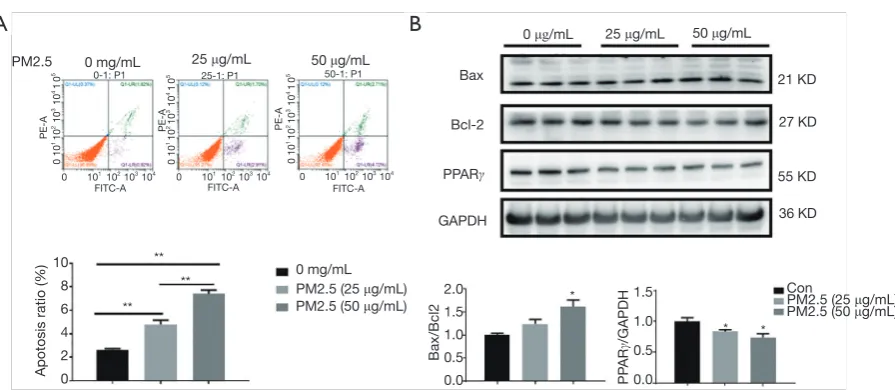
To mimic lung epithelial cell injury due to PM2.5 exposure, A549 cells were treated with 25 and 50 μg/mL PM2.5. Results demonstrated that 48 h of PM2.5 treatment led to potent cell apoptosis in A549 cells, as determined
by flow cytometry analysis (Figure 1A) and protein
level for Bax and Bcl2 (Figure 1B). Furthermore,
western blot also showed that PPARγ was significantly
downregulated in PM2.5-stimulated A549 cells
(Figure 1B). In addition, a dose-dependent increase of cell
apoptosis and decrease of PPARγ expression were also
observed (Figure 1). Thus, reduction of PPARγ might be
responsible for lung alveolar epithelial damage induced by PM2.5 exposure.
Rosiglitazone attenuates PM2.5-induced apoptosis in A549 cells
In order to further examine the functional effect of PPARγ
on lung epithelial cells during PM2.5 exposure, rosiglitazone
was used as the PPARγ agonist. Flow cytometry suggested
that rosiglitazone reduced PM2.5-mediated apoptosis in
A549 cells (Figure 2A). Meanwhile, rosiglitazone reduced
the expression of Bax/Bcl2 ratio as determined by western
blot (Figure 2B). These results suggested the protective
effects of rosiglitazone against apoptosis in human lung epithelial cells caused by PM2.5 treatment.
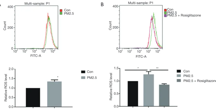
Rosiglitazone attenuates PM2.5-induced ROS production in A549 cells
Oxidative stress has been reported as a crucial pattern of cell toxicity induced by PM2.5. DCFH-DA was used to quantify the intracellular level of ROS. As shown in flow cytometry results, PM2.5 exposure elevated
ROS production in cells (Figure 3A), and rosiglitazone
significantly reduced ROS production in cells with PM2.5
exposure (Figure 3B). Thus, these data suggested that
[image:3.595.73.521.76.271.2]rosiglitazone protects against PM2.5-caused cell toxicity in human lung epithelial cells through reducing ROS generation.
Figure 1 Downregulation of PPARγ in PM2.5-treated A549 cells. (A) Cell apoptosis of A549 treated with PM2.5 (25 and 50 μg/mL, 48 h) was accessed with flow cytometry analysis (n=4); (B) protein level of Bcl-2, Bax and PPARγ in A549 cells treated with PM2.5 was accessed with western blot analysis (n=3). *, P<0.05; **, P<0.01. PPARγ, peroxisome proliferator-activated receptor gamma.
PM2.5 0 mg/mL
0 mg/mL 25 μg/mL
25 μg/mL 0 μg/mL
PM2.5 (25 μg/mL)
PM2.5 (25 μg/mL) 50 μg/mL
50 μg/mL
PM2.5 (50 μg/mL) PM2.5 (50 μg/mL)
10 8 6 4 2 0
Bax
Bcl-2
PPARγ
GAPDH
21 KD
27 KD
55 KD
36 KD
** ** **
Apotosis ratio (%)
2.0 1.5 1.0 0.5 0.0
1.5
1.0
0.5
0.0
Bax/Bcl2
Con *
* *
A
B
PP
AR
γ
/GAPDH
0 101 102 103 104 0 101 102 103 104 0 101 102 103 104
0 10
1 10 2 10 3 10 4 1
0
5
0 10
1 10 2 10 3 10 4 1
0
5
0 10
1 10 2 10 3 10 4 1
0
5
FITC-A FITC-A FITC-A
PE-A
0-1: P1 25-1: P1 50-1: P1
PM2.5
PM2.5
PM2.5 PM2.5
PM2.5 + Rosiglitazone
PM2.5 + Rosiglitazone
PM2.5 + Rosiglitazone PM2.5 +
Rosiglitazone
Bax
Bcl2
GAPDH
21 KD
27 KD
36 KD
Apotosis ratio (%)
2.0
1.5
1.0
0.5
0.0
Bax/Bcl2
Con
Con
Con Con
** * *
** **
C-3: P1 PM-3: P1 PM+PR–4: P1
0 101 102 103 104 0 101 102 103 104 0 101 102 103 104
0 10
1
10
2
10
3
10
4
1
0
5
0 10
1
10
2
10
3
10
4
1
0
5
0 10
1
10
2
10
3
10
4
1
0
5
FITC-A FITC-A FITC-A
PE-A PE-A PE-A
20
15
10
5
0
[image:4.595.62.537.86.281.2]A
B
Figure 2 Rosiglitazone attenuates PM2.5-induced apoptosis of A549 cells. (A) Cell apoptosis of A549 treated with PM2.5 (50 μg/mL, 48 h) with or without rosiglitazone (20 μM, 48 h) was accessed with flow cytometry analysis (n=4); (B) protein level of Bcl-2 and Bax in A549 cells treated with PM2.5 with or without rosiglitazone was accessed with western blot analysis (n=3). *, P<0.05; **, P<0.01.
FITC-A FITC-A
102 103 104 105 106
102 103 104 105 106
400
200
0
400
200
0
Count Count
Multi-sample: P1 Multi-sample: P1
PM2.5
PM2.5 PM2.5
PM2.5 Con
Con Con
Con
PM2.5 + Rosiglitazone
PM2.5 + Rosiglitazone 2.0
1.5
1.0
0.5
0.0
1.5
1.0
0.5
0.0
Relative ROS level Relative ROS level
*
* **
A
B
[image:4.595.75.525.363.591.2]Rosiglitazone inhibits PM2.5-activated ERK1/2 and STAT3 signaling in A549 cells
Given that ERK1/2 and STAT3 signaling pathways play critical roles in regulating cell apoptosis and oxidative stress (18-21), we next investigated the effect of rosiglitazone and PM2.5 on activation of ERK1/2 and STAT3. Our results showed that PM2.5 exposure increased the protein level of phosphorylated ERK1/2 and STAT3, whereas rosiglitazone suppressed the upregulated phosphorylated ERK1/2 and
STAT3 in PM2.5-treated cells (Figure 4A,B). These data
revealed that ERK1/2 and STAT3 inactivation might be responsible for the protective action of rosiglitazone in A549 cells during PM2.5 exposure.
Discussion
Urbanization and associated air pollution, in particular, PM2.5 increases the morbidity of various respiratory diseases (22). It is well-known that PM exposure played detrimental roles mostly in epithelial cells (4,22). However, the underlying molecular mechanism is largely unknown and the effective therapeutic methods are still lack. In present work, we verified that PM2.5 mediated ROS generation and cell apoptosis in A549 cells. Importantly, our
results demonstrated for the first time that PPARγ agonist rosiglitazone could protect against PM2.5-mediated A549 cell apoptosis and oxidative stress accumulation through inhibiting ERK1/2 and STAT2 signaling pathways.
Previous studies have linked several genes to airway epithelial cell damage induced by PM2.5, which exerting
their roles through regulating cell death and inflammatory
response (23-27). It is well-accepted that PPARγ is widely
expressed in almost all kinds of human tissues and plays central roles in early stage lung development (28-30).
PPARγ agonists prevent newborn mouse from
hypoxia-induced lung injury (31). Particularly, PPARγ has been
shown decreased in hepatic stellate cells exposed to PM2.5
during liver fibrosis (32). However, the expression changes
and roles of PPARγ in respiratory systems exposed to
PM2.5 are still unknown. This is the first study that reveals
the downregulation and the protective roles of PPARγ
in A549 cells treated with PM2.5. Given that PPARγ
deficiency is widely associated with pulmonary disorders including, asthma, cystic fibrosis lung cancer (29). Our results indicated that PM2.5 exposure might lead to
these pulmonary diseases through reducing the PPARγ
expression. Our results also suggested that PPARγ activation
is an alternative therapeutic strategy for treatment of lung injury caused by PM2.5.
ROS has diverse effects on cellular activities depending on its amount. In physiological conditions, the lung epithelial cells generate low levels of ROS and physiological
changes of ROS levels are essential for cell survival. PPARγ
agonist GW1929 elevated ROS levels and activated cell-cycle progression in primary cultured lung alveolar epithelial cells (33). However, in pathological conditions,
Con Con
Con Con
PM2.5 PM2.5
PM2.5 PM2.5
44KD 88KD
88KD 44KD
42KD
42KD
36KD 36KD
Rosiglitazone + PM2.5
Rosiglitazone + PM2.5
Rosiglitazone + PM2.5 Rosiglitazone + PM2.5
1.5
1.0
0.5
0.0
1.5
1.0
0.5
0.0
GAPDH GAPDH
P-ERK1/2
T-ERK1/2
P-STAT3
STAT3
* * * **
P-ERK1/2/T
-ERK1/2
P-ST
AT3/T
-ST
AT3
[image:5.595.91.511.87.254.2]A
B
prolonged oxidative stress leads to cell apoptosis and death.
Herein, we found that PPARγ agonist rosiglitazone reduced
ROS levels and inhibited the lung epithelial cell apoptosis
during PM2.5 exposure. Thus, PPARγ might have different
roles in ROS production due to the physiological and pathological conditions.
Numerous studies have suggested a central role of ERK1/2 and STAT3 signaling pathways in regulating oxidative stress and related cell apoptosis (18-21). As expected, phosphorylation of ERK1/2 and STAT3 were increased after PM2.5 exposure. Importantly, rosiglitazone stimulation repressed the activation of ERK1/2 and STAT3 pathways. Although further studies are required to clarify the detailed molecular mechanisms, results of present work indicated that rosiglitazone could protect against PM2.5-mediated cell damage via inhibiting ERK1/2 and STAT3 pathways.
Conclusions
In summary, our work demonstrated that PPARγ was
downregulated in A549 cells exposed to PM2.5. Application
of rosiglitazone, a PPARγ agonist, could relieve the
cytotoxic effect of PM2.5 on human lung epithelial A549 cells by inhibiting ERK1/2 and STAT3 activation. Our results suggested rosiglitazone as a potential therapeutic agent of PM2.5-induced lung diseases.
Acknowledgements
Funding: This study was supported by the CAMS
Innovation Fund for Medical Sciences (CIFMS, 2016-12M-1-006).
Footnote
Conflicts of Interest: The authors have no conflicts of interest
to declare.
References
1. Ma QY, Huang DY, Zhang HJ, et al. Exposure to particulate matter 2.5 (PM2.5) induced macrophage-dependent inflammation, characterized by increased Th1/Th17 cytokine secretion and cytotoxicity. Int Immunopharmacol 2017;50:139-45.
2. Liu F, Huang Y, Zhang F, et al. Macrophages treated with particulate matter PM2.5 induce selective neurotoxicity
through glutaminase-mediated glutamate generation. J Neurochem 2015;134:315-26.
3. Yang D, Ma M, Zhou W, et al. Inhibition of miR-32 activity promoted EMT induced by PM2.5 exposure through the modulation of the Smad1-mediated signaling pathways in lung cancer cells. Chemosphere 2017;184:289-98.
4. Gualtieri M, Mantecca P, Corvaja V, et al. Winter fine particulate matter from Milan induces morphological and functional alterations in human pulmonary epithelial cells (A549). Toxicol Lett 2009;188:52-62.
5. Wang W, Jariyasopit N, Schrlau J, et al. Concentration and photochemistry of PAHs, NPAHs, and OPAHs and toxicity of PM2.5 during the Beijing Olympic Games. Environ Sci Technol 2011;45:6887-95.
6. Cavanagh JA, Trought K, Brown L, et al. Exploratory investigation of the chemical characteristics and relative toxicity of ambient air particulates from two New Zealand cities. Sci Total Environ 2009;407:5007-18.
7. Bourgeois B, Owens JW. The influence of Hurricanes Katrina and Rita on the inflammatory cytokine response and protein expression in A549 cells exposed to PM2.5 collected in the Baton Rouge-Port Allen industrial corridor of Southeastern Louisiana in 2005. Toxicol Mech Methods 2014;24:220-42.
8. Lin ZQ, Xi ZG, Yang DF, et al. Oxidative damage to lung tissue and peripheral blood in endotracheal PM2.5-treated rats. Biomed Environ Sci 2009;22:223-8.
9. Corsini E, Budello S, Marabini L, et al. Comparison of wood smoke PM2.5 obtained from the combustion of FIR and beech pellets on inflammation and DNA damage in A549 and THP-1 human cell lines. Arch Toxicol 2013;87:2187-99.
10. Yan Z, Wang J, Li J, et al. Oxidative stress and endocytosis are involved in upregulation of interleukin-8 expression in airway cells exposed to PM2.5. Environ Toxicol 2016;31:1869-78.
11. He M, Ichinose T, Yoshida S, et al. PM2.5-induced lung inflammation in mice: Differences of inflammatory response in macrophages and type II alveolar cells. J Appl Toxicol 2017;37:1203-18.
12. Deng X, Zhang F, Rui W, et al. PM2.5-induced oxidative stress triggers autophagy in human lung epithelial A549 cells. Toxicol In Vitro 2013;27:1762-70.
Cite this article as:Pu XJ, Li J, Zhou QL, Pan W, Li YQ, Zhang Y, Wang J, Jiao Z. Rosiglitazone inhibits PM2.5-induced cytotoxicity in human lung epithelial A549 cells. Ann Transl Med 2018;6(8):152. doi: 10.21037/atm.2018.04.13
Toxicol 2017. [Epub ahead of print].
14. Lee JE, Park JH, Jang SJ, et al. Rosiglitazone inhibits chlorpyrifos-induced apoptosis via modulation of the oxidative stress and inflammatory response in SH-SY5Y cells. Toxicol Appl Pharmacol 2014;278:159-71. 15. Zhao J, Liu J, Pang X, et al. Rosiglitazone attenuates
angiotensin II-induced C-reactive protein expression in hepatocytes via inhibiting AT1/ROS/MAPK signal pathway. Int Immunopharmacol 2016;31:178-85. 16. An Z, Yu JR, Park WY. Rosiglitazone enhances
radiosensitivity by inhibiting repair of DNA damage in cervical cancer cells. Radiat Environ Biophys 2017;56:89-98.
17. Gong Y, Yin JY, Tong BD, et al. Low density lipoprotein - rosiglitazone - chitosan-calcium alginate/nanoparticles inhibition of human tenon's fibroblasts activation and proliferation. Oncotarget 2017;8:105126-36.
18. Tian Y, Xiao Y, Wang B, et al. Vitamin E and Lycopene Reduce Coal Burning Fluorosis-induced Spermatogenic Cell Apoptosis via Oxidative Stress-mediated JNK and ERK Signaling Pathways. Biosci Rep 2017. [Epub ahead of print].
19. Zhou XC, Dong SH, Liu ZS, et al. Regulation of gammaherpesvirus lytic replication by endoplasmic reticulum stress-induced transcription factors ATF4 and CHOP. J Biol Chem 2018;293:2801-14.
20. Rai A, Kumar U, Raj V, et al. Novel 1,4-benzothazines obliterate COX-2 mediated JAK-2/STAT-3 signals with potential regulation of oxidative and metabolic stress during colorectal cancer. Pharmacol Res 2017. [Epub ahead of print].
21. Rajamanickam V, Zhu H, Feng C, et al. Novel allylated monocarbonyl analogs of curcumin induce mitotic arrest and apoptosis by reactive oxygen species-mediated endoplasmic reticulum stress and inhibition of STAT3. Oncotarget 2017;8:101112-29.
22. Yang B, Chen D, Zhao H, et al. The effects for PM2.5 exposure on non-small-cell lung cancer induced motility and proliferation. Springerplus 2016;5:2059.
23. Zhou W, Yuan X, Zhang L, et al. Overexpression of
HO-1 assisted PM2.5-induced apoptosis failure and autophagy-related cell necrosis. Ecotoxicol Environ Saf 2017;145:605-14.
24. Nam HY, Choi BH, Lee JY, et al. The role of nitric oxide in the particulate matter (PM2.5)-induced
NFkappaB activation in lung epithelial cells. Toxicol Lett 2004;148:95-102.
25. Wang H, Guo Y, Liu L, et al. DDAH1 plays dual roles in PM2.5 induced cell death in A549 cells. Biochim Biophys Acta 2016;1860:2793-801.
26. Song L, Li D, Gu Y, et al. Let-7a modulates particulate matter (≤ 2.5 μm)-induced oxidative stress and injury in human airway epithelial cells by targeting arginase 2. J Appl Toxicol 2016;36:1302-10.
27. Huang Q, Chi Y, Deng J, et al. Fine particulate matter 2.5 exerted its toxicological effect by regulating a new layer, long non-coding RNA. Sci Rep 2017;7:9392.
28. Rubenstrunk A, Hanf R, Hum DW, et al. Safety issues and prospects for future generations of PPAR modulators. Biochim Biophys Acta 2007;1771:1065-81.
29. Heikkinen S, Auwerx J, Argmann CA. PPARgamma in human and mouse physiology. Biochim Biophys Acta 2007;1771:999-1013.
30. Wang Y, Santos J, Sakurai R, et al. Peroxisome
proliferator-activated receptor gamma agonists enhance lung maturation in a neonatal rat model. Pediatr Res 2009;65:150-5.
31. Takeda K, Okamoto M, de Langhe S, et al. Peroxisome proliferator-activated receptor-g agonist treatment increases septation and angiogenesis and decreases airway hyperresponsiveness in a model of experimental neonatal chronic lung disease. Anat Rec (Hoboken) 2009;292:1045-61.
32. Zheng Z, Zhang X, Wang J, et al. Exposure to fine airborne particulate matters induces hepatic fibrosis in murine models. J Hepatol 2015;63:1397-404.