Page 1 of 14
Should the left gastric artery lymph node be considered as the
predictive lymph node for extra-gastric lymph node metastases?
Weilin Sun1,2,3#, Jingyu Deng1,2,3#, Wenting He1,2,3, Jinyuan Liu1,2,3, Shiwei Guo1,2,3, Pengfei Gu1,2,3,
Zizhen Wu1,2,3, Han Liang1,2,3
1Department of Gastroenterology, Tianjin Medical University Cancer Institute and Hospital, National Clinical Research Center for Cancer, Tianjin,
China; 2Key Laboratory of Cancer Prevention and Therapy, Tianjin, China; 3Tianjin’s Clinical Research Center for Cancer, Tianjin, China
Contributions: (I) Conception and design: J Deng, H Liang; (II) Administrative support: H Liang; (III) Provision of study materials or patients: J Deng, H Liang; (IV) Collection and assembly of data: W Sun, W He, J Liu, S Guo, P Gu, Z Wu; (V) Data analysis and interpretation: W Sun, J Deng, W He; (VI) Manuscript writing: All authors; (VII) Final approval of manuscript: All authors.
#These authors equally contributed to the work.
Correspondence to: Jingyu Deng; Han Liang. Department of Gastroenterology, Tianjin Medical University Cancer Institute and Hospital, National Clinical Research Center for Cancer, Tianjin, China; Key Laboratory of Cancer Prevention and Therapy, Tianjin, China; Tianjin’s Clinical Research Center for Cancer, Tianjin, China. Email: dengery@126.com; tjlianghan@126.com.
Background: To validate the prognostic impacts of the left gastric artery lymph node (No. 7 LN) metastasis and investigate whether the No. 7 LN metastasis should be considered as the predictive LN for extra-gastric LN metastases.
Methods: Between January 2003 and December 2011, a total of 1,586 patients who underwent R0 gastrectomy were retrospected. Patients with LN metastases were divided into three groups: (I) patients with only peri-gastric LN metastases (peri-gastric group); (II) patients with peri-gastric and only No. 7 LN metastases (No. 7 group); and (III) patients with other extra-gastric LN metastases (extra-gastric group). Propensity score matching (PSM) was adopted to accurately evaluate prognoses of all patients after surgery. Results: Of 1,586 patients, 235 (14.82%) were pathologically identified to present with the No. 7 LN metastases. Patients with the No. 7 LN metastases presented the significantly lower survival rate both before and after adjustment by pTNM stage, compared to those without the No. 7 LN metastases. Patients in the No. 7 group were identified to present the significant lower survival rate than those in the peri-gastric group, and to present the similar median overall survival (OS) to those in the extra-gastric group. In addition, patients with extra-gastric LN except No. 7 LN metastases failed to show any superiority of survival outcomes, compared with those with extra-gastric LN metastases including the No. 7 LN metastasis. Conclusions: The No. 7 LN metastases had the crucial survival implications. Nevertheless, the No. 7 LN failed to be considered as the predictive LN for the extra-gastric LN metastases in gastric cancer (GC).
Keywords: Stomach; neoplasm; lymph node (LN); prognosis; left gastric artery
Submitted Jan 03, 2020. Accepted for publication May 09, 2020. doi: 10.21037/atm-19-4786a
View this article at: http://dx.doi.org/10.21037/atm-19-4786a
Introduction
Gastric cancer (GC) has a tendency toward lymphatic metastasis due to the abundant lymphatic vessels in the stomach wall. Lymphadenectomy has an important clinical impact, and the extent of lymphadenectomy may directly influence the patients’ survival outcome after radical
incidents occurring close to the peri-gastric LN stations (1,2). According to the latest Union for International Cancer Control/American Joint Committee on Cancer (UICC/AJCC) guidelines for GC (3), No. 7 station LNs should be considered while evaluating the extent of D2 lymphadenectomy. However, in the latest edition of the Japanese Gastric Cancer Treatment Guidelines (the 3rd
edition) and the 14th edition of the Japanese General Rules
for Gastric Cancer Study, the No. 7 LN station was assigned to the range of both D1 plus and D2 lymphadenectomy (4,5).That is to say, GC patients with cT1N0M0 stage disease might undergo different lymphadenectomies in different countries. Therefore, the clinical significance of the No. 7 station for GC patients remains controversial according to the current literature (1,6-9).
In order to evaluate precisely the range of LN metastasis, the concept of sentinel LNs (SLNs) arised, which was defined as the first draining LNs that obtain lymphatic flow from a primary tumor (10). The concept of SLNs is gradually being accepted and applied to GC, and novel techniques for SLN mapping have been developed, such as methods using dyes or radioisotopes (11-13). However, identifying specific SLNs in cases of GC is challenging, due to the complexity of lymphatic drainage from the gastric area (14,15). And SLNs seldom provides much benefit to predict the extra-gastric LN station or distant metastasis. Thus, new predictive factors are needed to identify the extra-gastric LN or distant metastasis. On the other hand, multiple recent studies have reported that the No. 7 LN station was the most common extra-gastric LN station to be involved in metastasis, regardless of tumor location (16-18). Taking anatomical location and high incidence of metastatic incidents of No. 7 LN station into account, we hypothesized that the No. 7 station should be on the main lymph routine and be a predictive LN for extra-gastric LN metastases. However, few studies have evaluated whether the No. 7 LN station might be considered as the predictive marker for determining the extent of lymphadenectomy in GC patients.
In this study, we aimed to demonstrate the prognostic impact of the No. 7 LN station and to validate whether the No. 7 LN should be considered as the predictive LN for other extra-gastric LN metastases in GC patients.
We present the following article in accordance with the STROBE reporting checklist (available at http://dx.doi. org/10.21037/atm-19-4786a).
Methods
Patients
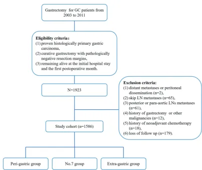
Between January 2003 and December 2011, a total of 1,923 GC patients who underwent R0 gastrectomy at Tianjin Medical University Cancer Institute and Hospital. The clinicopathologic date and fellow-up records of 1,923 GC patients were retrospectively reviewed after receiving Institutional Review Board approval. Eligibility criteria included: (I) proven histologically primary gastric carcinoma; (II) curative gastrectomy with pathologically negative resection margins (R0 resection); (III) remaining alive at the initial hospital stay and the first postoperative month. The exclusion criteria were: (I) distant metastases or peritoneal dissemination; (II) skip LN metastases; (III) posterior (No. 8p, No. 12b/p, No. 13, and No. 14v) or para-aortic (No. 16a2, and No. 16b1) LNs metastases; (IV) history of gastrectomy or other malignancies; (V) history of neoadjuvant chemotherapy; and (VI) loss of follow up.
Ultimately, 1,586 patients in total were included in this study (Figure S1). Of these 1,586 GC patients, 897 (56.56%) presented LN metastases, and 235 (14.82%) presented the No. 7 LN metastases. According to the range of LN involved, all included patients with LN involvement were divided into three groups of cases: (I) LN metastases limited to peri-gastric area (peri-gastric group), (II) peri-gastric LN metastases with only No. 7 LN metastases (No. 7 group), and (III) peri-gastric LN metastases with other extra-gastric LN metastases (extra-gastric group).
The study was approved by Tianjin Medical University Cancer Institute and Hospital ethics committees (No. bc2019087). All patients provided written informed consent before any enrolling procedures were initiated.
Surgical management
All included patients underwent the curative gastrectomy with lymphadenectomy for GC. Curative resection was defined as the complete absence of grossly visible tumor tissue and pathologically negative resection margins. The pT stage and pN stage were according to AJCC TNM staging system (19). The nodes staging system was defined according to the 13th edition of JCGC (Japanese Gastric
along the common hepatic artery (No. 8a), the celiac axis (No. 9), the splenic hilar(No. 10), splenic artery (No. 11) and the proper hepatic artery (No. 12a) were defined as extra-gastric LN stations. Skip LN metastases were defined as the presence of a metastatic LN in an extra-gastric area without peri-gastric LN involvement (21).
Follow-up
After curative surgery for GC, all patients were followed-up every 3 or 6 months for 2 years, and annually, thereafter, until death or deadline. The median follow-up time for the entire cohort was 33 months (range, 2 to 148 months). The deadline of follow-up in this study was December 2015. At every visit, patients underwent ultrasonography, computed tomography, chest radiography, and endoscopy. Overall survival (OS) served as the primary end-point, and was defined as the time interval between the date of surgery and the date of either death as a result of GC or the last follow-up. During the follow-up period, 1,229 patients (77.49%) died.
Propensity score matching (PSM)
To overcome possible selection bias, one-to-one matching using PSM was performed in this study (22,23).The propensity score, defined as the conditional probability of patients being treated given the covariates, could be used to balance the covariates in two groups and therefore reduce such bias (24,25).It had also been reported that potential confounding variables that could be unrelated to the exposure but related to the outcome should be included in the propensity score model, and that this would decrease the variance of an estimated exposure effect without increasing the bias (26).The propensity scores were estimated by using a non-parsimonious multiple logistic regression model. In this study, the No. 7 LN metastases were significant correlated with pN stage (spearman r=0.424, P<0.001). Therefore, the following covariates were selected for the calculation of the propensity score: gender, age, tumor location, tumor size, pT stage, Borrmann type, Lauren type, vasculolymphatic invasion, neurological invasion and adjuvant chemotherapy.
Statistical analysis
The χ2 or Fisher’s exact test used for categorical variables,
and a t test was used for continuous variables. Factors that
showed significant difference in the univariate analysis (P<0.05) were included in the multivariate analysis. Multivariate analysis was performed using a logistic regression model for the evaluation of the predictive risk factors. OS was determined using the Kaplan-Meier method, and a log-rank test was used to evaluate significance. Multivariate analyses of OS were performed to calculate the hazard ratios (HRs) and 95% confidence intervals (CIs) through the Cox regression model. In all statistical analyses, significance was defined as P<0.05 (two-sided). All statistical analyses were performed using the statistical analysis program package SPSS version 24.0 (SPSS, Chicago, IL, United States).
Results
Survival analysis of the No. 7 station LN metastases
The prognostic impact of the No. 7 station LN metastases in patients was determined. During the follow-up, 1,229 patients died and 357 patients remained alive. Kaplan-Meier analyses showed a significant difference in terms of prognosis between the No. 7 LN-negative (no metastasis) and the No. 7 LN-positive (metastases) patients (HR 1.795, 95% CI: 1.545–2.086, P<0.001, Figure 1A). The median survival time of No. 7 negative and No. 7 LN-positive patients was 38±1.757 vs. 18±1.730 months. That survival difference was also significant after stratification by pTNM stage (III stage with vs. without the No. 7 LN metastases: HR 1.225, 95% CI: 1.043–1.439, P=0.014,
Figure 1B). Although the small-scale samples resulted in the
non-significant difference in patients with II stage (II stage with vs. without the No. 7 metastases, HR 1.392, 95% CI: 0.763–2.539, P=0.281), the potential tendency of survival difference might be observed in the Figure 1B.
PSM among peri-gastric, the No. 7 and extra-gastric group
Table 1 showed the clinical characteristics of GC patients of
immensely reduced after PSM: tumor location (before vs. after: P<0.001 vs. P=0.125), tumor size (before vs. after: P=0.104
vs. P=0.286), pT stage (before vs. after: P=0.071 vs. P=0.106),
and Borrmann type (before vs. after: P<0.001 vs. P=0.207). Ultimately, 113 pair patients were enrolled after PSM.
Prognostic analysis before and after PSM
The prognostic analysis among peri-gastric, the No. 7 and extra-gastric groups was performed (Figure 2A). During the follow-up, the survival rates of these three groups were respectively: 15.4% (81/524), 13.04% (21/161) and 10.38% (22/212). And the median survival time were
[image:4.595.162.430.83.450.2]respectively: 24±1.381, 18±2.819, and 18±1.266 months. Before matching, Kaplan-Meier curve showed a significant difference in terms of prognosis between the No. 7 group and peri-gastric group (HR 1.227, 95% CI: 1.014–1.484, P=0.035, Figure 2B), but no significant difference in survival outcomes between the No. 7 group and extra-gastric group (HR 1.084, 95% CI: 0.872–1.349, P=0.467, Figure 2C). After PSM, the OS was also significantly poorer in the No. 7 group compared with peri-gastric group (HR 1.360, 95% CI: 1.051–1.761, P=0.020, Figure 2D). Similarly, the close survival rate between No. 7 group and extra-gastric group (HR 1.123, 95% CI: 0.851–1.482, P=0.411, Figure 2E) was observed after PSM.
Figure 1 Kaplan-Meier curves for overall survival (A) between patients with No. 7 LN metastasis and patients without No. 7 LN metastasis; (B) after adjustment by pTNM stage. LN, lymph node. No. 7 LN, LN along the left gastric artery. No. 7 (+), with No. 7 LN metastasis; No. 7 (−), without No. 7 LN metastasis; HR, hazard ratio; CI, confidence interval.
100
80
60
40
20
0
100
80
60
40
20
0
Cumulative pr
obability of survival (%)
Cumulative pr
obability of survival (%)
0 30 60 90 120 150
0 30 60 90 120 150
Time after surgery (months) with No. 7 LN metastasis without No. 7 LN metastasis I stage II stage without No. 7 LN metastases II stage with No. 7 LN metastases III stage without No. 7 LN metastases III stage with No. 7 LN metastases I stage 172 148 111 42 15 0
II stage with No. 7 (−) 488 335 207 62 13 0
II stage with No. 7 (+) 14 8 5 1 1 0
III stage with No. 7 (−) 691 273 135 49 12 0
III stage with No. 7 (+) 221 67 28 6 1 0
With No. 7 LN metastasis vs. without HR (95% CI) P value 1.795 (1.545−2.086) <0.001 With No. 7 LN metastasis vs. without HR (95% CI) P value II stage 1.392 (0.763−2.539) 0.281 Time after surgery (months) No. at risk No. at risk No. 7 (+) 235 75 33 7 2 0
No. 7 (−) 1,351 756 453 153 40 0
III stage 1.225 (1.043−1.439) 0.014
A
Table 1 Clinical characteristics of patients of peri-gastric group and No. 7 group before and after propensity score matching
Characteristics Entire cohort Propensity score matching
Peri-gastric (n=524) No. 7 (n=161) P value Peri-gastric (n=138) No. 7 (n=138) P value
Gender
Male 372 119 0.472 108 99 0.211
Female 152 42 30 39
Age (years)
<60 217 64 0.708 47 56 0.263
≥60 307 97 91 82
Tumor location
Upper 1/3 156 77 <0.001** 55 59 0.858
Middle 1/3 39 8 9 8
Lower 1/3 218 45 47 41
More than 2/3 111 31 27 30
Tumor size (cm)
≤5.0 254 82 0.585 74 69 0.547
>5.0 270 79 64 69
pT stage
Pt1a 1 0 0.660b 1 0 0.962b
Pt1b 2 2 1 2
Pt2 37 12 11 9
Pt3 32 7 6 5
Pt4a 436 137 116 119
Pt4b 16 3 3 3
Borrmann type
I 30 13 0.119 12 6 0.425
II 148 58 43 49
III 275 74 71 68
IV 71 16 12 15
Lauren typec
Intestinal 273 79 0.683 70 66 0.858b
Diffuse 230 78 67 71
Mixed 9 3 1 1
Vasculolymphatic invasion
No 519 157 0.273a 137 135 0.614a
Yes 5 4 1 3
Neurological invasionc
No 519 158 1.000b 135 136 0.481a
Yes 3 1 2 0
Adjuvant chemotherapy
No 204 60 0.704 61 55 0.464
Yes 320 101 77 83
Table 2 Clinical characteristics of patients of No. 7 group and extra-gastric group before and after propensity score matching
Characteristics Entire cohort Propensity score matching
No. 7 (n=161) Extra-gastric (n=212) P value No. 7 (n=113) Extra-gastric (n=113) P value
Gender
Male 119 156 0.943 84 86 0.758
Female 42 56 29 27
Age (years)
<60 64 97 0.246 54 46 0.284
≥60 97 115 59 67
Tumor location
Upper 1/3 77 35 <0.001** 34 33 0.125
Middle 1/3 8 28 5 12
Lower 1/3 45 96 45 50
More than 2/3 31 53 29 18
Tumor size (cm)
≤5.0 82 90 0.104 56 48 0.286
>5.0 79 122 57 65
pT stage
pT1a 0 1 0.071b 0 1 0.106b
pT1b 2 1 0 1
pT2 12 7 9 6
pT3 7 10 1 4
pT4a 137 179 102 95
pT4b 3 14 1 6
Borrmann type
I 13 8 <0.001** 4 8 0.207
II 58 44 34 22
III 74 113 62 66
IV 16 47 13 17
Lauren typec
Intestinal 79 90 0.250 48 43 0.170b
Diffuse 78 111 64 64
Mixed 3 9 1 6
Vasculolymphatic invasion
No 157 210 0.449a 111 112 1.000a
Yes 4 2 2 1
Neurological invasionc
No 158 210 1.000a 112 111 0.481a
Yes 3 2 0 2
Adjuvant chemotherapy
No 60 78 0.925 44 37 0.332
Yes 101 134 69 76
Figure 2 Kaplan-Meier curves for overall survival (A) among gastric group, No. 7 group and extra-gastric group; (B) between peri-gastric group and No. 7 group before PSM; (C) between No. 7 and extra-peri-gastric group before PSM; (D) between peri-peri-gastric group and No. 7 group after PSM; (E) between No. 7 and extra-gastric group after PSM. PSM, propensity score matching; LN, lymph node; No. 7 LN, LN along the left gastric artery; HR, hazard ratio; CI, confidence interval.
A
B
100 80 60 40 20 0 100 80 60 40 20 0 peri-gastric group No. 7 group extra-gastric groupperi-gastric group No. 7 group
P=0.035 P=0.467
0 30 60 90 120 150
0 30 60 90 120 150 Time after surgery (months)
Time after surgery (months) No. at risk
peri-gastric 524 225 121 39 11 0 No. 7 161 58 27 7 2 0
Cumulative pr
obability of survival (%)
Cumulative pr
obability of survival (%)
No. 7 vs. peri-gastric HR (95% CI) P value 1.227 (1.014−1.484) 0.035
C
100 80 60 40 20 0No. 7 group extra-gastric group
0 30 60 90 120 150 Time after surgery (months) No. at risk
No. 7 161 58 27 7 2 0 extra-gastric 212 67 31 11 1 0
Cumulative pr
obability of survival (%)
extra-gastric vs. No. 7 HR (95% CI) P value 1.084 (0.872−1.349) 0.467
E
100 80 60 40 20 0No. 7 group extra-gastric group
0 30 60 90 120 150 Time after surgery (months) No. at risk
No. 7 113 38 20 5 1 0 extra-gastric 113 35 12 5 0 0
Cumulative pr
obability of survival (%)
extra-gastric vs. No. 7 HR (95% CI) P value 1.123 (0.851−1.482) 0.411
D
100 80 60 40 20 0 peri-gastric group No. 7 group0 30 60 90 120 150
No. at risk
peri-gastric 138 61 38 12 4 0 No. 7 138 48 22 5 1 0
Time after surgery (months)
Cumulative pr
obability of survival (%)
Survival analysis for patients with extra-gastric LN metastases except the No. 7 LN
Patients in the extra-gastric group were further subdivided into two subgroups: 138 (37.00%) patients with extra-gastric LN except No. 7 LN metastases (No. 8a, No. 9, No. 10, No. 11, or No. 12a), and 74 (19.84%) presented with both the No. 7 LN and other extra-gastric LN metastases (Figure 3A). Patients without the No. 7 LN metastases failed to be elucidated to be significantly associated with the higher survival
rate compared to other subgroups of patients (Figure 3B), which indicated the No. 7 LN should not be considered as the predictive LN for the extra-gastric LN metastases.
Correlation analysis of risk factors for the No. 7 LN metastases
[image:8.595.92.474.82.491.2]Among 1,586 patients, 235 (14.82%) presented with the No. 7 LN metastases. The median number of the No. 7 LNs examined was 2 (range, 1 to 27). As shown in Table 3, Figure 3 Survival analysis for patients with extra-gastric LN metastases. (A) Patients with extra-gastric LN metastases were subdivided into three subgroups: patients with only No. 7 LN metastases, patients with extra-gastric LN except No. 7 LN metastases, and patients with both No. 7 LN and other extra-gastric LN metastases; (B) Kaplan-Meier curves for overall survival among three subgroups. LN, lymph node; No. 7 LN, LN along the left gastric artery; HR, hazard ratio; CI, confidence interval.
100
80
60
40
20
0
0 30 60 90 120 150
Time after surgery (months) both the No. 7 and other extra-gastric
only No. 7 LN metastases
extra-gastric LN except No. 7 LN metastases
both the No. 7 LN and other extra-gastric LN metastases extra-gastric except No. 7
only No. 7
74 (19.84%)
138 (37.00%)
161 (43.16%)
0 50 100 150 200
N
Cumulative pr
obability of survival (%)
only No. 7 157 56 27 7 2 0
other extra-gastric except No. 7 138 50 25 11 1 0
both No. 7 and other extra-gastric 72 17 6 0 0 0
No. at risk
HR (95%CI) P value
both vs. only No. 7 1.107 (0.955-1.283) 0.179
both vs. extra-gastric except No. 7 1.204 (0.889-1.630) 0.23
except No. 7 vs. only No. 7 1.017 (0.795-1.299) 0.895
A
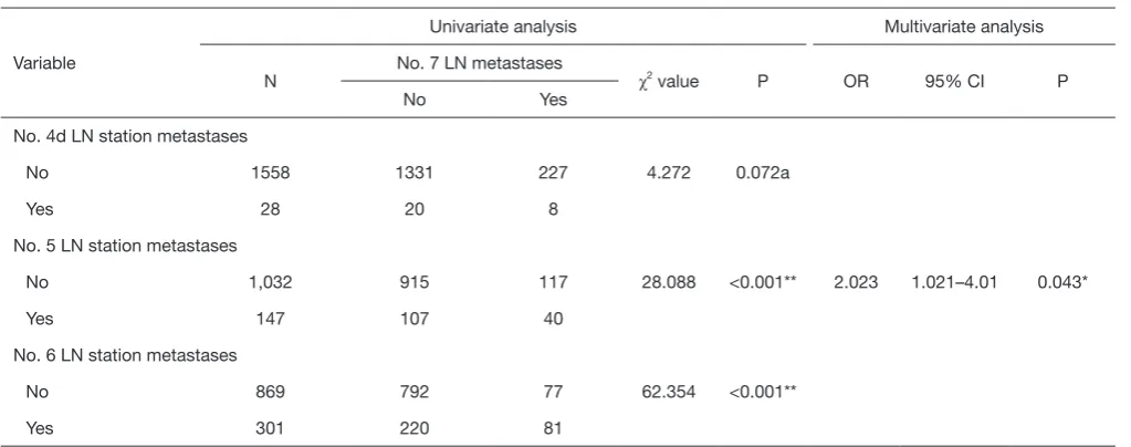
Table 3 Univariate and multivariate correlation analysis for the No. 7 LN metastases
Variable
Univariate analysis Multivariate analysis
N No. 7 LN metastases χ2 value P OR 95% CI P
No Yes
Gender
Male 1,144 974 170 0.006 0.938
Female 442 377 65
Age
Mean ± SD 60.68±11.48 61.68±11.19 0.217
<60 698 597 101 0.119 0.73
≥60 888 754 134
Tumor locationb
Upper 1/3 484 394 90 10.605 0.014*
Middle 1/3 134 112 22
Lower 1/3 655 578 77
More than 2/3 312 266 46
Tumor size
Mean ± SD 5.50±3.20 5.77±2.45 0.136
≤5.0 cm 881 766 115 4.885 0.027*
>5.0 cm 705 585 120
Number of LNs examined
Mean ± SD 14.79±10.00 19.13±10.52 <0.001**
≤15 916 812 104 22.31 <0.001**
16-30 536 436 100
More than 30 134 103 31
Pt stage
Pt1a 21 21 0 17.776 0.003*
Pt1b 31 29 2
Pt2 183 167 16
Pt3 107 98 9
Pt4a 1,195 995 200
Pt4b 49 41 8
Pn stage
Pn0 611 611 0 299.754 <0.001** 2.358 1.84–3.022 <0.001**
Pn1 288 270 18
Pn2 355 265 90
Table 3 (continued)
Variable
Univariate analysis Multivariate analysis
N No. 7 LN metastases χ2 value P OR 95% CI P
No Yes
Pn3a 248 156 92
Pn3b 84 49 35
Borrmann type
I 114 98 16 2.725 0.436
II 495 420 75
III 749 631 118
IV 228 202 26
Lauren typeb
Intestinal 825 718 107 7.49 0.024*
Diffuse 691 573 118
Mixed 32 24 8
Vasculolymphatic invasion
No 1,571 1,341 230 4.113 0.096a
Yes 15 10 5
Neurological invasionb
No 1,570 1,338 232 0.033 1.000a
Yes 8 7 1
No. 1 LN station metastases
No 1,358 1,212 146 123.736 <0.001**
Yes 228 139 89
No. 2 LN station metastases
No 688 580 108 20.317 <0.001**
Yes 161 111 50
No. 3 LN station metastases
No 1,033 960 73 140.995 <0.001** 2.089 1.097–3.98 0.025*
Yes 553 391 162
No. 4sa LN station metastases
No 806 669 137 5.607 0.018*
Yes 106 78 28
No. 4sb LN station metastases
No 1,379 1197 182 21.947 <0.001**
Yes 207 154 53
[image:10.595.54.553.104.683.2]Table 3 (continued)
Variable
Univariate analysis Multivariate analysis
N No. 7 LN metastases χ2 value P OR 95% CI P
No Yes
No. 4d LN station metastases
No 1558 1331 227 4.272 0.072a
Yes 28 20 8
No. 5 LN station metastases
No 1,032 915 117 28.088 <0.001** 2.023 1.021–4.01 0.043*
Yes 147 107 40
No. 6 LN station metastases
No 869 792 77 62.354 <0.001**
Yes 301 220 81
a, continuity correction analysis; b, some data missed; *, P<0.05; **, P<0.001. LN, lymph node.
the univariate analysis showed that the No. 7 LN metastases were significantly related with thirteen clinicopathologic characteristics: tumor location (P=0.014), tumor size (P=0.027), number of LNs examined (P<0.001), pT stage (P=0.003), pN stage (P<0.001), Lauren type (P=0.024), No. 1 LN metastatic status (P<0.001), No. 2 LN metastatic status (P<0.001), No. 3 LN metastatic status (P<0.001), No. 4sa LN metastatic status (P=0.018), No. 4sb LN metastatic status (P<0.001), No. 5 LN metastatic status (P<0.001) and No. 6 LN metastatic status (P<0.001). However, pN stage [odds ratio (OR) 2.358, 95% confidence interval (CI): 1.840 to 3.022, P<0.001], No. 3 LN metastatic status (OR 2.089, 95% CI: 1.097 to 3.980, P=0.025), and No. 5 LN metastatic status (OR 2.023, 95% CI: 1.021 to 4.010, P=0.043) were identified as independent risk factors for the No. 7 LN metastases by using the multivariate logistic analysis.
Discussion
In this study, we found that the OS rate of patients with metastases in the No. 7 station in addition to peri-gastric stations was significantly lower than that of patients with metastases in only peri-gastric LN stations. Nevertheless, the survival rate for patients with peri-gastric and No. 7 station metastases was not significantly different from the survival rate for patients with peri-gastric and other extra-gastric LN metastases. Furthermore, among patients with both peri-gastric and extra-gastric LN metastases, there
was no significant difference in survival rate between those with and without No. 7 station metastases. Metastasis in the No. 7 station did not appear to be essential for the development of other extra-gastric LN metastases, indicating that it should not be considered a predictive marker for predicting the invasion extent. Based on survival rate, the No. 7 station seems more closely aligned with the extra-gastric rather than peri-gastric stations.
[image:11.595.45.557.98.300.2]than that of NO. 7 group (HR 0.910, 95% CI: 0.839–0.986, P=0.021). Considering the specific clinical characteristic of skip metastases and the high frequency of skip metastases of No. 7 LN station, our study excluded this subgroup patients to obtain precise conclusion.
Currently, dissection of the No. 7 LN is deemed a part of the D1 LN dissection range (4), instead of the D2 dissection range (as per the previous definition) (20). In this study, the metastatic incident rate of the No. 7 LN station was 14.82% (235/1,586) in the entire cohort, which was the 3rd highest metastatic incident rate among all LN stations,
only ranking lower than the rates of the No. 3 (544/1,586) and No. 6 (301/1,170) LN stations. Therefore, the No. 7 LN station might be considered as the main route of lymphatic drainage from the gastric area. Our previous study also reported a high metastatic incident rate in the No. 7 LN station in GC patients (2). Other researchers have similarly reported that the metastatic incident rate of the No. 7 LN station was comparable to or even higher than that of the peri-gastric LN stations (1,31). This high metastatic incident rate might be the reason that the No. 7 LN station was reclassified in the range of D1 LN dissection in the 3rd edition of the Japanese Gastric Cancer
Treatment Guidelines and in the 14th edition of the Japanese
General Rules for Gastric Cancer Study (4,5). However, it is still controversial whether the prognostic implication of the No. 7 LN station is similar to that of the peri-gastric LN stations or other extra-gastric LN stations and whether the No. 7 station might be considered as an SLN for extra-gastric LN metastases in GC patients.
In the entire cohort, No. 7 LN metastases showed a significant impact on OS rate (P<0.001). After stratification by pTNM stage, we observed similar results in patients with pIII stage (P=0.014). We also observed a similar non-significant trend in pII stage patients (shown in the Figure 1B), which might be a result of the small sample size of these GC patient subgroups. Our results were consistent with those reported by Chen (32). Nevertheless, another study reported contrasting results after adjustment for pN stage, because two-thirds of their patient population received preoperative therapy to downstage the pN stage (33). Furthermore, the small sample size might limit the credibility of the results of that study. In despite of those limitations, we also observed some tendency of poor outcome in patients with No .7 LN metastases (with No. 7 LN metastases vs. without No. 7 LN metastases, 3-year survival rate: N1, 75% vs. 79%; N2, 40% vs. 80%; N3, 20% vs. 33%). Thus, we could not deny that No. 7 LN
station metastases might have a significant influence on the prognosis of GC patients.
To obtain more precise results, PSM was performed to balance the confounding factors between two groups. Both before and after PSM, the survival outcome of patients with No. 7 LN station metastases was similar to that of patients with extra-gastric LN station metastases (Figure 2C,E) and significantly poorer than that of patients with only peri-gastric LN station metastases (Figure 2B,D). Our results were consistent with those reported by Chen et al. (32). However, Murayama et al. reported that the prognostic impact of the No. 7 LN station was similar to that of peri-gastric LNs in patients with six or fewer positive LNs (6). This converse conclusion might be achieved result from enriching patients with lack of positive LNs. Based on our findings, we believe that the No. 7 LN station should be included in the range of D2 lymphadenectomy. If No. 7 LN involvement is highly suspected during the operation, D2 lymphadenectomy might be required. However, our study showed that metastases to extra-gastric LNs other than the No. 7 LN was observed in 37% (138/373) of patients, and this subgroup did not show a superior survival outcome. This result indicated that No. 7 LN metastasis was not essential for extra-gastric LN metastases and that the No. 7 LN should not be considered as the SLN for extra-gastric LN metastases. Further prospective large-scale studies are warranted to confirm this conclusion.
The results of multivariate analysis showed pN stage (P<0.001), No. 3 LN metastases (P=0.025), and No. 5 LN metastases (P=0.043) were independent risk factors for No. 7 LN metastases. Chen et al. also reported that metastases to the No. 7 LN station were associated with pN stage, pTNM stage, and No. 3 LN metastases, which is mostly consistent with our findings (32). In addition, previous studies have reported that No. 7 LN metastases are associated with aggressive biological behavior, such as large tumor size and vasculolymphatic invasion (34,35). These findings indicated that the No. 7 station might be a part of a crucial lymphatic route.
Conclusions
In conclusion, our study indicated the No. 7 LN station should be reclassified in the D2 dissection range due to its prognostic impact similar to that of extra-gastric LN station. If No. 7 LN involvement is highly suspected during the operation, D2 lymphadenectomy might be required. Nonetheless, our study proposed that the No. 7 LN station should not be considered a SLN as it does not appear to be essential for extra-gastric LN metastasis.
Acknowledgments
We would like to thank Editage (www.editage.com) for English language editing.
Funding: This study was supported by grants from the
Programs of National Natural Science Foundation of China [grant number 81572372 to JD], National Key Research and Development Program “major chronic non-infectious disease research” [grant number 2016YFC1303202 to HL], National Key Research and Development Program “precision medicine research” [grant number 2017YFC0908304 to JD].
Footnote
Reporting Checklist: The authors have completed the
STROBE reporting checklist. Available at http://dx.doi. org/10.21037/atm-19-4786a
Data Sharing Statement: Available at http://dx.doi.
org/10.21037/atm-19-4786a
Peer Review File: Available at http://dx.doi.org/10.21037/atm-19-4786a
Conflicts of Interest: All authors have completed the
ICMJE uniform disclosure form (available at http://dx.doi.
org/10.21037/atm-19-4786a). The authors have no conflicts
of interest to declare.
Ethical Statement: The authors are accountable for all
aspects of the work in ensuring that questions related to the accuracy or integrity of any part of the work are appropriately investigated and resolved. The study was approved by Tianjin Medical University Cancer Institute and Hospital ethics committees (No. bc2019087). All patients provided written informed consent before any enrolling procedures were initiated.
Open Access Statement: This is an Open Access article
distributed in accordance with the Creative Commons Attribution-NonCommercial-NoDerivs 4.0 International License (CC BY-NC-ND 4.0), which permits the non-commercial replication and distribution of the article with the strict proviso that no changes or edits are made and the original work is properly cited (including links to both the formal publication through the relevant DOI and the license). See:
https://creativecommons.org/licenses/by-nc-nd/4.0/.
References
1. Sun Z, Wang ZN, Zhu GL, et al. Advanced gastric cancer with early cancer macroscopic appearance: is it worthy of D2 lymphadenectomy? Ann Surg Oncol 2010;17:1278-90. 2. Liang J, Liang H, Deng J, et al. Clinical study on
lymph node metastasis regularity in 1456 patients with gastric cancer. Zhonghua Wei Chang Wai Ke Za Zhi 2018;21:1154-60.
3. Ajani JA, Bentrem DJ, Besh S, et al. Gastric cancer, version 2.2013: featured updates to the NCCN Guidelines. J Natl Compr Canc Netw 2013;11:531-46.
4. Japanese Gastric Cancer Association. Japanese classification of gastric carcinoma: 3rd English edition. Gastric Cancer 2011;14:101-12.
5. Japanese Gastric Cancer Association. Japanese Classification of Gastric cancer. 14th edition. Tokyo, Japan: Kanehara & Co. Ltd, 2010.
6. Murayama Y, Ichikawa D, Kubota T, et al. Prognostic impact of lymph node metastasis along the left gastric artery in gastric cancer. Hepatogastroenterology 2011;58:1603-6.
7. Ohdaira H, Nimura H, Takahashi N, et al. The possibility of performing a limited resection and a lymphadenectomy for proximal gastric carcinoma based on sentinel node navigation. Surg Today 2009;39:1026-31.
8. Mikami K, Hirano Y, Futami K, et al. Expansion of lymph node metastasis in mixed-type submucosal invasive gastric cancer. Asian J Surg 2018;41:462-6.
9. Sun ZQ, Zhou YB. Study on No. 7 lymph nodes micrometastasis in patients with node- negative gastric carcinoma by routine examination. Zhonghua Wei Chang Wai Ke Za Zhi 2005;8:339-42.
10. Morton DL, Wen DR, Wong JH, et al. Technical details of intraoperative lymphatic mapping for early stage melanoma. Arch Surg 1992;127:392-9.
nanoparticles for multimodal imaging and guided photothermal therapy of lymph node metastasis. Acta Biomater 2018;72:256-65.
12. Kim MC, Kim HH, Jung GJ, et al. Lymphatic mapping and sentinel node biopsy using 99mTc tin colloid in gastric cancer. Ann Surg 2004;239:383-7.
13. Farzaneh F, Moridi A, Azizmohammadi Z, et al. Value of Sentinel Lymph Node (SLN) Mapping and Biopsy using Combined Intracervical Radiotracers and Blue Dye Injections for Endometrial Cancer. Asian Pac J Cancer Prev 2017;18:431-5.
14. Kitagawa Y, Fujii H, Mukai M, et al. The role of the sentinel lymph node in gastrointestinal cancer. Surg Clin North Am 2000;80:1799-809.
15. Kitagawa Y, Kitajima M. Gastrointestinal cancer and sentinel node navigation surgery. J Surg Oncol 2002;79:188-93.
16. Ichikura T, Sugasawa H, Sakamoto N, et al. Limited gastrectomy with dissection of sentinel node stations for early gastric cancer with negative sentinel node biopsy. Ann Surg 2009;249:942-7.
17. Ohdaira H, Nimura H, Mitsumori N, et al. Validity of modified gastrectomy combined with sentinel node navigation surgery for early gastric cancer. Gastric Cancer 2007;10:117-22.
18. Kitagawa Y, Takeuchi H, Takagi Y, et al. Sentinel node mapping for gastric cancer: a prospective multicenter trial in Japan. J Clin Oncol 2013;31:3704-10.
19. Sobin L GM, Witterkind C, et al. TNM classification of malignant tumors. 7th edition. International Union Against Cancer (UICC). New York: Wiley, 2009. 20. Japanese Gastric Cancer Association. Japanese
Classification of Gastric Carcinoma - 2nd English Edition. Gastric Cancer 1998;1:10-24.
21. Choi YY, An JY, Guner A, et al. Skip lymph node metastasis in gastric cancer: is it skipping or skipped? Gastric Cancer 2016;19:206-15.
22. Zhao P, Su X, Ge T, et al. Propensity score and proximity matching using random forest. Contemp Clin Trials 2016;47:85-92.
23. Austin PC. The use of propensity score methods with survival or time-to-event outcomes: reporting measures of effect similar to those used in randomized experiments. Stat Med 2014;33:1242-58.
24. Han HS, Shehta A, Ahn S, et al. Laparoscopic versus open liver resection for hepatocellular carcinoma:
Case-matched study with propensity score matching. J Hepatol 2015;63:643-50.
25. Brookhart MA, Schneeweiss S, Rothman KJ, et al. Variable selection for propensity score models. Am J Epidemiol 2006;163:1149-56.
26. D'Agostino RB Jr. Propensity score methods for bias reduction in the comparison of a treatment to a non-randomized control group. Stat Med 1998;17:2265-81. 27. Su Z, Shu K, Zheng M, et al. Sentinel lymph node and skip metastases in gastric cancer: a prospective study. Hepatogastroenterology 2013;60:1513-8.
28. Zhao B, Mei D, Zhang J, et al. Impact of skip lymph node metastasis on the prognosis of gastric cancer patients who underwent curative gastrectomy. J BUON 2019;24:693-700.
29. Saito H, Tsujitani S, Ikeguchi M. Clinical significance of skip metastasis in patients with gastric cancer. Gastric Cancer 2007;10:87-91.
30. Kim DH, Choi MG, Noh JH, et al. Clinical significance of skip lymph node metastasis in gastric cancer patients. Eur J Surg Oncol 2015;41:339-45.
31. Zhu HT, Zhao YL, Wu YF, et al. Features of metastasis in different lymph node groups and their significance in lymph node dissection in total gastrectomy for gastric cancer. Zhonghua Zhong Liu Za Zhi 2008;30:863-5. 32. Chen JH, Cai SR, Zhai ET, et al. Survival prognosis and
clinicopathological features of the lymph nodes along the left gastric artery in gastric cancer: implications for D2 lymphadenectomy. Int J Clin Exp Pathol 2015;8:14365-73. 33. Ikoma N, Blum M, Estrella JS, et al. Left Gastric Artery
Lymph Nodes Should Be Included in D1 Lymph Node Dissection in Gastric Cancer. J Gastrointest Surg 2017;21:1563-70.
34. Park JM, Jang YJ, Kim JH, et al. Gastric cancer histology: clinicopathologic characteristics and prognostic value. J Surg Oncol 2008;98:520-5.
35. Liu E, Zhong M, Xu F, et al. Impact of lymphatic vessel invasion on survival in curative resected gastric cancer. J Gastrointest Surg 2011;15:1526-31.