62
A Survey On Skin Classification Methods Using Deep
Learning Techniques
Anisha Thangakani S
2, M Sornam
1Department of computer science, University of Madras, Guindy campus, Chennai [email protected], [email protected]
1-Corresponding Author
Abstract- Convolutional Neural Networks (CNN) is the most popular neural network model being used for skin classification problem. The big idea behind Convolutional Neural Networks is that a local understanding of skin is good enough. The practical profit is that having fewer parameters greatly improves the time it takes to be told still as reduces the quantity of information needed to coach the model. Skin classification research datasets are typically very large. Nevertheless, data augmentation is often used in order to improve generalization properties. It draws on similarities from nature, where neurons are interconnected, capable of learning and improving on performance, based on Convolutional Neural Networks classifiers
Index Terms-Convolution neural network, Local Binary Pattern, Bayesian Classifier, Support Vector Machine.
1. INTRODUCTION
Recently, skin classification and detection have been utilized in computer technology, medical imaging and also in computer aid diagnosis, the target of skin detection or classification very challenges. Skin classifier is mainly used to detect the skin problem like dryness, fungus, and allergic symptoms etching kind of problem that may lead to starting symptoms of malignant melanoma skin cancer. The correct identification of skin spots based on certain features is the key steps in detecting skin cancer disease in advance. A Bayesian classifier is based on the role of a (natural) class is to predict the values of features for members of that class. So that Convolutional Neural Networks used for both the classification and detection of skin. In this paper, we are going to see the concept of the individual Convolutional Neural Networks methods used to classify skin classifier is presented. Convolutional Neural Networks can be used to classify in different ways there are Bayesian classifier, a local binary pattern, k-nearest neighbors, and support vector machines, on convolutional neural network
2. RELATED WORK
The classification or detection of skin aims to find the true skin pixel from the image and frame. It also aims to divide the image into skin and non-skin pixel there are many approaches in literature, some of the approaches are briefly described below.
2.1. Skin detection on colored pixels
SonLanPhung et al. [1] proposed a new concept for skin colored region and detection an algorithm for
segmenting skin regions in color images using color and edge information is presented. Skin colored regions are first detected using the Bayesian model [2] of the human skin color. These regions are more segmented into skin region candidates that satisfy the homogeneity property of the human skin. The author shows that the Bayesian skin color model outperforms many other models such as the piece-wise linear models, Gaussian models. And the model of a multilayer perceptron. The data used in this work are taken from the ECU face detection database constructed at Edith Cowan University. The results established that the proposed segmentation algorithm reduces false detection caused by background pixels having skin colors, and more significantly it is capable of separating true skin regions from falsely detected regions for minimum cost and nonparametric density estimation.
Krishnan et al. [2] described the Skin Detection using Color Pixel Classification with Application to Face Detection Several algorithms have been proposed for
63
linear classifiers, the Bayesian classifier in Fig.2. with the histogram technique, Gaussian classifiers, and the Multi-layer perceptron A large set of XM2VTS face database is used to examine whether the selection of color space can enhance the compactness of the skin class and discriminability between skin and non-skin class in thirteen color spaces and six different skin color pixel classification algorithms An analysis of the pixel-wise skin segmentation approach that uses color pixel classification algorithms is presented. Multi-layer perceptron classifier produces good results. The Gaussian classifier with the histogram technique classifier had a maximum CDR of 99.10 percent. But the Bayesian classifier produces best results with 99.43. The piecewise linear classifiers applied for HSV and RGB color spaces produce still higher classification rates than all other classifiers
for an
image with good illumination conditions
Kai-Biao Ge et al. [3] described the skin on face detection algorithm based on AdaBoost has been implemented, but they extract MB-LBP features instead of Haar features, which are used for Training of AdaBoost classifier. In order to reduce the false alarm rate, and also combine skin color with AdaBoost. MB-LBP feature, to represent the facial image. The unique idea of MB-LBP is to encode rectangular regions by local binary pattern operator.
The MB-LBP features can also be calculated rapidly through an integral image, while these features capture more information about the image structure than Haar-like features and show more distinctive performance. Kai Biao Ge al. Selected 100 images from the Internet to test which contain different backgrounds, different sizes of face images. Experimental results show that the method is effective to detect human faces and the detection rate is more than 90%.
2.2.Melanoma detection on cancer cells usingLBP & SVM:
Raman deep et al. [4] proposed method uses the adaptive filter to find the threshold inspired by Swarm Intelligence (SI) optimization algorithms. The image processing techniques on the original image are implemented in primarily three streams: The gray scale format is used to compute the texture features in the GLCM domain. The original RGB image is used to generate the color moments like mean red, green and blue color component and standard deviation in red, green and blue color domain. The k-means clustering is used to extract the infected part and then local binary patterns are computed by thresholding the segmented part using the Otsu algorithm. The presented work results show the improvement in identifying the Melanoma skin cancer at different stages using image processing techniques based on textural feature analysis and SVM classifier and shows a fine distinction between different stages of the Melanoma skin cancer. However, in the case of border cases, the SVM classifier takes the guard to a good extent this is the vital information where the skin expert may get vital information at fine accuracy. Rashi Singh et al. [5] defined the Skin Texture Analysis Using Machine Learning the proposed work includes a set of image is obtained by capturing image using normal smartphone camera under proper illumination Implementing the proposed work includesacquisition of the skin images then preprocessing them to convert into gray scale using Irfan view software this image set using Law's filter and Local Binary Pattern (LBP) [5] feature extraction techniques. After obtaining these features, they are given as input to the classifier. The 197 input image set are used to training and testing set. The skin images were gathered from various areas and were rated as normal, good or bad skin from a standard dermatologist in the range of 1 to 10. The training phase consists of 70% of the total images and it was tested using the remaining 30% images. The accuracy of classifying the images into good, bad and normal skin is observed to be around 99.38%.
Priyadarshini et al. [6] described Local Binary Pattern (LBP) method is used for texture analysis; it has found that the powerful feature for texture feature extraction. Fig.2.piece-wise linear models
64
It determined that when LBP [5] is combined with the Histogram of oriented gradients (HOG) [classifier and it improves the detection performance on some datasets. The database consists of 20 digital images, previously diagnosed, 10 of them are benign and 10 are melanoma. The mechanisms proposed i.e., SVM and Local Binary Pattern is performed for 4 test skin samples and experimented to test the accuracy, Specificity, and Sensitivity. The features were fed automatically to a support vector machine classifier which achieved greater than 97% sensitivity and greater than 93%specificity. An SVM classifies data by finding the best hyper plane that separates all data points of one class from those of the other class and also obtain the trained system tested with lesion images found online and it’s able to achieve similar sensitivity.
2.3.Bayesian for Enhancing skin Recognition Accuracy:
2.3.1. Bayesian Decision Fusion
Maen Takruri et al [7] proposed the Bayesian Decision Fusion of a multiple of classifiers to enhance the melanoma detection rates.it results in improved recognition accuracy compared to standalone skin lesion classifier it proved comparable confidence interval and offer stable recognition rate. It includes 213 and 93digital images for benign and malignant skin lesions, respectively.Fig.4. 80% of the images are used for training. The remaining 20% is used for testing it is implemented using LIBSVM toolbox
.
Fold cross-validation is used to pick the best RBF kernel parameter. Similar to, the resulting 10-fold ross Validation Accuracies are 85.3% for Wavelet and Colorfeatures, 76.6% for Curvelet features and 78.7% for GLCM features.Probability Averaging Fusion resulted in 87.6 ± 0.89% and Bayesian Fusion resulted in 84.1 ± 0.31 %.The relation between confidence
distribution and accuracy over different classification systems is studied. Performance evaluations of the proposed Bayesian Decision fusion method show that it results in improved recognition accuracy compared to standalone Skin Lesion classifiers. It also provides comparable confidence intervals and can offer a stable recognition rate. Hence, it can lead to increased chances of non-invasive melanoma detection.
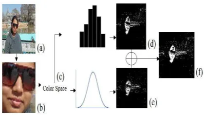
Varma et al. [8] defined the detection of skin region aims to find the presence of skin detection by dividing the image into the skin and non-skin pixel. HSV and YCbCr color spaces are used to transform pixel and Training. Histogram and Gaussian Mixture Model in Fig.5.(GMM) is used for classification of skin and non-skin pixels in an image. This is used to describe pixels as skin distribution. Fusion approach includes a grouping of several stages. Each stage has a certain number of features. Different stages are used to additive feature extraction for face detection systems. To increase the effectiveness of skin recognition fusion method is proposed. Here two single features
into one single feature. Detection of skin pixel is based on vote from both models. Product rule is applied to both models to fuse into one as given Skin classifier gives the threshold for skin for the given color space. The experiment is performed on three datasets namely ETHZ PASCAL dataset, Partheepan dataset, and SFA
Fig. 5
. Performance of 2D Histogram, GMM andFusion approach using Partheepan databset
Fig. 4
. The skin detection system: (a) Input image, (b) Face recognition,(c) Color feature, (d) 2D histogram processing, (e) GMM approach, and (f) [image:3.595.316.519.366.476.2] [image:3.595.72.282.495.613.2] [image:3.595.309.517.541.658.2]65
datasetThe hybrid approach is used for skin detection. The accuracy, F-Score, true positive rate, and the false positive rate have measured the performance of the system.Overall, the performance of a fusion approach over three different datasets using HSV is acceptable as compared to individual performances over YCbCr.
2.3.2.Training Bayesian Classifier with Scaling Unique Colors:
Dmitry et.al [9] analyzed to improve the quality of skin segmentation in images, the new training approach of the Bayesian classifier Fig.3. Has been proposed. The segmentation issues arise in several application of developing the intellectual systems based on supported video analysis.
One of the important tasks is the human skin segmentation for further high-level processing in Fig .8.The most used approach is based on naive Bayesian classifier to separate human skin (foreground) pixels from the background ones. The author proposed the new approach for preparing training dataset which considers only unique colors from each image to avoid normalization influence related to the size of the foreground area. It provides a stable improvement of the segmentation quality based on ROCcurves comparison
.
The reference data to obtain training and test sets were collected by combining public datasets:Pratheepan Dataset, Database for hand gesture recognition, UCI Skin Segmentation Data Set, IBDT Dataset for Skin Detection. The testing was conducted on the union of 4 biggest public datasetscontaining labeled pictures with human skin. The main result of this work is a new method of training the Bayesian classifier based on scaling unique colors of pixels collected from each training image. In comparison with the classical approach, it improves the quality of skin segmentation by 3-4% by TPR and reducing segmentation errors to 10% by FPR.
2.3.3.Bayesian classifier and connected component algorithm
Thao Nguyen et al. [10] proposed a new approach to improve the skin detection performance using the Bayesian classifier Fig.3. And the connected
component algorithm. It is utilized to identify "true skin" pixels using the first posterior probability threshold, which is approximate to 1, and to identify "skin candidate" pixels using the second posterior probability threshold. Other, the connected component algorithm is used to find all the connected components containing the "skin candidate" pixels. According to the skin pixel often connects with other skin pixels in an image, all pixels in a connected component are classified as "skin" if there is at least one "true skin" pixel in that connected component. Skin Detection Dataset (SDD) downloaded from the website (https://archive.ics.uci.edu/ml/datasets/skin+segmentat ion) used as the training set. We need to classify the skin or not Step 1. Compute the posterior probability that the pixel belongs to the skin class, using Bayes theorem Step 2. Find all connected components containing "skin candidate" pixels. Step 3. Classify the pixels with the subsequent rule: if the connected component contains at least minimum of one "true skin" pixel, then all pixels happinessto itelementarea unit component are classified as "skin" and vice versa in order to control the false positive rate at a low level, Thao Nguyen et al. choose 𝜀1 ≈ 1. For 𝜀2, the detection rate of the method is equal to or less than that of Bayesian classifier if 𝜀2 = 0.5. Therefore, a value of threshold 𝜀2 that is slightly less than 0.5 will increase the detection rate of the algorithm dataset are presented and compared with some other methods such as the Bayesian classifier (BC), linear discriminant analysis (LDA), binary logistic regression (BLR), and Adaptive Neuro-Fuzzy Inference System (ANFIS) The best method in terms of detection rate is the ANFIS with the detection rate over 87%.
2.4.Genetic Programming for Skin Cancer Detection
[image:4.595.324.507.153.242.2]Qurrat et al. [11] delineateda Genetic Programming (GP), associaterisingbiological processComputation technique, has the potential to evolve higher solutions for image classification issue compared to
Fig. 8. Segmentation results for image from new Proposed training approach with all unique colors
[image:4.595.108.259.490.580.2]66
severalexisting methods GP has been utilized to automatically evolve a classifier for skin cancer detection and also analyzed GP as a feature selection method They have compared their results with ANNs and reported that GP has outperformed the competitor method. ANNs gave 31:7% sensitivity and 92:2% specificity, whereas GP achieved 61:5% sensitivity and 99:2%specificity. A dataset of dermoscopic images namely PH2 acquired from Pedro Hispano Hospital Portugal, is used in the experiments they used GP to evolve solutions for this binary classification problem of detecting skin cancer. The LBP method used to extract features from images and also used features provided by domain experts. Three scenarios are considered in using different feature sets; using domain features, using LBP features and using a combination of both. They have also examined GP as a feature selection method and did experiments where GP-Selected features are used to evolve solutions using GP and other state-of-the-art classification methods.GP has achieved better or comparable performance in most cases as compared to other methods (k-NN, SVM, Naıve Bayes, Decision Trees, Random Forest, and Multilayer Perceptron). With all features, the highest accuracy achieved is 97:92% and GP-selected features, even 100% accuracy are achieved in some of GP.
2.5.Skin Detection dataset for training and assessment of skin Classifiers
Mohammad et al. [12] described a new concept for a dataset that is SDD (Skin Detection Dataset) is suitable for assessment of skin of skin classifier and also addresses the limitation of former image libraries. Because it's an extensive database it evaluates the effectiveness and characteristics of the database and also measure the performance on quality training images on ANN Classifier The proposed database has been compared to SFA through which Qualitatively and quantitatively, the appealing features of the SDD are confirmed the result of simulating several skin detection method based on the proposed database.it contains more than 20,000 color images and which give a reliable result for evaluation purpose, and well organized so that comparing the performance of different method rather than dataset finally it is shown that using SDD is more efficient than SFA
2.6.Skin Diagnosis Using multi-label deep neural network
However, it's onerousto spotskin cancerthroughoutits early to mid-stages by visual examination. Enhanced automated computer-aided model.
Sherin et al. [13] described for skin diagnosis using deep learning. The model integrates associateincreasedsegmentation section for locating the infected lesion of the skin and a Convolution Neural Network (CNN) is intended as a feature extractor. A classifier model has been designed based on multiclass linear Support Vector Machine (SVM) trained with CNN features extracted from the digital skin images dataset, First dataset from the publicly available online databases
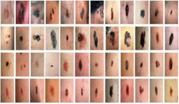
Dermatology Information System in Fig.7 The dataset comprises 69 images 43 of them represent melanoma and 26 for not melanoma skin cancer. The second dataset is DermQuest the dataset comprises 134
images 76 of them represent melanoma and 58 for not melanoma skin cancer in Fig.8 some sample images from the dermis dataset.Performance of classification, Different sets of experiments have been conducted. The performance is evaluated in terms of classification sensitivity, specificity, and accuracy
[image:5.595.331.504.278.388.2]from the confusion matrix of classification. The equation described below with the following conventions.TP (True Positive) is Positive samples classified as positive. Similarly TN is Negative anditssamples classified as negative. First dataset was able to correctly identify 94.12 % patients with the non-melanoma disease diagnosis 94.12 % of patients with melanoma correctly.
Fig. 9. Samples of DermIS dataset
[image:5.595.326.505.500.604.2]67
Finally, the overall accuracy of the correct diagnosis
patients is 94.12 %. Second dataset able to correctly identify all patients with non-melanoma disease diagnosis 87.5 % of patients with melanoma correctly. Finally, the overall accuracy of the correct diagnosis patients is 93.75%. The third dataset can produce high accuracy also when applied to multiclass skin disease. The results show that the proposed system correctly identifies all patients without Melanoma and Eczema diseases. Also, it willproperlyestablish all patients with the diseaseof the skin unwellness non-melanoma disease can also diagnose. Finally, melanoma skin disease has the highest accuracy compared with another disease.
Haof Liao et al. [14] proposed an investigation to achieve robust skin disease diagnosis. In this, they use two approaches:
i. A direct approach is to target the ground truth diagnosis labels;
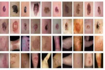
ii. Alternative approach instead focuses on determining skin lesion characteristics that are more visually consistent and discernible. Hoaf Liao et al. collected dermatology photos to further, use on convolution neural networks (CNN) for both the disease targeted and lesion-targeted classifications they have collected a dataset of 75; 665 skin disease images in Fig.7 then train and compare both disease-targeted and lesion-targeted classifiers, respectively. Disease-targeted classification, they achieved only 27:6% top-1 accuracy and 57:9% top-5 accuracy are with a mean average precision (mAP) of 0:42. In contrast, for lesion-targeted classification, they achieved a much higher mAP of 0:70.image retrieval result also confirmed CNN's trained using lesion tag learn the dermatology features very well. The Fig . 9,10,11. shown above.
Seema et al. [15] described a Skin disease detection is basically an image classification task. Convolution Neural Network (CNN) as a classifier. To achieve a lot of reliable and objective accuracy computer-aided diagnosing is also used. Recently deep learning is used in many image classification tasks. They used the
Convolution Neural Network (CNN) as a classifier and collected raw images dataset for two skin diseases mainly Leprosy and Warts from the Department of Skin and VD from KEM Hospital, Parel Mumbai. A CNN consists of different types of layers. Each layer
[image:6.595.97.274.117.234.2] [image:6.595.317.520.174.282.2]has a specific kind of function. It consists of 1 input and 1 output layer. In between these two layers, there may be multiple hidden layers. The hidden layers typically consist of convolution layers, pooling layers, fully connected layers and dropout layers image dataset was divided into training and testing dataset. CNN model was trained to fit training data. It outputs the probability of every test record being classified as either Leprosy or Warts. And confusion matrix for the testing set. The paper result indicates CNN has been feasibly for skin disease detection. They used convolution network as a classifier for skin disease detection from an input image. The overall accuracy of classification is 95% which shows that CNN successfully be used for the stated purpose. The major advantage of a system is that do not have to extract features from the set of input images separately. Convolution layer and pooling layer acts as feature extractor while dense layer works as a classifier. Fig. 11. Samples of DermNet dataset
68
3. Tables:Table 1: different method and classifier on skin classification
Author Name
Algorithm Dataset Outcomes
SonLanPhun g et al.
Bayesian model piece-wise linear models, Gaussian model multilayer perceptron ECU face detection database Reduces false detection caused by background pixels having skin colors. Krishnan et al. [2] Bayesian classifier with the histogram technique, Gaussian classifiers, Multi-layer perceptron XM2VTS face database Bayesian classifier produces best results with 99.43 Kai-Biao Ge et al. AdaBoost With MB-LBP features 100 images from the Internet Detection rate is more than
90%. Rashi Singh et al. Local Binary Pattern capturing image using normal smartphon e camera
The accuracy of classifying thephotographs
into good, bad, normal is observed to be around 99.38%. Priyadarshini et al. Local binary pattern (LBP) & Histogram of oriented gradients (HOG) DermQue st DermNet DermIS Achieved greater than 97% sensitivity and greater than 93%specificity.
To achieve similar sensitivity.
Maen Takruri et al
Bayesian Decision Fusion
of a multiple of classifiers epilumine scence microscop y images Bayesian Fusion resulted
in 84.1 ± 0.31 %. Raman deep et al. Swarm Intelligence (SI) optimization algorithms 100 images from the Internet Skin professionalmig
ht get very importantinfo at
fine accuracy.
Author Name
Algorithm Dataset Outcomes
Dmitry et.al Bayesian classifier UCI Skin Segmentation Data Set, IBDT Dataset for Skin Detection.
The quality of skin segmentation by 3-4% by TPR and
reducing segmentation errors to 10% by
FPR.
Thao Nguyen et al
Bayesian classifier & connected component algorithm Skin Detection Dataset (SDD)
ANFIS with the detection rate over
87%.
Varma et al. Fusion approach ETHZ PASCAL, Partheepan, & SFA dataset.
HSV is acceptable as compared to
individual performances over
YCbCr.
Qurrat et al.
Compare Genetic Programmin g & ANN classifier Dermoscopic images namely PH2 acquired from Pedro Hispano Hospital Portugal
ANN gave 31:7% sensitivity and 92:2% specificity, whereas GP achieved 61:5% sensitivity and 99:2%specificity. Mohammad et al. ANN Classifier SDD (Skin Detection Dataset)
SDD is more efficient than SFA
Sherin et al.
Convolution Neural Network DermQuest DermNet DermIS Correct diagnosis patients is 93.75% with high accuracy
Haof Liao et al. convolution neural networks DermQuest DermNet DermIS Disease-targeted classification, solely 27:6% top-1
accuracy and 57:9% top-5 Lesion-targeted classification, and
achieved a much higher mAP of
69
4. CONCLUSION
Convolutional Neural Network is a popular deep learning technique for current skin detection and classification. Deep learning techniques, CNN is very dependent on the size and quality of the training data. Given a well-prepared dataset, CNN's are capable for skin classification tasks. However, they are still not robust to detect and classify such as glare and noise, which humans are able to cope. The theory of CNN is still being developed and researchers are working to endow it with properties such as active attention and online memory, CNN's to evaluate new items that area unitimmenselytotally differentfrom what they were trained on. This better emulates the skin detection, thus moving towards a smarter.
REFERENCES
[1] Phung SL, Bouzerdoum A, Chai D. Skin segmentation using color and edge information. InSignal Processing and Its Applications, 2003. Proceedings. Seventh International Symposium on 2003 Jul 1 (Vol. 1, pp. 525-528). IEEE.
[2] Nallaperumal K, Ravi S, Babu CN, Selvakumar RK, Fred AL, Christopher S, Vinsley SS. Skin detection using color pixel classification with application to face detection: A comparative study. Iniccima 2007 Dec 13 (pp. 436-441). IEEE.
[3] Ge KB, Wen J, Fang B. Adaboost algorithm based on MB-LBP features with skin color segmentation for face detection. InWavelet Analysis and Pattern Recognition (ICWAPR), 2011 International Conference on 2011 Jul 10 (pp. 40-43). IEEE.
[4] Mahmoodi MR, Sayedi SM, Karimi F, Fahimi Z, Rezai V, Mannani Z. SDD: A skin detection dataset for training and assessment of human skin classifiers. InKnowledge-Based Engineering and Innovation (KBEI), 2015 2nd International Conference on 2015 Nov 5 (pp. 71-77). IEEE.
[5] Rashi Singh, Pankti Shah “Skin Texture Analysis
Using Machine Learning”, IEEE 2016
[6] Priyadarshini D, Rengini D “Automatic Melanoma Detection Using Local Binary Pattern And Support Vector Machine” International journal of Innovative Research in Computer And Communication Engineering
[7] Takruri M, Abubakar A. Bayesian decision fusion for enhancing melanoma recognition accuracy. In2017 International Conference on Electrical and Computing Technologies and Applications (ICECTA) 2017 Nov 21 (pp. 1-4). IEEE.
[8] Varma SL, Behera V. Human skin detection using histogram processing and Gaussian Mixture Model based on color spaces. In2017 International Conference on Intelligent
Sustainable Systems (ICISS) 2017 Dec 7 (pp. 116-120). IEEE.
[9] Vorotnev D, Golovanov R, Umnyashkin S. Training Bayesian classifier with scaling unique colors among image samples. InYoung Researchers in Electrical and Electronic Engineering (EIConRus), 2018 IEEE Conference of Russian 2018 Jan 29 (pp. 1835-1839). IEEE.
[10] Nguyen-Trang T. A New Efficient Approach to Detect Skin in Color Image Using Bayesian Classifier and Connected Component Algorithm. Mathematical Problems in Engineering. 2018;2018.
[11] Ain QU, Xue B, Al-Sahaf H, Zhang M. Genetic programming for skin cancer detection in dermoscopic images. InEvolutionary Computation (CEC), 2017 IEEE Congress on 2017 Jun 5 (pp. 2420-2427). IEEE.
[12] Shoieb DA, Youssef SM, Aly WM. Computer-aided model for skin diagnosis using deep learning. Journal of Image and Graphics. 2016 Dec;4(2):122-9.
[13] Ramandeep Kaur, Gurmeen Kaur” Enhancement
and Detection of Melanoma in Skin Images Using Local Binary Pattern (LBP)”, International journal of advanced Research
[14] Liao H, Li Y, Luo J. Skin disease classification versus skin lesion characterization: Achieving robust diagnosis using multi-label deep neural networks. InPattern Recognition (ICPR), 2016 23rd International Conference on 2016 Dec 4 (pp. 355-360). IEEE.