Original Article
Morphological chimeras of larvae and adults in a hydrozoan –
insights into the control of pattern formation and morphogenesis
MICHAEL KROIHER
Zoologisches Institut, Universität zu Köln, Germany
ABSTRACT In the marine hydroid Hydractinia echinata, metamorphosis transforms the spindle-shaped larva into a primary polyp. It bears a hypostome with a ring of tentacles at its apical end, a gastric region in the middle and stolons at the base. In nature, metamorphosis is induced in response to external stimuli provided by bacteria. These stimuli can be replaced by artificial inducers, one of which is heat shock. Among heat shock treated stages are those undergoing complete metamorphosis but also specimens forming chimeras of different developmental stages. In the chimeric larvae, the posterior is transformed into the apical hypostome of the adult polyp while the anterior part of the larva persists as larval tissue. After transverse sectioning, these stage chimeras regenerate the missing body parts with respect to the nature of the tissue at the wound surface. This shows that the decision to make larva or polyp morphology depends not on the majority of the tissue in the original body section, but on stage specificity within the regenerating animal part. Single cells can escape from this general rule, since RFamide nerve cells which usually differentiate in polyp tissue appear in regenerated larval tails of sectioned stage chimeras. The results indicate that the pattern-forming system of the larva and of the adult have features in common. The primary signals controlling patterning along the anterior-posterior axis in larvae and the apical-basal axis in polyps are thus likely to be the same while the interpretation of these primary signals by the individual cells changes during metamorphosis.
KEY WORDS:
Hydrozoa, Hydractinia, pattern formation, regeneration, metamorphosis, RFamide nerve cells.
0214-6282/2000/$20.00
© UBC Press Printed in Spain
www.ehu.es/ijdb
*Address correspondence to: Michael Kroiher Zoologisches Institut, Universität zu Köln, Weyertal 119, 50923 Köln, Germany. FAX: +49-221-470-5171. e-mail: [email protected]
Abbreviations used in this paper: FITC, fluorescein-isothiocyanate; RFamide, Arg-Phe-amide.
Introduction
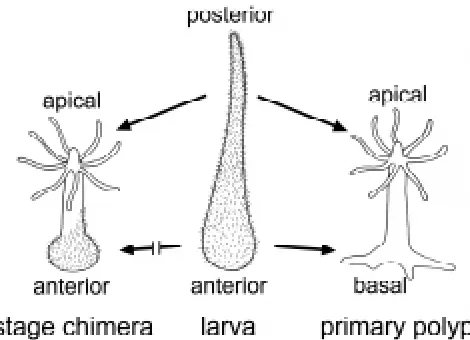
Most cnidarians have a life cycle involving developmental stages of distinct complex organisation. This is the case in species which have motile planula larvae, a sessile polyp and pelagic medusae. We are looking at the process of metamorphosis in the marine hydrozoan H. echinata to understand the mechanisms controlling pattern formation during these stages. Within three days, the fertilised egg of H. echinata develops into a spindle-shaped planula larva with a blunt anterior end and a tapered posterior tail. Morpho-genetically, the planula represents a resting state, since cell prolif-eration and cell differentiation are almost at a standstill (Plickert et al., 1988; Kroiher et al., 1990). Metamorphosis transforms the larva into the completely different shaped primary polyp bearing a mouth with tentacles, a gastric region and stolons at the base. The anterior of the larva gives rise to the basal parts of the polyp while the posterior develops into the apical parts (Schwoerer-Böhning et al., 1990). Metamorphosis involves a marked increase in cell prolifera-tion and the differentiaprolifera-tion of polyp specific cell types (Plickert et al., 1988; Plickert, 1989; Kroiher et al., 1990).
The onset of metamorphosis requires an external stimulus. Bacteria are the natural inducer (Müller, 1969), although there does not appear to be a requirement for a particular species since even E. coli can cause metamorphosis (Kroiher and Berking, 1999). Metamorphosis can also be initiated with a number of structurally unrelated substances (for review see Berking, 1998). Irrespective of the inducer, the larvae always transform into essen-tially identically shaped (primary) polyps.
the control system. Pieces from transverse-sectioned larvae regenerate the missing larval structures whereas pieces of trans-verse-sectioned polyps regenerate adult structures.
Interestingly, individual H. echinata consisting of larval tissue and adult tissue in combination (i.e. stage chimeras) have been observed. The occurrence of stage chimeras of basal metamor-phosis is a frequent event. In these chimeras the anterior part of the larva transforms into the respective adult polyp structure, namely the basal plate and the stolons. The posterior tail, how-ever, remains as a tail. The spontaneous occurrence of the counterpart, stage chimeras of apical metamorphosis is an infre-quent event (Plickert, 1987). In these chimeras, only the posterior tail undergoes metamorphosis into the respective apical adult structure, the head of the polyp. The anterior larval part remains in the larval state. In this study we make use of the frequent appearance of stage chimeras of the apical type after induction of metamorphosis by increased temperature or by a combination of increased temperature and low Mg2+ sea-water to discover
com-mon versus different features of the pattern forming systems of larva and adult.
It has been shown that the decision as to whether larva or polyp morphology regenerates after a bisection of such stage chimeras depends not on the majority of the tissue in the original body section, but on stage specificity within the regenerating animal part. However, nerve cells of the RFamide type escape from this rule, since such nerve cells, which usually differentiate in polyp tissue, appear in regenerated larval tails of sectioned stage chimeras.
Results
Larvae of Hydractinia echinata exposed to increased tem-perature undergo partial metamorphosis
When larvae were exposed to 28°C for 3 hours, instead of the 10-18°C at which they normally live, they were found to undergo
1996; M. Kroiher, unpublished observation). The first question that was addressed regarding the chimeras was whether the larva-like and polyp-like structures in these animals are capable of the functions of the respective stage-specific structures. Larvae are unable to take up food. Stage chimeras however, catch pray with their tentacles and take it up via their mouth just as polyps do. Polyps cannot move, larvae are motile. Chimeras are propelled by their larval anterior, whose ectoderm is covered with cilia as in the anterior of normal larvae. Larvae of H. echinata are positively phototactic. Similarly, the chimeras crawl to the source of light with the anterior tapered end, their larval part facing forward. A larva can undergo metamorphosis into a primary polyp. Similarly, the anterior (larval) part of the chimera transforms into a polyp structure (basal plate and stolons) after treatment with an induc-ing agent: stage chimeras were incubated for 3 hours with 100 mM Cs+. From 50 chimeras, 39 developed a basal plate and stolons.
Thus, in stage chimeras, the larva-like and polyp-like structures function like the respective structures in normal larvae and pol-yps.
In stage chimeras, the larval-like and the polyp-like struc-tures are covered by ectodermal epithelial cells which dis-play larval and polyp features, respectively
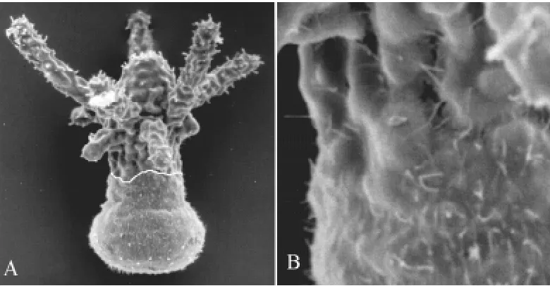
The Hydractinia larva is covered with cilia, while the polyp is not (Weiss et al., 1985). The cilia are lost immediately after induction of metamorphosis (Spindler and Müller, 1972; Weiss and Buss, 1987). In the chimeras, the ectodermal epithelial cells of the larva-like anterior retain their cilia, while those of the apical end with the polyp-like structures are free of cilia. The hypostome and the tentacles are thus restricted to regions not covered by cilia (Figs. 1, 2A). We conclude that the ectodermal epithelial cells in the anterior part remain in the larval stage and only the posterior cells transform into the apical polyp stage. Between the ciliated and the non-ciliated part of the animal there is not a gradual change in ciliation but rather a distinct border (Fig. 2B). The position of this border varies. The border is not strictly transversal or straight but rather irregular. Close to the border, ectodermal ciliated larval cells and unciliated polyp cells are intermingled. In a few cases, patches of cells with cilia were found within the apical polyp-like structure.
In stage chimeras, the distribution of a specific type of nerve cell correlates with the distribution of these cells in the larva and in the polyp
A group of neurosecretory nerve cells can be visualised by staining with an antibody against RFamide (Grimmelikhuijzen, 1985). In the larva, these cells are mostly restricted to the anterior part (Fig. 3A; Plickert, 1989; Kroiher and Plickert, 1992), while in the polyp they are restricted to the opposite end, the polyp’s head (Fig. 3B; Grimmelikhuijzen, 1985). In the stage chimeras, the RFamide-positive nerve cells are found in the anterior and in the apical part (Fig. 3C). This indicates that in stage chimeras, the RFamide-positive nerve cells are found in those structures in
which they occur in the larva and in the polyp. In several of those larvae which remained in the larval state after the heat treatment, RFamide-positive nerve cells were detected at the posterior end, too (in one experiment 6 out of 9 animals remaining in the larval state showed that unusual distribution).
Bisected stage chimeras provide insight into the control of axial regeneration versus the control of the stage specificity of the tissue that regenerates
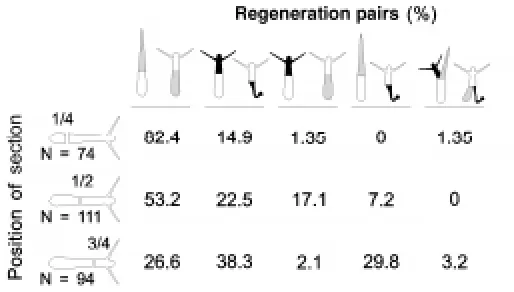
Both larvae and polyps of H. echinata are able to regenerate missing body parts following transverse sectioning. Stage chime-ras can also regenerate. For a further analysis of their regenera-tion capacity, chimeric larvae were bisected along the longitudinal axis. Because we could not locate the exact position of the border between larval and polyp tissue in the living stage chimeras, we examined the outcome after bisection at various positions (Fig. 4). In nearly all cases only one stage specific structure (either larval tissue or polyp tissue) was formed from a regenerating piece (Fig. 5A). In rare cases two different stage specific struc-tures (larval tissue and polyp tissue) were formed at the same wound surface from a single regenerating piece (Fig. 5B). How-ever, in all cases analysed the pieces regenerated with respect to the missing axial tissue structures. The anterior pieces regener-ated either a posterior larval end or an apical polyp end and the apical pieces regenerated either an anterior larval end or a basal polyp end. When two structures were formed from the same wound surface, one structure consisted of larval tissue and the other consisted of adult tissue. The anterior larval part regener-ated a posterior larval and an apical polyp end while the apical pieces regenerated an anterior larval and a basal polyp end. A regenerate with two structures from the same tissue type (i.e. larval or adult) has never been observed. Thus, from pieces of bisected stage chimeras we obtained either a normal shaped larva, a normal shaped polyp, a stage chimera or a stage chimera with an additional anterior or apical end.
In most cases, both pieces of a bisected larva regenerated structures of the same stage specific tissue type (Fig. 4). Both regenerates consisted of either larval tissue or polyp tissue. Most of the pairs regenerated larval tissue if the bisection was made in
the anterior region. An increased number of pairs regenerated polyp tissue if the bisection point was shifted to a more posterior (apical in the adult polyp) region (Fig. 4). In about 21% of all sectioned larvae, the regenerated stage-specific tissue type was different in the two regenerating pieces. One piece regenerated larval tissue while the other regenerated polyp tissue. This type of regeneration was dependent on the position of bisection. Bisec-tion at a more posterior point increased the frequency of cases in which the two pieces regenerated with different tissue types (Fig. 4). A sectioning at position 1/2XX caused the larval anterior piece to regenerate an apical polyp part while the apical polyp piece regenerated a larval anterior part. Hence the bisection of a single stage chimera generated two stage chimeras by regeneration. A sectioning at the most posterior position (3/4XX) caused a high number of pairs to regenerate tissue quality in accordance to the majority of tissue in the regenerating piece. In these cases, the larval anterior piece regenerated a larval posterior and the apical polyp piece regenerated a basal polyp part. Hence we obtained from a stage chimera a planula larva and a polyp (Fig. 4). In the rare cases where two different stage specific tissue types were regenerated at the same wound surface, both pieces of a pair regenerated in this way (see also Fig. 4).
To test if the cells at the wound surface decide which type of tissue the regenerate will have, stage chimeras were bisected. Immediately thereafter, the ectodermal epithelial cells at the wound were analysed for the presence of cilia as an indicator of larval tissue and the fate of the regenerate was subsequently monitored (Fig. 6). In cases where ciliated larval cells were present at the cut surface, the majority of the regenerates con-sisted of larval tissue. If no ciliated cells could be detected at the cut surface, most of the pieces regenerated polyp-specific tissue.
In bisected stage chimeras which regenerate a larval poste-rior, RFamide-positive nerve cells occur at unusually high density
In larvae, RFamide-positive nerve cells are concentrated in the anterior half and are rare in the posterior part of the animal. In the polyp, this distribution is reversed (see above). After bisection of a larva, the anterior part regenerates the missing posterior end. In
this new posterior end, RFamide-positive nerve cells are rarely found. In contrast, in all bisected stage chimeras which regener-ated a larval posterior, RFamide-positive nerve cells were present at high density in the regenerated larval tail (Table 1; see also Fig. 5A).
Discussion
Pattern of stage chimera
In H. echinata, several treatments were found to result in the formation of partially metamorphosed animals or stage chimeras (for a review see Berking, 1998). Apical metamorphosed animals, which have been observed only rarely, appeared following heat treatment (shown here) or at a much higher frequency following treatment with a combination of low Mg2+ seawater and elevated
temperature (Walther et al., 1996). In such stage chimeras (1) the larval part and the polyp part, respectively, behave as they do in complete animals. (2) Within the larval and polyp parts, at least the ectodermal epithelial cells display the stage specific morphol-ogy. (3) RFamide-positive nerve cells are located in those regions where they are found in the respective developmental stages of complete animals. Thus, it appears that in the stage chimeras the larval and the polyp tissues are very similar if not identical to the respective tissues in the normal larva and polyp.
Transition from larva to adult
The existence of partially metamorphosed animals indicate that it is possible for the transition to the polyp stage to occur in only some parts of the animal. Thus those parts which have acquired a polyp-like appearance are unable to trigger the other parts to do the same. On the other hand, the larva-like parts are unable to hinder the transition of neighbouring parts into the polyp stage. With respect to the ectodermal epithelial cells, the distribution pattern of cilia as an indicator for larval or polyp tissue shows a distinct border rather than a gradual change between these parts. The position of this border varies in different animals and is not strictly transversal or straight, but rather irregular. Close to the border, patches of larval cells or even single larval cells are intermingled in polyp
Regeneration of stage chimeras
The occurrence of apparently normal larvae or polyps following regeneration may indicate that the majority of the tissue deter-mines the fate of the regenerate: anterior larval parts regenerate posterior larval tails and apical polyp parts regenerate basal polyp parts. However, the larval anterior can regenerate an apical polyp part and the apical polyp part can regenerate a larval anterior. Thus, it is not the majority of the tissue within the isolated piece which determines the fate of the regenerate but rather the quality of the tissue at the wound. Close examination of both pieces of a bisected larva indicates the same: often both regenerates were of the same tissue type. Thus, in one of the two pieces the majority tissue type did not decide the fate of the regenerate.
We found pairs of pieces in which the anterior larval part regenerated an apical polyp structure and the apical polyp part regenerated an anterior larval structure. We suspect that initially both larval and polyp tissue exist at the wound site. The process of regeneration would initiate at a random position on the circum-ference of the wound. The tissue quality (larval or polyp) at the initiation site is proposed to determine the type of tissue in the regenerated structure, while other parts of the circumference will be prevented from forming this tissue type by lateral inhibition. Only in rare cases were larval and polyp structures observed to form simultaneously. In these cases, the process of regeneration may have started simultaneously at two distant positions on the circumference in polyp and in larva-like tissue. In all cases an anterior part regenerates a posterior/apical part and an apical part regenerates a basal /anterior part.
Control of pattern formation during regeneration
The existence of partial metamorphosis allows an insight into the control of pattern formation in H. echinata. It appears that the
Fig. 3. Localization of RFamide-positive nerve cells in (A) a larva, (B) a primary polyp and (C) a stage chimera. Note that in the stage chimera, the RFamide-positive nerve cells are located in the anterior part and in the apical part.
pattern forming system in Hydractinia is hierarchically organised. Signalling molecules control the positional value along the body axis, but these signalling molecules are not structure-specific. The positional value is believed to control local development by generation of secondary signalling molecules, which may be structure-specific (Berking, 1998). We present data showing that the mechanisms controlling regeneration follow this hierarchically organisation as well. In the first step of regeneration, the missing positional values are re-established, with the tissue identity being irrelevant for that decision. This first step may be controlled by signalling factors which are identical in larvae and polyps. What type of tissue is formed depends on both the local positional value and the developmental stage of the cells which happen to be at that position. If the majority of cells (larval or polyp) of the tissue determines the fate of the regenerate (axial fate and stage
Fig. 5. Localisation of RFamide-positive nerve cells after regeneration of bisected stage chimeras.(A) Regenerated pieces from a bisected stage chimera. The regenerated tissue is indicated by an arrow. Both sections regenerated larval tissue. The anterior larval section regenerated a larval posterior to become a “larva”. Note that the regenerated tail contains RFamide-positive nerve cells. The apical polyp section regenerated a larval anterior and hence a new stage chimera developed. (B) Section of a stage chimera, which regenerated an obvious larval posterior (p) and a polyp head (h) at the same sectioned surface. Note that in the larval posterior, RFamide-positive nerve cells are present and that ectopic tentacles (t) have developed. The white line indicates the position of bisection.
Fig. 6. Relationship between the presence of cilia at the wound surface and frequency of occurrence of regeneration pairs after bisection of stage chimeras at position 3/4 along the anterior-apical body axis. Bisection was carried out 3 days after induction of metamor-phosis and the regeneration pairs were observed for 5 days. Grey, regeneration of larval tissue; black, regeneration of polyp tissue.
TABLE 1
RFAMIDE-POSITIVE NERVE CELLS IN REGENERATED TAILS OF LARVAE AND STAGE CHIMERAS
Larva Stage chimera
n animals 71 32 50
Position of section at the body axis 1/3 1/3 2/3
n (%) regenerated posterior 14 (19.7%) 32 (100%) 49 (98%) larval tails with RFamide cells
mean number of RFamide cells 0.6 ±2.2 6 ±3.7 7.5 ±5 in regenerated larval tails
specificity), this would hint at a control of pattern formation in H. echinata by structure-specific signalling molecules (e.g. hypostome, tentacle, basal disc, larval anterior specific signalling molecules).
Nerve cells
obtain larvae were as described (Müller, 1969).
Induction of metamorphosis
Metamorphosis was induced with a treatment at 28°C for 3 hours (Kroiher et al., 1992) or a combined treatment with artificial seawater containing 2.5 mM Mg2+ (5% of the content of normal seawater) and 28°C
for 3 hours (Walther et al., 1996). For induction, 30-60 larvae (7 days of age) were incubated in a microtube in 600 µl medium at the appropriate temperature. Following treatment, the medium was changed 3 times and the larvae were transferred into Petri dishes containing 4 ml sea-water at 18°C.
Sectioning of animals
Larvae or stage chimeras were sectioned transversely with the edge of a small hypodermic needle. After sectioning, the seawater was changed 3 times and the specimens were transferred as pairs into 24 well plates. The sea-water was changed every day thereafter.
Immunohistochemistry
Nerve cells that contain neuropeptides of the Arg-Phe-amide type (RFamide) were detected immunocytochemically in whole mount prepara-tions with an antiserum against Arg-Phe-amide (RFamide; Grimmelikhuijzen, 1985). Fluorescein-isothiocyanate (FITC) conjugated anti-rabbit IgG (Sigma, München, FRG) was used as the secondary antibody.
Scanning electron microscopy
Animals were anaesthetised with 0.2 M MgCl2 in sea water for 20 minutes and fixed in 2% OsO4 in 0.1 M sodium cacodylate in distilled water at pH 7 for 1 hour. Thereafter the specimens were washed with 0.1 M Na2HPO4, pH 7 for 30 minutes and with 0.4 M glycine, pH 7.2 for 2 hours at room temperature. The specimens were then dehydrated in ethanol, critical point dried and sputter-coated with gold-palladium. The specimens were examined with a Hitachi S-520 scanning electron microscope.
Acknowledgements
I thank Dr. S. Berking for stimulating discussions and Alysia Runde, Drs. G. Plickert and R. Steele for critical reading of the manuscript.
References
BERKING, S. (1980). Commitment of stem cells to nerve cells and migration of nerve cells precursors in preparatory bud development in Hydra. J. Embryol. Exp. Morphol. 60: 373-387.
BERKING, S. (1998). Hydrozoa metamorphosis and pattern formation. Current Topics in Dev. Biol. 38: 81-131.
BODE, H.R. (1996). The interstitial cell lineage of hydra: a stem cell system that arose early in evolution. J. Cell Sci. 109:1155-64.
Helgol. Mar. Res. 53: 118-121.
KROIHER, M. and PLICKERT, G. (1992). Analysis of pattern formation during embryonic development of Hydractinia echinata. Roux’s Arch. Dev. Biol. 201: 95-104.
KROIHER, M., PLICKERT, G. and MÜLLER, W.A. (1990) Pattern of cell proliferation in embryogenesis and planula development of Hydractinia echinata predicts the postmetamorphic body pattern. Roux’s Arch. Dev. Biol. 199: 156-163.
KROIHER, M., WALTHER, M. and BERKING, S. (1992). Heat shock as inducer of metamorphosis in marine invertebrates. Roux’s Arch. Dev. Biol. 201: 169-172.
LEITZ, T. and WIRTH, A. (1991). Vanadate, known to interfere with signal transduc-tion, induces metamorphosis in Hydractinia (Coelenterata; Hydrozoa) and causes profound alterations of the larval and postmetamorphic body pattern. Differentia-tion 47: 119-127.
LEITZ, T., MORAND, K. and MANN, M. (1994). Metamorphosin A: a novel peptide controlling development of the lower metazoan Hydractinia echinata (Coelenterata, Hydrozoa). Dev. Biol. 163: 440-446.
MÜLLER, W. A. (1969) Auslösung der Metamorphose durch Bakterien bei den Larven von Hydractinia echinata. Zool. Jb. Anat. Ontog. 86: 84-95.
PLICKERT, G. (1987). Low-molecular-weight factors from colonial hydroids affect pattern formation. Roux’s Arch. Dev. Biol. 196: 248-256.
PLICKERT, G. (1989). Proportion-altering factor (PAF) stimulates nerve cell forma-tion in Hydractinia echinata. Cell Diff. Develop. 26: 19-28.
PLICKERT, G., KROIHER, M. and MUNCK, A. (1988). Cell proliferation and early differentiation during embryonic development and metamorphosis of Hydractinia echinata. Development 103: 795-803.
SCHWOERER-BÖHNING, B., KROIHER, M. and MÜLLER, W.A. (1990). Signal transmission and covert prepattern in the metamorphosis of Hydractinia echinata. Roux’s Arch. Dev. Biol. 198: 245-251.
SPINDLER, K.D. and MÜLLER, W.A. (1972). Induction of metamorphosis by bacteria and by a lithium-pulse in the larvae of Hydractinia echinata. Roux’s Arch. Dev. Biol. 169: 71-280.
TERAGAWA, C.K. and BODE, H.R. (1995). Migrating interstitial cells differentiate into neurons in hydra. Dev. Biol. 171: 286-293.
VAN DE VYVER, G. (1964). Etude histologique du dèveloppement d´Hydractinia echinata (Flem.). Cah. de Biol. Mar. 5: 295-310.
WALTHER, M., ULRICH, R., KROIHER, M. and BERKING, S. (1996). Metamorphosis and pattern formation in Hydractinia echinata, a colonial hydroid. Int. J. Dev. Biol. 40: 313-322.
WEIS, V.M. and BUSS L.W. (1987). Ultrastructure of metamorphosis in Hydractinia echinata. Postilla 199: 1-20.
WEIS, V.M., KEENE, D.R. and BUSS, L.W. (1985). Biology of the hydractiniid hydroids. 4: Ultrastructure of the planula of Hydractinia echinata. Biol. Bull. 168: 403-418.