Discovery of the congenital nephrotic syndrome gene discloses
the structure of the mysterious molecular sieve of the kidney
KARL TRYGGVASON
1*, VESA RUOTSALAINEN
2and JORMA WARTIOVAARA
31Division of Matrix Biology, Department of Medical Biochemistry and Biophysics, Karolinska Institute, Stockholm, Sweden, 2Biocenter and
Department of Biochemistry, University of Oulu, Oulu, Finland and 3Institute of Biotechnology, University of Helsinki, Helsinki, Finland
ABSTRACT The molecular nature of the glomerular slit diaphragm, the site of renal ultrafiltration, has until recently remained a mystery. However, the identification of the gene affected in congenital nephrotic syndrome has revealed the presence of a novel protein, possibly specific for the slit diaphragm. This protein, which has been termed nephrin, is a transmembrane protein that probably forms the main building block of an isoporous zipper-like slit diaphragm filter structure. Defects in nephrin lead to abnormal or absent slit diaphragm leading to massive proteinuria and renal failure. The discovery of nephrin sheds new light on the glomerular filtration barrier, provides new insight into the pathomechanisms of proteinuria, and even opens up possibilities for the development of novel therapies for this common and severe kidney complication.
KEY WORDS:
congenital nephrosis, nephrin, glomerular basement membrane, slit membrane
0214-6282/99/$15.00 © UBC Press
Printed in Spain www.ehu.es/ijdb
*Address for reprints: Division of Matrix Biology, Department of Medical Biochemistry and Biophysics, Karolinska Institute, S-171 77 Stockholm, Sweden. FAX: +46-8-31 61 65. e-mail: karl.tryggvason@mbb.ki.se
Glomerular filtration barrier
Ultrafiltration of blood during formation of the primary urine in the glomerulus is one of the central functions of the human kidney (Tisher and Madsen, 1996). Structurally, the glomerulus is a tuft of anastomosing capillary loops surrounded by the Bowman´s cap-sule leading the primary urine to the tubular system. The glomeru-lar filtration barrier is formed by three layers: the innermost fenes-trated vascular endothelium, the glomerular basement membrane (GBM), and the podocyte layer. The podocytes form a tight web on top of the GBM with their interdigitating foot processes joined by a slit diaphragm.
It is generally acknowledged that the molecules passing through the glomerular filtration barrier are selected according to their size, charge and shape (Bohrer et al., 1978; Brenner et al., 1978; Batsford et al., 1987; Kanwar et al., 1991; Ghitescu et al., 1992; Fujigaki et al., 1993; Remuzzi and Remuzzi, 1994). The exact locations of the various selective functions in the barrier are, however, more controversial. The charge-selective filter has been thought to be located in the GBM, a cross-linked meshwork of type IV collagen, laminin, nidogen and proteoglycans (Hudson et al., 1993; Yurchenco and O´Rear, 1993). The anionic charge of heparan sulfate side chains of proteoglycans is believed to hinder the traversal of anionic plasma proteins (Caulfield and Farquhar, 1978; Kanwar and Farquhar, 1979; Kanwar et al., 1991). The location of the size-selective property of the filtration barrier has been attributed to the GBM alone or, alternatively to the slit
Abbreviations used in this paper: GBM, glomerular basement membrane;
NPHS1, nephrotic syndrome 1; CNF, congenital nephrotic syndrome of the Finnish type (=NPHS1); APLP1, amyloid precursor-like protein 1; CAM, cell adhesion molecule.
diaphragm (Latta, 1970; Karnovsky and Ainsworth, 1972; Kanwar et al., 1991).
massive proteinuria already in utero. Electron microscopic exami-nation of NPHS1 patient kidneys reveals thinner lamina densa of the GBM than in controls, but no structural abnormality of the GBM has been detected (Autio-Harmainen, 1981; Autio-Harmainen and Rapola, 1983). In kidneys of patients with NPHS1, the podocyte foot processes are absent, and no slit diaphragms have been described in the podocyte cell-cell adhesions. This finding is typical for nephroses of any cause.
The gene mutated in NPHS1 codes for a putative transmem-brane protein termed nephrin that belongs to the immunoglobulin (Ig) superfamily. It has an extracellular portion containing eight Ig-motifs and one type III fibronectin domain (Kestilä et al., 1998). This, together with the sequence of the intracellular domain which contains eight tyrosines, suggested that nephrin is a signaling adhesion molecule. Using in situ hybridization, nephrin was shown to be exclusively expressed in glomerular podocytes (Kestilä et al., 1998).
Recently, we localized nephrin in immunofluorescence micros-copy to the GBM region of newborn human glomeruli using antibodies generated against recombinant antigen (Ruotsalainen et al., 1999). Moreover, we demonstrated by immunoelectron microscopy that nephrin is located in the podocyte slit region. We propose that nephrin is most likely a component of the slit
dia-phragm. The fact that the protein is mutated in NPHS1, further indicates an essential role for the slit diaphragm in the maintenance and size selectivity of the glomerular filtration barrier.
Congenital nephrotic syndrome of the Finnish type
Congenital nephrotic syndrome of the Finnish type, is a distinct entity among nephrotic syndromes. It is the first disease to be described to belong to the so-called Finnish disease heritage (Hallman et al., 1956). This Finnish variant of congenital nephrotic syndrome has traditionally been referred to in the literature as CNF, but has now been designated as NPHS1. It is an autosomal recessive disorder with an incidence of 1:10,000 births in Finland, but considerably less frequent in other countries (Norio, 1966; Huttunen, 1976). The disease manifests itself already at the fetal stage with heavy proteinuria in utero, demonstrating early lesions of the glomerular filtration barrier. The pathogenesis of NPHS1 has remained obscure. There are no pathognomonic pathologic fea-tures, the most typical histological finding of NPHS1 kidneys being dilation of the proximal tubuli (Huttunen et al., 1980). The kidneys are also large and have been found to contain a higher amount of nephrons than age-matched controls (Tryggvason and Kouvalainen, 1975). Electron microscopy reveals no abnormal features of the GBM itself, although there is a loss of foot processes of the glomerular epithelial cells, a finding characteristic for nephrotic syndromes of any cause. Chemical analyses carried out on the composition of the GBM of NHPS1 patients in the 1970s did not reveal any typical changes and later studies on GBM proteins, such as type IV collagen, laminin, and heparan sulfate proteoglycan, have not revealed abnormal findings in NPHS1 (Tryggvason, 1977, Ljungberg et al., 1993, Kestilä et al., 1994b). NPHS1 is a progressive disease, usually leading to death during the first two years of life, the only life-saving treatment being kidney transplan-tation (Holmberg et al., 1995).
An important feature with the disease is that most transplanted patients have not developed extrarenal complications. This sug-gested that the mutated gene product is highly specific for kidney development and/or glomerular filtration function. However, about 20% of the patients have developed post-transplantation nephro-sis, the cause of which is unknown (Laine et al., 1993; Holmberg et al., 1995). Because of the apparent kidney specificity of NPHS1 and, because the disease practically only seems to affect the filtration barrier, we considered it as a model for solving the nature of the actual kidney glomerular filter.
The hunt for the CNF gene
Since numerous studies on CNF had given no clues concerning the molecular defects in the diseases, we decided to use a molecular genetic approach. In 1989 we initiated the actual gene hunt. Due to the relatively high frequency of the disease in Finland and due to the homogeneity of the Finnish population, we decided to only use Finnish families for the gene search. For that purpose, we collected samples from all CNF families we could identify in Finland. DNA was isolated from peripheral blood, but we also established cell lines from all CNF patients to ensure that we had sufficient amounts of DNA at any time. Samples from a total of 29 CNF families were collected. Of those, only 17 were large enough to be suitable for genetic linkage analyses. This work was initially
in the hands of a graduate student, Marjo Kestilä, who was soon joined by another gradu-ated student, Minna Männikkö. Having col-lected the patient material they could now start the gene hunt.
Basically two possibilities were ahead of us for identifying the unknown CNF gene. The candidate gene approach or positional clon-ing. In the candidate gene approach one tries to make educational guesses about possible genes, and the obvious genes to examine in this regard, were genes coding for basement membrane proteins, as the disease seemed to primarily affect the filtration barrier. For this purpose, we first used genetic markers for chromosomal regions where all known BM genes were known to be located. However, we could soon demonstrate absence of linkage to all then known BM genes, such as those for the collagen IV alpha 1-4 chains, laminin alpha 1, 3, etc.
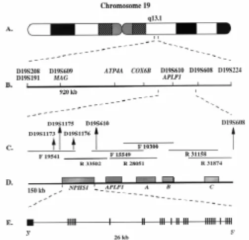
Since the candidate gene approach did not give us a lead, we initiated positional cloning, which basically means to carry out a genome wide screening with markers for all autosomal chromosomes and examine if linkage could be found between any chromosomal region and CNF. At that time, a panel of about 400 mark-ers had been established and after using about 60 of them, Kestilä and Männikkö could dem-onstrate significant lod scores with markers in the long arm of chromosome 19 (19q13.1) (Kestilä et al., 1994a; Männikkö et al., 1995). Having localized the gene to 19q13.1 in 1994 (Fig. 1), we searched data bases to find out if any groups were particularly involved in characterization of chromosome 19, and found out that a genomics group at the Lawrence Livermore Laboratory in California was map-ping the entire chromosome for eventual se-quencing. Surprisingly, a senior investigator in the group, Anne Olsen, had worked with Karl Tryggvason ten years earlier in the same labo-ratory at Rutgers Medical School, New Jersey, USA. It was therefore not difficult to establish collaboration with the Livermore group which provided us with overlapping cosmid clones from the critical region between markers D19S208 and D19S224 (Olsen et al., 1996).
D19S1176 and D19S610 (Fig. 1). The work now became very exciting and Southern hybridization analyses of CNF patient DNA with genomic clones did not reveal variations, suggesting that the mutations causing NPHS1 do not represent major genomic rear-rangements. Having the sequence of the 150 kilo base critical region, we searched for potential candidate genes using exon prediction programs and data base similarity searches. Based on those analyses, the critical region was estimated to include over 100 potential exons. Similarity searches revealed one previously known gene, i.e. APLP1 encoding an amyloid precursor-like
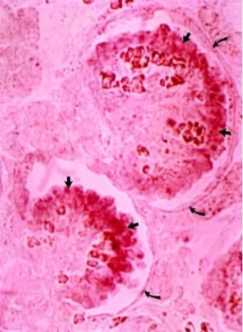
pro-Fig. 2. Expression of nephrin mRNA in human embryonic kidney by in situ hybridization. Expression was only observed in the glomerular regions. A high magnification strong expression was observed in the periphery of individual glomeruli (straight arrows), probably mainly in epithelial cells. No expression is observed in the Bowman’s capsule (bent arrow), proximal tubuli or endothelial cells of vessel walls.
tein (Lenkkeri et al., 1998) and eight distinct expressed sequence tags (ESTs). Together, the analyses indicated the presence of at least ten novel genes in the critical region.
Using GRAIL and GENSCAN exon prediction programs and sequences from cDNAs, the exon/intron structures of five of the genes, NPHS1 (Fig. 1), APLP1 (Lenkkeri et al., 1998), A, B and C (not shown) were determined. Although steady state transcript levels varied, northern analyses revealed expression of all the genes in kidney, and with the exception of NPHS1, also in other tissues (not shown). Therefore, none of them could be excluded as the NPHS1 gene, and all were subjected to mutation analysis.
The 17 exon APLP1 gene located distal to D19S610 did not show variations between patients and controls, and was excluded as the NPHS1 gene (Lenkkeri et al., 1998). Also, the novel genes A, B and C containing 9, 5 and 3 exons, respectively, did not have sequence variants segregating with NPHS1, and could similarly be excluded as genes causing NPHS1 (data not shown). A fourth novel gene located proximal to D19S610 encoding a transcript of about 4.3 kb was shown to be strongly expressed in human embryonic and adult kidneys, while no clear signals were observed above background in other tissues (Fig. 2). Therefore, this gene was a strong candidate for CNF. Full-length cDNA for the transcript was constructed using fetal kidney poly(A) mRNA and PCR prim-ers made based on the predicted exon structure. The gene was found to have a size of 26 kb and to contain 29 exons (Fig. 1).
Exon sequencing analyses revealed the presence of two major mutations in over 90% of NPHS1 chromosomes. The first muta-tion, a 2bp deletion in exon 2 causes a frame shift resulting in the generation of a stop codon within the same exon. The second sequence variant found in the NPHS1 gene was a nonsense mutation CGA➝TGA in exon 26 (R1109X, Finminor). A total of 108 parents and 54 healthy siblings were analyzed, none of them being homozygous or compound heterozygous for the two muta-tions identified here. One out of 83 control individuals was heterozygous for the Finmajor mutation.
The nine-year hunt for the CNF gene was now completed, as we had now definitely demonstrated that the NPHS1 gene was the disease gene. This gene has now been confirmed to be mutated in a large number of other patients worldwide with congenital neph-rotic syndrome (Lenkkeri et al., 1999), as we have now found a total of over 40 mutations in the gene.
Nephrin
Due to the high association of expression and pathology with glomeruli, the proximal part of the nephron, we have named the
NPHS1 gene product nephrin (Fig. 3). The role of nephrin is still hypothetical, but it is a transmembrane protein with a domain structure resembling that of a large group of cell adhesion recep-tors belonging to the immunoglobulin superfamily (Brümmendorf and Rathjen, 1994). It has eight extracellular Ig-like modules and one fibronectin type III-like module.
The Ig-like modules of nephrin are all of type C2, which is particularly found in proteins participating in cell-cell or cell-matrix interactions. The cytosolic domain has no significant homology with other known proteins. However, it contains nine tyrosines some of which could become phosphorylated during ligand binding of nephrin. The crucial role for the intracellular domain of nephrin is emphasized by the fact that the Finminor mutation, which results in the loss of 132 out of 155 residues of this domain, results in full blown NPHS1.
Nephrin is a component of the slit membrane
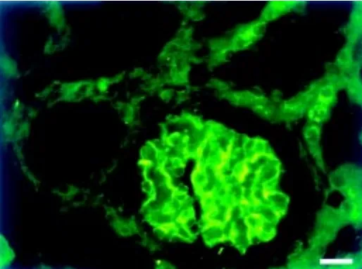
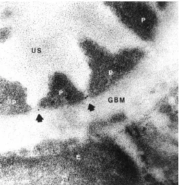
The remarkable interdigitating pattern of adjacent podocyte foot processes in the kidney glomerulus has implied an essential role of the podocytes for the integrity of the glomerular filtration barrier (Daniels, 1993; Kriz et al., 1994; Mundel and Kriz, 1995). Much interest in the study of this barrier has been focused on the cell junction between the foot processes, the so called slit diaphragm. However, the actual molecular structure of the diaphragm has remained unraveled. Vesa Ruotsalainen generated antibodies against nephrin using remobinant extracellular domain of the protein as antigen. By using these antibodies we could show strong immunostaining of the GBM region in light microscopy (Fig. 4). More importantly, Päivi Ljungberg and Jorma Wartiovaara could localize nephrin to the slit diaphragm region by immunoelectron microscopy (Fig. 5), providing the first evidence for a specific protein in this size-selective molecular filter of the kidney (Ruotsalainen et al., 1999). The fact that mutations in the nephrin
Fig. 3. Schematic domain structure of nephrin. The red box represents the transmembrane domain. The Ig repeats are shown by incomplete circles connected by disulfide bridges (C-C). The locations of free cysteine residues are indicated by a -C. N= extracellular amino terminal, C= intracellular carboxyl terminal.
gene lead to massive proteinuria (Kestilä et al., 1998; Lenkkeri et al., 1999), furthermore indicates that nephrin has a direct role or actual filtration function in this size-selective barrier.
Concerning nephrin localization, it should be stressed that, in its extracellular labeling, nephrin was found only between the foot processes. It was completely absent from the interspaces between the foot processes and the GBM, a site with very much larger potential labeling surface than present in the narrow slit. The occasional cases of gold label observed in the cytoplasm of foot processes may represent newly synthesized nephrin molecules.
The results of the immunolocalization studies raise the funda-mental question as to how a protein like nephrin, either alone or together with other slit membrane protein(s), can contribute to the molecular structure of a porous filter. The ultrastructure of the podocyte slit diaphragm has been studied extensively by electron microscopy (Mundel and Kriz, 1995). Rodewald and Karnovsky (1974) were the first to suggest a zipper-like organization of this structure. As these studies used the conventional thin-sectioning method requiring harsh chemical treatments in sample prepara-tion, it has later been suggested that the zipper-looking structure might be due to an artifact. Since then, the podocyte-podocyte junction has also been studied by several other electron micro-scopic methods (Furukawa et al., 1991; Ohno et al., 1992). These studies have indicated that the width of this junction varies between 20 and 50 nm. The actual slit diaphragm is considered to be a rather rigid structure. However, it has recently been suggested that at least the slit area might increase with increasing perfusion pres-sures of the glomerulus (Kriz et al., 1996; Yu et al., 1997). Since the actual molecular structure of the slit diaphragm is, as yet, unre-solved, it is precocious to speculate on a molecular assembly allowing change in the width of the slit. Our results, however, provide evidence that nephrin is a component of such an assembly. This calls for further studies on the molecular structure of the slit diaphragm.
Several lines of evidence indicate that nephrin may assemble into a zipper-like isoporous filter structure similar to that presented by Rodewald and Karnovsky (1974). First, the present study demonstrated that nephrin is specifically located at the slit dia-phragm. Second, nephrin must be crucial for the structural integrity of the slit diaphragm, as absence of the protein or different amino acid substitutions cause congenital nephrosis and lack of the slit diaphragm with massive proteinuria as a result (Lenkkeri et al., 1999). Third, nephrin molecules extending towards each other from two adjacent foot processes are likely to interact with each other in the slit through homophilic interactions, as has been shown for other Ig cell adhesion molecules, such as N-CAM (Kiselyov et al., 1997), C-CAM (Öbrink, 1997) and L1 (Sonderegger and Rathjen, 1992). Fourth, such homophilic assembly of nephrin molecules in the slit could have a zipper-like arrangement, essen-tially as that proposed based on electron microscopic studies.
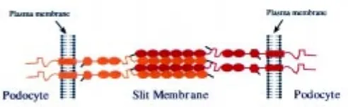
A hypothetical head-to-head assembly of nephrin through homophilic interactions is illustrated in Figure 6. The amino termi-nal extracellular domain of nephrin contains six consecutive Ig repeats, followed by a spacer domain, two additional Ig repeats, and one fibronectin type III -like domain (Fig. 3). Each Ig motif contains two cysteine residues that, similarly to corresponding motifs in other proteins (Chothia and Jones, 1997), can be as-sumed to form a disulfide bridge within the repeat structure (Fig. 3). Ig motifs have been shown to adopt a globular or ellipsoid structure
with an average axis length between 24 and 47 Å, averaging 35 Å (Holden et al., 1992). If the Ig repeats were to form a chain-like structure, as has been proposed for Ig cell adhesion molecules, all eight motifs would contribute to a length of about 28 nm. The region between Ig repeats 6 and 7 and fibronectin type III -like domain would add more length to the protein. Consequently, a single nephrin molecule can extend through most of the width of the 35-45 nm wide slit diaphragm.
In addition to the two cysteine residues in each Ig motif, nephrin contains three free cysteines, one in Ig motif 1, one in the spacer region between Ig motifs 6 and 7, and one in the fibronectin domain close to the plasma membrane. The three free cysteines are likely to have a function in forming intermolecular disulfide bridges that provide strength to the slit diaphragm. These cysteines are impor-tant as their absence results in proteinuria and congenital nephrotic syndrome (Lenkkeri et al., 1999). In the hypothetical model pre-sented here, the free cysteine of Ig motif 1 in one molecule interacts with the cysteine residue of the spacer in another nephrin mol-ecule. Such disulfide bonds could “lock” the homophilic unit of six Ig repeats of one nephrin molecule to similar units of two adjacent nephrin molecules. A centrally located aggregate of numerous nephrin molecules along the slit between two foot processes could constitute the central filament visualized by Rodewald and Karnovsky (1974). The width of the central aggregate would be 21 nm (6x35 Å) by simply assuming a linear chain arrangement of six
Ig repeats, 35 Å each, and this would not agree with the 11 nm width of the central filament reported by Rodewald and Karnovsky (1974). However, this difference could be attributed to the shrink-age of samples, extraction of components, or failure to stain. Also, the mode of packing of Ig modules may well be different from that presented in the model, so that it is still possible that nephrin forms the basis of the slit diaphragm through homophilic interactions.
In conclusion, the recent identification of nephrin and its present specific localization to the podocyte slit diaphragm may accelerate the elucidation of the molecular structure of the size-selective glomerular filtration barrier. The model for nephrin assembly into a slit diaphragm proposed in this study supports the model for slit diaphragm ultrastructure presented over two de-cades ago based on transmission electron microscopy. However, further studies are needed to validate this model and examine other proteins contributing to the slit diaphragm structure. Also, other functions of nephrin, such as its potential signaling role, need to be investigated. The elucidation of the molecular struc-ture of the filtration barrier can have significant clinical value. It not only explains the absence of slit diaphragms in NPHS1, but may also help to understand the pathogenic mechanisms of pro-teinuria in several other genetic and acquired kidney diseases that lead to proteinuria and renal failure. Considering the limited knowledge on the structure of the slit diaphragm, our recent results represent a noteworthy advance that may help to unravel the nature of this important extracellular structure.
References
AUTIO-HARMAINEN, H. (1981). Renal pathology of fetuses with congenital nephrotic syndrome of the Finnish type. 2. A qualitative and quantitative electron micro-scopic study. Acta Pathol. Microbiol. Scan. - Section A, Pathol. 89: 215-222.
AUTIO-HARMAINEN, H. and RAPOLA, J. (1983). The thickness of the glomerular basement membrane in congenital nephrotic syndrome of the Finnish type. Nephron 34: 48-50.
BATSFORD, S.R., ROHRBACH, R. and VOGT, A. (1987). Size restriction in the glomerular capillary wall: importance of lamina densa. Kidney Int. 31: 710-717.
BOHRER, M.P., BAYLIS, C., HUMES, H.D., GLASSOCK, R.J., ROBERTSON, C.R. and BRENNER, B.M. (1978). Permselectivity of the glomerular capillary wall.
Facilitated filtration of circulating polycations. J. Clin. Invest. 61: 72-78.
BRENNER, B.M., HOSTETTER, T.H. and HUMES, H.D. (1978). Molecular basis of proteinuria of glomerular origin. New Eng. J. Med. 298: 826-833.
BRÜMMENDORF, T. and RATHJEN, F.G. (1994). Cell adhesion molecules. 1: immunoglobulin superfamily. Protein Profile 1: 951-1058.
CAULFIELD, J.P. and FARQUHAR, M.G. (1978). Loss of anionic sites from the glomerular basement membrane in aminonucleoside nephrosis. Lab. Invest. 39: 505-512.
CHOTHIA, C. and JONES, E.Y. (1997). The molecular structure of cell adhesion molecules. Annu. Rev. Biochem. 66: 823-862.
DANIELS, B.S. (1993). The role of the glomerular epithelial cell in the maintenance of the glomerular filtration barrier. Am. J. Nephrol. 13: 318-323.
FUJIGAKI, Y., NAGASE, M., KOBAYASI, S., HIDAKA, S., SHIMOMURA, M. and HISHIDA, A. (1993). Intra-GBM site of the functional filtration barrier for endog-enous proteins in rats. Kidney Int. 43: 567-574.
FURUKAWA, T., OHNO, S., OGUCHI, H., HORA, K., TOKUNAGA, S. and FURUTA, S. (1991). Morphometric study of glomerular slit diaphragms fixed by rapid-freezing and freeze-substitution. Kidney Int. 40: 621-624.
GHITESCU, L., DESJARDINS, M. and BENDAYAN, M. (1992). Immunocytochemical study of glomerular permeability to anionic, neutral and cationic albumins. Kidney Int. 42: 25-32.
HALLMAN, N., HJELT, L. and AHVENAINEN, E.K. (1956). Nephrotic syndrome in newborn and young infants. Ann. Paediatr. Fenn. 2: 227-241.
HOLDEN, H.M., ITO, M., HARTSHORNE, D.J. and RAYMENT, I. (1992). X-ray structure determination of telokin, the C-terminal domain of myosin light chain kinase, at 2.8 A resolution. J. Mol. Biol. 227: 840-851.
HOLMBERG, C., ANTIKAINEN, M., RÖNNHOLM, K., ALA-HOUHALA, M. and JALANKO, H. (1995). Management of congenital nephrotic syndrome of the Finnish type. Pediatr. Nephrol. 9: 87-93.
HUDSON, B.G., REEDERS, S.T. and TRYGGVASON, K. (1993). Type IV collagen: structure, gene organization, and role in human diseases. Molecular basis of Goodpasture and Alport syndromes and diffuse leiomyomatosis. J. Biol. Chem. 268: 26033-26036.
HUTTUNEN, N.P. (1976). Congenital nephrotic syndrome of the Finnish type: Study of 75 patients. Arch. Dis. Child. 51: 344-348.
HUTTUNEN, N.P., VEHASKARI, M., VIIKARI, M. and LAIPIO, M.L. (1980). Pro-teinuria in congenital nephrotic syndrome of the Finnish type. Clin. Nephrol. 13: 12-19.
KANWAR, Y.S. and FARQUHAR, M.G. (1979). Anionic sites in the glomerular basement membrane. In vivo and in vitro localization to the laminae rarae by cationic probes. J. Cell Biol. 81: 137-153.
KANWAR, Y.S., LIU, Z.Z., KASHIHARA, N. and WALLNER, E.I. (1991). Current status of the structural and functional basis of glomerular filtration and proteinuria. Semin. Nephrol. 11: 390-413.
KARNOVSKY, M.J. and AINSWORTH, S.K. (1972). The structural basis of glomerular filtration. Adv. Nephrol. 2: 35-60.
KAWACHI, H., ABRAHAMSON, D.R., ST JOHN, P.L., GOLDSTEIN, D.J., SHIA, M.A., MATSUI, K., SHIMIZU, F. and SALANT, D.J. (1995). Developmental expression of the nephritogenic antigen of monoclonal antibody 5-1-6. Am. J. Pathol. 147: 823-833.
KESTILÄ, M., LENKKERI, U., MÄNNIKKÖ, M., LAMERDIN, J., MCCREADY, P., PUTAALA, H., RUOTSALAINEN, V., MORITA, T., NISSINEN, M., HERVA, R., KASHTAN, C.E., PELTONEN, L., HOLMBERG, C., OLSEN, A. and TRYGGVASON, K. (1998). Positionally cloned gene for a novel glomerular protein-nephrin is mutated in congenital nephrotic syndrome. Mol. Cell 1: 575-582.
KESTILÄ, M., MÄNNIKKÖ, M., HOLMBERG, C., GYAPAY, G., WEISSENBACH, C., SAVOLAINEN, E-R., PELTONEN, L. and TRYGGVASON, K. (1994a). Congeni-tal nephrotic syndrome of the Finnish type maps to the long arm of chromosome 19. Am. J. Hum. Genet. 54: 757-764.
KESTILÄ, M., MANNIKKÖ, M., HOLMBERG, C., KORPELA, K., SAVOLAINEN, E.R., PELTONEN, L. and TRYGGVASON, K. (1994b). Exclusion of eight genes as mutated loci in congenital nephrotic syndrome of the Finnish type. Kidney Int. 45: 986-990.
KISELYOV, V.V., BEREZIN, V., MAAR, T.E., SOROKA, V., EDVARDSEN, K., SCHOUSBOE, A. and BOCK, E. (1997). The first immunoglobulin-like neural cell adhesion molecule (NCAM) domain is involved in double-reciprocal interaction
with the second immunoglobulin-like NCAM domain and in heparin binding. J. Biol. Chem. 272: 10125-10134.
KRIZ, W., HACKENTHAL, E., NOBILING, R., SAKAI, T., ELGER, M. and HAHNEL, B. (1994). A role for podocytes to counteract capillary wall distension. Kidney Int. 45: 369-376.
KRIZ, W., KRETZLER, M., PROVOOST, A.P. and SHIRATO, I. (1996). Stability and leakiness: opposing challenges to the glomerulus. Kidney Int. 49: 1570-1574.
KURIHARA, H., ANDERSON, J.M. and FARQUHAR, M.G. (1992). Diversity among tight junctions in rat kidney: glomerular slit diaphragms and endothelial junctions express only one isoform of the tight junction protein ZO-1. Proc. Natl. Acad. Sci. USA 89: 7075-7079.
LAINE, J., JALANKO, H., HOLTHOFER, H., KROGERUS, L., RAPOLA, J., VON WILLEBRAND, E., LAUTENSCHLAGER, I., SALMELA, K. and HOLMBERG, C. (1993). Post-transplantation nephrosis in congenital nephrotic syndrome of the Finnish type. Kidney Int. 44: 867-874.
LATTA, H. (1970). The glomerular cappillary wall. J. Ultrastruct. Res. 32: 526-544
LENKKERI, U., KESTILA, M., LAMERDIN, J., MCCREADY, P., ADAMSON, A., OLSEN, A. and TRYGGVASON, K. (1998). Structure of the human amyloid-precursor-like protein gene APLP1 at 19q13.1. Hum. Genet. 102: 192-196.
LENKKERI, U., MÄNNIKKÖ, M., MCCREADY, P., LAMERDIN, J., GRIBOUVAL, O., NIAUDET, P., ANTIGNAC, C., KASHTAN, C.E., HOLMBERG, C., OLSEN, A., KESTILÄ, M. and TRYGGVASON, K. (1999). Structure of the gene for congenital nephrotic syndrome of the finnish type (NPHS1) and characterization of muta-tions. Am. J. Hum. Genet. 64: 51-61.
LJUNGBERG, P., JALANKO, H., HOLMBERG, C. and HOLTHOFER, H. (1993). Congenital nephrosis of the Finnish type (CNF): matrix components of the glomerular basement membranes and of cultured mesangial cells. Histochem. J. 25: 606-612.
MÄNNIKKÖ, M., KESTILÄ, M., HOLMBERG, C., NORIO, R., RYYNÄNEN, M., OLSEN, A., PELTONEN, L. and TRYGGVASON, K. (1995). Fine mapping and haplotype analysis of the locus for congenital nephrotic syndrome on chromosome 19q13.1. Am. J. Hum. Genet. 57: 1377-1383.
MUNDEL, P., and KRIZ, W. (1995). Structure and function of podocytes: an update. Anat. Embryol. 192: 385-397.
NORIO, R. (1966). Heredity in the congenital nephrotic syndrome: a genetic study of 57 families with a review of reported cases. Ann Paediatr Fenn 12 (Suppl. 27): 1-94.
ÖBRINK, B. (1997). CEA adhesion molecules: multifunctional proteins with signal-regulatory properties. Curr. Opin. Cell Biol. 9: 616-626.
OHNO, S., HORA, K., FURUKAWA, T. and OGUCHI, H. (1992). Ultrastructural study of the glomerular slit diaphragm in fresh unfixed kidneys by a quick-freezing method. Virchows Arch. B. (Cell Pathol.) (Including Mol. Pathol.) 61: 351-358.
OLSEN, A.S., GEORGESCU, A., JOHNSON, S. and CARRANO, A.V. (1996). Assem-bly of a 1-Mb restriction-mapped cosmid contig spanning the candidate region for Finnish congenital nephrosis (NPHS1) in 19q13.1. Genomics 34: 223-225.
ORIKASA, M., MATSUI, K., OITE, T. and SHIMIZU, F. (1988). Massive proteinuria induced in rats by a single intravenous injection of a monoclonal antibody. J. Immunol. 141: 807-814.
RAPOLA, J. (1987). Congenital nephrotic syndrome. Pediatr. Nephrol. 1: 441-446.
REMUZZI, A. and REMUZZI, G. (1994). Glomerular perm-selective function. Kidney Int. 45: 398-402.
RODEWALD, R. and KARNOVSKY, M.J. (1974). Porous substructure of the glomeru-lar slit diaphragm in the rat and mouse. J. Cell Biol. 60: 423-433.
RUOTSALAINEN, V., LJUNGBERG, P., WARTIOVAARA, J., LENKKERI, U., KESTILÄ, M., JALANKO, H., HOLMBERG, C. and TRYGGVASON, K. (1999). Nephrin is specifically located at the slit membrane of glomerular podocytes. Proc. Natl. Acad. Sci. USA. 96: 7962-7967.
SCHNABEL, E., ANDERSON, J.M. and FARQUHAR, M.G. (1990). The tight junction protein ZO-1 is concentrated along slit diaphragms of the glomerular epithelium. J. Cell Biol. 111: 1255-1263.
SONDEREGGER, P. and RATHJEN, F.G. (1992). Regulation of axonal growth in the vertebrate nervous system by interactions between glycoproteins belonging to two subgroups of the immunoglobulin superfamily. J. Cell Biol. 119: 1387-1394.
TISHER, C.C. and MADSEN, K.M. (1996). Anatomy of the kidney. The Kidney (eds. Brenner, B.M. and Rector, F.C.), W.B. Saunders Company, Philadelphia.
TRYGGVASON, K. (1977). Composition of the glomerular basement membrane in the congenital nephrotic syndrome of the Finnish type. Eur. J. Clin. Invest. 7: 177-180.
TRYGGVASON, K. and KOUVALAINEN, K. (1975). Number of nephrons in normal human kidneys and kidneys of patients with the congenital nephrotic syndrome. A study using a sieving method for counting of glomeruli. Nephron 15: 62-68.
YU, Y., LENG, C.G., KATO, Y. and OHNO, S. (1997). Ultrastructural study of glomerular capillary loops at different perfusion pressures as revealed by quick-freezing, freeze-substitution and conventional fixation methods. Nephron 76: 452-459.