Original Article
Heat shock factor 2 is activated during
mouse heart development
MINNA ERIKSSON
1,2, EERO JOKINEN
2, LEA SISTONEN
3and SIRPA LEPPÄ*
1,41Haartman Institute, Department of Pathology, University of Helsinki, Finland, 2HUCH Hospital for Children and Adolescents, University of Helsinki, Finland, 3Turku Centre for Biotechnology, University of Turku, Åbo Akademi University, Turku, Finland and 4Department of
Oncology, Helsinki University Central Hospital, Finland
ABSTRACT Two members of the heat shock transcription factor family, HSF1 and HSF2, have been identified as activators of mammalian heat shock gene expression. HSF1 acts as a classical stress-responsive factor, whereas HSF2 might play a role in embryogenesis, since it is active during pre-and post-implantation periods up to 15.5 days of mouse embryonic development. In this study, we analyzed HSF1 and HSF2 expression and activation during mouse heart formation. Our results show an abundant expression of HSF1 throughout heart development. In contrast, expression of the alternatively spliced HSF2-α and HSF2-β, and an additional higher molecular weight isoform is strongly upregulated in the developing mouse heart at E11.5-12.5, a stage after which tubular heart has looped and chambers formed, and the myocardial walls are maturating and the valves differentiating. At the same developmental stage, HSF2 DNA-binding activity is transiently induced, whereas the weak HSE-binding activity, which is detected throughout heart development, consists primarily of HSF1. Interestingly, heat shock gene expression shows no temporal or spatial correlation with HSF2 expression and activation. Taken together, our results indicate that HSF2 activation is associated with specific stages of heart formation but is not involved in the regulation of inducible heat shock gene expression.
KEY WORDS:
heat shock factor, heat shock protein, heart development.
0214-6282/2000/$20.00 © UBC Press
Printed in Spain
www.ehu.es/ijdb
*Address for correspondence: Sirpa Leppä. Haartman Institute. Department of Pathology, P.O. Box 21, FIN-00014 University of Helsinki, Finland. TEL.: 358 4 0755 8293. FAX: 358 9 1912 6700. e-mail: [email protected]
Abbreviations used in this paper: HSF, Heat Shock Factor; HSE, Heat Shock Element; HSP, Heat Shock Protein.
Introduction
Cells respond to extracellular stimuli by activating signal trans-duction pathways, which culminate in changes in gene expression. A critical step for gene expression is the activation of transcription factors, which do not only turn genes on and off, but also ensure that genes are expressed in a correct spatial, temporal and quantitative fashion. Heat shock factor, HSF, was first identified as a heat-inducible transcription factor that upon activation interacts with the heat shock element, HSE, within the promoter of heat shock genes, and induces transcription (Morimoto, 1998; Wu, 1995). To date, several distinct HSFs have been identified in human (HSF1, HSF2, and HSF4) and in mouse (HSF1 and HSF2) cells (Nakai et al., 1997; Rabindran et al., 1991; Sarge et al., 1991; Schuetz et al., 1991). Of these, HSF1 is the classical stress-induced factor. Studies in cell culture have indicated that upon exposure of cells to diverse forms of stress, such as elevated temperatures, heavy metals, and amino acid analogs, HSF1 is rapidly converted from a monomer to a trimer, hyperphosphorylated and translocated into the nucleus, where it activates heat shock
gene expression (Baler et al., 1993; Rabindran et al., 1993; Sarge et al., 1993). Interestingly, HSF1 activation is involved in the regulation of heat shock response also under in vivo pathological conditions, such as ischaemic and reperfusion injuries in the heart and the brain (Higashi et al., 1995; Nishizawa et al., 1996).
differen-tiation of K562 cells provides the only example of the transcriptional induction of heat shock genes in concert with HSF2 activation (Sistonen et al., 1992; Sistonen et al., 1994). Since direct evidence for HSF2 being responsible for heat shock gene expression is lacking (Yoshima et al., 1998), the functional significance of HSF2 DNA-binding activity remains unclear.
The molecular properties of HSF2 have been studied in detail in cultured cells. Unlike HSF1, HSF2 exists as a dimer in a non-DNA-binding form, but similarly to HSF1, is converted to a trimer upon activation (Murphy et al., 1994; Sistonen et al., 1994). In addition, two splice variants of HSF2, HSF2-α and HSF2-β, have been identified in mouse tissues (Fiorenza et al., 1995; Goodson et al., 1995). The ratio of HSF2-α and HSF2-β isoforms varies signifi-cantly between different adult tissues, such as brain, heart, and testis (Goodson et al., 1995), suggesting that these two proteins are functionally distinct. Indeed, HSF2-α has been shown to be a more potent transcriptional activator than HSF2-β in transiently overexpressing 3T3 cells (Goodson et al., 1995). In addition, stable expression of the HSF2-α and HSF2-β isoforms in K562 cells results in different cellular responses, as HSF2-α acts as an activator and HSF2-β as a repressor of heat shock gene expres-sion during hemin-induced erythroid differentiation of K562 cells (Leppä et al., 1997a).
During embryogenesis, HSF2 mRNA has been shown to be most abundantly expressed in the developing brain (Rallu et al., 1997), but the expression patterns for HSF1 or HSF2 proteins have not been reported. In the present study, we have investigated their expression and DNA-binding activities in relation to the heat shock gene expression during heart development. Heart formation be-gins soon after gastrulation by embryonic clustering of the meso-dermal precursor cells that are initially specified to become cardioblasts in response to inductive signals from the adjacent endoderm (Olson and Srivastava, 1996; Rossant, 1996). These cells form a cardiac tube, which is the earliest formed heart structure. Tubular heart initiates rhythmic contractions and under-goes looping, after which the atrial and ventricular chambers, and the inflow and outflow tracts are formed. Subsequent chamber maturation, valve, septa, and trabecula formation give rise to the morphologically mature heart. Our results reveal that both HSF1 and HSF2 are strongly expressed in the developing heart. Whereas HSF1 is constitutively expressed at high levels, HSF2 expression and DNA-binding activity are transiently upregulated after cardiac looping at later developmental stages when chambers maturate and valves are formed. Interestingly, HSF2 activation and heat shock gene expression are uncoupled, suggesting that HSF2 may regulate other than heat shock genes during heart formation.
Results
HSF1 and HSF2 are abundantly expressed in the developing heart
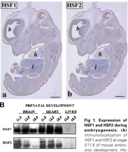
To investigate the expression and tissue distribution of HSF1 and HSF2 during mouse embryogenesis, we analyzed the devel-oping embryos by immunostaining using antibodies specific for HSF1 and HSF2 (Alastalo et al., 1998; Sarge et al., 1993). Recently, Rallu and coworkers (1997) showed that HSF2 mRNA was abundantly expressed in the developing brain. Our immunostaining results were consistent with the mRNA analysis and further demonstrated that in addition to brain, both HSF1 and HSF2 proteins were abundantly expressed in the developing heart (Fig. 1A). The tissue distribution of HSF1 and HSF2 was very similar. For example at stage E11.5, a prominent HSF1 (panel a) and HSF2 (panel b) immunoreactivity was detected in the heart in comparison to adjacent tissues, such as the lungs.
We then used SDS-PAGE and Western blotting to assess the levels of HSF1 and HSF2 in different tissues. Protein extracts were isolated from developing heart, liver, and brain at different stages of embryogenesis (Fig. 1B). Consistent with the immunostaining analysis, HSF1 and HSF2 were abundantly expressed in the developing heart and brain, whereas they were undetectable in the liver. Furthermore, we observed that expression levels of HSF2 varied in the heart and brain during prenatal development: the levels were abundant at E11.5 and E12.5, but at E15.5 the expression was downregulated. In comparison, the amounts of HSF1 remained relatively constant.
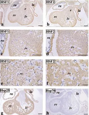
Having detected an abundant expression of HSF1 and HSF2 in the developing heart tissue, we next asked whether these proteins are localized in similar or distinct patterns. When sections of heart specimens from stage E11.5 embryos were immunostained with antibodies against HSF1 and HSF2, very similar although not identical staining patterns were seen (Fig. 2). The expression of HSF1 and HSF2 in the heart was uniform in the differentiating atrial and ventricular walls, trabeculations, and endocardial cushion
tological samples were stained with antibodies against (a) HSF1 (b) and HSF2. Shown are micrographs of vertical sections through whole mouse embryos. Positive staining appears brown. Brain (b) and the lungs (l) are also indicated. Bar, 100 µm. (B). Western analysis of HSF1 and HSF2 expression during embryogenesis. Protein extracts prepared from isolated heart, brain and liver at different developmental stages were analyzed by Western blotting using antibodies against HSF1 and HSF2.
Fig 1. Expression of HSF1 and HSF2 during embryogenesis. (A)
Immunolocalization of HSF1 and HSF2 at stage E11.5 of mouse embry-onic development.
His-A
area (panels a and b). However, the distribution of HSF1 and HSF2 within a single cell was slightly different. HSF1 signal appeared to localize more abundantly in the nucleus than in the cytoplasm (panel c and e), as compared to HSF2 staining, which showed more prominent cytoplasmic immunoreaction (panel d and f).
In comparison to HSF1 and HSF2, distribution of heat shock proteins Hsp25 and Hsp70 was analyzed from the serial sections of the same sample. Hsp25 was abundantly expressed in the atrial and ventricular walls, and trabeculations but it was not detectable in the endocardial cushion area (panel g). Interestingly, Hsp70 was not ex-pressed in the developing heart tissue (panel h).
The expression of HSF2 isoforms is stage-dependent during heart development
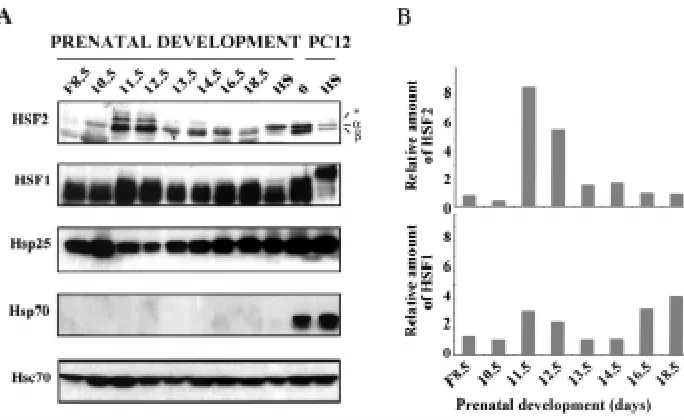
To assess the temporal expression of HSF2 protein in the developing heart in detail, tissue extracts from whole embryos at stage E8.5, and embryonic hearts at stages E10.5-E18.5 were prepared and analyzed by SDS-PAGE and Western blotting (Fig. 3). Untreated and heat-shocked rat PC12 cells were used as controls. Consistent with the results in Fig. 1B, the expression levels of HSF2 varied between different developmental stages (Fig. 3A). In the embryo at stage E8.5 and in the heart tissue at E10.5, HSF2 was expressed at low levels, but an increase in the HSF2 protein levels was observed at stages E11.5 and E12.5. The increases at these stages were 8 and 6 fold, respectively, as compared to the expression level of HSF2 at stage E18.5 (Fig. 3B, upper panel). Thereafter, HSF2 expression markedly decreased but remained detectable throughout the heart development.
The alternatively spliced isoforms of HSF2, HSF2-α and HSF2-β have been shown to be differently expressed in different adult organs and during spermatogenesis (Alastalo et al., 1998; Fiorenza et al., 1995; Goodson et al., 1995). Analogously, we were interested to compare the relative abundance of HSF2 isoforms at different stages of heart development. We found that the HSF2-β isoform was more abundant than HSF2-α. Moreover, we detected an addi-tional, previously unidentified higher molecular weight band, which was upregulated transiently at stages E11.5 and E12.5. The band was not heart-specific, as it was also detected in the brain samples (Fig. 1B).
play an important role in HSF1 activation (Chu et al., 1996; Cotto et al., 1996; Jurivich et al., 1995; Kline and Morimoto, 1997), we were puzzled to observe that HSF1 from untreated and heat-shocked heart tissue migrated identically in SDS-PAGE, whereas in PC12 cells exposure to heat shock resulted in a shift in HSF1 mobility typical of hyperphosphorylation (Cotto et al., 1996; Sarge et al., 1993). The basis for the difference in phosphorylation state of HSF1 between the heat-shocked heart tissue and PC12 cells is unclear and was not addressed in this study.
In addition to HSF1 and HSF2, the amounts of heat shock proteins were analyzed from the same samples (Fig. 3). The expression pattern of Hsp25 was opposite to that of HSF2, as high Hsp25 levels were observed at stage E8.5 embryo, and E10.5 and E13.5-E18.5 heart tissue. The constitutively expressed Hsp70 (Hsc70) was present throughout the heart development, increas-Fig. 2. Immunolocalization of HSF1, HSF2, Hsp25 and Hsp70 in the develop-ing mouse heart. Histological samples from stage E11.5 were stained with antibodies against (a) HSF1, (b) HSF2, (g) Hsp25 and (h) Hsp70. Shown are micrographs of horizontal sections through embryonic hearts. (c and e) and (d
and f) show high magnification views of a and b, respectively. Positive staining appears brown. (ra), (la), (rv) and (lv) indicate right and left atrial and ventricular chambers, respectively, (e) indicates endocardial cushion tissue.Bar,50 µm ( a-d, g and h); 10 µm (e and f).
To explore whether the higher molecular weight band was generated from an additional mRNA variant, we analyzed the existence of HSF2 mRNA isoforms by RT-PCR. For this analysis, we used various oligonucleotide primer pairs that span all identified 13 exons (Manuel et al., 1999). Depending on the primer pairs, the results revealed one or two HSF2 mRNA variants, which corre-sponded to previously identified HSF2-α and HSF2-β isoforms (Goodson et al., 1995). No additional mRNA isoforms were ob-served (data not shown). Based on this analysis, it seems unlikely that the higher molecular weight band was translated from addi-tional mRNA.
ing slightly at days E10.5-E11.5. Instead, consistent with the immunostaining data (Fig. 2f), expression of the inducible form of Hsp70 was not detectable in the developing heart.
HSF2 DNA-binding activity is induced during mouse cardiogenesis
Previous results in cell culture have indicated that high expres-sion levels of HSF2 precede its DNA-binding capacity, whereas the activation of HSF1 is not dependent on the amount of protein (Leppä et al., 1997; Murphy et al., 1994; Sistonen et al., 1994). To analyze the HSF DNA-binding activities at different stages of the mouse heart development, tissue extracts from whole embryos at stage E8.5, and embryonic hearts at stages E10.5-E18.5 were analyzed by gel mobility shift assay. A heat-shocked sample from an embryonic heart tissue at E14.5 was used as a positive control. A constitutive HSE-binding activity was detected throughout the heart development (Fig. 4). The activity was prominent in the whole embryo at E8.5 and in the heart tissue between the days E10.5-E12.5. Quantification revealed that the DNA-binding activity was highest (about 6-fold induction as compared to E15.5) at E12.5, after which the activity decreased and remained relatively low up to birth.
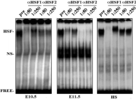
The composition of the HSE-binding activity was determined using antibody perturbation assays. Tissue extracts from hearts at differ-ent stages of developmdiffer-ent were incubated with polyclonal antisera raised against HSF1 and HSF2 (Sarge et al., 1993), and thereafter subjected to gel mobility shift analysis. Figure 5 (left panel) shows that at E10.5 the HSF1 antiserum clearly interfered with the HSF-HSE complex at 1:50 dilution, whereas HSF2 antiserum had only a slight effect on the complex formation. Instead, at stage E11.5, when the HSE-binding activity was strongly increased (Fig. 4), the situation was opposite: HSF1 antiserum had only a minor effect on HSE-binding activity (Fig. 5, middle panel), but the HSE-HSF complex formation was completely neutralized by the HSF2 antise-rum even at dilution 1:250. An identical pattern was obtained at stage E12.5 (data not shown). We also observed that the modest HSE-binding activity at E13.5-E18.5 consisted primarily of HSF1 (data not shown), and as expected, the heat-induced HSE-binding activity consisted solely of HSF1 (Fig. 5; right panel). These results
indicate that whereas HSF1 DNA-binding activity peaked at stage E10.5, and was then maintained relatively low throughout the cardiac development, HSF2 DNA-binding activity was induced at stages E11.5-E12.5.
HSF2 activation and heat shock gene expression are uncou-pled
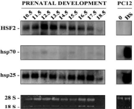
Immunostaining and Western analyses shown in Figures 2 and 3 suggested that the expression of heat shock proteins was not necessarily HSE-dependent during heart development. To inves-tigate this possibility further, we analyzed the heat shock gene expression by Northern blotting (Fig. 6). The expression of HSF2 mRNA was detectable throughout the heart development, increas-ing slightly at E12.5-E13.5. Consistent with the protein data (Figs. 2 and 3), hsp70 mRNA expression was not detectable in the developing heart, and the levels of hsp25 mRNA remained rela-tively constant throughout the cardiogenesis. The results indicate that expression of the inducible heat shock genes does not correlate with HSF2 expression and HSE-binding activity during heart development.
Fig. 3. Upregulation of the HSF2-α and HSF2-β isoforms in the developing heart.(A) Expres-sion of HSF2 and HSF1 during heart development. Protein extracts isolated from total embryos (F), untreated and heat-shocked (HS; 42°C, 1 h) heart tissue at indicated developmental stages were analyzed by Western blotting using antibodies against HSF2 and HSF1. Subsequently, the blots were reprobed with Hsp25, Hsp70, and Hsc70 antibodies. Untreated (0) and heat-shocked rat PC12 cells were used as controls. α, β and asterisk indicate the distinct forms of HSF2. A representa-tive blot of three experiments is shown. Note that the exposures for HSF1 and HSF2 are 4-6 fold longer than for heat shock proteins. (B) Quantifica-tion of HSF2 and HSF1. The bands of the HSF2-α + β and HSF1 shown in (A) were quantified using computerized image analysis. The values for fold induction of HSF2 and HSF1 are shown relative to the lowest detectable level of HSF2 and HSF1 at stages E18.5. and E10.5, respectively.
overexpressing HSF2-β, the hemin-mediated acquisition of HSF2 DNA-binding activity and subsequent induction of heat shock gene expression are prevented (Leppä et al., 1997a). Besides HSF2-α and HSF2-β, we detected an additional higher molecular weight isoform, the expression pattern of which correlated with the HSF2 DNA-binding activity. To this end, the role of several different HSF2 isoforms awaits further clarification, but based on the current findings it seems clear that the effects of different HSF2 isoforms on HSF2 activity are dependent on the cellular context.
Series of regulatory modifications must occur before a transcrip-tion factor can be considered fully activated. Common features for HSF1 and HSF2 are that, upon activation, they undergo trimerization, acquire DNA-binding activity, and translocate into the nucleus, where they transactivate target gene expression. In the present study, we observed that although HSF2 activity was induced, as determined by its ability to interact with HSE, the expression of the classical heat shock genes hsp70 and hsp25 was not upregulated. Hence, as previously shown to occur during mouse brain develop-ment (Rallu et al., 1997), no obvious correlation between the HSE-binding activity and the heat shock gene expression can be observed during heart formation. Considering the finding that HSF2-β is transcriptionally weaker than HSF2-α (Goodson et al., 1995), it is possible that the HSF2-β isoform is not transcriptionally active enough to upregulate heat shock gene expression. How-ever, a more likely explanation is that instead of activating classical heat shock genes, HSF2 regulates other target genes during development. Consistent with the latter hypothesis, genetic stud-ies in Drosophila indicate that the requirement of HSF for oogen-esis and early larval development is unrelated to the developmen-tal expression of heat shock genes and is likely to involve novel target gene expression (Jedlicka et al., 1997). Our previous studies suggest that in K562 cells undergoing erythroid differentiation, a potential HSF2 target gene is thioredoxin, whose expression is
Discussion
In the present study, we focused on the analysis of HSF1 and HSF2 expression and DNA-binding activity during the mouse heart development. HSF2 DNA-binding activity was found to be transiently induced at the developmental stages after looping and chamber formation, when chambers are maturating and endocardial cush-ions differentiating. Immunohistochemical analysis revealed that the upregulation of HSF2 protein levels coincides with HSF2 DNA-binding activity. Closer examination of the alternatively spliced HSF2-α and HSF2-β, and the additional, previously unidentified higher molecular weight isoform by Western blot analysis con-firmed the immunohistochemical studies and further demonstrated that HSF2-α and HSF2-β are highly expressed at E11.5-E12.5, after which the expression levels are markedly downregulated, yet remain present throughout the heart development. In contrast, the higher molecular weight HSF2 isoform is expressed transiently at E11.5-E12.5, at least at detectable levels. Interestingly, during spermatogenesis, expression of additional smaller molecular weight HSF2 isoforms is also upregulated in a stage-specific manner (Alastalo et al., 1998). To date, the identity of the additional bands remains unknown. However, based on our PCR-analysis it seems unlikely that the higher molecular weight isoform was generated from additional mRNA variant.
As far as the DNA-binding activity is concerned, HSF2 activation is associated with the abundant protein levels during the heart development. The results are in agreement with the previous findings in K562 cells, which show that the HSF2 DNA-binding activity is progressively induced with a corresponding increase in HSF2 protein levels during the hemin-mediated erythroid differen-tiation (Leppä et al., 1997a; Sistonen et al., 1994). Analogously, constitutive HSF2 DNA-binding activity associates with high HSF2 expression levels during the pre- and postimplantation stages of mouse embryogenesis (Christians et al., 1997; Mezger et al., 1994; Rallu et al., 1997). In the developing heart, the regulation of HSF2 expression appears to occur at multiple molecular levels. Since HSF2 mRNA expression was found to be slightly increased at stages E12.5-E13.5, the regulation of HSF2 levels may involve changes in HSF2 gene expression. Whether the expression of HSF2 is regulated at transcriptional or post-transcriptional level during heart development is not clear, but at least in K562 cells both transcriptional induction and mRNA stabilization are involved (Pirkkala et al., 1999). However, it seems unlikely that the relatively minor changes in the HSF2 mRNA levels would exclusively ac-count for the prominent alterations in the HSF2 protein amounts. Thus, we speculate that in the developing heart, HSF2 expression and activation involve increased protein synthesis and/or reduced degradation, both of which were recently shown to be required for the maintenance of HSF2 levels and activity in cell culture condi-tions (Mathew et al., 1998).
With respect to the relative abundance of the alternatively spliced HSF2-α and HSF2-β isoforms contributing to HSF2 activa-tion, we found that during heart development, the smaller HSF2-β is quantitatively the major isoform. Similarly, HSF2-β has been shown to be predominantly expressed in the embryonal carcinoma cells and developing brain tissue, which both exhibit constitutive HSF2 DNA-binding activity (Murphy et al., 1994; Rallu et al., 1997). On the other hand, HSF2-α prevails in K562 cells, and when the molar ratio of the HSF2-α and HSF2-β isoforms is inverted by
induced in response to hemin in an HSF2-dependent manner (Leppä et al., 1997b). It remains to be shown whether HSF2 regulates gene expression that contributes to cardiomyocyte matu-ration.
Heart formation requires precisely controlled gene expression. During the recent years, a number of molecules whose activity is required for the proper morphogenesis of the heart have been identified. Certain transcription factors have been found to be essential for the specific stages of heart development. For exam-ple, mutation of the Nkx2-5 and MEF2C genes results in arrest of morphogenesis at the looping stage (Lin et al., 1997; Lyons et al., 1995), whereas GATA-4 is required even earlier when the tubular heart is forming (Kuo et al., 1997; Molkentin et al., 1997). Our results demonstrate that HSF2 DNA-binding activity is associated with the later developmental stages, which are distinguished by the growth and differentiation of the myocardium, trabeculations, and the valves. Future studies will determine whether HSF2 activation at these specific stages is critical for the normal heart development.
Materials and Methods
Tissue preparation
Mouse embryos were obtained by mating NMRI male and female mice. The day when the copulation plug was found was considered as E0.5. The embryos were collected and processed for immunohistochemistry, or heart tissue was isolated and frozen in liquid nitrogen for further biochemical analyses.
Immunohistochemistry
For immunostainings, mouse embryos were fixed in 4% paraformaldehyde, embedded in paraffin and sectioned. The sections of mouse embryos were treated in microwave oven in 0.1 M sodium citrate (pH 5.0) for 10 minutes and washed with PBS. Subsequent staining was performed using polyclonal antibodies against HSF1 and HSF2 (Alastalo et al., 1998; Sarge et al., 1993), or Hsp25 (SPA-801, StressGen) and Hsp70
Fig. 6. Expression of HSF2 mRNA during heart development. Total RNA from isolated hearts at indicated developmental stages, and from untreated (0) and heat-shocked (HS) PC12 cells were analyzed by Northern blotting. Blots were probed with 32P-labeled cDNA probes for mHSF2, hsp70 and hsp25. The ethidium bromide stained gel showing ribosomal 28 S and 18 S RNAs is shown as a loading control. Note that the exposure for HSF2 mRNA is 3 fold longer in comparison to hsp70 and hsp25 mRNAs.
(SPA-810, StressGen). Immunohistochemistry was completed using the Vectastain ABC Kit reagents (Vector Laboratories, USA) according to the manufacturer’s instructions. The immunoreactions were visualized by using 3,3-diaminobenzidine tetrahydrochloride (0.25 mg/ml), ammonium nickel sulfate (10 mg/ml) and hydrogen peroxide (0.005%) in PBS.
Western blot analysis
Tissue extracts from whole mouse embryos (E8.5), isolated hearts (from E10.5 up to E18.5), livers, brains, or untreated and heat-shocked (42°C, 1 h) rat pheochromocytoma (PC12) cells were prepared by homogenization in two volumes of high salt buffer containing 20 mM Hepes, pH 7.9, 0.42 M NaCl, 25% (v/v) glycerol, 1.5 mM MgCl2, 0.2 mM EDTA, 0.5 mM dithiotreitol (DTT), and 0.5 mM phenylmethylsulfonyl fluoride (PMSF). Concentration of total soluble protein was measured using the Bradford method. Protein extracts (10 µg) were separated on an 8% SDS-PAGE and transferred to nitrocellulose membranes using a semi-dry blotting apparatus (BioRad). Membranes were blocked in PBS containing 5% (w/v) non-fat dry milk and 0.3% Tween-20 for 1 h at RT, followed by incubation with antibodies against HSF1 and HSF2 (Sarge et al., 1993), Hsp70 (SPA-810, StressGen), Hsc70 (SPA-815, StressGen), and Hsp25 (SPA-801, StressGen) for 1 h at RT. Blots were washed 3 times for 10 minutes in PBS containing 0.3% Tween-20 and further incubated with HRP-conjugated secondary antibodies for 1 h at RT. After washes the blots were subjected to immunodetection using ECL-method (Amersham). Quantification was performed using computer-ized image analysis (Microcomputer Imaging Device version M4, Imaging Research Inc.).
Gel mobility shift assay
To assay heat shock element (HSE)-binding activity, 15 µg of tissue extract was incubated for 20 minutes at room temperature (RT) in 20 µl reaction volumes containing 0.1 ng annealed [γ32P]dATP-labeled HSE
oligonucleotide (5’-CTAGAAGCTTCTAGAA GCTTCTAG-3’), 1 µg poly(dI-dC), 10 µg BSA, 10 mM Tris, pH 7.5, 50 mM NaCl, 1mM EDTA, 5% (v/v) glycerol, 0.5 mM DTT, and 0.5 mM PMSF (Mosser et al., 1988). Protein-DNA complexes were resolved on a 4% polyacrylamide gel containing 0.5 x TBE and visualized by autoradiography.
Perturbation assays were done by incubating tissue extracts (13 µg) with preimmune serum (dilution 1:10), or immunoserum against HSF1 and HSF2 (dilutions 1:50 and 1:250) (Sarge et al., 1993) for 15 minutes at RT. Subsequently, binding reactions were performed, and protein-DNA com-plexes resolved on a 4% polyacrylamide gel, followed by autoradiography.
Northern blot analysis
For Northern blot analysis, total cellular RNA was extracted from prenatal mouse hearts, non-treated and heat-shocked PC12 cells using Qiagen RNAeasy kit. Total RNA (10 µg) was separated on a 1% formaldehyde-agarose gel and transferred to nylon membranes (Hybond-N, Amersham). Filters were hybridized with [α32P]dCTP-labeled cDNAs coding for mouse
HSF2 (Sarge et al., 1991), human hsp70 (Wu et al., 1985), and mouse hsp25 (Gaestel et al., 1993). Hybridizations were performed according to the manufacturer’s instructions.
Acknowledgements
We thank O. Rahkonen and E. Abdelwahid for tissue samples and L.C. Andersson for help with the photographing. We are also grateful for T-P. Alastalo and L. Pirkkala for comments on the manuscript. This work was supported by the Academy of Finland, the Finnish Cultural Foundation, and Helsinki University Central Hospital.
References
BALER, R., DAHL, G. and VOELLMY, R. (1993). Activation of human heat shock genes is accompanied by oligomerization, modification, and rapid translocation of heat shock transcription factor HSF1. Mol. Cell. Biol. 13: 2486-2496.
CHRISTIANS, E., MICHEL, E., ADENOT, P., MEZGER, V., RALLU, M., MORANGE, M. and RENARD, J.P. (1997). Evidence for the involvement of mouse heat shock factor 1 in the atypical expression of the HSP70.1 heat shock gene during mouse zygotic genome activation. Mol. Cell. Biol. 17: 778-88.
CHU, B., SONCIN, F., PRICE, B.D., STEVENSON, M.A. and CALDERWOOD, S.K. (1996). Sequential phosphorylation by mitogen-activated protein kinase and glycogen synthase kinase 3 represses transcriptional activation by heat shock factor-1. J. Biol. Chem. 271: 30847-57.
COTTO, J.J., KLINE, M. and MORIMOTO, R.I. (1996). Activation of heat shock factor 1 DNA binding precedes stress-induced serine phosphorylation. Evidence for a multistep pathway of regulation. J. Biol. Chem. 271: 3355-3358.
FIORENZA, M.T., FARKAS, T., DISSING, M., KOLDING, D. and ZIMARINO, V. (1995). Complex expression of murine heat shock transcription factors. Nucleic. Acids. Res. 23: 467-474.
GAESTEL, M., GOTTHARDT, R. and MULLER, T. (1993). Structure and organisation of a murine gene encoding small heat-shock protein Hsp25. Gene 128: 279-283.
GOODSON, M.L., PARK-SARGE, O.K. and SARGE, K.D. (1995). Tissue-dependent expression of heat shock factor 2 isoforms with distinct transcriptional activities. Mol. Cell. Biol. 15: 5288-93.
HIGASHI, T., NAKAI, A., UEMURA, Y., KIKUCHI, H. and NAGATA, K. (1995). Activation of heat shock factor 1 in rat brain during cerebral ischemia or after heat shock. Brain Res. Mol. Brain Res. 34: 262-70.
JEDLICKA, P., MORTIN, M.A. and WU, C. (1997). Multiple functions of Drosophila heat shock transcription factor in vivo. EMBO J. 16: 2452-62.
JURIVICH, D.A., PACHETTI, C., QIU, L. and WELK, J.F. (1995). Salicylate triggers heat shock factor differently than heat. J. Biol. Chem. 270: 24489-95.
KLINE, M.P. and MORIMOTO, R.I. (1997). Repression of the heat shock factor 1 transcriptional activation domain is modulated by constitutive phosphorylation. Mol. Cell. Biol. 17: 2107-15.
KUO, C.T., MORRISEY, E.E., ANANDAPPA, R., SIGRIST, K., LU, M.M., PARMACEK, M.S., SOUDAIS, C. and LEIDEN, J.M. (1997). GATA4 transcription factor is required for ventral morphogenesis and heart tube formation. Genes Dev. 11: 1048-1060.
LEPPÄ, S., PIRKKALA, L., SAARENTO, H., SARGE, K.D. and SISTONEN, L. (1997a). Overexpression of HSF2-beta inhibits hemin-induced heat shock gene expression and erythroid differentiation in K562 cells. J. Biol. Chem. 272: 15293-15298.
LEPPÄ, S., PIRKKALA, L., CHOW, S.C., ERIKSSON, J.E. and SISTONEN, L. (1997b). Thioredoxin is transcriptionally induced upon activation of heat shock factor 2. J. Biol. Chem. 272: 30400-30404.
LIN, Q., SCHWARZ, J., BUCANA, C. and OLSON, E.N. (1997). Control of mouse cardiac morphogenesis and myogenesis by transcription factor MEF2C. Science 276: 1404-1407.
LYONS, I., PARSONS, L.M., HARTLEY, L., LI, R., ANDREWS, J.E., ROBB, L. and HARVEY, R.P. (1995). Myogenic and morphogenetic defects in the heart tubes of murine embryos lacking the homeo box gene Nkx2-5. Genes Dev. 9: 1654-1666.
MANUEL, M., SAGE, J., MATTEI, M.G., MORANGE, M. and MEZGER, V. (1999). Genomic structure and chromosomal localization of the mouse Hsf2 gene and promoter sequences. Gene 232: 115-24.
MATHEW, A., MATHUR, S.K. and MORIMOTO, R.I. (1998). Heat shock response and protein degradation: regulation of HSF2 by the ubiquitin-proteasome path-way. Mol. Cell. Biol. 18: 5091-5098.
MEZGER, V., RALLU, M., MORIMOTO, R.I., MORANGE, M. and RENARD, J.P. (1994). Heat shock factor 2-like activity in mouse blastocysts. Dev. Biol. 166: 819-22.
MOLKENTIN, J.D., LIN, Q., DUNCAN, S.A. and OLSON, E.N. (1997). Requirement of the transcription factor GATA4 for heart tube formation and ventral morphogen-esis. Genes Dev. 11: 1061-1072.
MORIMOTO, R.I. (1998). Regulation of the heat shock transcriptional response: cross talk between a family of heat shock factors, molecular chaperones, and negative regulators. Genes Dev. 12: 3788-96.
MOSSER, D.D., THEODORAKIS, N.G. and MORIMOTO, R.I. (1988). Coordinate changes in heat shock element-binding activity and HSP70 gene transcription rates in human cells. Mol. Cell. Biol. 8: 4736-4744.
MURPHY, S.P., GORZOWSKI, J.J., SARGE, K.D. and PHILLIPS, B. (1994). Charac-terization of constitutive HSF2 DNA-binding activity in mouse embryonal carci-noma cells. Mol. Cell. Biol. 14: 5309-17.
NAKAI, A., TANABE, M., KAWAZOE, Y., INAZAWA, J., MORIMOTO, R.I. and NAGATA, K. (1997). HSF4, a new member of the human heat shock factor family which lacks properties of a transcriptional activator. Mol. Cell. Biol. 17: 469-81.
NISHIZAWA, J., NAKAI, A., HIGASHI, T., TANABE, M., NOMOTO, S., MATSUDA, K., BAN, T. and NAGATA, K. (1996). Reperfusion causes significant activation of heat shock transcription factor 1 in ischemic rat heart. Circulation 94: 2185-92.
OLSON, E.N. and SRIVASTAVA, D. (1996). Molecular pathways controlling heart development. Science 272: 671-676.
PIRKKALA, L., ALASTALO, T-P., NYKÄNEN, P., SEPPÄ, L. and SISTONEN, L. (1999). Differentiation lineage-specific expression of human heat shock transcrip-tion factor 2. FASEB J. 13: 1089-1098.
RABINDRAN, S.K., GIORGI, G., CLOS, J. and WU, C. (1991). Molecular cloning and expression of a human heat shock factor, HSF1. Proc. Natl. Acad. Sci. USA 88: 6906-10.
RABINDRAN, S.K., HAROUN, R.I., CLOS, J., WISNIEWSKI, J. and WU, C. (1993). Regulation of heat shock factor trimer formation: role of a conserved leucine zipper. Science 259: 230-234.
RALLU, M., LOONES, M., LALLEMAND, Y., MORIMOTO, R., MORANGE, M. and MEZGER, V. (1997). Function and regulation of heat shock factor 2 during mouse embryogenesis. Proc. Natl. Acad. Sci. USA 94: 2392-2397.
ROSSANT, J. (1996). Mouse mutants and cardiac development: new molecular insights into cardiogenesis. Circ. Res. 78: 349-53.
SARGE, K.D., MURPHY, S.P. and MORIMOTO, R.I. (1993). Activation of heat shock gene transcription by heat shock factor 1 involves oligomerization, acquisition of DNA-binding activity, and nuclear localization and can occur in the absence of stress. Mol. Cell. Biol. 13: 1392-1407.
SARGE, K.D., ZIMARINO, V., HOLM, K., WU, C. and MORIMOTO, R.I. (1991). Cloning and characterization of two mouse heat shock factors with distinct inducible and constitutive DNA-binding ability. Genes Dev. 5: 1902-11.
SCHUETZ, T.J., GALLO, G.J., SHELDON, L., TEMPST, P. and KINGSTON, R.E. (1991). Isolation of a cDNA for HSF2: Evidence for two heat shock factor genes in humans. Proc. Natl. Acad. Sci. USA 88: 6911-6915.
SISTONEN, L., SARGE, K.D. and MORIMOTO, R.I. (1994). Human heat shock factors 1 and 2 are differentially activated and can synergistically induce hsp70 gene transcription. Mol. Cell. Biol. 14: 2087-99.
SISTONEN, L., SARGE, K.D., PHILLIPS, B., ABRAVAYA, K. and MORIMOTO, R.I. (1992). Activation of heat shock factor 2 during hemin-induced differentiation of human erythroleukemia cells. Mol. Cell. Biol. 12: 4104-11.
WU, B., HUNT, C. and MORIMOTO, R.I. (1985). Structure and expression of the human gene encoding major heat shock protein HSP70. Mol. Cell. Biol. 5: 330-341.
WU, C. (1995). Heat shock transcription factors: structure and regulation. Annu. Rev. Cell. Dev. Biol. 11: 441-69.
YOSHIMA, T., YURA, T. and YANAGI, H. (1998). Heat shock factor 1 mediates hemin-induced hsp70 gene transcription in K562 erythroleukemia cells. J. Biol. Chem. 273: 25466-71.