Original Article
Dynamic distribution of the replacement histone variant H3.3
in the mouse oocyte and preimplantation embryos
MARIA-ELENA TORRES-PADILLA, ANDREW J. BANNISTER
1, PAUL J. HURD
1, TONY KOUZARIDES
1and
MAGDALENA ZERNICKA-GOETZ*
The Wellcome Trust/Cancer Research UK Gurdon Institute, and 1Department of Pathology, University of Cambridge, U.K.
ABSTRACT Upon fertilization, the gametes undergo a drastic reprogramming that includes changes in DNA methylation and histone modifications. Currently, it is not known whether replacement of the major histones by histone variants is also involved in these processes. Here we have examined the expression and localization of the histone variant H3.3 in early mouse embryogenesis. We show that H3.3 is present in the oocyte as a maternal factor. It is then incorporated preferentially into the male pronucleus before genome activation, pointing towards an asymmetry in histone composition between the two pronuclei. This is in line with the male pronucleus bearing transcriptional activation first. The same distribution was observed when we followed the localisation of a tagged version of H3.3. We detected H3.3 in the nuclei of mouse embryos in all of the stages analysed, from the zygote to the blastocyst stage, suggesting that the epigenetic mechanisms in the early embryo not only involve changes in histone modifications but may also include histone replacement.
KEY WORDS: histone variant, early mouse embryo, reprogramming, replacement histone
Introduction
The removal and acquisition of new epigenetic marks during preimplantation development is essential to ensure the totipo-tency required for sustaining further development. In the zygote, changes in the epigenetic status of the parental genomes may also be needed for the activation of zygotic transcription. Covalent modifications of histones and DNA methylation are considered as the most important epigenetic marks in preimplantation develop-ment (Li, 2002, Morgan et al., 2005).
In the early zygote, histones H3 and H4 are synthesized from maternal mRNA, whereas the synthesis of histones H2A, H2B and H1, is delayed until the end of the first cell cycle (Wiekowski et al., 1997). Covalent modifications of histones contribute to the formation and maintenance of transcriptionally active or inactive chromatin domains. Acetylation of lysine (K) residues is generally associated with transcriptional activation whereas methylation of lysine residues can have opposing outcomes (repression or activation) depending on the precise methylation site. Apart from the major core histones, whose synthesis is tightly linked to the S-phase of the cell cycle, some histones are synthesized from orphan genes throughout the cell cycle and are incorporated into the chromatin independently of replication (Kamakaka and Biggins, 2005). These ‘replacement’ histones are also covalently modified
*Address correspondence to: Dr. Magdalena Zernicka-Goetz. The Wellcome Trust/Cancer Research UK Gurdon Institute, University of Cambridge, Tennis Court Road, Cambridge CB2 1QR, U.K. Fax +44-1223-634089. e-mail: mzg@mole.bio.cam.ac.uk
Abbreviations used in this paper: GV, germinal vesicle; HA tag, hemagglutin tag; PN, pronuclear stage; h phCG, hours after hCG injection.
0214-6282/2006/$25.00
© UBC Press Printed in Spain www.intjdevbiol.com
and can also be used to ‘mark’ the chromatin and hence regulate gene expression. Moreover, they can elicit different chromatin conformations upon assembly into the nucleosomes (Gautier et al., 2004). One interesting variant is H3.3, which can replace histone H3. The replication-independent deposition of histone variants allows the turnover of modification marks and hence a change of the heritable chromatin states. For example, upon gene activation, H3 carrying the repressive methyl H3K9 mark be-comes replaced by H3.3 unmethylated at K9. Moreover, preferen-tial deposition of H3.3 in transcriptionally active regions would result in perpetuation of epigenetic memory (Ahmad and Henikoff, 2002b, Schwartz and Ahmad, 2005). Although a growing number of reports support a role for the histone variants in regulating chromatin activity, their specific functions in vivo are not fully understood.
preimplantation development. Here, we have characterized the localization of the replacement histone variant H3.3 in mouse oocytes and early embryos. The histone variant H3.3 was present in all the stages examined. In the zygote, it showed an asymmetric distribution between the male and female pronucleus. We also observed that the pattern of localization of H3.3 changes from a punctate to a more disperse nuclear staining after the 2-cell stage.
Results
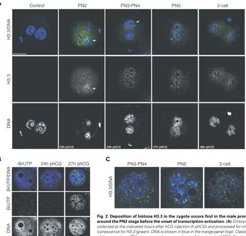
Here, we were interested in examining the distribution of the replacement histone variant H3.3 in oocytes and preimplantation mouse embryos. The characterisation of the H3.3 antibody used in this study is shown in Figure 1A. Only the specific H3.3 peptide competes the antibody. Importantly, the H3.3 antibody does not recognise a recombinant purified H3.2 variant, testifying for the specificity of the antibody. Using this H3.3 specific antibody we analysed the distribution of H3.3 in the oocyte. The H3f3a gene, encoding H3.3, was previously reported to be expressed in the growing oocyte (Couldrey et al., 1999). H3.3 clearly accumulates in the germinal vesicle (GV), but it is also detectable in the cytoplasm (Fig. 1B, left panel). After fertilisation, the maternal genome resumes meiosis from metaphase II and extrudes the second polar body containing the second set of haploid chromo-somes. During this time, which corresponds to the pronuclear stage PN0 (Adenot et al., 1997), H3.3 is only weakly dispersed in the cytoplasm with no obvious localisation in either the maternal or the paternal pronuclei (Fig. 1B, right panel).
The pronuclei are subsequently subject to cycles of decondensation and migrate towards the centre of the zygote. We first detect an enrichment of H3.3 in the pronuclei during decondensation around the PN2 stage. This enrichment is asym-metric between the two pronuclei: it is evident in the male pronucleus, but not in the female one. At this stage, a weak staining of the cytoplasm suggests localisation of H3.3 in the cytoplasm, probably reflecting translation, storage or association with chaperone molecules (Fig. 2A). Thus, the maternal genome
is associated with H3.3 in the GV, but H3.3 is then undetectable in the maternal chromatin during pronuclear formation and its incorporation starts earlier in the male pronucleus. In PN3-PN4 zygotes the pronuclei increase in size and locate close to each other in the centre of the cell. At this stage, the enrichment of H3.3 is still slightly brighter in the male pronucleus, but the female pronucleus also shows accumulation of H3.3 (the signal/size ratio of the male pronucleus compared to the female one was 1.8
±
0.17, n=7, as quantified with the Volocity software, Improvision)(Fig. 2A). Before mitosis, the pronuclei become apposed (PN5), at this stage H3.3 is present in the two pronuclei and a difference in H3.3 accumulation between the male and the female pronucleus is no longer detected (Fig. 2A). At the 2-cell stage H3.3 is present in the two nuclei of the two blastomeres (Fig. 2A).H3.3 can be deposited via a replication-independent pathway that is triggered upon transcriptional activation (Janicki et al., 2004, Schwartz and Ahmad, 2005). We therefore wished to know whether the time when we first saw deposition of H3.3 in the zygote corresponded to the time when the genome activation occurs. Thus, we followed the incorporation of BrUTP in the zygote by immunofluorescence. When examined at 24h phCG (approximately PN3-PN4 stages), we do not detect BrUTP in either pronucleus, indicating that the zygote is not yet transcrip-tionally active at this stage (Fig. 2B). However, when examined at 27h phCG (PN5), we detect BrUTP staining throughout the nucleoplasm with sites of brighter intensity around the nucleolar-like bodies in the two pronuclei (Fig. 2B). This is consistent with the observation that the zygote undergoes transcriptional activa-tion between 26h and 29h phCG (Aoki et al., 1997, Bouniol et al., 1995). H3.3 exhibits a characteristic punctate staining throughout the nucleoplasm at these early stages and in the oocyte, which persists from the PN3 zygote stage up to the 2-cell stage (Fig. 2C). This pattern is different to the one exhibited by the BrUTP incorporated in the zygote (Fig. 2B). Thus, our results suggest that histone H3.3 accumulates in the male pronucleus before the onset of transcription activation.
We performed another series of experiments to confirm our
Fig. 1. Western blot confirming specificity of the H3.3 antibody (A) and immunochem-istry revealing presence of Histone H3.3 in the germinal vesicle stage oocyte (B). (A) 2 micrograms of calf thymus core histones (CH; 500 ng each of histones H2A, H2B, H3 and H4) and 1 microgram of bacterially puri-fied and produced histone H3.2 (rH3) were resolved by SDS-PAGE, Western blotted to nitrocellulose and probed with the anti-H3.3 antibody. Antibody probing was performed in the absence or presence of competitor block-ing peptide (1 µg/ml) as indicated above each lane on the panel.(B) Single confocal sec-tions of a germinal vesicle stage (GV) oocyte and a zygote at the PN0 stage (Adenot et al., 1997) stained with the H3.3 antibody (green) and TOTO-3 (DNA, blue). In the oocyte, H3.3 localises mainly to the germinal vesicle with some accumulation in the cytoplasm. After fertilization, before the extrusion of the second polar body during anaphase of the second meiotic division, H3.3 is dispersed in the cytoplasm. The head of the sperm (blue) is visible on the right hand side of the zygote. Merge images (top) and grayscale micrographs for H3.3 (bottom) are shown. Scale bar represents 40 µm.
A
B
C
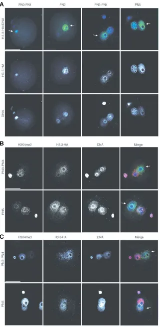
results. We injected mRNA for HA-tagged H3.3 into zygotes at the fertilisation cone stage, that is, right after fertilisation and before pronuclear formation. We then analysed the distribution of H3.3-HA at similar pronuclear stages as above. The results shown in Figure 3A demonstrate that the tagged version of H3.3 recapitu-lates the distribution pattern of the endogenous H3.3. Namely, we first observe a clear enrichment of H3.3-HA in the male
results also suggest that the preferential incorpora-tion in the male pronucleus is inherent to H3.3, regardless of whether it is the endogenous protein or a tagged injected one. This indicates that there is no obvious requirement for pre-existing covalent modified H3.3 for its appropriate deposition.
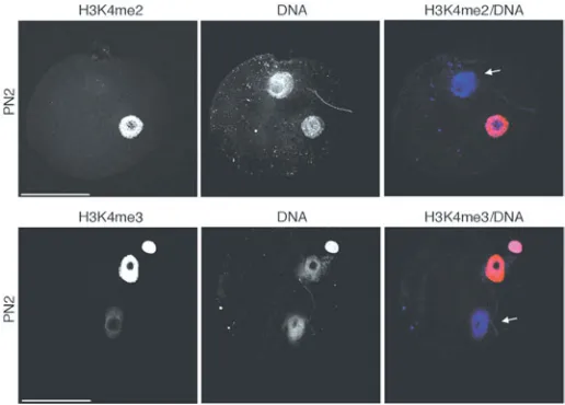
The female pronucleus is enriched in mono and trimethylated H3K4, defining an epigenetic asym-metry between the two pronuclei, which disappears at the PN5 stage (Lepikhov and Walter, 2004)(Fig. 3C). To unequivocally distinguish between the male and female pronucleus, we performed double immunostaining for H3.3-HA and H3K4me3 from PN3 through PN5 stages. These experiments con-firmed that the male pronucleus is enriched in H3.3-HA (Fig. 3C). We also analysed the distribution of H3K4me2 from PN3 through PN5. We find that the levels of H3K4me2 are much higher in the female pronucleus, but in contrast to H3K4me3, they re-main asymmetric throughout all the pronuclear stages analysed (Fig. 3B). Unexpectedly thus, the female pronucleus is enriched in H3K4 mono, di and trimethylation, which are ‘active’ marks, whilst H3.3 is enriched in the male pronucleus. Moreover, we find that at the PN2 stage, when H3.3 is clearly restricted to the male pronucleus, H3K4me2 is not detected in the paternal chromatin and the levels of H3K4me3 are low (Fig. 4). This data suggests that in the mouse embryo, H3.3 is hypomethylated at K4 and that H3.3 may progressively become methy-lated as the first cell cycle progresses.
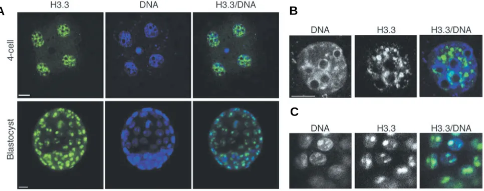
We then analysed the expression of histone H3.3 in 4-cell stage embryos and in the blastocyst. The histone H3.3 is readily detected in the four nuclei of the 4-cell stage embryos and the fluores-cent signal is much stronger than in 2-cell stage embryos (Fig. 5A). H3.3 exhibits a widespread nuclear distribution with some accumulation in a few dots in the nucleoplasm and is absent from the densely DNA-stained heterochromatic regions and from around the nucleolar-like bodies (Fig. 5B). Thus, its localization is predominantly euchromatic at the 4-cell stage. In the blastocyst, H3.3 is also
Fig. 3. HA tagged H3.3 is incorporated first in the male pronucleus and the female pronucleus reaches equivalent levels only at the PN5 stage.(A) Distribu-tion of HA-tagged H3.3 in the mouse zygote at different pronuclear stages. Zygotes were microinjected at the fertilisation cone stage with HA.H3.3 mRNA, cultured to the indicated pronuclear stages, fixed and processed for immunofluorescence using an anti HA antibody (green).
(B,C)Embryos were processed as in (A) but they were also stained with an antibody specific for dimethyl- (B) or trimethyl (C) H3K4 (red in the merge panel). The female pronucleus is distinguishable due to the enrichment in methylated H3K4. In all panelsDNA is shown in blue in the merge composites (top). Shown are stack Z-series projections of representative embryos. Scale bar repre-sents 50 µm. The male pronucleus is indicated with an arrow and the pronuclear stages are indicated.
A
B
widely expressed in the nuclei of all cells. We do not detect any obvious difference in the H3.3 content between the inner cell mass and the trophectoderm (Fig. 5A). Analysis of blastocyst cells under higher magnification revealed that H3.3 was widespread in the nucleoplasm but accumulated within two foci of much greater intensity in each nucleus of both the inner cell mass (not shown) and the trophectoderm (Fig. 5C). Thus, in the blastocyst, the pattern of nuclear distribution of histone H3.3 is also different compared to the one in the 4-cell stage embryo.
Discussion
The mouse embryo undergoes reprogramming of its epigenetic marks during the first cell cycles (Li, 2002, Morgan et al., 2005). The epigenetic status of each cell within the embryo will be determined not only by differences in DNA methylation and histone modifica-tions, but also by the content of histone and histone variants. Here we have shown that the histone H3 variant, H3.3, is deposited specifically in the male pronucleus. Moreover, there seems to be a dicotomy since the male pronucleus does not have active methylation marks in K4 of histone H3 at the early stages of pronuclear formation.
The zygote gradually acquires a permissive state for transcrip-tion (Latham et al., 1992). Thus, the alterations in chromatin structure that follow fertilisation could contribute to rendering the zygote transcriptionally competent. The observed incorporation of H3.3, which has been reported to be enriched at active genes and to contain active marks (McKittrick et al., 2004, Schwartz and Ahmad, 2005), could contribute to the acquisition of this state.
Asymmetries in the epigenetic status of the parental genomes occur in the zygote. These include active DNA demethylation in the paternal genome but not in the maternal one and accumulation of H3K4me1, H3K9me2 and H3K27me3 in the female pronucleus (Arney et al., 2002, Lepikhov and Walter, 2004, Santos et al., 2005). In contrast, the male pronucleus displays higher levels of histone H4 acetylation than the maternal chromatin (Adenot et al., 1997). However, histone H4 acetylation and methylation of H3K4 reach similar levels in both pronuclei at the end of the zygote stage and the main differences between them remain in heterochromatic structures and imprinting (Santos et al., 2005). Here we show, consistent with the differences in histone modifications, that there is also an asymmetry in the incorporation of H3.3 into the pronuclei. This asymmetry disappears at the late zygote stage. Our results are in line with recent work by van der Heijden and colleagues, who reported enrichment of H3.1 in the female pronucleus but not in the male one, leading the authors to suggest that the male pronucleus would be enriched in H3.3 (van der Heijden et al., 2005). Changes in the histone composition in the male pronucleus may be neces-sary for the first wave of transcriptional activation that starts first in the paternal genome, which supports higher levels of transcription than the maternal pronucleus (Aoki et al., 1997, Bouniol et al., 1995, Bouniol-Baly et al., 1997). Additionally, the differential incor-poration of H3.3 into the pronuclei may also contribute to ‘delay’ the incorporation of repressive marks into the paternal genome (Arney et al., 2002, Lepikhov and Walter, 2004, Santos et al., 2005).
We observed changes in the pattern of nuclear distribution of histone H3.3. A characteristic punctate pattern found at the 2-cell stage changes into a more widely dispersed at the 4-cell stage. In the blastocyst, we observed a characteristic enrichment in two
spots in the nucleoplasm. It is interesting to note that these changes correlate with the three major changes in transcript profile in the embryo: the first one up to the 2-cell stage, the second one after the 4-cell stage and the third one that leads to cellular differentiation in the blastocyst (Hamatani et al., 2004, Wang et al., 2004).
Deposition of histone H3.3 in somatic cells can occur in both replication-dependent or independent mechanisms (Ahmad and Henikoff, 2002a, Ahmad and Henikoff, 2002b). Histones are first detected in the male pronucleus at the early PN1 stage (Adenot et al., 1997, Arney et al., 2002, Lepikhov and Walter, 2004). We observed incorporation of H3.3 in the male pronucleus around the PN2 stage, before the onset of transcription activation, suggesting that the deposition of H3.3 in the zygote may not (at least initially) be coupled to genome activation. Indeed, HIRA, which is known to mediate H3.3 deposition in somatic cells (Tagami et al., 2004) was recently reported to be present in the sperm nucleus during decondensation immediately after fertilization and before genome activation (van der Heijden et al., 2005). Given that replication in the zygote starts at ~21h phCG and that BrdU incorporation is not detected in PN1 and PN2 embryos (Adenot et al., 1997, Bouniol-Baly et al., 1997), it is possible that H3.3 incorporation is also independent of DNA synthesis. Morever, the punctate localization pattern that we observed for H3.3 in the zygote persists until the late 2-cell stage when two rounds of replication have already occurred. So it is conceivable that the incorporation of H3.3 in the zygote occurs by a different mechanism independent of replication and transcription. A recent report has indeed documented that in Drosophila, HIRA is required for removal of the protamines (Jayaramaiah Raja and Renkawitz-Pohl, 2005) and for chromatin assembly in the male pronucleus (Loppin et al., 2005). Moreover, maternal H3.3-Flag specifically accumulates in the male pro-nucleus, suggestive of a replication independent nucleosome assembly at the genome wide level.
Genetic ablation of some of the histone modifiers that are involved in the establishment of epigenetic asymmetries in the zygote such as Enhancer of Zeste 2 results in impaired develop-ment (Erhardt et al., 2003, Santos et al., 2002). A hypomorphic mutation of the H3f3a gene, coding for H3.3, results in male sub-fertility (Couldrey et al., 1999). Thus, it will be interesting to determine whether H3.3 has a role in the reprogramming of the parental genomes that follow fertilisation. Investigating not only the changes in chromatin modifications but also histone composition during this period, will help to understand the mechanisms under-lying these events. Our results, which analyse the distribution of histone H3.3 throughout preimplantation development make a contribution towards this goal.
Experimental Procedures
Embryo collection and culture
Embryos were collected from F1 (C57BL/6 x CBA/H) ~6 weeks old superovulated females that were crossed with F1 males as described (Hogan et al., 1994). Zygotes and cleavage stage embryos were collected
at the indicated hours post-hCG (h phCG). Oocytes were recovered from ~6 weeks old F1 females as described by Hogan et al (Hogan et al., 1994).
Experiments with animals were carried out according to the Home Office regulation. Pronuclear stages (PN) were classified according to Adenot et al (1997; TableI and Fig. 3 therein, according to both, post-hCG time and the distance between pronuclei) and corresponded approximately to the following times after hCG injection in our strain and Animal Facility: PN0-PN1, 18h phCG; PN2, 20h phCG; PN3-PN4, 24h phCG; PN5, 27h phCG. For the BrUTP experiments, we refer to the hCG timing throughout the text so that the experimental design is clearer to understand.
Immunostaining and confocal analysis
After removal of the zona pellucida with acid Tyrode’s solution (Sigma), embryos were washed three times in PBS and fixed in 5% paraformalde-hyde, 0.04% Triton, 0.3% Tween and 0.2% sucrose in PBS for 20 minutes
at 37°C. After permeabilisation with 0.5% Triton in PBS for 20 minutes, the embryos were washed three times in PBS-T (0.1% Tween in PBS), blocked in 3% BSA in PBS-T and incubated with the primary antibodies (H3.3 ab4263, abcam, 1:75 dilution; anti histone H3 dimethyl K4 ab7766, abcam, 1:150 dilution; anti histone H3 trimethyl K4 ab8580, 1:150 dilution; anti HA antibody clone 3F10, Roche, 1:500 dilution) for ~12 h at 4°C. It is essential to note that we used an early batch of the H3.3 antibody, which was affinity purified from the first serum. This batch was fully characterized (Fig. 1A) and was shown to recognise nucleosomal histone H3.3 in vivo in ChIP
assays by two independent laboratories (Johnson et al., 2004; Daujat, S. personnal communication). Embryos were then washed twice in PBS-T, blocked for 30 minutes and incubated for 2 h at 25°C with the corresponding secondary antibodies (FITC-conjugated donkey anti rabbit IgG, Jackson ImmunoResearch). After 2 washes in PBS-T, the DNA was stained with TOTO-3 (Molecular Probes) and the embryos were mounted in Vectashield (Vector Laboratories). Confocal microscopy was performed using a 40x oil objective in a BioRad 1024 inverted microscope (Blastocysts) or a 60x oil objective in a BioRad Radiance Upright Confocal Laser Microscope using the BioRad LaserSharp 2000 software (all the other stages). All the stainings were repeated independently at least two times with at least 10 embryos analysed per stage.
BrUTP labelling
BrUTP labelling was performed as described (Borsuk and Maleszewski, 2002)(Torres-Padilla and Zernicka-Goetz, submitted). Embryos were collected at 20h phCG and microinjected using an Eppendorf Transjector 5246 with 1-2 pl of 100 mM BrUTP (5-bromo UTP, Sigma) in 2mM PIPES, 140mM KCl, pH 7.4. Embryos were fixed after culture at the time equivalent to 24 and 27h phCG and processed for immunostaining using an anti-BrdU antibody (1:100 dilution, Sigma).
mRNA microinjection
Zygotes were collected at the fertilisation cone stage (~17h phCG) and microinjected with 1-2 pl of 500 ng/µl of C-terminal double HA-tagged (human) histone H3.3 mRNA capped and transcribed in vitro. Zygotes were then cultured in KSOM under a %5 CO2 atmosphere at 37°C, fixed at the different pronuclear stages as indicated in the figure legends and
Fig. 5. Distribution of H3.3 in mouse 4-cell stage embryos and blastocyst.(A)Embryos were collected from the oviduct, processed for immunofluorescence with the H3.3 antibody and analysed under confocal microscopy. H3.3 is shown in green, DNA in blue. Shown are stack, single channel and merge confocal sections of representative embryos at the indicated stages. Scale bar represents 10 µm.(B) Higher magnification of one of the nuclei of the 4-cell stage embryo shown in (A). Note that H3.3 is distributed throughout the nucleoplasm but is absent from the densely stained heterochromatic regions. Shown are single confocal sections from single channel acquisitions and the corresponding merge image. Scale bar represents 10 µm. (C) Trophectoderm cells of the blastocyst shown in (A). Representative sections derived from single channel acquisition and merge image are shown.
A
B
processed for immunofluorescence. At least 20 embryos per stage from two independent experiments were analysed.
Western blot
Two micrograms of core histone preparation (500 ng of each, SIGMA) or 1 µg of purified bacterially produced histone H3.2 were resolved by SDS-PAGE, blotted to nitrocellulose and probed O.N. with the anti H3.3 antibody (1:1000 dilution). Competitor peptides were added as indicated at a concentration of 1 µg/ml. The H3.3 antibody used (ab4263, Batch 34119) was obtained from Abcam Ltd., UK. It was thoroughly characterised with regards to its specificity. Due to production issues, it is no longer commercially available.
Acknowledgments
This work was supported by the Wellcome Trust. M.Z-G. is a Wellcome Senior Research Fellow. M-E.-T-P. is a Long Term EMBO fellow.
References
ADENOT, P.G., MERCIER, Y., RENARD, J.P. and THOMPSON, E.M. (1997). Differential h4 acetylation of paternal and maternal chromatin precedes DNA replication and differential transcriptional activity in pronuclei of 1-cell mouse embryos. Development 124: 4615-25.
AHMAD, K. and HENIKOFF, S. (2002a). Histone h3 variants specify modes of chromatin assembly. Proc Natl Acad Sci USA 99 Suppl 4: 16477-84.
AHMAD, K. and HENIKOFF, S. (2002b). The histone variant h3.3 marks active chromatin by replication-independent nucleosome assembly. Mol Cell 9: 1191-200.
AOKI, F., WORRAD, D.M. and SCHULTZ, R.M. (1997). Regulation of transcrip-tional activity during the first and second cell cycles in the preimplantation mouse embryo. Dev Biol 181: 296-307.
ARNEY, K.L., BAO, S., BANNISTER, A.J., KOUZARIDES, T. and SURANI, M.A. (2002). Histone methylation defines epigenetic asymmetry in the mouse zygote. Int J Dev Biol 46: 317-20.
BORSUK, E. and MALESZEWSKI, M. (2002). DNA replication and rna synthesis in thymocyte nuclei microinjected into the cytoplasm of artificially activated mouse eggs. Zygote 10: 229-38.
BOUNIOL, C., NGUYEN, E. and DEBEY, P. (1995). Endogenous transcription occurs at the 1-cell stage in the mouse embryo. Exp Cell Res 218: 57-62.
BOUNIOL-BALY, C., NGUYEN, E., BESOMBES, D. and DEBEY, P. (1997). Dynamic organization of DNA replication in one-cell mouse embryos: Relation-ship to transcriptional activation. Exp Cell Res 236: 201-11.
COULDREY, C., CARLTON, M.B., NOLAN, P.M., COLLEDGE, W.H. and EVANS, M.J. (1999). A retroviral gene trap insertion into the histone 3.3a gene causes partial neonatal lethality, stunted growth, neuromuscular deficits and male sub-fertility in transgenic mice. Hum Mol Genet 8: 2489-95.
ERHARDT, S., SU, I.H., SCHNEIDER, R., BARTON, S., BANNISTER, A.J., PEREZ-BURGOS, L., JENUWEIN, T., KOUZARIDES, T., TARAKHOVSKY, A. and SURANI, M.A. (2003). Consequences of the depletion of zygotic and embryonic enhancer of zeste 2 during preimplantation mouse development. Development 130: 4235-48.
GAUTIER, T., ABBOTT, D.W., MOLLA, A., VERDEL, A., AUSIO, J. and DIMITROV, S. (2004). Histone variant h2abbd confers lower stability to the nucleosome. EMBO Rep 5: 715-20.
HAMATANI, T., CARTER, M.G., SHAROV, A.A. and KO, M.S. (2004). Dynamics of global gene expression changes during mouse preimplantation development. Dev Cell 6: 117-31.
HOGAN, B.L., BEDDINGTON, R., COSTANTINI, F. and LACY, E. (1994). Manipu-lating the mouse embryo. Cold Spring Harbor Laboratory Press.
JANICKI, S.M., TSUKAMOTO, T., SALGHETTI, S.E., TANSEY, W.P., SACHIDANANDAM, R., PRASANTH, K.V., RIED, T., SHAV-TAL, Y., BERTRAND, E., SINGER, R.H. et al. (2004). From silencing to gene expres-sion: Real-time analysis in single cells. Cell 116: 683-698.
JAYARAMAIAH RAJA, S. and RENKAWITZ-POHL, R. (2005). Replacement by drosophila melanogaster protamines and mst77f of histones during chromatin condensation in late spermatids and role of sesame in the removal of these proteins from the male pronucleus. Mol Cell Biol 25: 6165-77.
KAMAKAKA, R.T. and BIGGINS, S. (2005). Histone variants: Deviants? Genes Dev 19: 295-310.
LATHAM, K.E., SOLTER, D. and SCHULTZ, R.M. (1992). Acquisition of a transcrip-tionally permissive state during the 1-cell stage of mouse embryogenesis. Dev Biol 149: 457-62.
LEPIKHOV, K. and WALTER, J. (2004). Differential dynamics of histone h3 methylation at positions k4 and k9 in the mouse zygote. BMC Dev Biol 4: 12.
LI, E. (2002). Chromatin modification and epigenetic reprogramming in mammalian development. Nat Rev Genet 3: 662-73.
LOPPIN, B., BONNEFOY, E., ANSELME, C., LAURENCON, A., KARR, T.L. and COUBLE, P. (2005). The histone h3.3 chaperone hira is essential for chromatin assembly in the male pronucleus. Nature 437: 1386-90.
MCKITTRICK, E., GAFKEN, P.R., AHMAD, K. and HENIKOFF, S. (2004). Histone h3.3 is enriched in covalent modifications associated with active chromatin. Proc Natl Acad Sci USA 101: 1525-30.
MORGAN, H.D., SANTOS, F., GREEN, K., DEAN, W. and REIK, W. (2005). Epigenetic reprogramming in mammals. Hum Mol Genet 14 Spec No 1: R47-58.
SANTOS, F., HENDRICH, B., REIK, W. and DEAN, W. (2002). Dynamic reprogram-ming of DNA methylation in the early mouse embryo. Dev Biol 241: 172-82.
SANTOS, F., PETERS, A.H., OTTE, A.P., REIK, W. and DEAN, W. (2005). Dynamic chromatin modifications characterise the first cell cycle in mouse embryos. Dev Biol 280: 225-36.
SARMENTO, O.F., DIGILIO, L.C., WANG, Y., PERLIN, J., HERR, J.C., ALLIS, C.D. and COONROD, S.A. (2004). Dynamic alterations of specific histone modifica-tions during early murine development. J Cell Sci 117: 4449-59.
SCHWARTZ, B.E. and AHMAD, K. (2005). Transcriptional activation triggers deposition and removal of the histone variant h3.3. Genes Dev 19: 804-14.
TAGAMI, H., RAY-GALLET, D., ALMOUZNI, G. and NAKATANI, Y. (2004). Histone H3.1 and H3.3 complexes mediate nucleosome assembly pathways dependent or independent of DNA synthesis. Cell 116: 51-61.
VAN DER HEIJDEN, G.W., DIEKER, J.W., DERIJCK, A.A., MULLER, S., BERDEN, J.H., BRAAT, D.D., VAN DER VLAG, J. and DE BOER, P. (2005). Asymmetry in histone h3 variants and lysine methylation between paternal and maternal chromatin of the early mouse zygote. Mech Dev 122: 1008-22.
WANG, Q.T., PIOTROWSKA, K., CIEMERYCH, M.A., MILENKOVIC, L., SCOTT, M.P., DAVIS, R.W. and ZERNICKA-GOETZ, M. (2004). A genome-wide study of gene activity reveals developmental signaling pathways in the preimplanta-tion mouse embryo. Dev Cell 6: 133-44.
WIEKOWSKI, M., MIRANDA, M., NOTHIAS, J.Y. and DEPAMPHILIS, M.L. (1997). Changes in histone synthesis and modification at the beginning of mouse development correlate with the establishment of chromatin mediated repres-sion of transcription. J Cell Sci 110 (Pt 10): 1147-58.