From field to gel blot: teaching a holistic view of
develop-mental phenomena to undergraduate biology students
at the University of Tokyo
TAKASHI ARIIZUMI
1and MAKOTO ASASHIMA*
,1,21SORST, Japan Science and Technology Corporation, The University of Tokyo, and 2Department of Life Sciences (Biology), Graduate School of Arts and Sciences, The University of Tokyo, Japan
ABSTRACT We present here an outline of the lectures and laboratory exercises for undergraduate developmental biology students at the University of Tokyo. The main aim of our course is to help students fill the gap between natural history, classical embryology and molecular developmental biology. To achieve this aim, we take up various topics in the lectures, from fertilization and early development to developmental engineering. Our laboratory exercises begin with an introduction to the natural history of the organism. The entire class and the instructors collect newts in the field and discuss features of their mating behavior and so on. In the laboratory, students are absorbed by exercises such as a lampbrush chromosome preparation and an in vitro beating heart induction. After that, students choose their own research projects for which they will employ both classical embryological and modern molecular biological techniques. At the end of our course, the connec-tivity principle from field to gel blot will be part of the students’ understanding.
KEY WORDS:
developmental biology, teaching, connectivity, newt
0214-6282/2003/$25.00 © UBC Press
Printed in Spain www.ijdb.ehu.es
*Address correspondence to: Dr. Makoto Asashima. Dept. of Life Sciences (Biology), Graduate School of Arts and Sciences, The University of Tokyo, Tokyo 153-8902, Japan. Fax: +81-3-5454-4330. e-mail: asashi@bio.c.u-tokyo.ac.jp
Bacground Information
Scholarly Interests of the Authors
1) Characterization of the cellular and molecular basis of em-bryonic induction, body axis formation, and organogenesis. 2) Establishment of the in vitro organ induction systems using
embryonal pluripotent cells.
Representative Publications
ARIIZUMI, T. and ASASHIMA, M. (2001). In vitro induction systems for analyses of amphibian organogenesis and body patterning. Int. J. Dev. Biol. 45: 273-279. ARIIZUMI, T., KOMAZAKI, S., ASASHIMA, M. and MALACINSKI, G.M. (1996).
Activin-treated urodele ectoderm: A model experimental system for cardiogenesis. Int. J. Dev. Biol. 40: 715-718.
ASASHIMA, M. Mesoderm induction during early amphibian development. (1994). Dev. Growth Differ. 36: 343-355.
ASASHIMA, M., NAKANO, H., SHIMADA, K., KINOSHITA, K., ISHII, K., SHIBAI, H. and UENO, N. (1990). Mesodermal induction in early amphibian embryos by activin A (erythroid differentiation factor). Roux’s Arch. Dev. Biol. 198: 330-335.
ASASHIMA, M., NAKANO, H., UCHIYAMA, H., SUGINO, H., NAKAMURA, T., ETO, Y., EJIMA, D., NISHIMATSU, S., UENO, N., and KINOSHITA, K. (1991). Presence of activin (erythroid differentiation factor) in unfertilized eggs and blastulae of Xenopus laevis. Proc. Natl. Acad. Sci. USA 88: 6511-6514.
ASASHIMA, M., KINOSHITA, K., ARIIZUMI, T., and MALACINSKI, G.M. (1999). Role of activin and other peptide growth factors in body patterning in the early amphibian embryo. Int. Rev. Cytol. 191: 1-52.
General Teaching Philosophy
Our philosophy is based on the premise that all developmen-tal phenomena studied in the classroom should be viewed in the context of the natural circumstances under which they evolved. Similarly, all the developmental concepts and principles which emerge from observations in the artificial circumstances of the laboratory should be interpreted in terms of the environment in which the organism evolved. It is our personal opinion, there-fore, that a continuum exists between the pond in which the newt lays its eggs and the timing of gene expression in an egg which is artificially inseminated on the laboratory bench. Our aim is to help undergraduate students understand that connec-tivity principle. We attempt to achieve that aim by (1) taking students on field trips to collect the amphibia they will use as model organisms in classroom exercises; and (2) by introduc-ing students to both classical embryology and modern molecu-lar gene expression technology for learning about developmen-tal mechanisms.
General Features of the Course
of Tokyo, and their average age is 20 years. Prior to taking this course, they have studied several aspects of biology, including cell biology, molecular genetics, ecology, taxonomy, plant biol-ogy, physiolbiol-ogy, and biochemistry. Thus, they are well pre-pared for a rigorous lecture and laboratory schedule. In addi-tion, due to the strict entrance requirements of the University of Tokyo, they are—on average—very effective at learning and understanding the detailed features of the complex phenomena (e.g., organogenesis) that we present in this course. Following the completion of the formal schedule of this course (see Table 1), students work on individual research projects in our re-search laboratory. Those projects often represent one or an-other aspect of a subject discussed in the formal course sched-ule.
The course schedule consists of 2 h of lecture per week for each of 15 weeks. At the end of the course, there is one examination. Associated with the lecture section of the course is a set of laboratory exercises, which are designed to introduce students to laboratory animal maintenance methods, traditional as well as modern methodologies, and the independent study strategies which are common to professional research endeav-ors.
Fig. 1. Students and professor collecting newts as a class project in Niigata prefecture in Northern Japan.
Lecture Topics
A main agenda of our course is to bridge the gap between natural history, classical embryology, and molecular develop-mental biology. A list of lecture topics is included in Table 1. Lectures are guided by a pair of textbooks (both in Japanese): Developmental Biology (Asashima, 1996) and Molecular Devel-opmental Biology: Body Plan Formation in Animal Development (Asashima and Komazaki, 2000).
Examination Questions
The single examination at the end of the course takes 90 min to complete. Typical questions include the following:
(1) When we compare the germ cells, egg and sperm, their size and formation processes are different.
Q1. What events will occur during oogenesis and spermato-genesis? Describe in detail the morphology of these germ cells and the molecular events with which they are associated.
Q2. Establishment of polarity in unfertilized egg is very important. Explain the meaning and importance of polarity in
embryonic development.
(2) Cell divisions begin after fertiliza-tion. Explain and illustrate the cell cycle using the following words:. M phase, S phase, G1 phase, G2 phase, G0 phase, Ca2+, cyclin, MPF, cdc-2, phosphorylation, dephosphorylation, cell differentiation, CSF, mitosis
(3) Embryonic induction is very im-portant for the establishment of the fundamental body plan.
Q1. Illustrate H. Spemann’s and H. Mangold’s organizer experiment and explain the meaning and importance of their experiment in the field of de-velopmental biology.
Q2. The processes of cleavage and gastrulation are important for the em-bryo to become “a whole organism.” Explain by refering to the following two points: (1) gradient and (2) axis formation.
Field Trip to collect
Experimen-tal Material
Japan. It is our firm belief that significant understanding of several aspects of developmental biology can be gained by viewing a life cycle from the point of view of the subject organism itself, in its natural environment. In a sense, there-fore, our laboratory exercises begin with an introduction to the natural history of the organism before proceeding to an analysis of the organism’s developmental processes in the laboratory.
Laboratory Exercises
Several student-oriented goals are built into the laboratory exercise schedule. Included is attainment of an understanding of the discipline of developmental biology as encompassing a wide variety of investigative strategies, including traditional observa-tions made with various microscopes, microsurgical operaobserva-tions and manipulations, and modern molecular biology assay meth-ods.
Another goal is for students to become acquainted with the concept of “change.” As simplistic as that may appear, we find that most of our students have had only limited experience with the concept of change in a “real-time” sense. Although most general biology textbooks and Internet sites illustrate stage series of developing embryos, grasping the concept of change in size, shape, and function as something more than an abstract notion is best achieved by observing living embryos. In this way, change can be understood to be multidimensional, with several phases taking place simultaneously. Thus, the apparent sim-plicity of the concept of change does indeed become more complex to the student as increase in size (growth); enhance-ment of body form (morphogenesis); gain of function (move-ment); and appearance of specialized features (differentiation) are observed in live specimens to be occurring at the same time.
These laboratory exercises are carried out over a period of 2 weeks. Students are scheduled to work in the laboratory each day of the week from 1:00 P.M. until 4:00 P.M. They do, however,
routinely remain working in the laboratory until mid-evening (7-8 P.M.) or later. Table 2 lists typical exercises. For most
stu-dents, these exercises represent the first time in their under-graduate training that they have manipulated living material. Thus, their enthusiasm is high, and motivation to work on a
TABLE 1
LECTURE (30 H TOTAL) TOPICS FOR THE UNDERGRADUATE DEVELOPMENTAL BIOLOGY COURSE
1. Background and history of modern developmental biology 2. Gametogenesis: formation of egg and sperm
3. Reproduction and fertilization: initiation of development 4. Cleavage and early development: origin of multicellularity 5. Body axis formation: gastrulation and neural morphogenesis 6. Organogenesis
7. Metamorphosis
8. Regeneration and tumor formation 9. Aging: development and evolution 10. Plant developmental biology 11. Molecular developmental biology 12. Developmental engineering
project until completion is strong. After all, for many students, it was their fascination with living organisms and life’s pro-cesses that attracted them to study biology in college! Conse-quently, a student will occasionally spend most of a night in the laboratory making extra observations. Detailed laboratory re-ports are required for each of the exercises.
In order to make this initial laboratory encounter with living organisms as complete as possible, students are first trained in laboratory animal husbandry methods. Taking care of the am-phibia, for example, becomes a component of the typical labora-tory routine. The response of students is predictable: At first, they are hesitant to handle the animals (e.g., Xenopus frogs). But by the end of the course they feel so confident about their relation-ship to laboratory animals that they often take embryos home and raise them as pets.
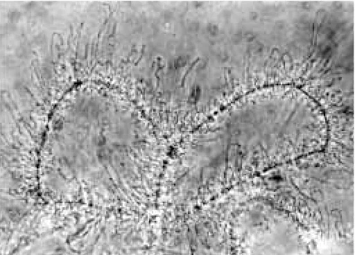
Fig. 2. Example of a lampbrush chromosome preparation prepared by undergraduate students. Germinal vesicles were isolated from newts, opened gently with watchmaker forceps and allowed to settle in micro-scope wells. The observations were made at 800× by using a phase-contrast microscope equipped with black-and-white camera film. Students are often surprised at the large size and complex morphology of these chromosomes. They take photos of their preparations and mount them in their laboratory report notebooks.
TABLE 2
LABORATORY EXERCISES USUALLY PERFORMED DURING OUR INTENSIVE (DAILY) LABORATORY COURSE
Protocols for these exercises are included as a section of a large laboratory manual (in Japanese), which is used for several biology courses, titled Life and Cognitive Science Experiment (Dept. of Life Sciences, 2001).
1. Artificial insemination of anuran (Xenopus) and urodele (newt) eggs 2. Membrane removal, fixation, and observation of internal embryo 3. Developmental staging and temperature effects
4. Microsurgery: primary embryonic organizer 5. Observation of lampbrush chromosomes 6. Nuclear (chromosome) division in larvae
7. Experimental manipulation: nuclear equivalence with egg ligation 8. Animal cap tissue culture differentiation
One of the laboratory exercises which students find most remarkable is the wet-mount observation of lampbrush chromo-somes (e.g., Komazaki and Asashima, 1979, 1981; Mori and Asashima, 1984). Seeing the chromosomes spread out gener-ates both a feeling of accomplishment and a sense of awe. A typical preparation is illustrated in Fig. 2.
Of all the laboratory exercises, however, the one students find the most exciting is the “animal cap assay.” Under proper circum-stances (e.g., Ariizumi and Asashima, 2001; Ariizumi et al., 1996, 1999), several students will succeed in generating blastula-stage animal cap organ cultures, which develop—after several days— into a beating heart. Treatment of animal caps in a high concen-tration of activin solution yields (for some students) cultures which rhythmically beat at the same rate as the heart beats in whole, untreated embryos (e.g., Ariizumi et al., 1996). The experimental protocol is diagrammed in Fig. 3.
Independent Research Projects
From an assortment of topics (including embryonic induction, cell differentiation, organogenesis, regeneration, etc.) students
TABLE 3
EXAMPLES OF TYPICAL 1-YEAR STUDENT RESEARCH PROJECTS
Project 1 Cloning and analysis of nodal-related genes in Xenopus tropicalis embryos
Technology cDNA library formation, DNA sequencing, microinjection technique
Project 2 Cloning and functional analysis of pronephros-related genes from the Xenopus animal cap treated with activin and retinoic acid Technology Animal cap assay, cDNA library formation, DNA sequencing,
micro-injection technique
Project 3. Formation of beating heart in vitro from Xenopus animal cap cells. Technology Animal cap assay, RT-PCR, histology
Project 4 Formation of sensory organs in vitro and transplantation into host embryos.
Technology Animal cap assay, microsurgery, transplantation technique
full-time research in our laboratory. Just as the regular laboratory exercises de-scribed above represent an extension of our normal research activities, these projects also build upon expertise avail-able in our research programs. Each of these projects lasts 1 year, and is drawn from a range of topics (Table 3). Among the many techniques they employ are nucleic acid extraction and hybridiza-tion, gel blots, vector ligahybridiza-tion, DNA clon-ing, and egg injection.
A Rewarding Experience for Both
the Student and the Professor
Like many life science professors, our interests in biology can be traced back to childhood. We began by observing the changing seasons in local ponds and by collecting insects and other live speci-mens, all the while admiring both their simplicities and their complexities. As we interact with students—especially in the field—we are able to recapitulate those early days and recapture some of the excitement and enthusiasm which origi-nally attracted us to a career in biology. In the laboratory, viewing the unfolding of embryonic patterns is exhilarating, for we are reminded of the splendor of life’s humble beginnings. Being able to share those experiences, over and over again with each new class, provides us with a continual spiritual renewal. This renewal comes from observing the enthusiasm students display as much as from ob-serving the embryos themselves. choose a subject for experimentation. Once a suitable topic is chosen, students develop a project in consultation with the various graduate students and postdoctoral fellows who perform
References
ARIIZUMI, T. and ASASHIMA, M. (2001). In vitro induction systems for analyses of amphibian organogenesis and body patterning. Int. J. Dev. Biol. 45: 273-279. ARIIZUMI, T., TAKANO, K., ASASHIMA, M. and MALACINSKI, G.M. (1999).
Bioas-says of inductive interactions in amphibian development. In Methods in Molecular Biology, Vol. 135: Developmental Biology Protocols, Vol. I (Eds. Tuan, R.S. and Lo, C.W.). Humana Press, Totowa, NJ, pp. 89-112.
ARIIZUMI, T., KOMAZAKI, S., ASASHIMA, M. and MALACINSKI, G.M. (1996). Activin treated urodele ectoderm: A model experimental system for cardiogenesis. Int. J. Dev. Biol. 40: 715-718.
ASASHIMA, M. (1996). Developmental Biology. Asakura Press, Tokyo.
ASASHIMA, M. and KOMAZAKI, S. (2000). Molecular Developmental Biology: Body Plan Formation in Animal Development. Shokabo Press, Tokyo.
DEPT. OF LIFE SCIENCES (BIOLOGY) (Ed.) (2001). Life and Cognitive Science Experiments. Dept. of Life Sciences (Biology), The University of Tokyo, Tokyo.
KOMAZAKI, S. and ASASHIMA, M. (1979). Gene expression during amphibian oogenesis. Bull. Yokohama City Univ. 29(2): 63-82. (in Japanese)
KOMAZAKI, S. and ASASHIMA, M. (1981). Chromosome condensation factors on the lampbrush chromosome of the newt oocyte. Bull. Yokohama City Univ. 32(1): 61-76. (in Japanese)