A systematic molecular genetic approach to study
mammalian germline development
KUNIYA ABE
1, MINORU S. H. KO
2and
GRANT R. MACGREGOR
3*
1Institute of Molecular Embryology and Genetics, Kumamoto University School of Medicine, Kumamoto, Japan, 2Center for Molecular Medicine and Genetics, Wayne State University School of Medicine, Detroit MI,
and 3Center for Molecular Medicine, Emory University School of Medicine, Atlanta, GA, USA
ABSTRACT It is difficult to study gene expression in mammalian embryonic germ cells as PGCs constitute only a minor proportion of the mouse embryo. We have overcome this problem by using a novel combination of established molecular and transgenic approaches. A line of mice has been generated in which the cells of the germ lineage express the β-galactosidase reporter gene during embryogenesis. Using this line, germ cells have been purified to near homogeneity from embryos at discrete stages during germline development by use of a stain for β-gal activity and a fluorescence activated cell sorter. Subsequently, cDNA libraries have been constructed from each germ cell population using a modified lone-linker PCR strategy. These combined cDNA libraries represent genes expressed in PGCs during mammalian germline development. To facilitate a molecular genetic approach to studying mammalian germline development, these cDNA libraries will be pooled to form an arrayed, addressed reference embryonic germ cell cDNA library. In parallel with large-scale cDNA sequencing efforts, genes that are differentially expressed in germ cells will be identified by screening the reference library with probes generated by subtractive hybridization. Complementary DNAs identified using this approach will be analyzed by sequencing, database comparison, genomic mapping and in situ hybridization to ascertain the potential functional importance of each gene to germline development. In addition to providing a wealth of novel information regarding patterns of gene expression during mammalian germline development, these results will form the basis for future experiments to determine the function of these genes in this process.
KEY WORDS:
germline, embryogenesis, mammalian, molecular genetic
0214-6282/98/$10.00
© UBC Press Printed in Spain
Introduction
Development of the germline is essential to the survival of a species. The primordial germ cells (PGCs) formed during mamma-lian embryogenesis ultimately give rise, in the male, to spermato-gonia, the only self-renewing cell type in an adult capable of making a genetic contribution to the next generation. Several important advances have recently been made in the ability to cryopreserve (Avarbock et al., 1996) and transplant (Brinster and Avarbock, 1994; Brinster and Zimmermann, 1994; Russell et al., 1996) this stem cell population to a host testis. Remarkably, apparently normal spermatogenesis can be generated following spermatogo-nial transplantation to testes of xenobiotic species (Clouthier et al., 1996; Russell and Brinster, 1996). Thus, contemporary mamma-lian germ cell research is of importance to both basic and applied
*Address for reprints: Center for Molecular Medicine, Emory University School of Medicine, 1462 Clifton Road, NE, Atlanta, GA 30322, USA. FAX: (404) 727.8367. e-mail: gmacg@bimcore.emory.edu
Abbreviations used in this paper: AIS, analytical imaging software; cDNA, complementary DNA; PGC, primordial germ cell; EEM, extra-embryonic mesoderm; MGF, mast cell growth factor; TNF-α, tumour necrosis factor -alpha; OSM, oncostatin M; LIF, leukemia inhibitory factor; IL-11, interleukin-11; RT-PCR, reverse transcriptase polymerase chain reaction; TNAP, tissue non-specific alkaline phosphatase; FACS, fluorescence activated cell sorter; MACS, magnetic antibody cell sorting; EST, expressed sequence tag; e7.5, embryonic day 7.5; FDG, fluorescein di-galactoside; MHC, major histocompatibility complex; LL-PCR, lone-linker - PCR; CCD, charge coupled device.
In this review we give a brief summary of the biology of germline formation during mouse embryonic development including current experimental approaches to identify genes whose function is required for this process. We conclude by describing a novel approach we have developed to facilitate a systematic molecular genetic study of mammalian germline development.
Origin and development of the mammalian germ
lineage
Germline formation in the mouse
Most of our current knowledge about the embryological origin and genetic basis for mammalian germline development has come from studies using the mouse. Cell transplantation or cell marking approaches have been used to assess the developmental potency of cells from the early mouse embryo. Results of these studies have provided insight about the location and time of formation of the mammalian germ lineage. Analysis of potency of cells from the pre-and early post-implantation embryo indicated that while the de-scendants of these cells can contribute to the germline, they do not do so exclusively (Gardner et al., 1985; Gardner and Beddington, 1988). Results of fate-mapping experiments involving labeling of individual cells from the proximal epiblast of egg-cylinder stage embryos indicated that the descendant of these cells could contrib-ute to both somatic and germ lineages (Lawson and Hage, 1994). Significantly, distal epiblast cells of an egg-cylinder stage embryo could also contribute to the germline upon grafting to a proximal epiblast location although the converse was not true (Tam and Zhou, 1996). Collectively, these results support the theory that the murine germline is not determined before gastrulation.
One of the difficulties in studying germline development in mammals concerns the paucity of markers specific for this lineage. In the mouse, PGCs can first be distinguished from somatic cells on the basis of their relatively high level of expression of tissue non-specific alkaline phosphatase (TNAP) within the extra-embryonic mesoderm (EEM) (McKay et al., 1953; Chiquoine, 1954; Ozdzenski, 1967; Ginsburg et al., 1990; MacGregor et al., 1995). Although alkaline phosphatase is a marker of the developing germ lineage in many species, its activity is not required for development of PGCs in mouse (MacGregor et al., 1995). While within the EEM, the PGCs are not tightly clustered but appear to be interspersed with additional cells that do not express TNAP. Approximately 50 TNAP positive cells can be found in this location at this time (Ginsburg et al., 1990; MacGregor et al., 1995). This figure is very similar to an estimate for the founding population size of the mouse germline of 45 that was generated from fate-mapping studies of Lawson and Hage (Lawson and Hage, 1994). These findings are consistent with a model in which the mouse germline is specified from approximately 50 cells within the EEM shortly after the onset of gastrulation.
The EEM in which the PGCs are found is closely bordered by extra-embryonic visceral endoderm. Local embryonic environ-ment appears to play a role in many inductive events during vertebrate embryogenesis. For example, visceral endoderm is involved in the induction of cardiac mesoderm in several species (Kelley et al., 1993; Schultheiss et al., 1995; Soudais et al., 1995; Narita et al., 1997). As with formation of hematopoietic cells within the EEM component of the yolk sac, it is interesting to speculate that the over-lying visceral endoderm may play some part in the
specification of the PGCs within the EEM. Snow (1981) demon-strated that PGCs would form in fragments isolated from the extreme posterior of e7 and e7.5 embryos following their culture in vitro. As discussed (Copp et al., 1986; Ginsburg et al., 1990), these fragments were likely to have been composed of EEM with over-lying extra-embryonic visceral endoderm. It would be of interest to determine if PGCs would still form in EEM in culture following removal of the associated endoderm or alternatively if heterotopic EEM could be induced to form PGCs following co-culture with visceral endoderm isolated from the region just posterior to the primitive streak.
Migration of PGCs during embryogenesis
After specification, the PGCs follow guidance cues that will ultimately lead them to the developing genital ridges. In addition, between the time of specification and arrival in the genital ridges, the PGCs must not only survive, but proliferate. Finally, during this period the PGCs must not respond to any inappropriate positional cues that would compromise their totipotency.
At around e8.0 (1-5 somite stage), the PGCs are associated with the hindgut pocket. Snow observed that pieces dissected from the posterior of early gastrulation stage embryos that were subse-quently cultured in vitro could form an allantois containing PGCs (Snow, 1981). However, if the cultured portion of the embryo also formed a hindgut, all PGCs would be localized around the endo-derm. This suggests that the PGCs have an affinity for the hindgut endoderm. Similar phenomena are also observed during Dro-sophila (Jaglarz and Howard, 1995), Xenopus (Whitington and Dixon, 1975) and avian (Fujimoto et al., 1976) embryonic develop-ment.
During the next day and a half, the growth of the hindgut appears responsible for the passive movement of the PGCs to an internal location within the developing embryo. However, at around the 5-15 somite stage (e8.5-9.0), PGCs begin an active migration from the hindgut endoderm, through the newly forming dorsal mesen-tery to the genital ridges which they begin to colonize at around the 20-30 somite stage (e9.5 to e10.0). Several lines of evidence suggest that the PGCs are guided to the developing gonadal ridge at least in part by chemotropic means (Kuwana et al., 1986; Godin et al., 1990; Godin and Wylie, 1991). During this migratory phase, the PGCs extend long filopodial processes that link them together in extensive arrays (Gomperts et al., 1994a). These filopodia disappear following the arrival of the PGCs in the genital ridge. The filopodia may play an adhesive role between PGCs during migra-tion as after arrival in the genital ridges, the PGCs are found to be tightly associated (Gomperts et al., 1994b).
to laminin in the early mouse embryo may involve expression of integrins and a heparin sulfate proteoglycan on the surface of the PGCs (Garcia-Castro et al., 1997). The migratory process is essentially completed by the 45 somite stage (e11.5-e12.0).
During movement from the hindgut pocket to the genital ridges, the number of PGCs increases from 100 to around 2500 (Tam and Snow, 1981; Buehr et al., 1993). Following their arrival in the genital ridges, the PGCs continue to divide until around e13.5 at which time they number approximately 25,000 (Chiquoine, 1954; Tam and Snow, 1981; Buehr et al., 1993). Results of in vitro culture and genetic studies suggest that several growth factors and other factors can influence the survival and/or proliferation of PGCs during embryogenesis. These include mast cell growth factor (MGF), leukemia inhibitory factor (LIF), tumor necrosis factor-α (TNF-α), interleukin-11 (IL-11), and oncostatin M (OSM) (Besmer et al., 1993; Wylie and Heasman, 1993; Cheng et al., 1994; De Felici and Pesce, 1994; Donovan, 1994; Kawase et al., 1994; Koshimizu et al., 1996).
Sexual differentiation of primordial germ cells
Following the onset of morphologic gonadal differentiation at around e13.5, PGCs differentiate into oogonia in the fetal ovary or M (mitotic)-prospermatogonia in the fetal testis. In females, a proportion of the oogonia enter meiotic prophase almost immedi-ately after the mitotic proliferation at e13.5 to form oocytes while in males the prospermatogonia continue to proliferate for an addi-tional day prior to entering mitotic arrest around e14.5. Most oogonia have formed oocytes in meiotic prophase by e15.5 while T (transition) - prospermatogonia continue to be held in G1-phase mitotic arrest until after birth (McLaren, 1988).
It is well established that the somatic component of the fetal testis produces a local factor(s) that inhibits meiosis in PGCs as well as directing the germ cells to develop as prospermatogonia. Experimental evidence to support this includes the finding that PGCs of either sex that end up in the adrenal instead of gonad enter meiosis before birth (Zamboni and Upadhyay, 1983). In addition, studies using inter-sex chimeric mice indicate that XX germ cells are subject to meiotic and mitotic arrest and differentiate into prospermatogonia in the fetal testis while XY PGCs enter meiosis in the fetal ovary and attempt to produce oocytes (McLaren, 1983,1993). Results of more recent studies involving tissue re-aggregation experiments indicated that the fetal testis produces a factor at around e12 that is responsible for inhibiting entry of prospermatogonia into meiosis (McLaren and Southee, 1997).
Genetic basis for germline development in the mouse
The molecular basis for germline development is perhaps best understood in Drosophila and Caenorhabditis where sophisticated genetic screens have provided the mutants required to help elucidate this process (Ellis and Kimble, 1994; Williamson and Lehmann, 1996). Strategies to perform large-scale mutagenesis in the mouse have been greatly assisted by the exploitation of simple sequence length polymorphisms (SSLP) that facilitate rapid ge-netic mapping. In addition, retroviral gene trapping is an efficient approach of mutagenesis that provides information about the pattern of expression of mutated genes as well a method to clone them (Gossler et al., 1989; Friedrich and Soriano, 1991; Skarnes et al., 1992,1995; Wurst et al., 1995; Forrester et al., 1996; Baker et al., 1997; Holzschu et al., 1997). However, despite such
techni-cal advances, the cost and space required to conduct a mutagen-esis screen for germline defects in the mouse is considerable as such a screen requires test breeding of F2 animals. This has forced the use of alternative experimental strategies to identify genes that have an essential function in mammalian germline development. Molecular genetic studies of early aspects of mouse germline development are particularly problematic as the lineage consti-tutes a relatively small population of cells within the embryo. Without sufficient quantities of pure PGCs, it is difficult to use a molecular approach to identify the genes whose function is re-quired for germline development. However, several different ge-netic approaches have been used to study genes that are ex-pressed in PGCs during murine embryogenesis. These have included empirical approaches e.g., RT-PCR using small amounts of PGC containing material or in situ hybridization of candidate genes to germ cell containing sections; RT-PCR based approaches using degenerate oligonucleotides that have specificity for differ-ent classes of gene products; or by cloning murine homologs of genes known to be expressed in embryonic germ cells in other species. Reverse-genetic approaches (i.e., knock-outs) have also been used to provide information regarding function of known genes in germline development. Finally, in a genetic and arguably most direct approach, the cloning of genes associated with the relatively few existing mutations that are known to affect germline
development can identify genes that a priori have an essential function during germline development.
Perhaps the best examples of the latter approach are mice mutant at the dominant white-spotting (W) and steel (Sl ) loci which have overlapping mutant phenotypes that include drastic reduc-tions in the number of gonadal PGCs found during embryogenesis (Besmer et al., 1993; Buehr et al., 1993; Donovan, 1994). Molecu-lar genetic analyses have revealed that the W locus encodes the c-kit tyrosine kinase transmembrane receptor while the product of the Sl locus, mast cell growth factor (MGF) is a natural ligand for the c-kit receptor. Further analyses have shown that c-kit is expressed by PGCs during germline development while the surrounding somatic mesenchyme expresses MGF. Although soluble MGF can act through the c-kit receptor to enhance survival of PGCs in culture, it is the transmembrane form of MGF that appears to be more important for germ cell survival in vivo as Sl d mice which
make the secreted form of MGF but lack the transmembrane form display a severe germ cell deficiency (Donovan, 1994; Marziali et al., 1993). Hertwig’s anemia (an), atrichosis (at), Teratoma (Ter) and germ cell deficient (gcd) are four additional mutations that affect germ cell development during embryogenesis although to date none of the genes affected in these mutants has been cloned (Lyon et al., 1996).
Reverse genetic approaches have also been used to analyze the role of growth factors in PGC proliferation and survival. As described earlier, LIF, OSM, TNF-α and IL-11 have all been shown to stimulate survival and/or proliferation of PGCs in culture. How-ever, mice deficient in LIF are not agametic (Stewart et al., 1992) and there is no overt difference in numbers of PGCs found in LIF deficient embryos compared to control animals (P. Donovan and C. Stewart, personal communication) As these cytokines appear to be both structurally and functionally related, it is possible that functional redundancy may mask the effects of loss of a single growth factor on germline development. Significantly, all of these cytokines appear to signal through receptor complexes that in-cludes a common subunit, gp130 (Kishimoto et al., 1994). Moreo-ver, mutation of gp130 results in a 20-60% reduction in PGC in vivo (Koshimizu et al., 1996). One interpretation of these findings is that gp130 mediated cytokine signaling may be more important in proliferation rather than survival of PGCs in vivo.
A reverse genetic approach has been used to analyze the function of additional genes in germ cell development. Examples of genes expressed in PGCs include the transcription factor Oct-3/4 (Scholer et al., 1990), TNAP (Hahnel et al., 1990), the Pem homeobox gene product (Lin et al., 1994), alpha 3, 5, 6 and beta 1 integrins (De Felici et al., 1992) and a mouse homolog (Mvh) of
Fig. 2. (A). Purification of PECs from TNAPβ-geo embryos. FACS profile of disaggregated gonads isolated from e12.5 wild-type embryos (left) or em-bryos obtained from a cross involving a TNAP β-geo parent (right).The cells were stained with FDG to detect lacZ activity. X-axis indicates fluorescence, Y-axis indicates viability (propidium iodide exclusion). The F(+) population is boxed in red (right panel). (B)
Alkaline phosphatase (AP) staining of the F(+) sorted cells from the e12.5 embryonic gonads. Inset – higher magnification of a single AP(+) cell. (C) AP staining of the F(-) sorted cells from e12.5 gonads.
the Vasa gene that is required for germline development in Dro-sophila (Fujiwara et al., 1994). TNAP is not required for PGC development (MacGregor et al., 1995), germline development appears to be normal in Pem deficient embryos (J. Pitman and C. MacLeod, personal communication) and although analyses of α− 5 integrin deficient animals has been reported, the status of PGCs in mutants was not addressed (Yang et al., 1993; Goh et al., 1997). RT-PCR based approaches have been used to survey the expression of known genes in differentiating germ cells (Coucouvanis and Jones, 1993). In addition, degenerate oligonu-cleotide probes with homology to functional domains of known proteins have been used as a method to identify novel genes that are potentially important in germline development. Sky encodes a receptor tyrosine kinase that is expressed in PGCs and which can bind Gas-6 as a ligand (Matsubara et al., 1996). A similar approach was also used to identify Gek1 which encodes a novel serine -threonine kinase expressed in PGCs (Yanagisawa et al., 1996). Gek-1 maps to proximal chromosome 11 in mouse (Yanagisawa et al., 1996). Of potential interest, the as yet uncloned gcd insertional transgenic mutation in which embryos display significant reduction in numbers of PGCs (Pellas et al., 1991) also maps to this chromosomal region (Duncan et al., 1995).
Epigenetic aspects of embryonic germ cell development
PGCs represent a unique mammalian embryonic cell lineage as they provide the source of genetic information for a subsequent generation. Several epigenetic features that regulate gene expres-sion are modified during development of this unique lineage including genomic imprinting and X-inactivation. During germ cell development the parental imprint is erased and a new imprint established. The precise timing of erasure of the parental imprint has not yet been established for any of the imprinted genes although Szabo and Mann (Szabo and Mann, 1995) have demon-strated that inherited imprinting is either erased, or not recognized, in PGCs prior to the time of genital ridge colonization. Thus, a functional generalized neutralization of imprinting is associated with the totipotent state of this unique cell lineage. Tam and co-workers have suggested that X-inactivation occurs within the PGCs during their migration through the dorsal mesentery while re-activation may occur shortly after arrival within the genital ridge (Tam et al., 1994). The molecular basis for control of either of these processes is currently poorly understood.
Many of the aforementioned studies have provided important information about the genetic basis for germline development in
the mouse. However, several important fundamental questions remain largely unanswered. These include (a) precisely when and where is the germline specified during embryogenesis and how is this achieved? (b) What protects germ cells from responding to inappropriate developmental cues during germline development? (c) How is the migration of PGCs to the endoderm and subse-quently the embryonic gonad regulated? (d) How is survival and proliferation of PGCs regulated during embryogenesis? (e) When and how is genomic imprinting and X-inactivation modified? (f) Which genes are required to regulate sexual differentiation of both male and female germ cells?
A systematic molecular genetic approach to the study
of mammalian germline development
Overview
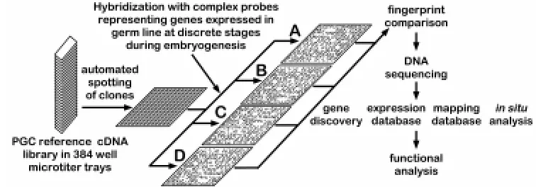
Recent advances in genome analysis technology now make a large-scale systematic analysis of gene expression during devel-opment a practical endeavor. One such approach involves exami-nation of patterns of gene expression by hybridization of complex probes to high density arrays of cDNA clones. This approach was used over 15 years ago to examine gene expression during Xenopus and Aspergillus development although at the time the expression of only around 1000 cDNAs were examined (Dworkin and Dawid, 1980; Timberlake, 1980). The concept of creating an arrayed cDNA library or a cDNA catalog as a permanent reference and using hybridization for the simultaneous monitoring of expres-sion of many genes has been described by several groups (Ko, 1990; Lennon and Lehrach, 1991). Although originally involving the spotting of cDNA clones at high density onto nylon filters (Gress et al., 1992; Meier-Ewert et al., 1993; Harrison et al., 1995; Zhao et al., 1995), more advanced technology has recently been devel-oped that permits spotting of cDNAs on glass plates at even greater densities (Schena et al., 1995). A similar method has also been developed to synthesize oligonucleotides in arrays on glass wafers (Fodor et al., 1993). One significant advantage of the latter tech-nique is the dramatic increase in sensitivity of detection of low abundance mRNAs that is achieved when used in conjunction with a laser-based multi-color fluorescence detection system.
A second approach that has been used with success is high through-put, single-pass sequencing of cDNA clones (Adams et al., 1991; Hoog, 1991; Khan et al., 1992; Okubo et al., 1992). With current rates of sequence through-put and quality of automated DNA sequencers, this approach has been successful in generating
more than 1 million ESTs from various cell types and organisms (Aaronson et al., 1996; Hillier et al., 1996). Furthermore, the ability of this approach to compare expression profiles has been validated for many cell types (Okubo et al., 1995). One variant of this approach is serial analysis of gene expression (SAGE) that in-volves sequencing of large numbers of cDNAs each of which is a concatenate of shorter cDNA fragments generated from many independent cDNA clones (Velculescu et al., 1995). Both tech-niques can rapidly provide large amounts of information about expression profiles of genes in different cell types.
We have devised a unique strategy to facilitate a systematic analysis of gene expression during mammalian germline develop-ment. A line of transgenic mice has been created in which the developing germline is marked by expression of a beta-galactosi-dase reporter gene. With these animals, PGCs can be purified from embryos at discrete stages of germline development and cDNA libraries generated from each stage of PGCs. These cDNA librar-ies can then be analyzed by a large-scale systematic approach using genome analysis methodology. This approach benefits from a low inherent bias in that all genes expressed in embryonic germ cells are theoretically amenable to analysis. While we are in essence constructing a large scale EST database, our approach is unique in that the library is from a single cell type – i.e., embryonic germ cells. Moreover, the reference library represents genes expressed at multiple stages during the process of germline development. In addition to generating valuable novel markers of the germ lineage and providing an addressed database of differen-tial gene expression during germline development, this permanent resource will facilitate future experiments to determine the function of these novel genes in this fundamental biological process.
Generation of transgenic mice that express a reporter gene within the developing germline.
The main barrier to a direct analysis of gene expression in the mammalian germline during embryogenesis has been in obtaining sufficient quantities of pure populations of PGCs. Several tech-niques have been described to purify PGCs from developing mouse embryos using antibodies that define markers of PGCs and either fluorescence activated cell sorting (FACS) (McCarrey et al.,
1987) or para-magnetic beads (MACS) (Pesce and De Felici, 1995). In both cases, PGCs were identified using monoclonal antibodies (EMA-1 or TG-1 respectively) that recognize epitopes expressed by PGCs during embryonic development. By definition use of these antibodies is limited to the period during embryonic development that the target epitope is expressed by germ cells. TNAP is currently the PGC marker with the widest window of expression during germline development in the mouse (Cooke et al., 1993; Enders and May, 1994). Thus, use of this marker to purify PGCs should facilitate an analysis of gene expression in the developing germline at the greatest number of stages during mouse embryonic development. However, purification of PGCs from early post-gastrulation stage embryos using (tissue non-specific) alkaline phosphatase as a marker is confounded by the expression of an additional isoenzyme, embryonic alkaline phos-phatase (Ginsburg et al., 1990; Hahnel et al., 1990). Therefore, an alternative strategy was required to purify PGCs on the basis of TNAP expression.
The bacterial lacZ (beta-galactosidase) gene has been used successfully as a reporter gene in several diverse species. Tech-niques have also been developed to purify mammalian cells expressing lacZ using a FACS and the purified lacZ expressing cells are viable following this procedure (Nolan et al., 1988; MacGregor et al., 1991). Consequently, a line of transgenic mice was generated that expressed lacZ under control of the TNAP locus, in the developing germ lineage. The rationale for choosing this method to facilitate purification of germ cells was that in addition to generating high purities of germ cells at a wide range of developmental stages, the purified cells could also be used for either in vitro culture experiments, or for experiments involving grafting to host embryos. The developmental potency of the purified and marked germ cells could then be assessed following culture of the embryos in vitro or after transfer of the host embryo to pseudo-pregnant female recipients for development in vivo.
Analysis of patterns of lacZ activity assessed by X-gal staining of embryos transgenic for different TNAP promoter – lacZ mini-genes failed to identify a line of mice in which the developing germline was marked by lacZ expression (Escalante-Alcalde et al., 1996). The reason for this appeared to involve a requirement for several cis-acting sequences that regulate TNAP expression and which are dispersed throughout the TNAP locus (Escalante-Alcalde et al., 1996). To minimize risk of experimental failure due to absence of such cis-elements in a transgene construct, we used homologous recombination in mouse embryonic stem (ES) cells to ‘knock-in’ a lacZ gene at the TNAP (Akp2) locus (MacGregor et al., 1995). In TNAP β-geo embryos, β-galactosidase expression in
embryonic germ cells precisely follows the expression of the endogenous TNAP gene although germline development appears to be unaffected by this mutation (MacGregor et al., 1995) (Fig. 1).
Isolation and purification of PGCs from TNAP βgeo embryos.
We have modified and optimized the use of FACS-gal (MacGregor et al., 1991; Nolan et al., 1988) to purify PGCs from TNAP β-geo embryos (Abe et al., 1996). Gonads or gonadal ridges
prior to sexual differentiation were isolated from TNAP βgeo
em-bryos and the tissues dissociated by incubation in trypsin and trituration using a siliconized pipette until a single cell suspension was achieved (Abe et al., 1996; Buehr and McLaren, 1993; Cooke et al., 1993). After loading of a β-gal fluorescent substrate, fluorescein
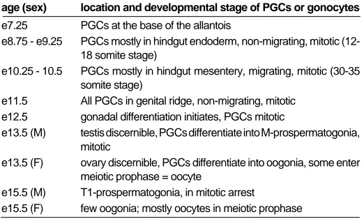
TABLE 1
DISCRETE STAGES IN GERM LINE DEVELOPMENT DURING MOUSE EMBRYOGENESIS
age (sex) location and developmental stage of PGCs or gonocytes
e7.25 PGCs at the base of the allantois
e8.75 - e9.25 PGCs mostly in hindgut endoderm, non-migrating, mitotic (12-18 somite stage)
e10.25 - 10.5 PGCs mostly in hindgut mesentery, migrating, mitotic (30-35 somite stage)
e11.5 All PGCs in genital ridge, non-migrating, mitotic e12.5 gonadal differentiation initiates, PGCs mitotic
e13.5 (M) testis discernible, PGCs differentiate into M-prospermatogonia, mitotic
e13.5 (F) ovary discernible, PGCs differentiate into oogonia, some enter meiotic prophase = oocyte
e15.5 (M) T1-prospermatogonia, in mitotic arrest
di-galactoside (FDG) into dissociated cells by osmotic shock and enzymatic conversion of FDG by incubating cells on ice for 1-2 h, the germ cells were purified by FACS-gal as described (Abe et al., 1996).
As seen in Figure 2A, a cell population displaying high fluores-cence appeared only in samples prepared from embryos derived from a cross involving a TNAP β-geo parent. Cell viability is routinely
approximately 90% following the FACS-gal procedure and the purified PGCs can be maintained in culture (K. Abe; unpublished observations). To confirm that the fluorescein positive [F(+)] cells were PGCs, they were examined both for morphology and status for existing markers of mouse PGCs (Fig. 2B-E). Microscopically, the sorted F(+) cells were relatively large, round cells with a large nuclear-cytoplasmic ratio, each of which is a characteristic of PGCs (Eddy and Hahnel, 1983). When stained for AP activity, 96
± 2% of the F(+) cells are routinely found to be AP positive (Abe et al., 1996) (Fig. 2B). In contrast, only a few percent of fluorescent negative cells are AP(+) (Fig. 2C). These cells are presumptive wild-type PGCs (i.e., not expressing a TNAP β-geo allele) that would
not fluoresce and therefore would not be detected by the FACS. On average we can routinely identify approximately 2000 AP positive cells in the gonads of a e12.5 embryo and approximately 3500 AP positive cells from an e13.5 stage embryo with approximately 2-5% of the total input cells being sorted on the basis of their high fluorescence intensity. This estimate for total numbers of germ cells at this stage is lower than those previously reported (Tam and Snow, 1981; Pesce and De Felici, 1995). This might be due to factors such as the procedure for counting cell numbers, the precise developmental stage of embryos used, the genetic back-ground of the mice in each study or the stochastic nature of β-gal expression in mammalian cells (MacGregor et al., 1987; Ko et al., 1990b).
An antibody against c-kit tyrosine kinase, that is expressed in PGCs reacted specifically with the e12.5 and e13.5 sorted F(+) cells at a similar rate as for AP (Fig. 2D). Interestingly, the EMA-1 epitope was expressed in only a subset of PGCs at e12.5 and even fewer at e13.5 (Fig. 2E). In agreement with previous studies (Hahnel and Eddy, 1986,1987), this suggests that the epitope defined by the EMA-1 antibody is down-regulated shortly after PGCs have colonized the genital ridge. These data demonstrate that the FACS-gal method can isolate highly pure populations of PGCs from mouse embryos. More recently we have used FACS-gal to purify PGCs from e11.5 and e10.5 embryos and oogonia and prospermatogonia from e13.5 and e14.5 male and female gonads. Prior to e10.5, TNAP β-geo embryos are dissected and the
hindgut or base of allantois separated from contaminating tissues by mechanical or enzymatic means. Following trypsinization of the isolated tissues, the cells are loaded with FDG. However, there are relative few PGCs present in embryos at these earlier developmen-tal stages. Consequently, instead of purification using a FACS, these early stages of PGCs are collected manually using a pipette controlled with micro-manipulator, in conjunction with an inverted microscope equipped with a stage-cooler, a CCD-camera, Nomarski optics and epifluorescence (G.M., unpublished observations). As we routinely construct cDNA libraries from as few as 200 cells (M.S.H. Ko and K. Abe, unpublished observations) sufficient PGCs can easily be isolated from around twenty embryos using this technique.
Table one lists the embryonic stages from which germ cells will be purified. At each stage listed the germ cells are developmentally distinct from other stages. It follows that cDNA libraries generated at each stage will contain genes whose function is required during that particular step of the developmental process. For example, a proportion of cDNAs found in the male e13.5 library but not the
female e13.5 library should encode products required for prospermatogonial differentiation and meiotic arrest while the complementary analysis will identify genes whose function is associated with oogonial differentiation and entry into meiosis. To facilitate identification of genes in the germ cell reference cDNA library that are not germ cell specific, cDNA libraries have also been constructed from e6.5 epiblast and from non-PGC cells isolated from e12.5 gonads.
Amplification and validation of cDNA generated from limiting amounts of germ cell RNA
FACS-gal can be used to generate pure populations of embry-onic germ cells. However, it would require prohibitively large numbers of embryos to produce sufficient germ cell RNA to construct a cDNA library by conventional means. To circumvent this type of problem, the lone linker-PCR (LL-PCR) technique was developed that enables unbiased amplification of complex cDNA mixtures (Ko et al., 1990a; Abe, 1992) from small amounts of input material. This technique can be routinely used to generate suffi-cient cDNA for construction of plasmid based cDNA libraries from as little as 200 cells (M.S.H.Ko and K.Abe, unpublished observa-tions). LL-PCR or similar methods have been applied to construct cDNA libraries from various RNA sources (Ko et al., 1990a; Froussard, 1993; Takahashi and Ko, 1994). Such cDNA libraries have been successfully used by several large scale cDNA sequencing projects including the HHMI-Washington University Mouse EST project and the ERATO-Wayne State University Mouse EST project.
RNA is isolated from the purified germ cells and cDNA synthe-sized and amplified using LL-PCR as described (Abe, 1992). The PGCs are lysed immediately after purification in Trizol (BRL) to isolate total RNA. Following DNase treatment to eliminate contami-nating genomic DNA, double strand cDNA is synthesized by conventional methods from 10-100ng of total template RNA using an oligo dT primer containing a Not I site. An asymmetrical double stranded DNA adaptor having a non-palindromic protruding end and a blunt end is ligated to the ends of the double stranded cDNA such that only a single adaptor (i.e., a ‘Lone Linker’) is added to each end with high efficiency. Subsequently, the entire pool of cDNA is amplified using a single oligonucleotide primer in a PCR. With a refined ‘Long PCR protocol’, LL-PCR can amplify DNA
fragments greater than 2 kb in length with unbiased efficiency, generating cDNA pools that are representative of the original population (Ko et al., 1990a) (K. Abe, unpublished).
Prior to construction of cDNA libraries, it is essential to validate the synthesized cDNA as being representative of genes expressed in germ cells during embryogenesis. Consequently, the individual cDNA reactions were analyzed using PCR to determine if each cDNA pool contained clones representing genes known to be expressed during mammalian germline development. Individual cDNA pools were synthesized using total RNA isolated using PGCs from e11.5, e12.5, e13.5 (both mixed and sex-specific) embryos as well as from both e6.5 epiblast and the somatic cells isolated as a by-product of PGC sorting from e12.5 gonads. The latter two samples were generated for use as negative controls for the validation process. In addition, they will also be used as a source of non-germ cell specific cDNA driver DNA in subtractive hybridization experiments. Each cDNA pool was examined for the presence of the following sequences –Mvh the mouse homolog of the Drosophila vasa gene that is expressed in murine germ cells (Fujiwara et al., 1994), TNAP (Hahnel et al., 1990; MacGregor et al., 1995), Oct-3/4 (Scholer et al., 1990), β-geo (in TNAP β-geo mice)
(MacGregor et al., 1995), SF-1 / Ad4BP (Hatano et al., 1994), WT1 (Kreidberg et al., 1993) and Hprt. All of these genes are known to be expressed in germ cells during embryogenesis with the excep-tion of Ad4BP and WT-1 which are expressed in the somatic component of the gonad.
Each of the genes known to be expressed in the germ cells was indeed found to be represented in the synthesized cDNA pools (data not shown). In addition, the somatically expressed genes were absent from the germ cell specific cDNA pools, being repre-sented only in the e12.5 gonadal somatic cell samples. Thus, the synthesized cDNA appeared to being of sufficient quality to pro-ceed with individual cDNA library construction. As the cDNA represents genes transcribed in germ cells during embryogenesis, it is in itself a valuable reagent that can be used to evaluate whether a candidate gene is expressed in the developing germline.
Construction of embryonic germ cell cDNA libraries
Having validated the individual cDNA syntheses, the amplified cDNA is digested with Not I then Sal I and is ligated to Not I -Sal I digested pBluescript prior to electro-transformation of E.coli DH10B.
Results of preliminary sequencing analyses of individual cDNA clones from libraries constructed in this manner indicate that this force cloning method is efficient in generating high quality cDNA libraries from starting material of total RNA. Specifically, no inserts with homology to rRNA were found in this library in contrast to similar libraries that were constructed using cDNA digested with Sal I alone. The cDNAs for the reference library will be stored as ordered arrays, created by robotic picking of clones from the individual libraries into 384 well micro-titer trays.
Analysis of embryonic germ cell cDNA libraries
High through-put sequencing of cDNA clones
The initial analysis of the embryonic germ cell library will be conducted by large scale, single pass sequencing of cDNA clones. This is a proven strategy to rapidly obtain information about gene expression in a particular tissue or organ. In addition, relative abundance of expressed genes or ‘expression profiles’ can be monitored by compiling and analyzing the sequence information (Okubo et al., 1995; Kawamoto et al., 1996; Yokoyama et al., 1996). Furthermore, as the cDNA libraries in this case are made from purified germ cells, a priori, each gene is expressed in germ cells at some stage during germline development. Moreover, we can identify genes whose expression is germline specific by comparing this information with databases of somatic expressed genes. With the compiled data of PGC-expressed sequences, database searches can also be performed to identify homologs in other model organisms e.g., yeast and worm. This method is rapid and does not suffer from the technical difficulties that can be encountered using hybridization methods. Functional studies with homologs can then be performed in organisms with sophisticated
genetics which can provide insight regarding the biochemical pathway in which the gene functions (Kuwabara, 1997). Finally, this method is less prone to bias and should enable identification of differentially expressed genes that might be overlooked by the hybridization approach.
Differential hybridization
The second method of analysis involves screening reference of cDNA library filters by differential hybridization, focusing on iden-tification of genes that are differentially expressed during discrete stages of germline development (listed in Table 1). The rationale for doing so is that genes whose function is required during a particular period of germline development (e.g., during migration of PGCs from the hindgut to the genital ridges) should be expressed in germ cells during this stage. Two principal methods will be used to perform the analysis. Each method uses hybridization filters onto which cDNA clones have been spotted at high density in an addressed, arrayed manner. The spotted clones represent a mixture of all the individual cDNA libraries that were constructed from the discrete populations of purified germ cells. Thus the filters will contain clones that represent the majority of genes expressed in germ cells during several stages of germline development.
Construction of full scale PGC reference cDNA library
The addressed embryonic germ cell reference cDNA library will be made by gridding cDNA clones on filters at high density in ordered arrays (Meier-Ewert et al., 1993). These filters will be screened with the complex cDNA probes. As all the clones on these filters have defined addresses in permanent storage, analysis of the hybridization signals using image analysis software enables
TABLE 2
EXAMPLE OF BLASTN SEARCH RESULTS OF CDNA CLONES FROM MIXTURE OF MALE AND FEMALE E13.5 PGCS
Clone # Reads Ns Putative ID (Blastn score > 800) or Similar to (Blastn score >300) Accession Score P value
H0501A01-3 648 7 UNKNOWN D10471 132 8.30E-00
H0501A04-3 567 0 UNKNOWN X07077 135 5.60E-00
H0501A12-3 564 6 UNKNOWN D10444 130 8.70E-00
H0501B01-3 613 10 Mouse mRNA for ubiquitin, score = 1490 X51703 1490 0.00E+01
H0501B02-3 490 6 Rat mRNA for ribosomal protein L27, score = 1313 X07424 1313 0.00E+01
H0503A03-3 641 3 Human peptidyl-prolyl isomerase and essential mitotic, score = 1448 U49070 1448 0.00E+01
H0503A07-3 723 87 UNKNOWN X63234 194 1.10E-04
H0503A11-3 291 0 UNKNOWN M99063 122 9.90E-00
H0503A12-3 568 0 UNKNOWN, similar to Human splicing factor SRp55-1 (SRp-55) mRNA, complete cds. U30883 390 0.00E+01
H0503B01-3 670 86 UNKNOWN 0 0.00E+01
H0503B03-3 282 22 UNKNOWN, similar to M.musculus mRNA for 17 beta-hydroxysteroid dehydrogenase X89998 331 0.00E+01 H0503B04-3 614 26 Mouse single stranded DNA binding protein p9 mRNA, complete, score = 1394 J03750 1394 0.00E+01
H0503B07-3 522 2 UNKNOWN U12235 144 1.30E-00
H0503B12-3 595 46 UNKNOWN, similar to H.sapiens mRNA for Drosophila female sterile homeotic (FSH) X62083 487 0.00E+01
H0504A07-3 162 0 UNKNOWN, similar to Rat matrin 3 mRNA. M63485 471 0.00E+01
H0504A09-3 677 71 UNKNOWN, similar to R.rattus mRNA for ribosomal protein L23a X65228 519 0.00E+01
H0504A10-3 758 38 UNKNOWN, similar to H.sapiens mRNA for DLG2 X82895 594 0.00E+01
H0504A12-3 702 18 M96 = metal response element DNA-binding protein S78454 1938 0.00E+01
H0505A03-3 604 19 UNKNOWN 0 0.00E+01
H0505A05-3 421 36 UNKNOWN 0 0.00E+01
H0505A06-3 530 18 Rat mRNA for ribosomal protein S13, score = 2015 X53378 2015 0.00E+01
the simultaneous identification of clones in addition to quantitation of their relative expression levels (Meier-Ewert et al., 1993; Nguyen et al., 1995; Zhao et al., 1995).
Based on an estimate of 100,000 expressed sequences in the mammalian genome, we estimate that a reference cDNA library composed of 2x10⁵ independent clones, spotted on a total of seven
filters will have approximately 95% probability of containing rare transcripts (Sambrook et al., 1989; Ausbel et al., 1994). The clones will be propagated, DNA purified and each plasmid cDNA clone will be spotted in duplicate (to generate consistency for treatment of individual filters) in 5x5 arrays onto hybridization membranes at a density of approx. 28,000 individual clones per 20 cm x 20 cm filter. In total, approximately 200,000 clones (seven filters) will be picked to constitute the embryonic germ cell reference cDNA library. This will be performed by a commercial company so that additional sets of filters of this library can be constructed and distributed to the international research community at minimal cost.
The number of independent cDNA clones required to give complete representation of genes expressed in the developing germ line could theoretically be reduced by construction of an ‘equalized’ cDNA library, i.e. a library with minimized redundancy (Ko, 1990,1995; Takahashi and Ko, 1994). However, in our expe-rience, this approach is technically more complex and requires significantly greater amounts of input germ cell RNA. One also loses information about the expression level of individual genes that is provided by the frequency a cDNA is observed in a non-equalized library. For these reasons we will construct the reference library from non-equalized libraries.
The hybridization signature approach, using high density cDNA filters and labeled complex probes can provide extremely high throughput measurement of relative gene transcriptional activity (Fig. 3). Such application of established genome analysis technology has gained rapid acceptance as a method to analyze differential gene expression during development on a large scale basis (Harrison et al., 1995; Nguyen et al., 1995; Takahashi et al., 1995; Lashkari et al., 1997). Image analysis software called AIS (Analytical Imaging System) with the capacity to locate and quantitate hybridization signals from arrayed, high density filters has been developed in collaboration with Fuji Film Co., Japan (Fig. 4). Each set of filters will be analyzed following hybridization with a particular complex probe mixture. To facilitate accumulation and retrieval of information, the data will be stored in the Wayne State University –Mouse EST database on the ORACLE relational database system that operates on a UNIX platform (M.S.H.Ko, unpublished). In addition to the AIS software, comparison of expression levels of all genes between different cDNA library will be performed using an independent analysis package –the EXPANAL, X-Window program, that is dy-namically linked to the ORACLE database (M.S.H.Ko, unpublished). In the first approach, independent sets of such filters will be screened with complex probes that have been enriched for genes expressed in germ cells at specific embryonic stages. These probes will be prepared by subtraction using different cDNA libraries. Subtracted cDNA probes will be generated by enzymatic degradation subtraction (EDS) (Zeng et al., 1994).This is a highly efficient method of subtractive hybridization that involves phenol-emulsion reassociation of lone linker amplified cDNA followed by exonuclease digestion of tester/driver or driver/driver hybrids. The technique can be used to identify DNA sequences that are differ-entially represented between two populations of highly complex molecules with great sensitivity (as low as 2.5 fold difference) and
efficiency (Zeng et al., 1994). In addition, with this method the subtracted probes can be made without converting cDNA into conventional libraries. The use of subtractive hybridization to generate probes enriched for genes expressed at specific stages during germline development has two primary advantages over conventional differential screening. First, in addition to depleting common clones, the enrichment increases the sensitivity of detec-tion, permitting identification of cDNAs representing rarer mRNAs (Harrison et al., 1995). Secondly, the use of subtraction enriched probes in combination with high-density addressed cDNA filters can reduce the required data analysis to less than 1% of that generated using non-subtracted probes (Harrison et al., 1995).
While subtractive hybridization has been successfully used by several groups to identify genes that are differentially expressed during a developmental process, this technique may not identify all transcripts whose expression levels differ subtly during develop-ment. In addition, as genes that play an important role in germline development (e.g., c-kit, Mgf) can be expressed in additional embryonic tissues or in germ cells for an extended period during germline development, genes in this category could be lost in a specific subtraction style screen.
To address this issue, the reference library will also be screened using a second, complementary experimental approach, i.e. con-ventional differential hybridization (Nguyen et al., 1995). This will involve hybridization of complex probes synthesized from each independent cDNA library to independent sets of reference library filters. In addition, the e6.5 epiblast and e12.5 gonadal somatic cell libraries will also be used as complex probes to screen the library. The results of the hybridization will be analyzed using the AIS and EXPANAL software packages each of which provides both quan-titative and qualitative information about the expression of each clone in the reference cDNA library at specific stages during germline development. Clones displaying differential expression during germline development will be sequenced and the informa-tion analyzed to assist in determining which clones will be selected for further analysis.
Pilot experiments
High through-put automated DNA sequencing and PCR-based cDNA mapping mapping technologies are powerful genome analy-sis tools that can be applied to provide direct information regarding gene expression during a developmental process (Takahashi and Ko, 1993; Ko et al., 1994 and submitted). In our experience, between 400-600bp of sequence information from each end of a clone is routinely obtained by single pass sequencing. The 3’-end cDNA sequence information can be used to identify the signature of genes by performing a homology search of the current data bases. PCR primers can be made from specific genes and these and the individual cDNA libraries screened to determine if each clones expression is sex-specific in nature. The 3’-end cDNA sequence information is used to design a PCR primer pair to facilitate gene mapping (Takahashi and Ko, 1993; Ko et al., 1994) using an interspecific species backcross panel (Rowe et al, 1994). An example of the data generated is shown in Figure 5.
sequences were either identical or displayed significant homology to known genes in the mouse, rat or human. PCR was used to confirm whether each of these genes was indeed expressed in PGCs and whether their expression was restricted to PGCs. Figure 5A indicates that the three genes analyzed, M96, Dlg 2 like and fsh-related are indeed represented in the cDNA synthesized from the purified germ cells although each is also expressed in somatic cells. Interestingly, the fsh-related gene product may be alterna-tively spliced in e6.5 epiblast and in e13.5 germ cells.
All three genes are homologs of genes known to be required for Drosophila development. M96 is a homolog of Polycomb-like while Dlg2 is a homolog of Disc large. The fsh (female sterile homeotic) gene product is a positive regulator of the bithorax complex in Drosophila (Haynes et al., 1989) and a human homolog (Ring3 ) has also been identified (Beck et al., 1992). Ring3 is known to function as nuclear kinase (Denis and Green, 1996). To determine the potential association of the fsh-related mouse homolog of fsh/ Ring3 with existing mutations known to affect germline develop-ment, the expression pattern of fsh-related in embryos and adult tissues was analyzed (Fig. 5B) and the gene mapped (Fig. 5C). In the tissues analyzed, the gene appears to be expressed at highest levels in the testis (Rhonda H. Nicholson and M.S.H. Ko, unpub-lished results). Fsh-related is also expressed at different stages of embryogenesis. The fsh-related gene maps to chromosome 17 close to the H-2 locus which is consistent with the conserved chromosomal location of its human homolog Ring3 within the human MHC class II locus (Beck et al., 1992). Interestingly, this gene maps to the t-complex tw5 developmental mutation (Xueqian
Wang and M.S.H. Ko) that is associated with a cell proliferation defect specific to e6.5 embryonic ectoderm (Bennett and Dunn, 1958; Bennett, 1975) (K. Abe, unpublished observations).
This brief example serves to illustrate how this approach will provide enormous amounts of novel information regarding genes expressed in the germline during embryonic development. In essence, this data will represent a mammalian germline EST database. Importantly however, in contrast to many current EST projects, the embryonic germ cell reference cDNA library is unique as the database represents genes that are expressed in a single cell type during its development. Once identified, specific germ cell expressed genes will be mapped to identify regions of germline-specific or stage-germline-specific gene clustering on the mouse genome. Such clustering within the mouse genome, has been discovered for genes expressed within extra-embryonic tissues (Ko et al., submit-ted). Lastly, the combined sequencing and mapping information will be used to screen for candidate cDNAs for the genes affected by the an, at, Ter, and gcd mutations.
More recently, we have completed sequencing of 4000 clones from independent male and female e13.5 PGC libraries. A total of 1600 sequences from each library have been compared to the existing database by BLAST analysis. While many sequences were found with homology to known genes, approximately one-third of the sequences appeared to be novel (M.S.H. Ko and K. Abe.; unpublished observations). Significantly, a preliminary analy-sis of the known sequences indicated the presence of transcripts whose expression is sex-specific.
Criteria for selection of cDNA clones from the PGC reference library for in depth characterization
One of the major problems that arises from the generation of such large amounts of primary data is the choice of criteria that will
be used to define a subset of cDNA clones for subsequent in depth molecular analyses. The criteria we have adopted are (a) the requirement for a sequence motif that implies functional impor-tance (e.g. known DNA/RNA binding motif such as zinc finger, DEAD, Y-box, bHLH domains or motif related to cell signaling such as tyrosine kinase domains; (b) sequence homology to previously characterized genes whose function is relevant to germline devel-opment in other species; (c) a germ cell specific expression pattern as revealed by whole mount in situ analysis or high through-put in situ analysis of embryonic tissue sections mounted in 96 well plates (Komiya et al., 1997) or (d) a chromosomal location supportive of candidacy for an existing mutation known to affect germline devel-opment. In these cases, full length cDNAs will be derived, sequenced and the cognate genes will be analyzed in the appropriate mutant strains. These results will lay the foundation for future gene disruption experiments to ascertain the function of these genes during germline development.
Future perspectives
The major barrier to studying the molecular basis for mamma-lian germline development has been the inability to obtain sufficient quantities of pure embryonic germ cells with which to conduct molecular analyses. We have developed and validated an experi-mental strategy that has successfully overcome this problem. This technology will be used to generate an arrayed addressed refer-ence cDNA library of genes expressed in the germline during mammalian development. By combining several novel techniques, we will be able to provide a unique and valuable resource with which to perform a systematic molecular genetic analysis of mammalian germline development. By making this library avail-able to the international research community, any research group with basic molecular biology skills will be able to take advantage of this resource and in turn contribute to the study of the molecular basis for mammalian germline development. The permanent ad-dressed nature of the library will minimize redundant analyses and facilitate long term accumulation of information regarding each clone in the library.
The reference library will also facilitate identification of genes that are genetically downstream of the loci affected in an, gcd, Ter, at and any additional mutants that may be identified in the future in which germline development is affected. This can be accom-plished by crossing the TNAP β-geo marker into each of these
mutant backgrounds and using PGCs purified from the mutant animals as a source of complex cDNA probes to ‘fingerprint’ the reference library. Downstream loci affected by these mutations would then be identified by differences in the ‘fingerprints’ between normal and mutant complex probes.
germ cell development. Finally, this study epitomizes the growing utility of ‘genome analysis’ technology to study developmental proc-esses at the molecular level (Harrison et al., 1995; Bernard et al., 1996; Granjeaud et al., 1996; Heller et al., 1997; Lashkari et al., 1997; Wodicka et al., 1997; Zhang et al., 1997).
Summary
Development of the germ lineage is essential for maintenance of a species. Despite this importance, relatively little is known about the molecular basis for mammalian germline development. The paucity of reports concerning gene expression in mammalian embryonic germ cells is in part the result of technical considera-tions as PGCs constitute only a minor proportion of the mouse embryo. Thus, in the absence of a suitable long term in vitro culture system, it has been difficult to isolate sufficient quantities of pure germ cells with which to conduct molecular genetic studies. We have overcome this problem by using a novel combination of established molecular and transgenic approaches. A line of mice has been generated in which the cells of the germ lineage express the β-galactosidase reporter gene during embryogenesis. Using this line, germ cells have been purified to near homogeneity from embryos at discrete stages during germline development by use of a viable stain for b-gal activity and a fluorescence activated cell sorter (FACS). Subsequently, cDNA libraries have been con-structed from each germ cell population using a modified lone-linker PCR strategy. These combined cDNA libraries represent genes that are expressed in PGCs during mammalian germline development. To facilitate a molecular genetic approach to study-ing mammalian germline development, these cDNA libraries will be pooled to form an arrayed, addressed reference embryonic germ cell cDNA library. In parallel with large-scale cDNA sequencing efforts, genes that are differentially expressed in germ cells either prior to gonadal differentiation or in a sexually dimorphic manner following gonadal differentiation will be identified by screening the reference library with probes generated by subtractive hybridiza-tion. Complementary DNAs identified using this approach will be analyzed by sequencing, database comparison, genomic mapping and in situ hybridization to ascertain the potential functional impor-tance of each gene to germline development. In addition to providing a wealth of novel information regarding patterns of gene expression during germline development, these results will form the basis for future experiments to determine the function of these genes in this process. The addressed reference embryonic germ cell cDNA library will constitute a valuable, permanent shared resource that will enable the entire international research commu-nity to study the molecular basis for germline development during mammalian embryogenesis.
Acknowledgments
We thank members of Ko laboratory for providing preliminary results, especially Rhonda H. Nicholson for the northern analyses in Figure 5, Marija J. Grahovac and Meng K. Lim for the large scale cDNA sequencing analyses, and Xueqian Wang for the mapping of Fsh -related gene. We also thank Colin Stewart, Peter Donovan, Jeff Pitman and Carol MacLeod for communicating results prior to publication. The work described is sup-ported in part by grants to K.A. from the Science and Technology Agency of Japan, with Special Coordinating Funds for Promoting Science and Technology and by grants from the Ministry of Education, Science and Culture of Japan, and to M.S.H.K. (R01HD32243) and G.R.M. (R01HD36437), from the NIH, USA.
References
AARONSON, J.S., ECKMAN, B., BLEVINS, R.A., BORKOWSKI, J.A., MYERSON, J., IMRAN, S. and ELLISTON, K.O. (1996). Toward the development of a gene index to the human genome: an assessment of the nature of high-throughput EST sequence data. Genome Res. 6: 829-845.
ABE, K. (1992). Rapid isolation of desired sequences from lone linker PCR amplified cDNA mixtures: Application to identification and recovery of expressed sequences in cloned genomic DNA. Mammal. Genome 2: 252-259.
ABE, K., HASHIYAMA, M., MACGREGOR, G., YAMAMURA, K.-I. and ABE, K. (1996). Purification of primordial germ cells from TNAPβ-geo mouse embryos using FACS-gal. Dev. Biol. 180: 468-472.
ADAMS, M.D., KELLEY, J.M., GOCAYNE, J.D., DUBNICK, M., POLYMEROPOULOS, M.H., XIAO, H., MERRIL, C.R., WU, A., OLDE, B., MORENO, R.F. and ET AL. (1991). Complementary DNA sequencing: expressed sequence tags and human genome project. Science 252: 1651-1656.
ALVAREZ-BUYLLA, A. and MERCHANT-LARIOS, H. (1986). Mouse primordial germ cells use fibronectin as a substrate for migration. Exp. Cell Res. 165: 362-8.
ANSTROM, K.K. and TUCKER, R.P. (1996). Tenascin-C lines the migratory path-ways of avian primordial germ cells and hematopoietic progenitor cells. Dev. Dynamics 206: 437-446.
AUSBEL, F.M., BRENT, R., KINGSTON, R.E., MOORE, D.D., SEIDMAN, J.G., SMITH, J.A. and STRUHL, K. (1994). Current Protocols in Molecular Biology. Wiley Interscience, Brooklyn, N Y.
AVARBOCK, M.R., BRINSTER, C.J. and BRINSTER, R.L. (1996). Reconstitution of spermatogenesis from frozen spermatogonial stem cells. Nature Med. 2: 693-696.
BAKER, R.K., HAENDEL, M.A., SWANSON, B.J., SHAMBAUGH, J.C., MICALES, B.K. and LYONS, G.E. (1997). In vitro preselection of gene trapped ES cell clones for characterizing novel developmentally regulated genes in the mouse. Dev. Biol. 185: 201-214.
BECK, S., HANSON, I., KELLEY, A., PAPPIN, D.J.C. and TROWSDALE, J. (1992). A homolog of the Drosophila female sterile homeotic (fsh) gene in the class II region of the human MHC. J. DNA Seq. Map. 2: 203-210.
BENNETT, D. (1975). The T-locus of the mouse. Cell 6: 441-454.
BENNETT, D. and DUNN, L.C. (1958). Effects on embryonic development of a group of genetically similar lethal alleles derived from different populations of wild housed mice. J. Morphol. 103: 135-158.
BERNARD, K., AUPHAN, N., GRANJEAUD, S., VICTORERO, G., SCHMITT-VERHULST, A.M., JORDAN, B.R. and NGUYEN, C. (1996). Multiplex messenger assay: simultaneous, quantitative measurement of expression of many genes in the context of T cell activation. Nucleic Acids Res 24: 1435-1442.
BESMER, P., MANOVA, K., DUTTLINGER, R., HUANG, E.J., PACKER, A., GYSSLER, C. and BACHVAROVA, R.F. (1993). The kit-ligand (steel factor) and its receptor c-kit/W: pleiotropic roles in gametogenesis and melanogenesis. Development (Suppl.) : 125-137.
BRINSTER, R.L. and AVARBOCK, M.R. (1994). Germline transmission of donor haplotype following spermatogonial transplantation. Proc. Natl. Acad. Sci. USA 91: 11303-11307.
BRINSTER, R.L. and ZIMMERMANN, J.W. (1994). Spermatogenesis following male germ-cell transplantation. Proc. Natl. Acad. Sci. USA 91: 11298-11302.
BUEHR, M. and MCLAREN, A. (1993). Isolation and culture of primordial germ cells. In Methods in Enzymology: Guide to techniques in mouse development. (P.M. Wassarman and M.L. DePamphilis, Eds.). Academic Press, San Diego. Vol. 225, pp. 58-77.
BUEHR, M., MCLAREN, A., BARTLEY, A. and DARLING, S. (1993). Proliferation and migration of primordial germ cells in We/We mouse embryos. Dev. Dynamics 198: 182-189.
CHENG, L., GEARING, D.P., WHITE, L.S., COMPTON, D.L., SCHOOLEY, K. and DONOVAN, P.J. (1994). Role of leukemia inhibitory factor and its receptor in mouse primordial germ cell growth. Development 120: 3145-3153.
CHIQUOINE, A.D. (1954). The Identification, origin, and migration of the primordial germ cells in the mouse embryo. Anat. Rec. 118: 135-146.
CLOUTHIER, D.E., AVARBOCK, M.R., MAIKA, S.D., HAMMER, R.E. and BRINSTER, R.L. (1996). Rat spermatogenesis in mouse testis. Nature 381: 418-421.
Enzymology: Guide to techniques in mouse development. (P. M. Wassarman and M. L. DePamphilis, Eds.). Academic Press, San Diego. Vol. 225, pp. 37-57.
COPP, A.J., ROBERTS, H.M. and POLANI, P.E. (1986). Chimerism of primordial germ cells in the early post-implantation mouse embryo following microsurgical grafting of posterior primitive streak cells in vitro. J. Embryol. Exp. Morphol. 95: 95-115.
COUCOUVANIS, E.C. and JONES, P.P. (1993). Changes in protooncogene expres-sion correlated with general and sex-specific differentiation in murine primordial germ cells. Mech. Dev. 42: 49-58.
DE FELICI, M. and DOLCI, S. (1989). In vitro adhesion of mouse fetal germ cells to extracellular matrix components. Cell Differ. Dev. 26: 87-96.
DE FELICI, M. and PESCE, M. (1994). Growth factors in in mouse primordial germ cell migration and proliferation. Prog. Growth Factor Res. 5: 135-143.
DE FELICI, M., DOLCI, S. and PESCE, M. (1992). Cellular and molecular aspects of mouse primordial germ cell migration and proliferation in culture. Int. J. Dev. Biol. 36: 205-213.
DENIS, G.V. and GREEN, M.R. (1996). A novel, mitogen-activated nuclear kinase is related to a Drosophila developmental regulator. Genes Dev. 10: 261-71.
DONOVAN, P.J. (1994). Growth factor regulation of mouse primordial germ cell development. Curr. Top. Dev. Biol. 29: 189-225.
DONOVAN, P.J., STOTT, D., CAIRNS, L.A., HEASMAN, J. and WYLIE, C.C. (1986). Migratory and postmigratory mouse primordial germ cells behave differently in culture. Cell 44: 831-838.
DUNCAN, M.K., LIEMAN, J. and CHADA, K.K. (1995). The germ cell deficient locus maps to mouse chromosome 11A2-3. Mammal. Genome 6: 697-699.
DWORKIN, M.B. and DAWID, I.B. (1980). Use of a cloned library for the study of abundant poly(A)+RNA during Xenopus laevis development. Dev. Biol. 76: 449-64.
EDDY, E.M. and HAHNEL, A.C. (1983). Establishment of the germ cell line in mammals. In Current problems in germ cell differentiation. (A. McLaren and C.C. Wylie, Eds.). Cambridge University Press, Cambridge. pp. 41-69.
ELLIS, R.E. and KIMBLE, J. (1994). Control of germ cell differentiation in Caenorhabditis elegans. Ciba Found Symp 182: 179-88; discussion 189-92.
ENDERS, G.C. and MAY, J.J. (1994). Developmentally regulated expression of a mouse germ cell nuclear antigen examined from embryonic day 11 to adult in male and female. Dev. Biol. 163: 331-340.
ENGLAND, M.A., SWAN, A.P. and DANE, P. (1986). The migration of amphibian primordial germ cells in the chick embryo. Scan. Electron Microsc. Pt3: 1175-1182.
ESCALANTE-ALCALDE, D., RECILLAS-TARGA, F., HERNANDEZ-GARCIA, D., CASTRO-OBREGON, S., TERAO, M., GARATTINI, E. and COVARRUBIAS, L. (1996). Retinoic acid and methylation cis-regulatory elements control the mouse tissue non-specific alkaline phosphatase gene expression. Mech. Dev. 57: 21-32.
FFRENCH-CONSTANT, C., HOLLINGSWORTH, A., HEASMAN, J. and WYLIE, C.C. (1991). Response to fibronectin of mouse primordial germ cells before, during and after migration. Development 113: 1365-1373.
FODOR, S.P., RAVA, R.P., HUANG, X.C., PEASE, A.C., HOLMES, C.P. and ADAMS, C.L. (1993). Multiplexed biochemical assays with biological chips. Nature 364: 555-556.
FORRESTER, L.M., NAGY, A., SAM, M., WATT, A., STEVENSON, L., BERNSTEIN, A., JOYNER, A.L. and WURST, W. (1996). An induction gene trap screen in embryonic stem cells: Identification of genes that respond to retinoic acid in vitro. Proc. Natl. Acad. Sci. USA 93: 1677-1682.
FRIEDRICH, G. and SORIANO, P. (1991). Promoter traps in embryonic stem cells: a genetic screen to identify and. Genes Dev. 5: 1513-1523.
FROUSSARD, P. (1993). rPCR: a powerful tool for random amplification of whole RNA sequences. PCR Methods Appl. 2: 185-190.
FUJIMOTO, T., UKESHIMA, A. and KIYOFUJI, R. (1976). The origin, migration and morphology of the primordial germ cells in the chick embryo. Anat. Rec. 185: 139-145.
FUJIMOTO, T., YOSHINAGA, K. and KONO, I. (1985). Distribution of fibronectin on the migratory pathway of primordial germ cells in mice. Anat. Rec. 211: 271-278.
FUJIWARA, Y., KOMIYA, T., KAWABATA, H., SATO, M., FUJIMOTO, H., FURUSAWA, M. and NOCE, T. (1994). Isolation of a DEAD-family protein gene that encodes a murine homolog of Drosophila vasa and its specific expression in germ cell lineage. Proc. Natl. Acad. Sci. USA 91: 12258-12262.
GARCIA-CASTRO, M.I., ANDERSON, R., HEASMAN, J. and WYLIE, C. (1997). Interactions between germ cells and extracellular matrix glycoproteins during migration and gonad assembly in the mouse embryo. J. Cell Biol. 138: 471-480.
GARDNER, R.L. and BEDDINGTON, R.S.P. (1988). Multi-lineage ‘stem’ cells in the mammalian embryo. J. Cell Sci. (Suppl.) 10: 11-27.
GARDNER, R.L., LYON, M.F., EVANS, E.P. and BURTENSHAW, M.D. (1985). Clonal analysis of X-chromosome inactivation and the origin of the germ line in the mouse embryo. J. Embryol. Exp. Morphol. 88: 349-363.
GINSBURG, M., SNOW, M.H.L. and MCLAREN, A. (1990). Primordial germ cells in the mouse during gastrulation. Development 110: 521-528.
GODIN, I. and WYLIE, C.C. (1991). TGF beta 1 inhibits proliferation and has a chemotropic effect on mouse primordial germ cells in culture. Development 113: 1451-1457.
GODIN, I., WYLIE, C.C. and HEASMAN, J. (1990). Genital ridges exert long range effects on lmouse primordial germ cell numbers and direction of migration in culture. Development 108: 357-363.
GOH, K.L., YANG, J.T. and HYNES, R.O. (1997). Mesodermal defects and cranial neural crest apoptosis in a5 integrin-null embryos. Development 124: 4309-4319.
GOMPERTS, M., GARCIA-CASTRO, M., WYLIE, C. and HEASMAN, J. (1994a). Interactions between primordial germ cells play a role in their migration in mouse embryos. Development 120: 135-141.
GOMPERTS, M., WYLIE, C. and HEASMAN, J. (1994b). Primordial germ cell migration. Ciba Found. Symp. 182: 121-34; discussion 134-9.
GOSSLER, A., JOYNER, A.L., ROSSANT, J. and SKARNES, W.C. (1989). Mouse embryonic stem cells and reporter constructs to detect developmentally regulated genes. Science 244: 463-465.
GRANJEAUD, S., NGUYEN, C., ROCHA, D., LUTON, R. and JORDAN, B.R. (1996). From hybridization image to numerical values: a practical, high throughput quantification system for high density filter hybridizations. Genet Anal 12: 151-62.
GRESS, T.M., HOHEISEL, J.D., LENNON, G.G., ZEHETNER, G. and LEHRACH, H. (1992). Hybridization fingerprinting of high-density cDNA-library arrays with cDNA pools derived from whole tissues. Mammal. Genome 3: 609-619.
HAHNEL, A.C. and EDDY, E.M. (1986). Cell surface markers of mouse promordial germ cells defined by two monoclonal antibodies. Gamete Res. 15: 25-34.
HAHNEL, A.C. and EDDY, E.M. (1987). The distribution of two cell surface determi-nants of mouse embryonal carcinoma and early embryonic cells. J Reprod. Immunol. 10: 89-110.
HAHNEL, A.C., RAPPOLEE, D.A., MILLÁN, J.L., MANES, T., ZIOMEK, C.A., THEODOSIOU, N.G., WERB, Z., PEDERSON, R.A. and SCHULTZ, G.A. (1990). Two alkaline phosphatase genes are expressed during early development in the mouse embryo. Development 110: 555-564.
HARRISON, S.M., DUNWOODIE, S.L., ARKELL, R.M., LEHRACH, H. and BEDDINGTON, R.S.P. (1995). Isolation of novel tissue-specific genes from cDNA libraries representing the individual tissue constituents of the gastrulating mouse embryo. Development 121: 2479-2489.
HATANO, O., TAKAYAMA, K., IMAI, T., WATERMAN, M.R., TAKAKUSU, A., OMURA, T. and MOROHASHI, K. (1994). Sex-dependent expression of a transcription factor, Ad4BP, regulating steroidogenic P-450 genes in the gonads during prenatal and postnatal rat development. Development 120: 2787-97.
HAYNES, S.R., MOZER, B.A., BHATIA-DEY, N. and DAWID, I.B. (1989). The Drosophila fsh locus, a maternal effect homeotic gene encodes apparent mem-brane proteins. Dev. Biol. 134: 246-257.
HEASMAN, J., HYNES, R.O., SWAN, A.P., THOMAS, V. and WYLIE, C.C. (1981). Primordial germ cells of Xenopus embryos: the role of fibronectin in their adhesion during migration. Cell 27: 437-447.
HELLER, R.A., SCHENA, M., CHAI, A., SHALON, D., BEDILION, T., GILMORE, J., WOOLLEY, D.E. and DAVIS, R.W. (1997). Discovery and analysis of inflamma-tory disease-related genes using cDNA microarrays. Proc. Natl. Acad. Sci. USA 94: 2150-2155.