Scholarship@Western
Scholarship@Western
Electronic Thesis and Dissertation Repository
11-2-2012 12:00 AM
Evaluation of the Utility of Decomposition-Enhanced
Evaluation of the Utility of Decomposition-Enhanced
Spike-Triggered Averaging Motor Unit Number Estimation as an
Triggered Averaging Motor Unit Number Estimation as an
Outcome Measure for the Study of Amyotrophic Lateral Sclerosis
Outcome Measure for the Study of Amyotrophic Lateral Sclerosis
Colleen T. Ives
The University of Western Ontario
Supervisor
Dr. Timothy Doherty
The University of Western Ontario
Graduate Program in Health and Rehabilitation Sciences
A thesis submitted in partial fulfillment of the requirements for the degree in Doctor of Philosophy
© Colleen T. Ives 2012
Follow this and additional works at: https://ir.lib.uwo.ca/etd
Part of the Neurosciences Commons
Recommended Citation Recommended Citation
Ives, Colleen T., "Evaluation of the Utility of Decomposition-Enhanced Spike-Triggered Averaging Motor Unit Number Estimation as an Outcome Measure for the Study of Amyotrophic Lateral Sclerosis" (2012). Electronic Thesis and Dissertation Repository. 944.
https://ir.lib.uwo.ca/etd/944
This Dissertation/Thesis is brought to you for free and open access by Scholarship@Western. It has been accepted for inclusion in Electronic Thesis and Dissertation Repository by an authorized administrator of
TRIGGERED AVERAGING MOTOR UNIT NUMBER ESTIMATION AS AN OUTCOME MEASURE FOR THE STUDY OF AMYOTROPHIC LATERAL
SCLEROSIS
(Spine title: Motor Unit Number Estimation in Amyotrophic Lateral Sclerosis)
(Thesis format: Integrated Article)
by
Colleen T. Ives
Graduate Program in Health and Rehabilitation Sciences
A thesis submitted in partial fulfillment of the requirements for the degree of
Doctor of Philosophy
The School of Graduate and Postdoctoral Studies The University of Western Ontario
London, Ontario, Canada
ii
THE UNIVERSITY OF WESTERN ONTARIO School of Graduate and Postdoctoral Studies
CERTIFICATE OF EXAMINATION
Joint-Supervisor
______________________________ Dr. Timothy Doherty
Joint-Supervisor
______________________________ Dr. Jayne Garland
Supervisory Committee
______________________________ Dr. Christen Shoesmith
Examiners
______________________________ Dr. K. Ming Chan
______________________________ Dr. Charles Rice
______________________________ Dr. Denise Connelly
______________________________ Dr. Kara Patterson
The thesis by
Colleen T. Ives
entitled:
Evaluation of the Utility of Decomposition-Enhanced Spike-Triggered Averaging Motor Unit Number Estimation as an Outcome Measure for the Study of Amyotrophic Lateral
Sclerosis
is accepted in partial fulfillment of the requirements for the degree of
Doctor of Philosophy
iii
ABSTRACT
OBJECTIVES: The objectives of this thesis were to review the use of outcome
measures systematically across amyotrophic lateral sclerosis (ALS) clinical trials, and
evaluate the utility of decomposition-enhanced spike-triggered averaging (DE-STA)
motor unit number estimation (MUNE) as an outcome measure, with a particular focus
on its application to the upper trapezius (UT).
METHODS: First, a systematic review quantified the frequency of use of outcome
measures in ALS randomized controlled trials (Chapter 2). Next, the intra- and
inter-rater reliability of DE-STA MUNE was evaluated in the UT of control subjects (Chapter
3), followed by the intra-rater reliability of the technique in the UT and biceps brachii of
subjects with ALS (Chapter 4). To assess validity, the results of the technique in the UT
were compared between subjects with ALS and control subjects (Chapter 4). The
sensitivity to change of DE-STA MUNE in the UT was compared with that of various
clinical outcome measures in a longitudinal study of subjects with ALS (Chapter 5).
Finally, the influence of needle electrode depth on the results of the technique in the UT
was evaluated in control subjects (Chapter 6).
RESULTS: The heterogeneity in the use of outcome measures across ALS randomized
controlled trials was demonstrated, in addition to the infrequent use of MUNE. MUNE
results demonstrated moderate intra- and inter-rater reliability for control subjects in the
UT, although less favorable results were found overall for inter-rater reliability.
Application of DE-STA MUNE to the UT in subjects with ALS demonstrated
consistently high intra-rater reliability, the ability to detect the underlying
iv
MUNE results. Further evaluation found needle electrode depth to significantly influence
the results of the technique, with suggestions made for improved standardization of the
protocol.
SIGNIFICANCE: These studies were novel in their evaluation of MUNE in the
proximal, potentially clinically relevant UT. The studies mark the first evaluations of the
reliability and sensitivity to change of DE-STA MUNE in subjects with ALS, finding
application to the UT to be practical and promising for use as an outcome measure.
Implementation of proposed improvements to the protocol may aid in further establishing
DE-STA MUNE for use as an outcome measure in studies of ALS.
KEYWORDS: amyotrophic lateral sclerosis (ALS), decomposition-enhanced
spike-triggered averaging (DE-STA), electromyography (EMG), motor unit number estimation
v
CO-AUTHORSHIP
This thesis contains material from one published manuscript (Chapter 3) and one
manuscript submitted for publication (Chapter 4). Manuscripts from Chapters 2, 5 and 6
are in preparation to be submitted for publication. Colleen Ives was the first author and
Dr. Timothy J. Doherty was a co-author on each of the manuscripts. For the manuscript
from Chapter 2, Dr. Christen Shoesmith was a co-author. For the manuscript from
Chapter 5, Ms. Karen Findlater and Dr. Christen Shoesmith were co-authors.
vi
DEDICATION
This thesis is dedicated to my uncle and godfather, Ian Russell Quinn. In the face
of ALS, you lived with unimaginable positivity and strength, all the while maintaining
your sense of humor. You are my inspiration and source of passion for the career path I
vii
ACKNOWLEDGMENTS
I would like to gratefully acknowledge the many individuals who made this thesis
possible by contributing their knowledge, skills, and support along my journey as a
graduate student.
Thank you to my supervisor, Dr. Tim Doherty. Tim, I greatly appreciated your
mentorship throughout this research process, and graduate school in general. Thank you
for always instilling in me a sense of pride in my work, and offering your genuine and
insightful guidance in our discussions about my future plans.
I owe much gratitude to the additional members of my advisory committee for
their invaluable assistance in the preparation of this thesis. Thank you to Dr. Jayne
Garland for ensuring that my graduate school journey got off to an excellent start, and
fostering a sense of team spirit in our lab. A special thank you to Dr. Christen
Shoesmith; your tireless assistance with patient recruitment was the key to the success of
the ALS studies. I am also sincerely grateful for having had the opportunity to attend
ALS conferences with you. These were exceptional learning opportunities that further
sparked my interest in clinical ALS research.
Many thanks to our collaborators: Ms. Karen Findlater, for kindly taking the time
to share your expertise in various measurement techniques and Ms. Norine Foley, for
guiding me through the process of conducting a systematic review. To my lab mates and
peers in the HRS and Kinesiology graduate programs: I have truly valued your
companionship and benefitted greatly from our shared learning experiences. I wish you
viii
My friends and family have played a most important role in my graduate school
experience. I would like to thank my best friend and ‘sister’, Heather, for being there
through all of the ‘ups’ and ‘downs’ of graduate school. Your wholehearted support has
meant the world to me. As well, a very special thank you to Brenton for always being a
patient source of support for my computer questions.
Mom and Dad, I couldn’t have done this without your constant love and support.
I feel so fortunate to have been able to share my experiences with you and Brenton every
step of the way. With the opportunities and unconditional love that you have given me, I
have had the freedom to explore and discover my dreams, and set out toward achieving
them. I love you so much, and can never thank you enough.
Most importantly, I would like to thank the participants who volunteered to take
part in this research. In particular, I extend my deepest gratitude to the participants with
ALS and their families who gave so selflessly of their time and energies to contribute.
My time spent with all of you was the most rewarding aspect of my degree, and what you
ix
TABLE OF CONTENTS
CERTIFICATE OF EXAMINATION ... ii
ABSTRACT ... iii
CO-AUTHORSHIP ... v
DEDICATION ... vi
ACKNOWLEDGMENTS ... vii
TABLE OF CONTENTS ... ix
LIST OF TEXT BOXES ... xv
LIST OF TABLES ... xvi
LIST OF FIGURES ... xvii
LIST OF APPENDICES ... xviii
LIST OF ABBREVIATIONS ... xix
CHAPTER 1: GENERAL INTRODUCTION AND OVERVIEW 1.0 GENERAL INTRODUCTION ... 1
1.0.1 The motor unit ... 1
1.0.2 Amyotrophic lateral sclerosis ... 1
1.0.3 Motor unit number estimation ... 2
1.0.3.1 Equation 1 ... 3
1.0.4 Outcome measures and collateral reinnervation ... 3
1.0.5 Motor unit number estimation techniques ... 4
1.0.6 Quantitative electromyography ... 5
1.0.7 Decomposition-based quantitative electromyography ... 6
1.0.8 Upper trapezius muscle ... 11
1.0.9 Evaluation of decomposition-enhanced spike-triggered averaging MUNE ... 14
1.1 OVERVIEW OF THESIS CHAPTERS ... 16
1.1.1 Chapter 2 study objectives ... 16
1.1.2 Chapter 3 study objective ... 16
1.1.3 Chapter 4 study objectives ... 17
1.1.4 Chapter 5 study objective ... 17
1.1.5 Chapter 6 study objective ... 17
x
CHAPTER 2:
USE OF OUTCOME MEASURES IN ALS RANDOMIZED CONTROLLED TRIALS: A SYSTEMATIC REVIEW
2.0 INTRODUCTION ... 22
2.1 METHODS ... 27
2.1.1 Inclusion criteria ... 29
2.1.2 Exclusion criteria ... 29
2.2 RESULTS ... 30
2.2.1 Literature search results ... 30
2.2.2 Primary outcome measures ... 31
2.2.3 Secondary outcome measures ... 31
2.3 DISCUSSION ... 37
2.3.1 Primary outcome measures ... 37
2.3.1.1 Survival ... 37
2.3.1.2 Revised Amyotrophic Lateral Sclerosis Functional Rating Scale ... 38
2.3.1.3 Maximal voluntary isometric contraction ... 39
2.3.2 Secondary outcome measures ... 39
2.3.2.1 Forced vital capacity ... 40
2.3.2.2 Manual muscle testing ... 40
2.3.2.3 Maximal voluntary isometric contraction and survival ... 41
2.3.2.4 Functional rating scales/timed functional tests ... 41
2.3.2.5 Quality of life ... 42
2.3.2.6 Motor unit number estimation ... 42
2.3.2.7 Use of motor unit number estimation in ALS clinical trials ... 43
2.3.3 Limitations and future study ... 45
2.3.4 Summary ... 46
2.4 REFERENCES ... 47
CHAPTER 3: RELIABILITY OF DECOMPOSITION-ENHANCED SPIKE-TRIGGERED AVERAGING MOTOR UNIT NUMBER ESTIMATION IN THE UPPER TRAPEZIUS IN CONTROL SUBJECTS 3.0 INTRODUCTION ... 54
3.1 METHODS ... 55
xi
3.1.2 Electromyographic data collection protocol ... 56
3.1.3 Electromyographic signal decomposition and analysis ... 58
3.1.4 Intra-rater reliability ... 59
3.1.5 Inter-rater reliability ... 60
3.1.6 Statistics ... 60
3.1.6.1 Equation 1 ... 61
3.2 RESULTS ... 61
3.2.1 Subjects ... 61
3.2.2 Data collection results and S-MUP frequency distributions ... 61
3.2.3 Maximum CMAP, mean S-MUP, and MUNE ... 62
3.2.3.1 Intra-rater reliability ... 62
3.2.3.2 Inter-rater reliability ... 62
3.2.4 Motor unit potential parameters ... 63
3.2.4.1 Intra-rater reliability ... 63
3.2.4.2 Inter-rater reliability ... 63
3.3 DISCUSSION ... 67
3.4 REFERENCES ... 71
CHAPTER 4: EVALUATION OF DECOMPOSITION-ENHANCED SPIKE-TRIGGERED AVERAGING MOTOR UNIT NUMBER ESTIMATION IN SUBJECTS WITH AMYOTROPHIC LATERAL SCLEROSIS 4.0 INTRODUCTION ... 74
4.1 METHODS ... 76
4.1.1 Subjects ... 76
4.1.2 Electromyographic data collection and analysis ... 76
4.1.3 Intra-rater reliability ... 79
4.1.4 Statistics ... 80
4.1.4.1 Equation 1 ... 80
4.1.4.2 Equation 2 ... 81
4.1.4.3 Equation 3 ... 81
4.2 RESULTS ... 82
4.2.1 Subjects ... 82
xii
4.2.2.1 Upper trapezius ... 82
4.2.2.2 Biceps brachii... 82
4.2.3 Intra-rater reliability of maximum CMAP, mean S-MUP, and MUNE ... 83
4.2.3.1 Upper trapezius ... 83
4.2.3.2 Biceps brachii... 83
4.2.4 Intra-rater reliability of motor unit potential parameters ... 84
4.2.4.1 Upper trapezius ... 84
4.2.4.2 Biceps brachii... 84
4.2.5 Comparison of upper trapezius data between ALS and control subjects ... 90
4.3 DISCUSSION ... 94
4.3.1 Data collection and S-MUP frequency distributions ... 94
4.3.2 Intra-rater reliability ... 95
4.3.2.1 Upper trapezius ... 95
4.3.2.2 Biceps brachii... 97
4.3.3 Comparison of upper trapezius data between ALS and control subjects ... 99
4.3.4 Limitations and future study ... 101
4.3.5 Summary ... 102
4.4 REFERENCES ... 103
CHAPTER 5: SENSITIVITY TO CHANGE OF DECOMPOSITION-ENHANCED SPIKE-TRIGGERED AVERAGING MOTOR UNIT NUMBER ESTIMATION IN SUBJECTS WITH AMYOTROPHIC LATERAL SCLEROSIS 5.0 INTRODUCTION ... 106
5.1 METHODS ... 109
5.1.1 Subjects ... 109
5.1.2 Experimental protocol ... 109
5.1.2.1 Manual muscle testing ... 110
5.1.2.2 Hand-held dynamometry ... 111
5.1.2.3 Equation 1 ... 112
5.1.2.4 Motor unit number estimation ... 112
5.1.2.5 Equation 2 ... 115
5.1.2.6 Forced vital capacity ... 115
xiii
5.1.2.8 Revised Amyotrophic Lateral Sclerosis Functional Rating Scale ... 117
5.1.3 Evaluator training and reliability assessment ... 117
5.1.3.1 Manual muscle testing ... 118
5.1.3.2 Hand-held dynamometry ... 118
5.1.3.3 Forced vital capacity and sniff nasal inspiratory pressure ... 118
5.1.3.4 Revised Amyotrophic Lateral Sclerosis Functional Rating Scale ... 119
5.1.4 Statistics ... 119
5.1.4.1 Equation 3 ... 119
5.1.4.2 Equation 4 ... 119
5.1.4.3 Equation 5 ... 120
5.2 RESULTS ... 121
5.2.1 Subjects ... 121
5.2.2 Missing data ... 121
5.2.3 Hand-held dynamometry data collection ... 121
5.2.4 Results of data analysis... 122
5.3 DISCUSSION ... 132
5.3.1 Overall sensitivity to change results ... 132
5.3.2 Subjects ... 134
5.3.3 Hand-held dynamometry data collection ... 134
5.3.4 Longitudinal variability in MUNE, maximum CMAP, and mean S-MUP .... 135
5.3.5 S-MUP and MUP data collection results ... 138
5.3.6 Trends across outcome measures ... 139
5.3.7 Mean percentage change data ... 139
5.3.8 Limitations and future study ... 140
5.3.9 Summary ... 141
5.4 REFERENCES ... 143
CHAPTER 6: INFLUENCE OF NEEDLE ELECTRODE DEPTH ON DECOMPOSITION-ENHANCED SPIKE-TRIGGERED AVERAGING MOTOR UNIT NUMBER ESTIMATION IN THE UPPER TRAPEZIUS 6.0 INTRODUCTION ... 147
6.1 METHODS ... 149
xiv
6.1.2 Electromyographic data collection and analysis ... 149
6.1.3 Statistics ... 152
6.2 RESULTS ... 153
6.2.1 Subjects ... 153
6.2.2 Data collection results and S-MUP frequency distributions ... 153
6.2.3 Maximum CMAP, mean S-MUP, and MUNE: Needle electrode depth effects ... 155
6.2.4 Motor unit potential parameters: Needle electrode depth effects ... 155
6.3 DISCUSSION ... 159
6.4 REFERENCES ... 164
CHAPTER 7: GENERAL DISCUSSION AND SUMMARY 7.0 GENERAL DISCUSSION ... 166
7.1 LIMITATIONS ... 169
7.2 FUTURE STUDIES... 170
7.3 SUMMARY ... 171
7.4 REFERENCES ... 173
APPENDIX A ... 175
APPENDIX B ... 181
APPENDIX C ... 182
APPENDIX D ... 184
APPENDIX E ... 187
xv
LIST OF TEXT BOXES
2.1 Search terms
2.2 Frequency of use of primary outcome measures reported in ALS RCTs
xvi
LIST OF TABLES
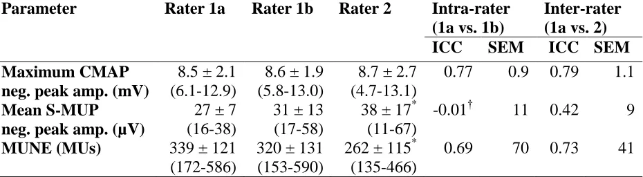
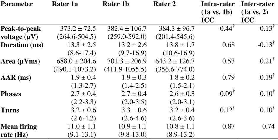
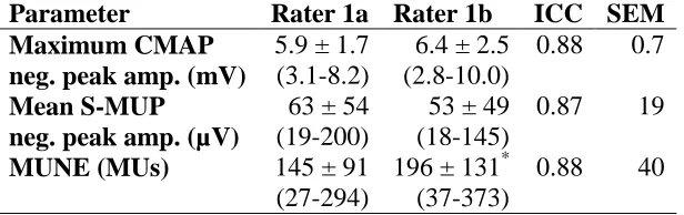
3.1 Intra- and inter-rater reliability of maximum CMAP, mean S-MUP, and MUNE
3.2 Intra- and inter-rater reliability of MUP parameters
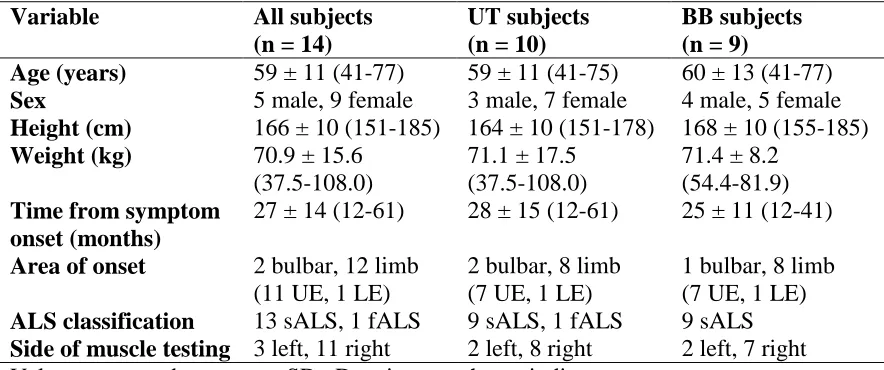
4.1 Subject demographics and clinical summary
4.2 Intra-rater reliability of maximum CMAP, mean S-MUP, and MUNE in the upper
trapezius
4.3 Intra-rater reliability of maximum CMAP, mean S-MUP, and MUNE in the biceps
brachii
4.4 Intra-rater reliability of MUP parameters in the upper trapezius
4.5 Intra-rater reliability of MUP parameters in the biceps brachii
5.1 Subject demographics and clinical summary at T0
5.2 Longitudinal change in outcome measures for those subjects with complete T0 and T6
data
5.3 Longitudinal change in outcome measures for those subjects with complete data
across all time points
xvii
LIST OF FIGURES
1.1 Collateral reinnervation
1.2 Spike-triggered averaging
1.3 Decomposition summary depicting the results of DQEMG from a single voluntary
contraction
1.4 Innervation of the upper trapezius by the spinal accessory nerve
2.1 Flow of studies through the systematic review
3.1 Frequency distributions of S-MUP data for rater 1a, rater 1b, and rater 2
4.1 Frequency distributions of S-MUP data for the UT, BB, and comparing UT data
between subjects with ALS and control subjects
4.2 Comparison of UT data between control subjects and subjects with ALS
5.1 Comparison between T0 and T6 for each electrophysiological outcome measure
5.2 Comparison between T0 and T6 for each measure of muscle strength
5.3 Comparison between T0 and T6 for measures of pulmonary function and the
ALSFRS-R
5.4 Frequency distributions of S-MUP data comparing results between T0 and T6
6.1 Frequency distributions of S-MUP data comparing results between superficial,
intermediate, and deep needle electrode depths
6.2 Maximum CMAP, mean S-MUP, and MUNE across superficial, intermediate, and
xviii
LIST OF APPENDICES
APPENDIX A: Summary of included clinical trials
APPENDIX B: Manual muscle testing grading scale
APPENDIX C: Revised Amyotrophic Lateral Sclerosis Functional Rating Scale
APPENDIX D: Ethical approval
xix
LIST OF ABBREVIATIONS
AAR: area-to-amplitude ratio
ALS: amyotrophic lateral sclerosis
ALSFRS: Amyotrophic Lateral Sclerosis Functional Rating Scale
ALSFRS-R: Revised Amyotrophic Lateral Sclerosis Functional Rating Scale
ANOVA: analysis of variance
BB: biceps brachii
CMAP: compound muscle action potential
DE-STA: decomposition-enhanced spike-triggered averaging
DQEMG: decomposition-based quantitative electromyography
EMG: electromyography
fALS: familial amyotrophic lateral sclerosis
FVC: forced vital capacity
HHD: hand-held dynamometry
ICC: intraclass correlation coefficient
LE: lower extremity
LMN: lower motor neuron
MDD: minimal detectable difference
me: margin of error
MMT: manual muscle testing
MU: motor unit
MUNE: motor unit number estimation
xx
MVC-RMS: maximal voluntary contraction - root mean square
MVIC: maximal voluntary isometric contraction
QEMG: quantitative electromyography
QoL: quality of life
RCT: randomized controlled trial
sALS: sporadic amyotrophic lateral sclerosis
SEM: standard error of measurement
SF-36: 36-item Short-Form survey
S-MUP: surface-detected motor unit potential
SNIP: sniff nasal inspiratory pressure
SRM: standardized response mean
STA: spike-triggered averaging
UE: upper extremity
UMN: upper motor neuron
CHAPTER 1
GENERAL INTRODUCTION AND OVERVIEW
1.0 GENERAL INTRODUCTION
1.0.1 The motor unit
A motor unit (MU) is defined as a single motor neuron (located in the brain stem
or spinal cord), its peripheral axon and the muscle fibers innervated by that axon.1, 2
Responsible for the production of force via reflex and voluntary contractions of skeletal
muscle, MUs are considered the elementary functional component of the motor system.3
Losses of MUs occur in aging, many peripheral neuropathies, and motor neuron diseases
such as amyotrophic lateral sclerosis (ALS).4
1.0.2 Amyotrophic lateral sclerosis
ALS is a progressive neurodegenerative disease characterized by the selective
death of upper and lower motor neurons.5 The disease has a population incidence of 1.5
to 2.5 per 100 000 persons per year and a gender ratio of roughly 2:1 male to female,
although there have been reports of a trend towards unity.6 While 5 to 10% of ALS cases
are familial, 90 to 95% of cases are considered sporadic, characterized by an absence of
family history.5 Cases are classified by region of symptom onset, with limb onset as the
most common presentation (e.g. foot drop), followed by bulbar onset (e.g. dysphagia,
dysarthria), and the relatively uncommon respiratory onset ALS (e.g. orthopnea).7
Degeneration of LMNs (and a corresponding decline in the number of functioning
MUs) results in denervation of skeletal muscle fibers and progressive muscle atrophy and
weakness.5 The progression of the disease is highly variable from patient to patient, and
3-5 years of symptom onset.5, 8 In general, death occurs as a result of respiratory failure.9
Many unknowns remain with regard to the etiology and pathogenesis of ALS. However,
a common overarching hypothesis for the etiology of ALS is that it involves the coupling
of exposure to a risk factor (proposed risk factors include strenuous physical activity,
exposure to heavy metals, and trauma with skeletal fracture) with genetic susceptibility to
the disease.10, 11 Many potentially convergent hypotheses exist as to the underlying
causes of motor neuron degeneration, including oxidative damage, glutamate
excitotoxicity, and aberrant RNA metabolism.12 With the exception of the drug riluzole
(which has been shown to prolong survival modestly),13 the interventions tested in
clinical trials thus far have not been found to modify the progression of the disease.14
1.0.3 Motor unit number estimation
Highly relevant for application to this disease population, motor unit number
estimation (MUNE) was developed by McComas et al. (1971)15 as a quantitative,
electrophysiological method for estimating the number of functioning MUs within a
muscle or group of muscles. This quantification may be useful to evaluate the severity
and natural history of the disease. Furthermore, it may be especially useful as an
outcome measure in ALS clinical trials to assess the efficacy of interventions by
monitoring disease progression.16
MUNE uses electromyography (EMG), which detects and records the electrical
activity of active MUs using surface and/or intramuscular electrodes. The action
potentials propagated along the individual muscle fibers of a MU following the discharge
of the associated motor neuron correspond with waveforms termed muscle fiber action
waveform termed a motor unit potential (MUP) that is unique for each MU.17 Certain
parameters associated with a surface-detected motor unit potential (S-MUP) or
intramuscularly-detected MUP (hereafter simply referred to as a MUP) are related to the
morphological and physiological characteristics of the underlying MU. For example, the
amplitude or area of an S-MUP is representative of the number of muscle fibers within
the MU (i.e. the innervation ratio, or size of the MU).3, 15, 17
Based on these principles, a MUNE is calculated through the division of a size
parameter of the maximum compound muscle action potential (CMAP), representative of
the activation of all of the motor axons in the nerve in response to supramaximal
electrical stimulation, by the same size parameter of the mean S-MUP, representative of
the average single MU size (Equation 1).1, 18
1.0.3.1 Equation 1
MUNE = maximum CMAP size mean S-MUP size
Where MUNE, motor unit number estimate; CMAP, compound muscle action potential;
S-MUP, surface-detected motor unit potential.
1.0.4 Outcome measures and collateral reinnervation
The selection of primary and secondary outcome measures (also termed
endpoints) for use in ALS clinical trials to determine the efficacy of interventions has
been highly heterogeneous. However, a quantitative review of ALS clinical trials has yet
to be conducted to identify the most commonly used and underutilized measures.
In general terms, ALS clinical trials commonly employ outcome measures that
assess function and muscle strength.19, 20 However, these outcome measures are
chronic denervating disorders such as ALS, whereby new nerve sprouts grow out from
surviving nerve axons to supply denervated muscle fibers (Figure 1.1). This process, by
maintaining muscle mass and strength, may allow these measures to remain relatively
stable until MU loss has surpassed a critical threshold.21
In contrast to these clinical outcome measures, MUNE takes into account the
effects of collateral reinnervation, as mean S-MUP size is incorporated into its
calculation. Its subsequent ability to monitor the underlying progression of the disease
(MU loss as well as collateral reinnervation) makes MUNE a valuable addition as an
outcome measure for use in ALS clinical trials.16, 22
1.0.5 Motor unit number estimation techniques
Various types of MUNE have been developed and modified since its introduction
over 40 years ago, with manual incremental stimulation,15 multiple point stimulation,23, 24
the statistical method,1 spike-triggered averaging (STA),25, 26 and
decomposition-enhanced spike-triggered averaging (DE-STA)18, 27 used most commonly.16, 22 While the
basic principles are the same for all techniques, they differ in their underlying
assumptions, as well as their inherent benefits and limitations.
A key difference among the techniques is the way in which the mean S-MUP is
obtained, which influences the range of muscle groups to which the protocol can be
applied. Manual incremental stimulation, multiple point stimulation, and the statistical
method (along with modifications to these techniques) employ percutaneous electrical
stimulation of the motor nerve in order to collect a sample of S-MUPs, whereas STA and
DE-STA utilize voluntary contractions. MUNE techniques utilizing electrical stimulation
muscles with easily accessible nerve supplies, and in the case of multiple point
stimulation, a certain length of the motor nerve available for stimulation. In contrast, the
use of voluntary contractions to collect a sample of S-MUPs allows for STA and
DE-STA MUNE to be applied to any muscle from which a maximum CMAP can be elicited.
Thus, these techniques can be applied to not only distal, but proximal muscles.16, 18, 22
1.0.6 Quantitative electromyography
A further advantage of STA and DE-STA MUNE stems from the introduction of
a needle electrode, which allows for an intramuscular EMG signal to be collected
simultaneously with the surface EMG signal during each voluntary contraction (Figure
1.2).16, 18, 22 The subsequent application of quantitative electromyography (QEMG)
techniques allows for quantitative MUP analysis in addition to the results yielded through
MUNE. QEMG techniques allow for the isolation of the activity of individual MUP
trains, representative of the repetitive firing of single MUs over a given period of time,28
and subsequent determination of the prototypical MUP associated with each MUP train.
Quantification of the distributions of various parameters characterizing the sizes, shapes,
and firing patterns of a representative sample of MUPs from a given muscle allows for
the evaluation of corresponding morphological and physiological features of the
associated MU pool. As such, quantitative MUP analysis is able to yield information
complementary to MUNE corresponding with the severity and progression of processes
affecting the neuromuscular system such as ALS.29, 30
QEMG facilitates the process of MUNE, with STA and DE-STA utilizing
different techniques. The process of STA involves the application of a level or
activity of an individual, voluntarily activated MUP train. Each MUP firing that meets or
falls within the limits of the discriminator acts as a trigger to isolate the time-locked
S-MUP for that specific S-MUP (Figure 1.2). Next, the S-S-MUPs associated with each S-MUP
firing for a given MUP train are ensemble-averaged to derive the S-MUP template
associated with a given MU.25, 26 First described as a method of MUNE by Boe et al.
(2004),27 DE-STA improves upon conventional STA MUNE by incorporating a series of
computer-based algorithms for intramuscular EMG signal decomposition, termed
decomposition-based quantitative electromyography (DQEMG).18, 28, 31
1.0.7 Decomposition-based quantitative electromyography
While STA is limited to the collection of one MUP train from each low-level
voluntary contraction, the addition of DQEMG allows for the resolution of composite
EMG signals produced from stronger voluntary contractions into their constituent MUP
trains. The individual MUP firings from each MUP train then serve as triggers to be used
in STA, resulting in the collection of S-MUP templates for numerous MUs from a single
contraction. By collecting more than one MUP train at a time from stronger voluntary
contractions, DE-STA MUNE both eases the level of focus required of the subject, and
allows for more efficient collection of a sample of S-MUPs. Furthermore, in line with
the “size principle” of MU recruitment,32
the use of stronger voluntary contractions
allows DE-STA MUNE to include not only low-threshold, generally smaller MUs but
also higher-threshold, larger MUs. This may allow for the determination of a more
representative mean S-MUP from which to calculate a MUNE.18, 31
The DQEMG algorithms involve MUP detection, clustering, and supervised
EMG signal. The composite signal is first bandpass filtered using a first-order difference
filter. Subsequently, the MUPs exceeding a certain threshold are detected and considered
to represent a significant MUP occurrence. Next, a shape- and temporal-based clustering
algorithm is applied to the detected MUPs from an interval of the EMG signal in order to
elucidate the number of contributing MUs, and the prototypical MUP shape for each MU.
Supervised classification involves the assignment of each MUP detected initially to its
corresponding MUP train. Assignments are made based on shape and firing pattern
information and utilize a certainty algorithm, with only classifications that exceed a
specific threshold of assignment certainty being made. Lastly, algorithms that examine
the temporal relationships between MUP trains allow for the splitting and merging of
MUP trains.28, 33
EMG signal decomposition and subsequent STA yields a decomposition summary
for each voluntary contraction, as depicted in Figure 1.3. Quantitative MUP analysis
involves the calculation of various parameters characterizing the MUP template, MUP
shimmer plot, S-MUP template, and firing pattern associated with each MUP train using
standard algorithms. Finally, descriptive statistics are calculated automatically for each
parameter based on the entire sample of accepted MUPs, S-MUPs, and MUP trains from
A B C
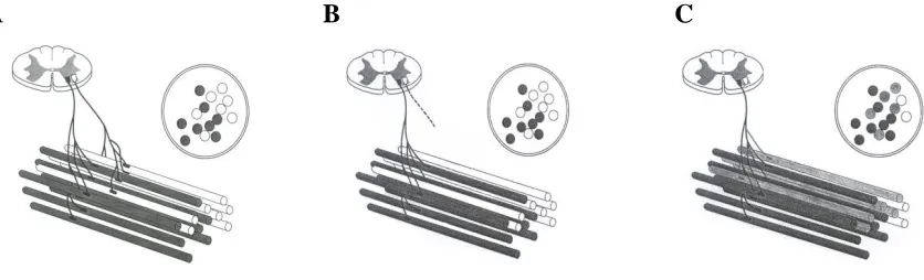
Figure 1.1 Collateral reinnervation. Two functioning MUs are depicted with their motor neurons located in the anterior horn of a cross-section of the spinal cord and their
peripheral axons innervating respective groups of muscle fibers. The muscle fibers innervated by the MU on the left are represented as shaded circles in the inset (A). Degeneration of one of the MUs occurs, resulting in the denervation of its associated skeletal muscle fibers (B). Collateral reinnervation is depicted, with new nerve sprouts from the axon of the surviving MU reinnervating many of the orphaned muscle fibers. The inset depicts the subsequent increase in the innervation ratio of the surviving MU (C). (Modified from Stålberg E, Falck B. The role of electromyography in neurology. Electroencephalogr Clin Neurophysiol 1997; 103:579-598).34
A
B
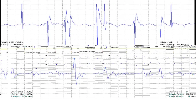
Figure 1.2 Spike-triggered averaging. An example of the simultaneous collection of an intramuscular EMG signal (top panel) and surface EMG signal (bottom panel) from a voluntary contraction, as occurs in STA and DE-STA MUNE techniques (A). Following the isolation of the activity of an individual MUP train from the intramuscular EMG signal using QEMG, individual MUP firings act as triggers to isolate time-locked S-MUPs. An S-MUP (thin line) is shown superimposed on its corresponding MUP firing (thick line) (B). ([B] Modified from Doherty T, Simmons Z, O'Connell B, Felice KJ, Conwit R, Chan KM, et al. Methods for estimating the numbers of motor units in human muscles. J Clin Neurophysiol 1995; 12:565-584).18
Figure 1.3 Decomposition summary depicting the results of DQEMG from a single voluntary contraction. Each row represents one MUP train. The first column depicts a MUP template and the number of isolated MUPs used for its estimation. The second column presents a MUP shimmer plot of the isolated MUPs making up the MUP train. The third column depicts an S-MUP template and the number of firings used for its estimation. The fourth column displays an interdischarge interval histogram, as well as the mean interdischarge interval, and standard deviation and coefficient of variation of the interdischarge interval. The fifth column depicts a firing graph, with vertical lines representing MU firing times and the top tracing representing the instantaneous firing rate plot. (Modified from Stashuk D. EMG signal decomposition: How can it be
accomplished and used? J Electromyogr Kinesiol 2001; 11:151-173).33
1.0.8 Upper trapezius muscle
Thus far, the use of MUNE as an outcome measure in ALS clinical trials has been
limited. Taking into consideration the many advantages associated with DE-STA
MUNE, this technique may be very useful in such a role.
In evaluating the potential utility of DE-STA MUNE as an outcome measure, the
role of the muscle under study becomes an important consideration. As previously
mentioned, respiratory failure is generally the cause of death in ALS.9 Thus, it would be
clinically relevant to study muscles of respiration in this population. While the principal
muscles of respiration (i.e. diaphragm, intercostal muscles) are not feasible to study using
MUNE, the upper trapezius (UT) muscle is an accessory muscle of respiration9 that
would be practical to assess, given its superficial position and easily accessible nerve
supply.
The UT (descending fibers), together with the middle (transverse fibers) and
lower (ascending fibers) trapezii, compose the trapezius muscle. The UT originates from
the external occipital protuberance, the medial third of the superior nuchal line, and the
ligamentum nuchae, and inserts into the posterior border of the lateral third of the
clavicle. This muscle acts with the levator scapulae to perform scapular elevation.35 The
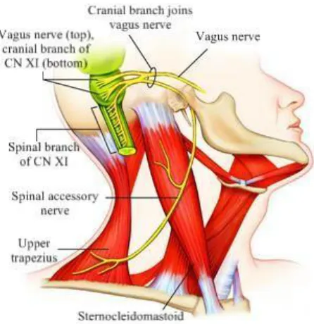
primary source of motor innervation for the UT is the spinal accessory nerve, which is the
spinal branch of cranial nerve XI, termed the accessory nerve.35-38 The spinal accessory
nerve is a motor nerve that supplies both the trapezius and sternocleidomastoid muscles,
and originates from motor neurons lying in the lateral portion of the gray matter of the
upper cervical spinal cord (C1-C6). Rootlets exit the lateral aspect of the spinal cord,
through the jugular foramen. The spinal accessory nerve then travels inferiorly,
innervating the SCM, and on through the posterior triangle of the neck to innervate the
UT (Figure 1.4).35, 36
As an accessory muscle of respiration, the UT aids with inspiration in the
presence of impaired diaphragmatic function, as occurs with progressive denervation in
ALS.9 Quantification of the number and characteristics of MUs associated with a muscle
related to respiratory function may offer insight into the severity and progression of the
disease, as well as the potential efficacy of interventions. Additionally, as a proximal
muscle, examination of the UT would be novel, as the majority of previous studies using
MUNE have utilized techniques that limited their evaluations to distal muscles.
A preliminary study by Lewis (2009)39 examined the application of DE-STA
MUNE to the UT in a small sample that included three control subjects and one subject
with ALS. Their results suggest that the application of DE-STA MUNE to this proximal
Figure 1.4 Innervation of the upper trapezius by the spinal accessory nerve. (Modified from Mosby’s medical dictionary. Mosby/Elsevier: St. Louis; 2009. 2056 p.).40
1.0.9 Evaluation of decomposition-enhanced spike-triggered averaging MUNE
In order to assess the utility of DE-STA MUNE as an outcome measure, its
reliability, validity, and sensitivity to change must be evaluated.41, 42 With respect to
reliability, the results of any useful measurement technique must be reproducible, both in
the hands of the same evaluator at two or more points in time (intra-rater reliability) and
of different evaluators (inter-rater reliability).43 Previous studies have established the
intra-27, 44 and inter-rater reliability45 of DE-STA MUNE in various muscle groups in
control subjects, in addition to the intra-rater reliability of subsets of data collected using
DQEMG46, and the intra- and inter-rater reliability46, 47 of the analysis of this data.
However, the reliability of DE-STA MUNE as applied to the UT in control subjects
remains to be assessed. Additionally, the reliability of DE-STA MUNE has yet to be
studied in any muscle group in subjects with ALS.
In addition to reliability, a critical property of any outcome measure is its validity.
That is, its ability to measure what it purports to measure in an accurate fashion.43 We
currently lack a gold standard technique to quantify the number of MUs in a given
muscle group in a living subject, and normative anatomical data for many muscle groups
with which to compare the results of DE-STA MUNE. A single previous study
compared the results of DE-STA MUNE between subjects with ALS and control
subjects, demonstrating the ability of the technique to detect the underlying
pathophysiology of the disease in the biceps brachii (BB) and first dorsal interosseous.48
However, such an evaluation of the validity of the technique in the UT has yet to be
Finally, an important property of any outcome measure is sensitivity to change,
which is the ability of an instrument to detect minimal change over time, and thus
monitor the potential efficacy of an intervention.43 A cross-sectional study by Boe et al.
(2009)49 compared DE-STA MUNE in the first dorsal interosseous and BB alongside
several clinical outcome measures in subjects with ALS to normative results for the same
measures. The study demonstrated the ability of DE-STA MUNE to detect underlying
pathophysiological features of the disease despite better-preserved functional
performance. However, a longitudinal study comparing the sensitivity to change between
DE-STA MUNE and clinical outcome measures in subjects with ALS has yet to be
conducted.
Sources of error associated with a measurement technique may negatively
influence its reliability, validity, and sensitivity to change.43 Thus, investigation into
potential sources of error associated with DE-STA MUNE may lead to improvements in
these properties of the technique that are essential to its utility as an outcome measure. It
is well established that the position of the detecting electrode relative to the muscle fibers
of a MU influences the characteristics of the corresponding EMG signal.17 Thus, the
influence of the depth of the needle electrode on the results of DE-STA MUNE may be
important to assess. This component of the technique’s data collection protocol has not
been evaluated systematically.
It is the overall objective of this thesis to review the use of outcome measures
systematically across ALS clinical trials, and to evaluate the utility of DE-STA MUNE as
1.1 OVERVIEW OF THESIS CHAPTERS
The objectives of this thesis were addressed through the conduct of a series of five
studies. First, a review of the frequency of use of outcome measures across ALS clinical
trials, in addition to the advantages and disadvantages of select measures was conducted
(Chapter 2). In order to assess the utility of DE-STA MUNE as an outcome measure, the
reliability (Chapters 3 and 4), validity (Chapter 4), and sensitivity to change (Chapter 5)
of the technique was evaluated. The findings from the study reported in Chapter 5
indicated the need for further study regarding potential sources of error associated with
the technique. Thus, continuing the assessment of the utility of DE-STA MUNE as an
outcome measure, the influence of the depth of the needle electrode on the results of the
technique was evaluated (Chapter 6).
1.1.1 Chapter 2 study objectives
To conduct a systematic review quantifying the frequency of use of outcome
measures as primary and secondary endpoints in ALS randomized controlled
trials published since 1990, classifying these outcome measures according to
common categories from the ALS literature.
To briefly review the advantages and disadvantages associated with select
outcome measures identified by the review.
1.1.2 Chapter 3 study objective
To assess the intra- and inter-rater reliability of DE-STA MUNE and quantitative
1.1.3 Chapter 4 study objectives
To assess the intra-rater reliability of DE-STA MUNE and quantitative MUP
analysis in the UT and BB of subjects with ALS.
To compare the results of DE-STA MUNE and quantitative MUP analysis in the
UT of subjects with ALS to data obtained previously (study 2) in control subjects
in the same muscle.
1.1.4 Chapter 5 study objective
To compare the sensitivity to change of DE-STA MUNE and quantitative MUP
analysis in the UT to that of various clinical outcome measures in subjects with
ALS. More specifically, to make comparisons with manual muscle testing in five
upper extremity muscle groups, scapular elevation and elbow flexion peak force
measured using hand-held dynamometry, forced vital capacity, sniff nasal
inspiratory pressure, and the Revised Amyotrophic Lateral Sclerosis Functional
Rating Scale.
1.1.5 Chapter 6 study objective
To evaluate the influence of needle electrode depth on the results of DE-STA
MUNE and quantitative MUP analysis in the UT of control subjects by comparing
the results obtained across superficial, intermediate, and deep needle electrode
1.2 REFERENCES
1. Daube JR. Estimating the number of motor units in a muscle. J Clin Neurophysiol 1995; 12:585-594.
2. Sherrington CS. Some functional problems attaching to convergence. Proc R Soc Lond B 1929; 105:332-362.
3. Doherty TJ, Chan KM, Brown WF. Motor neurons, motor units, and motor unit recruitment. In: Brown WF, Bolton CF, Aminoff MJ, editors. Neuromuscular function and disease: Basic, clinical, and electrodiagnostic aspects. W.B. Saunders Company: Philadelphia; 2002. Vol. 1, 247-273 p.
4. Doherty TJ, Brown WF. Motor unit number estimation: Methods and applications. In: Brown WF, Bolton CF, Aminoff MJ, editors. Neuromuscular function and disease: Basic, clinical, and electrodiagnostic aspects. W.B. Saunders Company: Philadelphia; 2002. Vol. 1, 274-290 p.
5. Rowland LP, Shneider NA. Amyotrophic lateral sclerosis. N Engl J Med 2001; 344:1688-1700.
6. Logroscino G, Traynor BJ, Hardiman O, Chió A, Couratier P, Mitchell JD, et al. Descriptive epidemiology of amyotrophic lateral sclerosis: New evidence and unsolved issues. J Neurol Neurosurg Psychiatry 2008; 79:6-11.
7. Cwik V. ALS clinical motor signs and symptoms. In: Mitsumoto H, Przedborski S, Gordon PH, editors. Amyotrophic lateral sclerosis. Taylor & Francis: New York; 2006. 99-116 p.
8. Shoesmith CL, Strong MJ. Amyotrophic lateral sclerosis: Update for family physicians. Can Fam Physician 2006; 52:1563-1569.
9. Gregory SA. Evaluation and management of respiratory muscle dysfunction in ALS. NeuroRehabilitation 2007; 22:435-443.
10. Brooks BR. Clinical epidemiology of amyotrophic lateral sclerosis. Neurol Clin 1996; 14:399-420.
11. Shoesmith C. A recipe for ALS. Can J Neurol Sci 2008; 35:125-126.
12. Rothstein JD. Current hypotheses for the underlying biology of amyotrophic lateral sclerosis. Ann Neurol 2009; 65:S3-S9.
14. Beghi E, Chiò A, Couratier P, Esteban J, Hardiman O, Logroscino G, et al. The epidemiology and treatment of ALS: Focus on the heterogeneity of the disease and critical appraisal of therapeutic trials. Amyotroph Lateral Scler 2011; 12:1-10.
15. McComas AJ, Fawcett PR, Campbell MJ, Sica RE. Electrophysiological estimation of the number of motor units within a human muscle. J Neurol Neurosurg Psychiatry 1971; 34:121-131.
16. Rashidipour O, Chan KM. Motor unit number estimation in neuromuscular disease. Can J Neurol Sci 2008; 35:153-159.
17. Stashuk DW, Doherty TJ. Normal motor unit action potential. In: Brown WF, Bolton CF, Aminoff MJ, editors. Neuromuscular function and disease: Basic, clinical, and electrodiagnostic aspects. W.B. Saunders Company: Philadelphia; 2002. Vol. 1, 291-310 p.
18. Doherty T, Simmons Z, O'Connell B, Felice KJ, Conwit R, Chan KM, et al. Methods for estimating the numbers of motor units in human muscles. J Clin Neurophysiol 1995; 12:565-584.
19. Shefner JM. Designing clinical trials in amyotrophic lateral sclerosis. Phys Med Rehabil Clin N Am 2008; 19:495-508.
20. Miller RG. Classical and emergent measures for clinical trials in ALS. Amyotroph Lateral Scler Other Motor Neuron Disord 2002; 3:1-2.
21. McComas AJ, Sica RE, Campbell MJ, Upton AR. Functional compensation in partially denervated muscles. J Neurol Neurosurg Psychiatry 1971; 34:453-460.
22. Bromberg MB, Brownell AA. Motor unit number estimation in the assessment of performance and function in motor neuron disease. Phys Med Rehabil Clin N Am 2008; 19:509-532.
23. Doherty TJ, Brown WF. The estimated numbers and relative sizes of thenar motor units as selected by multiple point stimulation in young and older adults. Muscle Nerve 1993; 16:355-366.
24. Kadrie HA, Yates SK, Milner Brown HS, Brown WF. Multiple point electrical stimulation of ulnar and median nerves. J Neurol Neurosurg Psychiatry 1976; 39:973-985.
26. Lee RG, Ashby P, White DG, Aguayo AJ. Analysis of motor conduction velocity in the human median nerve by computer simulation of compound muscle action potentials. Electroencephalogr Clin Neurophysiol 1975; 39:225-237.
27. Boe SG, Stashuk DW, Doherty TJ. Motor unit number estimation by decomposition-enhanced spike-triggered averaging: Control data, test-retest reliability, and contractile level effects. Muscle Nerve 2004; 29:693-699.
28. Stashuk DW. Decomposition and quantitative analysis of clinical electromyographic signals. Med Eng Phys 1999; 21:389-404.
29. Stashuk DW, Brown WF. Quantitative electromyography. In: Brown WF, Bolton CF, Aminoff MJ, editors. Neuromuscular function and disease: Basic, clinical, and
electrodiagnostic aspects. W.B. Saunders Company: Philadelphia; 2002. Vol. 1, 311-348 p.
30. Stålberg E, Nandedkar SD, Sanders DB, Falck B. Quantitative motor unit potential analysis. J Clin Neurophysiol 1996; 13:401-422.
31. Doherty TJ, Stashuk DW. Decomposition-based quantitative electromyography: Methods and initial normative data in five muscles. Muscle Nerve 2003; 28:204-211.
32. Henneman E, Mendell LM. Functional organization of motor unit pool and its inputs. In: Brooks VB, editor. Handbook of physiology. American Physiological Society:
Bethesda; 1981. Vol. 2, 423-508 p.
33. Stashuk D. EMG signal decomposition: How can it be accomplished and used? J Electromyogr Kinesiol 2001; 11:151-173.
34. Stålberg E, Falck B. The role of electromyography in neurology. Electroencephalogr Clin Neurophysiol 1997; 103:579-598.
35. Standring S, Gray H. Gray's anatomy: The anatomical basis of clinical practice. Churchill Livingstone/Elsevier: Edinburgh; 2008. 1551 p.
36. Campos D, Rieger A, Mohr H, Ellwanger JH, Borba Junior AM. Anatomy and evolution of accessory nerve: Cranial or spinal origins? A review. Journal of Morphological Sciences 2011; 28:222-227.
37. Pu YM, Tang EY, Yang XD. Trapezius muscle innervation from the spinal accessory nerve and branches of the cervical plexus. Int J Oral Maxillofac Surg 2008; 37:567-572.
39. Lewis RA. Motor unit number estimation in the upper trapezius muscle. Suppl Clin Neurophysiol 2009; 60:131-134.
40. Mosby's medical dictionary. Mosby/Elsevier: St. Louis; 2009. 2056 p.
41. Husted JA, Cook RJ, Farewell VT, Gladman DD. Methods for assessing
responsiveness: A critical review and recommendations. J Clin Epidemiol 2000; 53:459-468.
42. Guyatt G, Walter S, Norman G. Measuring change over time: Assessing the usefulness of evaluative instruments. J Chronic Dis 1987; 40:171-178.
43. Portney LG, Watkins MP. Foundations of clinical research: Applications to practice. Prentice Hall: New Jersey; 2008. 892 p.
44. Boe SG, Stashuk DW, Doherty TJ. Within-subject reliability of motor unit number estimates and quantitative motor unit analysis in a distal and proximal upper limb muscle. Clin Neurophysiol 2006; 117:596-603.
45. Boe SG, Dalton BH, Harwood B, Doherty TJ, Rice CL. Inter-rater reliability of motor unit number estimates and quantitative motor unit analysis in the tibialis anterior muscle. Clin Neurophysiol 2009; 120:947-952.
46. Calder KM, Agnew MJ, Stashuk DW, McLean L. Reliability of quantitative EMG analysis of the extensor carpi radialis muscle. J Neurosci Methods 2008; 168:483-493.
47. Boe SG, Antonowicz NM, Leung VW, Shea SM, Zimmerman TC, Doherty TJ. High inter-rater reliability in analyzing results of decomposition-based quantitative
electromyography in subjects with or without neuromuscular disorder. J Neurosci Methods 2010; 192:138-145.
48. Boe SG, Stashuk DW, Doherty TJ. Motor unit number estimates and quantitative motor unit analysis in healthy subjects and patients with amyotrophic lateral sclerosis. Muscle Nerve 2007; 36:62-70.
CHAPTER 2
USE OF OUTCOME MEASURES IN ALS RANDOMIZED CONTROLLED
TRIALS: A SYSTEMATIC REVIEW
2.0 INTRODUCTION
Clinical trials aimed at identifying interventions to modify the progression of
amyotrophic lateral sclerosis (ALS) are underway constantly. To date, only one drug
(riluzole) has been shown to prolong survival modestly.1 The process of drug
development typically begins with basic science investigations, followed by sequential
phases of human clinical trials. Early phase trials (phase I and II) focus on examination
of pharmacokinetic characteristics, the clinical safety and toxicity profile, feasibility, and
dosing of the pharmacologic agent, with some phase II trials opting to include a
preliminary assessment of efficacy.2
If the results of these early phase trials show promise for treatment efficacy, a
potential intervention would then be studied in a phase III clinical trial. A phase III
clinical trial is conducted in a large sample of patients, and the primary aim of these trials
is to assess the efficacy and safety of an intervention. A randomized controlled trial
(RCT) is the gold standard design for a phase III clinical trial. These trials are typically
randomized, double-blind, and placebo-controlled in order to promote high levels of
internal validity.3
As previously stated, the vast majority of interventions studied in RCTs for ALS
thus far have not been shown to alter the course of the disease significantly. This may be
due to their genuine ineffectiveness or, alternatively, to the design or conduct of the
measures, which are the variables used to monitor the efficacy of an intervention.
Outcome measures are also termed ‘endpoints’ and are designated as either the primary
or secondary outcomes in clinical trials. While more than one may occasionally be
selected, trials typically employ a single primary outcome measure, which is used in the
power calculation to determine the required sample size and whether the end result of the
trial is ‘positive’ or ‘negative’. Secondary outcome measures are considered to be less
clinically important than the primary outcome measure for a particular trial.2
The power of a clinical trial may be impacted by the chosen primary outcome
measure. Calculation of the sample size required in order for a trial to reach a desired
level of statistical power is dependent in large part on the sensitivity to change and
variability of the chosen primary endpoint. In addition, the degree of complexity and
burden added to study visits by the primary and secondary measures may influence the
amount of missing data and subject dropouts. Missing data may also result if the measure
becomes more difficult to assess with progression of the disease; an important issue in
ALS trials. The selected primary outcome measure also impacts the required duration of
the trial, thus acting as a critical determinant in the cost and time required for a single
study. These are particularly important issues when studying a relatively rare disease for
which resources (subjects and money) are often limited. The amount of time spent on
individual trials is also of key importance given the sense of urgency accompanying the
fatal nature of the disease, and considering the large number of potential agents waiting
to be tested.4, 5
While survival is obviously a clinically meaningful outcome for a fatal disease, its
order for studies to reach statistical power.4 The additional consideration of other
disadvantages associated with the use of survival (as will be discussed in detail) has led
to the utilization of other outcome measures in ALS clinical trials. These outcome
measures fall under categories of muscle strength, pulmonary function, functional rating
scales/timed functional tests, electrophysiological indices, biomarkers, imaging and
quality of life (QoL).2, 6
Assessment of muscle strength is a highly relevant outcome measure given that
muscle wasting and weakness are core features of disease progression. The strength of a
muscle or group of muscles during a voluntary contraction can be assessed quantitatively
with a strain-gauge, determining maximal voluntary isometric contraction (MVIC) or
with hand-held dynamometry, as well as by manual muscle testing (MMT) using the
Medical Research Council scale.7
Given that respiratory failure as a result of progressive respiratory muscle
weakness is generally the cause of death in ALS, clinical trials also commonly employ
outcome measures of pulmonary function. As the diaphragm is the major muscle
responsible for inspiration, these measures largely aim to monitor changes in the strength
of this muscle over time, and include such measures as vital capacity and forced vital
capacity (FVC).8
Functional outcome measures such as timed functional tests and functional rating
scales (e.g. Revised Amyotrophic Lateral Sclerosis Functional Rating Scale
[ALSFRS-R]) are measures of the impact of an intervention on activities of daily living and other
functional tasks which may be specifically affected by the disease process. These types
importance to the patient and their family.9 Functional rating scales can be generic or
disease-specific, and utilize the patient’s self-report or a clinician’s report.10
Unlike measures of muscle strength and function,11 electrophysiological indices
assess the underlying pathophysiology and progression of the disease, providing
quantitative information on the upper motor neuron (UMN) or lower motor neuron
(LMN) component. The quantitative techniques assessing the LMN rely on
electromyography (EMG) (e.g. motor unit number estimation [MUNE],
neurophysiological index, various needle EMG parameters) and those assessing the UMN
component of the disease currently utilize transcranial magnetic stimulation.12, 13
Biomarkers are objective laboratory measurements (e.g. cerebrospinal fluid levels
of prostaglandin E2, blood markers of oxidative damage) that function as potential
indicators of pathogenesis and biological response to pharmacologic intervention. These
markers may be highly sensitive to disease progression and early therapeutic effects as
outcome measures in clinical trials.14, 15 Although in their relatively early stages of use as
outcome measures, neuroimaging techniques such as magnetic resonance spectroscopy
may be highly valuable given their unique capacity to detect and monitor the UMN
component of the disease non-invasively and objectively.16
Incorporation of measures of QoL as outcome measures for an ALS clinical trial
allows for a more complete evaluation of the effect of an intervention, and may be
especially important to the patient given the fatal nature of the disease.2, 17 QoL
questionnaires can be generic, meaning that their items assess general health concepts not
[SF-36]), or specific, having been developed for the ALS patient population (e.g. Sickness
Impact Profile/ALS-19).19
Selection of primary and secondary endpoints to be used in a clinical trial may be
aided by the consensus guidelines on ALS trial design and execution published by the
World Federation of Neurology20 or by guidelines published by Leigh et al. (2004)9.
Although outdated, the World Federation of Neurology guidelines suggest the use of
change in muscle strength or survival as the primary outcome measure, and make a broad
recommendation for the inclusion of measures of muscle strength (recommending
quantitative myometry [i.e. MVIC]), pulmonary function (recommending FVC), bulbar
function and time to death in every trial. The guidelines by Leigh et al. (2004)9
recommend the use of either survival or, preferentially, the ALSFRS-R as the primary
outcome measure, with both sets of guidelines recommending the inclusion of a
disease-specific QoL measure alongside the SF-36 generic measure as secondary endpoints.
Heterogeneity in the use of outcome measures as a result of: a) the absence of an
unequivocal primary outcome measure selection; b) the presence of a multitude of
options available for secondary outcome measures; and c) limited guidelines for the use
of these measures, is widely apparent. However, a systematic review quantifying the
frequency of use of available outcome measures across ALS RCTs has yet to be
completed.
Various reviews have documented the use of primary and, less commonly,
secondary outcome measures across only select ALS clinical trials.3, 9, 21, 22 A systematic
review of ALS RCTs was conducted by Beghi et al. (2011)23, reporting on various
measures. Lastly, a systematic review of ALS RCTs by de Carvalho et al. (2005)12 with
highly selective inclusion criteria chose to focus on the sensitivity to change of various
primary outcome measures, but not on their frequency of use.
A systematic, quantitative synthesis of the literature would allow for identification
of all of the outcome measures that have been used in ALS clinical trials to date and
serve to pinpoint the most commonly used as well as underutilized outcome measures.
This would act as a resource to facilitate discussion regarding the selection of outcome
measures for future ALS clinical trials; a critical component of trial design impacting
largely on their potential for success.
Thus, the objectives of this systematic review were to quantify the frequency of
use of outcome measures as primary and secondary endpoints in ALS RCTs published
since 1990, classifying these outcome measures according to the previously mentioned
categories common in the ALS literature. A secondary objective was to briefly review
the advantages and disadvantages of select outcome measures identified by the review.
2.1 METHODS
Relevant studies were identified through a literature search encompassing the time
period from January 1990 to July 2011. The following databases were searched:
MEDLINE, EMBASE and Cochrane Central Register of Controlled Trials. Search terms
Box 2.1 Search terms
amyotrophic lateral sclerosis OR ALS OR MND OR motor neuron disease OR (MEDLINE only) motor neurone disease OR (MEDLINE only) motoneuron disease OR motoneurone disease OR Lou Gehrig's disease OR Charcot's disease
AND
placebo OR placebos
AND
double blind OR double blinded OR double-blind*
AND
2.1.1 Inclusion criteria
Studies were included if they: i) purported to assess an intervention for
efficacy/effectiveness solely in patients with ALS, regardless of phase (i.e. phase II trials
including assessments of efficacy were included); ii) compared subjects receiving the
intervention to a control group receiving placebo (in any form, frequency, dosage or in
combination with other products); and were iii) randomized or quasi-randomized (i.e.
alternate allocation); iv) double-blind; and v) prospective clinical trials. There were no
restrictions made with respect to study design (e.g. parallel group, cross-over), and
studies with multiple intervention and/or control groups were included.
2.1.2 Exclusion criteria
Review articles, editorials and other non-clinical trials were excluded, as were
abstracts that were published subsequently as full studies. Abstracts which lacked
sufficient reporting detail and abstracts/full-text articles that were unobtainable were also
excluded. Additionally, studies pertaining to symptomatic therapies for ALS (e.g. for
pseudobulbar affect, sialorrhoea, muscle cramps), ongoing studies, and non-English
studies were excluded.
Review of all search results and data extraction was performed by one author
(CI). First, titles and abstracts identified by the literature search were screened for
eligibility. For all retained abstracts (including all those for which eligibility remained in
question) an attempt was made to obtain the full-text articles and either the full-text
article or abstract (when the full-text was unavailable) was reviewed in detail to
determine eligibility. Upon determination of the eligible studies to be included in the
author, year of publication, sample size (n), trial duration, and primary and secondary
outcome measures. The methodological quality of the studies was not assessed, as it was
not relevant to the assessment of outcome measure selection. If the study did not clearly
identify which outcome measures were primary and which were secondary, or listed
more than one endpoint as the primary outcome measure, the variable for which the study
was powered statistically (where identified) was considered to be the primary outcome
measure.
Each identified outcome measure was classified into one of the following
common categories of outcome measures used in ALS clinical trials: survival, muscle
strength, pulmonary function, functional rating scales/timed functional tests,
electrophysiological indices, biomarkers, imaging, or QoL.2, 6
2.2 RESULTS
2.2.1 Literature search results
The results of the literature search and the reasons for exclusion are presented in
Figure 2.1. From a total of 1127 titles and abstracts identified by the search strategy, 85
potentially relevant papers were identified from the initial screening and subsequently
reviewed in detail. Fifty-seven of these papers were ultimately determined to be eligible
for inclusion in the review. One of these papers reported the results of 2 RCTs24 and
thus, 58 clinical trials were identified in total. A summary of all of the included clinical
trials is presented in Appendix A. The trials were published from 1992 to 2011. The
average sample size of these trials was 207 ± 278 participants (median: 99), ranging from
With the exception of 5 studies,28-32 all trials were assessments of pharmaceutical
compounds.
2.2.2 Primary outcome measures
The primary outcome measure was clearly stated in 45 (78%) of the clinical trials,
and these trials were incorporated into the descriptive analysis in Box 2.2. From these
clinical trials, 13 different primary outcome measures were identified. Twenty trials
(44%) utilized a primary outcome measure falling under the functional rating
scales/timed functional tests classification, 13 trials (29%) used survival, and 10 trials
(22%) used an outcome measure that assessed muscle strength. None of the trials in the
review utilized a primary outcome measure that evaluated pulmonary function, an
electrophysiological index, a biomarker, or QoL. Examining the most frequently used
individual outcome measures within these categories, survival was the most often used
primary outcome measure overall (13 trials; 29%), followed by MVIC – upper extremity
(UE) and the ALSFRS-R in 6 trials (13%) each. A megascore that incorporated more
than one outcome measure was used as the primary endpoint in 2 trials.33, 34
2.2.3 Secondary outcome measures
Forty-eight trials (83%) made it clear which endpoints (if any) were used as
secondary outcome measures, and these trials were incorporated into the descriptive
analysis in Box 2.3. On average, these trials utilized 4 ± 3 (median: 4) secondary
outcome measures, ranging from 034-37 to 10 outcome measures.38
Sixty different secondary outcome measures were identified across these 48 trials.
Measures classified as functional rating scales/timed functional tests were used most
function (34 times), and QoL (16 times). Imaging was not used as a secondary outcome
in any of the trials. Examining the most frequently used individual outcome measures
within these categories, FVC was the most often used secondary outcome measure
overall, employed in 21 trials (44%), followed by MMT in 19 trials (40%), MVIC and
survival in 13 trials (27%), respectively, patient visual analogue scales and the
ALSFRS-R in 9 trials (19%), respectively, Amyotrophic Lateral Sclerosis Functional ALSFRS-Rating Scale
(ALSFRS) in 8 trials (17%), and the Sickness Impact Profile in 6 trials (13%). The
modified Norris score, Norris score, Global Clinical Impression of Change scale, and
vital capacity were used in 5 trials (10%) each, while the SF-36 and MUNE were used in