1071-412X/03/$08.00⫹0 DOI: 10.1128/CDLI.10.4.506–513.2003
Copyright © 2003, American Society for Microbiology. All Rights Reserved.
Immunological Methods for Detection and Identification of
Infectious Disease and Biological Warfare Agents
Anne Harwood Peruski* and Leonard F. Peruski, Jr.*
Indiana University School of Medicine, Northwest Center, Gary, Indiana
NATURE OF THE PROBLEM
BW agents.The release of a biological weapon (BW) agent by a terrorist group or military force would likely be silent and undetectable or nearly so. As shown by anthrax attack during the fall of 2001 in the eastern United States, patients would begin appearing at hospitals and clinics within several days of exposure, most presenting with nonspecific flu-like symptoms. The first days of the outbreak might not even cause undue concern. However, depending on the type of agent and the method of dispersal, the public healthcare system would rap-idly be stretched to capacity and beyond.
The qualities that make a good BW agent are its relationship between aerosolization, infectivity, or toxicity and the amount of agent required to produce an effect (48). In addition, criteria such as environmental stability, ease of production, disease severity, and communicability determine which agents are the most likely to be utilized. For maximum effect, an optimal agent should be highly lethal and easily produced in large quantities and have limited options for preventive or prophy-lactic treatment. Given that the respiratory route is the most effective for most BW agents, stability in an aerosol form and the capability to be readily dispersed also in an aerosol (1- to 10-m particle size) are necessary. When potential agents are reviewed for these characteristics,Bacillus anthracis(anthrax) and variola major virus (smallpox) are considered to have the greatest potential for mass casualties and civil disruption. Also high on a prospective list of agents are botulinum neurotoxins,
Yersinia pestis, andFrancisella tularensis(48, 91, 92). Lower on
the prospective list are Burkholderia pseudomallei and
Burk-holderia mallei, Rickettsia sp., Coxiella burnetii, Venezuelan
equine encephalitis virus, Marburg and Ebola viruses, and in-fluenza viruses (48, 63, 91, 92).
Emerging infectious disease agents.In addition to diseases caused by intentional epidemics, there are several emerging infectious diseases (ID) with the potential for significant public health consequences, including dengue fever, West Nile fever, and Rift Valley fever as well as the recent reemergence of malaria in the eastern United States (48, 63, 91, 92). As with BW agents, emerging ID agents may be directly transmissible or vector borne (63). A complex interplay of factors can influ-ence disease emerginflu-ence, including genetic variation, environ-mental changes, and population pressures. Further compound-ing this already complicated situation, are the estimated 600
million international tourists annually, many with the potential to the spread disease globally in a matter of hours (63). Clearly, the challenges facing modern clinical microbiologists and im-munologists are daunting enough without the added difficulties posed by the intentional release of BW/ID agents!
Because of the threat posed by both BW and emerging or reemerging ID agents, there is a need to rapidly identify such agents in the clinical setting in order to treat the individuals at risk and to improve public health surveillance and epidemiol-ogy. To meet this challenge, a vast array of assay strategies has been developed for use in clinical diagnostics and environmen-tal detection. Over the past 20 years, technologies have been developed or adapted to the challenges posed by these agents, permitting detection and identification in several minutes to hours. In particular, the development of improved reagents and detection equipment has led to dramatic improvements in the sensitivity and specificity of immunoassay systems, allowing an ever-increasing range of analytes to be identified and quan-titated. Recent developments in molecular biology techniques have made possible the production of fusion antibody conju-gates, which may lead to further improvements in the sensitiv-ity and cost of reagents, as well as possibly revolutionizing the production of monoclonal antibodies. At the same time, sim-ple-to-use, inexpensive assay systems have been developed with the necessary reliability, accuracy, and sensitivity to bring immunoassay technology to much more diverse areas such as outpatient monitoring, large screening programs in developing countries, and remote environmental surveillance. As a result of these continual improvements, immunoassays are now the most widely used analytical technique in laboratory medicine, embracing a vast repertoire of analytes that are analyzed by an increasingly diverse range of devices. This brief review will detail the current state of the art in rapid immunological assays for the detection and identification of BW/ID agents and also offer a look into the future of the technology and its applica-tion.
ANTIBODY GENERATION
Antibodies—the most critical reagent.The single most crit-ical reagent in immunologcrit-ical assays remains the antibody. Extensively used as diagnostic tools, they are key components in the vast array of tests used by clinical laboratories. The phenomenal growth in the range and scope of immunoassays has largely arisen through the exquisite specificity that can be achieved by different types of antibodies. Even the nature of the antibody reagents critical to such assays is evolving in technological complexity. Over the last quarter century, poly-clonal antibodies have been largely replaced in clinical assays by monoclonal antibodies while recombinant antibodies may * Corresponding author. Mailing address: Department of
Microbi-ology and ImmunMicrobi-ology, Indiana University School of Medicine, North-west Center for Medical Education, 3400 Broadway, Gary, Indiana 46408. Phone: (219) 980-6609. Fax: (219) 980-6656. E-mail for Leo-nard F. Peruski, Jr.: lperuski@iun.edu. E-mail for Anne Harwood Peruski: aperuski@iun.edu.
506
on August 17, 2020 by guest
http://cvi.asm.org/
be poised as the next-generation reagent. While each type of antibodies has its own advantages and disadvantages in terms of generation, cost, and overall utility, monoclonal and recom-binant antibodies have a key advantage over their polyclonal brethren: the potential of an unlimited supply of uniform re-agents, enabling broad standardization of methodologies.
As might be expected, all currently fielded rapid immuno-logical assays for the detection and identification of BW/ID agents share one common trait: they require the use of anti-bodies. In most cases, antibody affinity and specificity are the limiting factors of these assays. As a result, the antibodies selected for use in an assay must be chosen with great care. Criteria for selection include, but are not limited to, the ability to bind to a desired target with high affinity while at the same time retaining high specificity. These attributes are determined empirically based on extensive testing with a robust panel of target agents, molecular and environmental mimics, and po-tential interferents. Such a specificity panel usually includes (or should include) genetic near neighbors as well as material or agents from environmental or biological sources likely to be contaminants of samples.
Antibody development.Suitable polyclonal and monoclonal antibodies can be developed in a number of different ways. The most straightforward is to inject host animals, such as mice for monoclonal development or rabbits or goats for polyclonal development, with live or inactivated material and then fuse the spleens or collect the blood of those animals that have high titers to the target agent (31). If done correctly, this method tends to produce high-affinity antibodies with neutralizing ti-ters (31). However, antibodies produced in this manner do not tend to be overly specific for a particular organism but rather broadly cross-reactive to many related species. An effective screening procedure is thus critical for obtaining specific anti-bodies that are not cross-reactive. As a result, this approach works the best with purified toxins or other large proteins that have unique antigenic sites readily accessible for antibody binding and maturation. A second approach involves the im-munization of animals with selected fractions of viral and bac-terial lysates (31). For example, to detect an intact live agent from clinical or environmental specimens, the best antibodies are usually generated by using membrane-bound or -associated protein fractions as immunogens. Suitable antigens can be solubilized with detergent or left as an insoluble matrix (31) and then used to immunize an appropriate host. A third ap-proach involves the immunization of animals with highly puri-fied proteins (31). Such proteins are selected on the basis of being unique to the target organism as well as based on im-munogenic potential. These antigens can be either biochemi-cally purified or genetibiochemi-cally engineered and expressed by stan-dard methods. A final approach to antibody development is genetic immunization (5, 38, 83). Genetic immunization can be used to generate specific antibodies by delivering the gene, which encodes the target protein in a eukaryotic expression vector, rather than the protein itself (5, 38, 83). This approach is well suited to cases in which the protein is difficult to purify or when a gene, but not the protein, has been obtained.
Once antibodies of the desired specificity and affinity have been developed, they can be utilized in a broad range of im-munoassays (Table 1). We will discuss four imim-munoassays that are the most widely used for the detection and identification of
potential BW/ID agents: immunochromatographic lateral flow assays, enzyme-linked immunosorbent assays (ELISA), time-resolved fluorescence (TRF) assays, and immunomagnetic sep-aration-electrochemiluminescence (IMS-ECL) assays.
IMMUNOCHROMATOGRAPHIC LATERAL FLOW ASSAYS
Current assay technology and limitations. Immunochro-matographic assays were first described in the late 1960s and were originally developed to assess the presence of serum proteins (47). Other early assays that used an immunochro-matographic technique include those for the quantification of drugs in biological fluids (100), theophylline in whole blood (57), and mouse immunoglobulin (36). Over the past decade many immunochromatographic assays have been reported for the detection of infectious diseases (2–4, 8, 9, 13, 15–17, 21, 22, 27, 29, 37, 42, 59–61, 69, 74–76, 84, 94), cancer (35, 72), car-diovascular problems (54, 65, 66, 73), pancreatitis (43), and illicit drugs (7, 95). Other promising areas for the use of such assays are drug monitoring (93), food safety (10), and veteri-nary medicine (45, 50).
Assays using this format are rapid, taking approximately 15 min to run, and are also simple to use, requiring only the dilution of the test agent in a sample buffer and applying several drops (⬃200 l) to the test strip (Fig. 1A). Typical
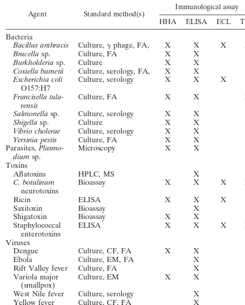
TABLE 1. Examples of BW/ID agents, accepted detection and identification methods, and available rapid immunological assaysa
Agent Standard method(s)
Immunological assay
HHA ELISA ECL TRF
Bacteria
Bacillus anthracis Culture,␥phage, FA, X X X X
Brucellasp. Culture, FA X X
Burkholderiasp. Culture X X
Coxiella bumetii Culture, serology, FA, X X
Escherichia coli
O157:H7
Culture, serology X X X
Francisella tula-rensis
Culture, FA X X X
Salmonellasp. Culture, serology X X
Shigellasp. Culture X X
Vibrio cholerae Culture, serology X X
Yersinia pestis Culture, FA X X Parasites,
Plasmo-diumsp.
Microscopy X X
Toxins
Aflatoxins HPLC, MS X
C. botulinum
neurotoxins
Bioassay X X X X
Ricin ELISA X X X
Saxitoxin Bioassay X
Shigatoxin Bioassay X X
Staphylococcal enterotoxins
ELISA X X X X
Viruses
Dengue Culture, CF, FA X X
Ebola Culture, EM, FA X
Rift Valley fever Culture, FA X
Variola major (smallpox)
Culture, EM X X
West Nile fever Culture, serology X
Yellow fever Culture, CF, FA X
a
Abbreviations: CF, complement fixation; EM, electron microscopy; FA, flu-orescent antibody; HHA, hand held assay; HPLC, high-performance liquid chro-matography; MS, mass spectroscopy; X, assay available.
on August 17, 2020 by guest
http://cvi.asm.org/
handheld assay devices contain a colloidal gold (or other)-labeled antibody dried onto a filter pad affixed to a nitrocellu-lose strip. A capture antibody is applied in a line on the strip and dried. To perform the test, a specimen is suspended in buffer and added to the pad containing the colloidal gold-labeled antibody. The antibody specifically binds to antigen present in the specimen, and the resulting complex wicks down the membrane where it binds to the capture antibody. A pos-itive reaction is visualized as a red line created by the bound colloidal gold. Similar assays using different detection systems have been described in the literature, including those based on latex particles and upconverting phosphatases (30, 64).
The present generation of handheld assays have several lim-itations. First, only one agent can be detected per assay strip. Thus, if an unknown sample needs to be characterized, several handheld assays must usually be run to obtain a presumptive identification. The second limitation is that each of the assays have varying sensitivity levels to their respective target agents. Assays for bacterial agents tend to be the most sensitive, able to detect from 2 ⫻ 105 to 2 ⫻ 106 CFU/ml while those for toxins have sensitivities ranging from 50 pg/ml to 50 ng/ml. Assays specific for viruses usually have the lowest sensitivities,
ranging from 2⫻105to 5⫻107PFU/ml. Third, since these assays are visualized as a red line created by the bound colloi-dal gold, the sensitivity is limited to what can be seen by the unaided (and uncalibrated) human eye. Typically, an arbitrary quantitation of the detection sensitivity of these assays is done by assigning a number between 0 and 5, with the increasing intensity of the red line assigned a higher value. Besides the somewhat arbitrary nature of this process, numeric values can vary based on the skill of the technician responsible for vali-dating a given lot of assays.
Enhanced labeling and detection approaches. Recent ad-vances in detection and labeling technologies that would in some instances improve the sensitivities of assays by at least an order of magnitude and make detection quantitative, not merely subjective, may offset the disadvantages inherent to present handheld assays. To detect very low levels of antigen, which may be present at low concentrations in vivo or in en-vironmental samples, the sensitivity of conventional gold-la-beled lateral flow assays can be enhanced up to one order of magnitude by using a silver enhancement step. Lateral flow assays are run, as described above, washed in phosphate-buff-ered saline and a Tween 20 solution, and then immersed in a FIG. 1. Principles of the four primary immunological assays. (A) Lateral flow immunochromatographic assay (handheld assay); (B) ELISA; (C) ECL; (D) TRF.
on August 17, 2020 by guest
http://cvi.asm.org/
silver enhancer reagent for 5 min. Horton and coworkers re-ported sensitivities of 100 ng/ml before enhancement and 100 pg/ml after enhancement (36). This system has the advantage of greater sensitivity but does not need a specialized mecha-nism to read the assay, which is advantageous for use in a field setting.
Alternative approaches to antibody labeling coupled with specialized quantitative readers can also lead to significant improvements in the sensitivity of lateral flow immunoassays. For example, superparamagnetic nanoparticles comprising ei-ther iron oxide (Fe3O4) or iron oxide and a polysaccharide matrix can be used to label antibodies in place of gold. Their broad potential for laboratory applications has been demon-strated by their use in detection of typhoid-specific antibodies, cell separation, and antibody sorting (53, 88–90). More re-cently, systems using paramagnetic particle labels have been described that make it possible to use iron oxide particles as labels in place of gold (6, 49, 70, 71). The labeled antibody-antigen mixture wicks up the membrane, as described above, and is deposited at the site of the solid-phase antibody, and the magnetic flux is measured in the antigen capture zone. This technology has three advantages. First, the signal is permanent and can be read more than one time. Second, the signal is quantitative and can be assigned a value in millivolts. Third, the signal generated is comparable to the detection limits seen with radionucleotide labels or nephelometric techniques (70, 71).
ELISAS
In the early 1970s, the search for simple but sensitive meth-ods for the quantitative detection of antigen and antibody that did not rely upon particle agglutination or radiolabeled re-agents led to the development of solid-phase enzyme-coupled reagent assays (23). In principal, the labeling by chemical con-jugation of an enzyme bound to either antigen or antibody allows detection of immune complexes formed on a solid phase as the fixed enzyme, once washed free of excess reagents, and on subsequent substrate interaction, yields a colored product that is directly visualized and/or quantitatively measured by optical density. The resulting assays, ELISAs, are economical, versatile, robust, and simple assays that achieve separation of bound and free moieties by use of a solid-phase support. Be-cause of their combined simplicity and sensitivity, ELISAs can be used reliably for screening large numbers of small-volume test samples in the simplest of laboratory environments. This technical advance has had its greatest impact in epidemiology and in the diagnosis of ID (41, 67, 96).
In general, two-site antigen-capture assays are used for the detection of BW/ID agents. This assay format is simple, spe-cific, sensitive, and readily converted to the lateral flow assay format previously described above. As illustrated in Fig. 1B, a capture antibody (often a monoclonal of high affinity) affixed to the solid phase is exposed to a test sample (as well as to positive- and negative-control samples) and, after washing, the complex is exposed further to diluted detector antibody spe-cific to the same antigen. Finally, a conjugate antibody is added and the reaction is visualized. In this system, the antigen must have multiple epitopes for antibody binding, or a repeating, spatially distant, single epitope. Such an assay can be very
sensitive and specific. The use of a monoclonal antibody as the capture antibody usually results in a finished assay of high specificity and low background. However, the addition of a polyclonal antibody can greatly increase the breadth of the assay to detect multiple isolates of the same species of bacteria, virus, or fungus. With minor modifications, this assay can de-tect serum immune complexes. The antibody isotypes of the immune complexes can be determined and quantified by using a panel of isotype-specific antiglobulin conjugates on the same test samples in repeated assays.
TRF ASSAYS
Assays based on TRF use lanthanide chelate labels with unique fluorescence properties (24, 25, 33, 34, 55, 56, 58, 78, 79). Key among these properties is a very long fluorescence decay time and an exceptionally large Stokes’ shift. The long fluorescence decay time allows the user to measure fluores-cence after the background has fully subsided. Additionally, the label, a lanthanide chelate, is dissociated from the antibody or reporter molecule into a new, highly fluorescent chelate within a protective micelle. Most importantly, these lanthanide chelates can be used in place of other labels typically used in ELISA-based procedures. Cumulatively, these factors contrib-ute to the high sensitivity and low backgrounds characteristic of immunologic assays based on TRF.
TRF assays are set up in a traditional 96-well two-site anti-gen-capture ELISA format. As illustrated in Fig. 1C, a capture antibody (most often a monoclonal of high affinity) affixed to the solid phase is exposed to a test sample (as well as to positive- and negative-control samples) and, after washing, the complex is exposed further to diluted detector antibody spe-cific to the same antigen which is labeled with a lanthanide chelate (europium, samarium, terbium, or dysprosium). Eu-ropium, Eu3⫹, is the label that is mainly used. A low-pH
enhancement solution is then added, which causes the lan-thanide to dissociate from the labeled compound. This form of the lanthanide is highly fluorescent. As in an ELISA, the an-tigen must have multiple epitopes for antibody binding, or a repeating, spatially distant, single epitope. TRF assays have many of the same limitations as ELISAs; primarily, these are a function of the antibodies used. Additionally, special care must be taken with these assays to avoid lanthanide contamination, including the use of dedicated measuring devices and rigorous washing techniques.
A key advantage of TRF assays is that they can be up to one order of magnitude more sensitive because of the highly fluo-rescent nature of the lanthanide label (68). Recently several papers have been published which report the use of this re-porter technology in clinical specimens for the detection of low-level proteins that are below the sensitivity limit of tradi-tional ELISAs as well as BW agents in both clinical and envi-ronmental specimens (18–20, 26, 39, 40, 68, 85, 97). Also, as with ELISAs, these assays can be modified to detect serum immune complexes (1). The antibody isotypes of the immune complexes can be determined and quantified by using a panel of isotype-specific antiglobulin conjugates on the same test samples in repeated assays.
on August 17, 2020 by guest
http://cvi.asm.org/
IMS-ECL ASSAYS
Recent advances in IMS and ECL have led to the develop-ment of several related technologies and systems including the ORIGEN immunoassay system (Igen, Inc., Rockville, Md.), QPCR 5000 (Applied Biosystems, Foster City, Calif.), and magnetic ECL detection systems (11, 98, 99).
IMS has been used for soluble and particulate antigen cap-ture, separation, purification, and concentration efficiently with high-affinity antibodies for several decades (32, 51, 86). A key feature of this technology is its ability to capture and concen-trate antigens from a variety of complex biological matrices. One of the major advantages of IMS is the increased reaction kinetics as a result of the potentially greater surface area on the magnetic beads compared to conventional ELISA and im-munological reaction within a turbulent bead suspension (62, 80). Additionally, beads can be mixed rapidly or slowly to encourage rapid capture of soluble antigens or gentle docking with particulate antigens. A further advantage of the magnetic beads is the rapid separation of antibody-captured materials from the surrounding milieu when placed in a magnetic field. These beads contain paramagnetic magnetite (FE3O4) that is magnetizable in the presence of an external field but not in its absence. The beads have a large number of different sizes, ranging from a few nanometers to several micrometers. They are usually spherical, but the shape is dependent on the man-ufacturing process and the needs of the end user.
The format and principle of an ECL assay is similar to that of assays based on ELISA and TRF technologies (Fig. 1D). Detection is accomplished by the heavy metal chelate ruthe-nium (II) tris-bipyridal Ru(bpy)3
2⫹conjugated to a detector
antibody. Initially Ru(bpy)3
2⫹and tripropylamine (TPA) are
oxidized at the surface of an anode. TPA immediately loses a proton, becoming a powerful reducer. This causes Ru(bpy)3
3⫹
to enter a high-energy state by a high-energy electron transfer from the electron carrier, TPA. Relaxation to the ground state results in light emission detectable at 620 nm. Ru(bpy)3
2⫹is
not consumable during the reaction and may be oxidized and excited again due to excess TPA used in the buffer (98). The ECL assay format has been used by several groups to detect BW agents, includingB. anthracisand staphylococcal entero-toxin B (28, 44, 99).
As with TRF assays, assays based on IMS and ECL technol-ogy have limitations similar to those of ELISA, and again, these are primarily a function of the antibodies used—the better the avidity and affinity of the antibody, the more sensi-tive and specific the assay. As with other highly sensisensi-tive assays, signal-to-noise ratios need to be closely studied and the limit of detection needs to be carefully analyzed (68, 77).
FUTURE TRENDS
The advances in immunological reagents and assay formats are being matched by improvements in complementary labo-ratory technology, ranging from automated analyzers and mi-croarrays that facilitate the analysis of large numbers of sam-ples, to self-contained miniaturized devices that enable an immunoassay to be performed at the point of care or in a field setting. Together, these novel reagents and new technologies are likely to transform diagnostic medicine over the next
dec-ade as much as our recognition of the civilian public health threat posed by BW/ID agents has.
On the horizon are even further advances in sensitivity and throughput. Most are based a combination of existing immu-nological systems coupled with electronic sensing modules (12). An automated system has been described that utilizes solid-phase ELISA coupled with a multichannel optical flow cell with a sensor composed of a light-emitting diode and a photodetector (46). Simultaneous detection of staphylococcal enterotoxin B, bacteriophage M13, and Escherichia coli has been accomplished with this immunosensor. Another proto-type system that uses a light-addressable potentiometric sensor and a flowthrough immunofiltration enzyme assay can detect eight agents simultaneously within 15 min (87). While the limit of specific detection is considerably higher than that of more mature assays, the speed and multiplexing offered by this ap-proach are promising. Other apap-proaches, based on microflu-idic arrays and photosensors, have also been reported and show promising improvements in sensitivity with no loss in specificity (81, 82). The main advantages offered by these ex-perimental systems is that in addition to automation, multi-plexing, and throughput, they offer a quantitative assessment of the agent present, detecting in some instances fewer than 50 copies of target agent.
While the systems described above are all fairly elaborate, laboratory-based systems, similar improvements in handheld immunoassays are also on the horizon. Recently, a self-con-tained handheld biosensor has been described that uses immu-noaffinity for specificity and fluorescence for quantitation (14). The prototype is automated and requires no special storage. It is also multiuse: approximately 100 measurements can be made before refurbishment is required. Sensitivity is also promising. Using aflatoxins, a detection limit has been demonstrated at concentrations from 0.1 to 50 ppb in less than 2 min. The design flexibility suggests that it could be readily adapted for the detection of other biological analytes.
Besides these advances in automation and throughput, an-other technique has been demonstrated that may significantly improve the sensitivity of current solid-phase immunoassays. This approach, based on force differentiation, subjects a la-beled antigen-antibody complex to a magnetic field of defined magnitude and orientation, displacing weakly bound nonspe-cific particles while leaving the spenonspe-cific immunochemical com-plex intact (52). The number of antigen-antibody comcom-plexes bound to the surface after applying the differentiation force was related to the analyte concentration, permitting the devel-opment of an optical detection scheme to count the number of such complexes. In this prototype, the sensitivity of the force differentiation assay was one to two orders of magnitude higher than conventional solid-phase immunoassay techniques, while retaining 99% specificity.
Although new and improved assays and sensor formats will continue to be developed, the most critical component of any immunological test will likely remain the antibody itself. Ulti-mately, improvements in the affinity, specificity, and mass pro-duction of antibodies will dictate the success or failure of a given immunoassay technology. Sensitive and specific detec-tion of BW and ID agents by immunoassays has improved by several orders of magnitude over the past 30 years. If recent scientific progress is a fair indicator, the future promises
on August 17, 2020 by guest
http://cvi.asm.org/
tinued improvements in immunoassays with an ever-increasing array of applications.
REFERENCES
1.Aggerbeck, H., B. Norgaard-Pedersen, and I. Heron.1996. Simultaneous
quantitation of diphtheria and tetanus antibodies by double antigen, time-resolved fluorescence immunoassay. J. Immunol. Methods190:171–183.
2.Aidoo, S., W. K. Ampofo, J. A. Brandful, S. V. Nuvor, J. K. Ansah, N.
Nii-Trebi, J. S. Barnor, F. Apeagyei, T. Sata, D. Ofori-Adjei, and K.
Ishi-kawa.2001. Suitability of a rapid immunochromatographic test for
detec-tion of antiboies to human immunodeficiency virus in Ghana, West Africa. J. Clin. Microbiol.39:2572–2575.
3.Allwinn, R., C. Schieferstein, S. Clauke, and H. W. Doerr.1999. Rapid
diagnosis of primary dengue fever by the immunochromatographic test and by electron microscopy-a case report. Infection27:365–367.
4.Araz, E., M. Tanyuksel, N. Ardic, and C. Tabuk.2000. Performance of a
commercial immunochromatographic test for the diagnosis of vivaz malaria in Turkey. Trans. R. Soc. Trop. Med. Hyg.94:55–56.
5.Barry, M. A., M. E. Barry, and S. A. Johnston.1994. Production of
mono-clonal antibodies by genetic immunization. BioTechniques16:616–618, 620.
6.Baselt, D. R., G. U. Lee, M. Natesan, S. W. Metzger, P. E. Sheehan, and
R. J. Colton.1998. A biosensor based on magnetoresistance technology.
Biosens. Bioelectron.13:731–739.
7.Beck, O., M. Kraft, M. R. Moeller, B. L. Smith, S. Schneider, and R.
Wennig.2000. Frontline immunochromatographic device for on-site urine
testing of amphetamines: laboratory validation using authentic specimens. Ann. Clin. Biochem.37:199–204.
8.Berdal, B. P., R. Mehl, H. Haaheim, M. Loksa, R. Grunow, J. Burans, C.
Morgan, and H. Meyer. 2000. Field detection ofFrancisella tularensis.
Scand. J. Infect. Dis.32:287–291.
9.Bhaskar, S., S. Singh, and M. Sharma.1996. A single-step
immunochro-matographic test for the detection ofEntamoeba histolyticain stool samples. J. Immunol. Methods196:193–198.
10. Bird, C. B., R. L. Miller, and B. M. Miller.1999. Reveal forSalmonellatest system. J. AOAC Int.82:625–633.
11. Blackburn, G. F., H. P. Shah, J. H. Kenten, J. Leland, R. A. Kamin, J. Link, J. Peterman, M. J. Powell, A. Shah, D. B. Talley, S. K. Tyagi, E. Wilkens,
T. Wu, and R. J. Massey.1991. Electrochemiluminescence detection for
development of immunoassays and DNA probe assays for clinical diagnos-tics. Clin. Chem.37:1534–1539.
12. Bossi, A., S. A. Piletsky, P. G. Righetti, and A. P. Turner.2000. Capillary electrophoresis coupled to biosensor detection. J. Chromatogr. A892:143– 153.
13. Buser, J., L. Risch, T. Rutz, S. Mannang, and J. Munzinger.2001.
Com-parison of a rotavirus latex agglutination test with two rapid immunochro-matographic test devices for detection of rotavirus in human feces. Eur. J. Clin. Microbiol. Infect. Dis.20:295–296.
14. Carlson, M. A., C. B. Bargeron, R. C. Benson, A. B. Fraser, T. E. Phillips,
J. T. Velky, J. D. Groopman, P. T. Strickland, and H. W. Ko.2000. An
automated, handheld biosensor for aflatoxin. Biosens. Bioelectron.14:841– 848.
15. Chakravarti, A., R. Gur, N. Berry, and M. D. Mathur.2000. Evaluation of
three commercially available kits for serological diagnosis of dengue hae-morrhagic fever. Diagn. Microbiol. Infect. Dis.36:273–274.
16. Chanteau, S., L. Rahalison, M. Ratsitorahina, Mahafaly, M.
Rasoloma-haro, P. Biosier, T. O’Brien, J. Aldrich, A. Keleher, C. Morgan, and J.
Bu-rans.2000. Early diagnosis of bubonic plague using F1 antigen capture
ELISA assay and rapid immunogold dipstick. Int. J. Med. Microbiol.290:
279–283.
17. Ching, W. M., D. Rowland, Z. Ahang, A. L. Bourgeois, D. Kelly, G. A.
Dasch, and P. L. Devine.2001. Early diagnosis of scrub typhus with a rapid flow assay using recombinant major outer membrane protein antigen (r56) ofOrientia tsutsugamushi. Clin. Diagn. Lab. Immunol.8:409–414.
18. Daijo, J. E., and J. R. Sportsman. 1999. A time-resolved fluorescence
immunoassay for insulin in rodent plasma. J. Pharm. Biomed. Anal.19:
335–342.
19. Diamandis, E. P.1991. Multiple labeling and time-resolvable fluorophores. Clin. Chem.37:1486–1491.
20. Diamandis, E. P., and T. K. Christopoulos.1991. Time-resolved
immu-nofluorometric detection of antigens separated by high-performance liquid chromatography and coated to polystyrene. BioTechniques10:646–648.
21. Dominguez, J., N. Gali, S. Blanco, P. Pedroso, C. Prat, L. Matas, and V.
Ausina.2001. Detection ofStreptococcus pneumoniaeantigen by a rapid
immunochromatographic assay in urine samples. Chest119:243–249.
22. Dominguez, J., N. Gali, L. Matas, P. Pedroso, A. Hernandez, E. Padilla,
and V. Ausina.1999. Evaluation of a rapid immunochromatographic assay
for the detection ofLegionella antigen in urine samples. Eur. J. Clin. Microbiol. Infect. Dis.18:896–898.
23. Engvall, E., and P. Perlmann.1971. Enzyme-linked immunosorbent assay
(ELISA). Quantitative assay of immunoglobulin G. Immunochemistry
8:871–874.
24. Evangelista, R. A., A. Pollak, B. Allore, E. F. Templeton, R. C. Morton, and
E. P. Diamandis.1988. A new europium chelate for protein labelling and
time-resolved fluorometric applications. Clin. Biochem.21:173–178. 25. Evangelista, R. A., A. Pollak, and E. F. Templeton.1991. Enzyme-amplified
lanthanide luminescence for enzyme detection in bioanalytical assays. Anal. Biochem.197:213–224.
26. Fiet, J., F. Giton, P. Boudou, J. M. Villette, H. Soliman, G. Morineau, A.
Boudi, and H. Galons.2001. A new specific and sensitive time
resolved-fluoroimmunoassay of 11-deoxycortisol in serum. J. Steroid Biochem. Mol. Biol.77:143–150.
27. Garcia, L. S., and R. Y. Shimizu.2000. Detection ofGiardia lambliaand
Cryptosporidium parvumantigens in human fecal specimens using the ColorPAC combination rapid solid-phase qualitative immunochromato-graphic assay. J. Clin. Microbiol.38:1267–1268.
28. Gatto-Menking, D. L., H. Yu, J. G. Bruno, M. T. Goode, M. Miller, and
A. W. Zulich.1995. Sensitive detection of biotoxoids and bacterial spores using an immunomagnetic electrochemiluminescence sensor. Biosens. Bio-electron.10:501–507.
29. Grunow, R., W. Splettstoesser, S. McDonald, C. Otterbein, T. O’Brien, C.
Morgan, J. Aldrich, E. Hofer, E. J. Finke, and H. Meyer.2000. Detection of
Francisella tularensisin biological specimens using a capture enzyme-linked immunosorbent assay, an immunochromatographic handheld assay, and a PCR. Clin. Diagn. Lab. Immunol.7:86–90.
30. Hampl, J., M. Hall, N. A. Mufti, Y. M. Yao, D. B. MacQueen, W. H. Wright,
and D. E. Cooper.2001. Upconverting phosphor reporters in
immunochro-matographic assays. Anal. Biochem.288:176–187.
31. Harlow, E., and D. Lane.1988. Antibodies: a laboratory manual. Cold
Spring Harbor Laboratory, Cold Spring Harbor, N.Y.
32. Haukanes, B. L., and C. Kyam.1993. Application of magnetic beads in
bioassays. Bio/Technology11:60–63.
33. Hemmila, I.1988. Lanthanides as probes for time-resolved fluorometric
immunoassays. Scand. J. Clin. Lab. Investig.48:389–399.
34. Hemmila, I., S. Dakuba, V. M. Mukkala, H. Siitari, and T. Lovgren.1984.
Europium as a label in time-resolved immunofluorometric assays. Anal. Biochem.137:335–343.
35. Hochmeister, M. N., B. Budowle, O. Rudin, C. Gehrig, U. Borer, M. Thali,
and R. Dirnhofer. 1999. Evaluation of prostate-specific antigen (PSA)
membrane test assays for the forensic identification of seminal fluid. J. Fo-rensic Sci.44:1057–1060.
36. Horton, J. K., S. Swinburne, and M. J. O’Sullivan.1991. A novel, rapid,
single-step immunochromatographic procedure for the detection of mouse immunoglobulin. J. Immunol. Methods140:131–134.
37. Jelinek, T., S. Eichenlaub, and T. Loscher.1999. Sensitivity and specificity of a rapid immunochromatographic test for diagnosis of visceral leishman-iasis. Eur. J. Clin. Microbiol. Infect. Dis.18:669–670.
38. Johnston, S. A., and D. C. Tang.1994. Gene gun transfection of animal cells and genetic immunization. Methods Cell Biol.43(Pt A):353–365.
39. Kahan, I., A. Papanastasiou-Diamandi, G. Ellis, S. K. Makela, J.
Mc-Laurin, M. D’Costa, and E. P. Diamandis.1990. Sensitive time-resolved
fluorescence immunoassay of somatotropin in serum. Clin. Chem.36:503– 508.
40. Kakabakos, S. E., T. K. Christopoulos, and E. P. Diamandis.1992.
Multi-analyte immunoassay based on spatially distinct fluorescent areas quanti-fied by laser-excited solid-phase time-resolved fluorometry. Clin. Chem.
38:338–342.
41. Katti, M. K.2001. Are enzyme-linked immunosorbent assay and
immuno-blot assay independent in immunodiagnosis of infectious diseases? Clin. Infect. Dis.32:1114.
42. Kaur, H., and A. Mani.2000. Evaluation and usefulness of a
immunochro-matographic test for rapid detection ofPlasmodium falciparuminfection. Ind. J. Med. Sci.54:421–424.
43. Kemppainen, E. A., J. I. Hedstrom, P. A. Puolakkainen, V. S. Sainio, R. K. Haapianen, V. Perhoniemi, S. Osman, E. O. Kivilaakso, and U. H. Sten-man.1997. Rapid measurement of urinary trypsinogen-2 as a screening test for acute pancreatitis. N. Engl. J. Med.336:1788–1793.
44. Kijek, T. M., C. A. Rossi, D. Moss, R. W. Parke, and E. A. Henchal.2000. Rapid and sensitive immunomagnetic-electrochemiluminescent detection of staphylococcal enterotoxin B. J. Immunol. Methods236:9–17. 45. Klingenberg, K. D. V., and J. Esfandiari.1996. Evaluation of a one-step test
for rapid, in practice detection of rotavirus in farm animals. Vet. Rec.
138:393–395.
46. Koch, S., H. Wolf, C. Danapel, and K. A. Feller.2000. Optical flow-cell
multichannel immunosensor for the detection of biological warfare agents. Biosens. Bioelectron.14:779–784.
47. Kohn, J.1968. An immunochromatographic technique. Immunology15:
863–865.
48. Kortepeter, M. G., and G. W. Parker.1999. Potential biological weapons
threats. Emerg. Infect. Dis.5:523–527.
49. Kriz, K., J. Gehrke, and D. Kriz.1998. Advancements toward magneto
immunoassays. Biosens. Bioelectron.13:817–823.
50. Laitinen, M. P. A., and M. Vuento.1996. Immunochromatographic assay
for quantitation of milk progesterone. Acta Chem. Scand.50:141–145.
on August 17, 2020 by guest
http://cvi.asm.org/
51. Lea, T., F. Vartdal, K. Nustad, S. Funderud, A. Berge, T. Ellingsen, R.
Schmid, P. Stenstad, and J. Ugelstad.1988. Monosized, magnetic polymer
particles: their use in separation of cells and subcellular components, and in the study of lymphocyte function in vitro. J. Mol. Recognit.1:9–18. 52. Lee, G. U., S. Metzger, M. Natesan, C. Yanavich, and Y. F. Dufrene.2000.
Implementation of force differentiation in the immunoassay. Anal. Bio-chem.287:261–271.
53. Lim, P. L., F. C. Tam, Y. M. Cheong, and M. Jegathesan.1998. One-step
2-minute test to detect typhoid-specific antibodies based on particle sepa-ration in tubes. J. Clin. Microbiol.36:2271–2278.
54. Lou, S. C., C. Patel, S. Ching, and J. Gordon.1993. One-step competitive immunochromatographic assay for semiquantitative determination of li-poprotein(a) in plasma. Clin. Chem.39:619–624.
55. Lovgren, T., I. Hemmilia, K. Pettersson, and P. Halonen. 1985.
Time-resolved fluorometry in immunassay, p. 203–217.InT. Collins and W. Hoh (ed.), Alternative immunoassays. Wiley and Sons Ltd., Chichester, United Kingdom.
56. Lovgren, T., L. Merio, K. Mitrunen, M. Makinen, K. Blomberg, T.
Palen-ius, and K. Pettersson.1996. One-step all-in-one dry reagent immunoassay with fluorescent europium chelate label and time-resolved fluorometry. Clin. Chem.42:1196–1201.
57. McKenzie, S.1988. Whole blood assay of theophylline concentrations using immunochromatographic stick. Arch. Dis. Child.63:571–572.
58. Mikola, H., T. Takalo, and I. Hemmilia.1995. Synthesis and properties of luminescent lanthanide chelate labels and labeled haptenic antigens for homogeneous immunoassays. Bioconjug. Chem.6:235–241.
59. Mills, C. D., D. C. H. Burgess, H. J. Taylor, and K. C. Kain.1999. Evalu-ation of a rapid and inexpensive dipstick assay for the diagnosis of Plasmo-dium falciparummalaria. Bull. W. H. O.77:553–559.
60. Miwa, H., S. Akamatsu, T. Tachikawa, T. Sogabe, K. Ohtaka, A. Nagahara,
Y. Sugiyama, and N. Sato.2001. On-site diagnosis ofH. pyloriinfection by urine. Diagn. Microbiol. Infect. Dis.39:95–97.
61. Mohanty, S., S. K. Mishra, A. Mohanty, and B. S. Das.1999.
Immunochro-matographic test for the diagnosis of Falciparum malaria. J. Assoc. Physi-cians India47:201–202.
62. Nguyen, V., N. Leclerc, C. Wolff, P. Kennel, P. Fonteneau, R. Deyes, J.
Warter, and P. Poindron.1999. Protection of immunoreactivity of dry
immobilized proteins on microtitration plates in ELISA application for detection of autoantibodies in myasthenia gravis. J. Biotechnol.72:115–125. 63. Nichol, S. T., J. Arikawa, and Y. Kawaoka.2000. Emerging viral diseases.
Proc. Natl. Acad. Sci. USA97:12411–12412.
64. Ortega-Vinuesa, J. L., and D. Bastos-Gonzalez.2001. A review of factors
affecting the performance of latex agglutination tests. J. Biomater. Sci. Polym. Ed.12:379–408.
65. Pagani, F., C. Serena, C. Bosio, C. Cuccia, and M. Panteghini. 2001.
Evaluation of a rapid bedside immunochromatographic assay for detection of cardiac troponin I in whole blood. Clin. Chem. Lab. Med.39:458–459.
66. Panteghini, M., and F. Pagani.1996. Characterization of a rapid
immuno-chromatographic assay for simultaneous detection of high concentrations of myoglobin and CK-MB in whole blood. Clin. Chem.42:1292–1293.
67. Payne, J. W. J., D. L. Marshall, R. K. Shockley, and W. J. Martin.1988.
Clinical laboratory applications of monoclonal antibodies. Clin. Microbiol. Rev.1:313–329.
68. Peruski, A. H., L. H. Johnson, III, and L. F. Peruski, Jr.2002. Rapid and sensitive detection of biological warfare agents using time-resolved fluores-cence assays. J. Immunol. Methods263:35–41.
69. Pillai, D. R., and K. C. Kain.1999. Immunochromatographic strip-based
detection ofEntamoeba histolytica-E. disparandGiardia lamblia coproan-tigen. J. Clin. Microbiol.37:3017–3019.
70. Richardson, J., P. Hawkins, and R. Luxton.2001. The use of coated
para-magnetic particles as a physical label in a magneto-immunoassay. Biosens. Bioelectron.16:989–993.
71. Richardson, J., A. Hill, R. Luxton, and P. Hawkins.2001. A novel measur-ing system for the determination of paramagnetic particle labels for use in magneto-immunoassays. Biosens. Bioelectron.16:1127–1132.
72. Sanchez-Carbayo, M., E. Herrero, J. Megias, A. Mira, and F. Soria.1999.
Initial evaluation of the new urinary bladder cancer rapid test in the de-tection of transitional cell carcinoma of the bladder. Urology54:656–661. 73. Schouten, Y., R. J. D. Winter, J. P. Gorgels, R. W. Koster, R. Adams, and
G. T. Sanders.1998. Clinical evaluation of the CARDIAC STATus, a rapid
immunochromatographic assay for simultaneous detection of elevated con-centrations of CK-MB and myoglobin in whole blood. Clin. Chem. Lab. Med.36:469–473.
74. Schramm, W., S. E. Wade, B. B. Angulo, P. C. Torres, and A.
Burgess-Cassler.1998. A simple whole-blood test for detecting antibodies to human immunodeficiency virus. Clin. Diagn. Lab. Immunol.5:263–265. 75. Schrier, W. H., R. J. Schoengold, J. T. Baker, J. L. Norell, C. L. Jaseph, Y.
Okin, J. Y. Doe, and H. Chandler.1998. Development of FlexSure HP-an
immunochromatographic method to detect antibodies againstHelicobacter pylori. Clin. Chem.44:293–298.
76. Shin, H. S., C. K. Kim, K. S. Shin, H. K. Chung, and T. R. Heo.2001.
Pretreatment of whole blood for use in immunochromatographic assays for hepatitis B virus surface antigen. Clin. Diagn. Lab. Immunol.8:9–13.
77. Smith, D. R., C. A. Rossi, T. M. Kijek, E. A. Henchal, and G. V. Ludwig.
2001. Comparison of dissociation-enhanced lanthanide fluorescent immu-noassays to enzyme-linked immunosorbent assays for detection of staphy-lococcal enterotoxin B,Yersinia pestis-specific F1 antigen, and Venezuelan equine encephalitis virus. Clin. Diagn. Lab. Immunol.8:1070–1075.
78. Soini, E., and I. Hemmila.1979. Fluoroimmunoassay: present status and
key problems. Clin. Chem.25:353–361.
79. Soini, E., and H. Kojola.1983. Time-resolved fluorometer for lanthanide
chelates–a new generation of nonisotopic immunoassays. Clin. Chem.29:
65–68.
80. Stenberg, M., and H. Nygren.1988. Kinetics of antigen-antibody reactions at solid-liquid interfaces. J. Immunol. Methods113:3–15.
81. Stokes, D. L., M. J. Sepaniak, and T. Vo-Dinh.1997. Development of a new capillary electrophoresis-based fibre optic sensor. Biomed. Chromatogr.
11:187–192.
82. Szurdoki, F., K. L. Michael, and D. R. Walt.2001. A duplexed microsphere-based fluorescent immunoassay. Anal. Biochem.291:219–228.
83. Tang, D. C., M. DeVit, and S. A. Johnston.1992. Genetic immunization is
a simple method for eliciting an immune response. Nature 356:152– 154.
84. Tjitra, E., S. Suprianto, M. Dyer, B. J. Currie, and N. M. Anstey.1999.
Field evaluation of the ICT malaria P.f/P.v immunochromatographic test for detection ofPlasmodium falciparumandPlasmodium vivaxin patients with a presumptive clinical diagnosis of malaria in eastern Indonesia. J. Clin. Microbiol.37:2412–2417.
85. Tschop, M., H. M. Behre, E. Nieschlag, R. A. Dressendorfer, and C. J.
Strasburger.1998. A time-resolved fluorescence immunoassay for the
mea-surement of testosterone in saliva: monitoring of testosterone replacement therapy with testosterone buciclate. Clin. Chem. Lab. Med.36:223–230. 86. Ugelstad, J., P. Stenstad, L. Kilaas, W. S. Prestvik, R. Herie, A. Berge, and
E. Hornes.1993. Monodisperse magnetic polymer particles. New
biochem-ical and biomedbiochem-ical applications. Blood Purif.11:349–369.
87. Uithoven, K. A., J. C. Schmidt, and M. E. Ballman.2000. Rapid
identifi-cation of biological warfare agents using an instrument employing a light addressable potentiometric sensor and a flow-through immunofiltration-enzyme assay system. Biosens. Bioelectron.14:761–770.
88. Vaccaro, D. E., and J. E. Markinac.1995. Use of monoclonal antibodies
with magnetic particles to separate cell subpopulations by negative selec-tion. Methods Mol. Biol.45:245–252.
89. Vaccaro, D. E., and J. E. Markinac.1995. Use of monoclonal antibodies
with magnetic particles to separate cell subpopulations by positive selection. Methods Mol. Biol.45:253–259.
90. Valenti, S., A. Sarkissian, G. Giordano, and K. D. Dahl.1995. A technique for sorting rat gonadotropes using anti-LH or anti-FSH antibodies co-valently attached to magnetic beads. J. Neuroendocrinol.7:673–679.
91. Vorobjev, A. A., B. L. Cherkassey, A. V. Stepanow, A. A. Kyuregyan, and
Y. M. Fjedorov.1994. “Criterion rating” as a measure of portable use of
bioagents as biological weapons. Report to the Working Group on Biolog-ical Weapons Control of the Committee on International Security and Arms Control (CISAC). National Academy of Sciences, Washington, D.C.
92. Vorobjev, A. A., B. L. Cherkassey, A. V. Stepanow, A. A. Kyuregyan, and
Y. M. Fjedorov.1997. Key problems of controlling especially dangerous
infections, p. 71–76.InProceedings of the International Symposium of Severe Infectious Disease: epidemiology, express-diagnostics and preven-tion. State Scientific Institution, Volgo-Vyatsky Center of Applied Biotech-nology, Kirov, Russia.
93. Wannamaker, B., L. Denio, W. E. Dodson, F. Dreifuss, C. Crosby, N.
Santilli, P. Duffner, P. Ryan-Dudeck, C. Conboy, and E. Ellis.1989.
Im-munochromatographic measurement of phenobarbital in whole blood with a non-instrumented assay. Neurology39:1215–1218.
94. Weaver, P. C., E. P. Yzerman, K. J. Kuijper, P. Speelman, and J. Dankert.
2000. Rapid diagnosis of Legionnaires disease using an immunochromato-graphic assay for Legionella pneumophilaserogroup 1 antigen in urine during an outbreak in The Netherlands. J. Clin. Microbiol.38:2738–2739.
95. Wennig, R., M. R. Moeller, J. M. Haguenoer, A. Marocchi, F. Aoppi, B. L.
Smith, R. D. L. Torre, C. A. C. Goerlach-Graw, J. Schaeffler, and R.
Leinberger. 1998. Development and evaluation of
immunochromato-graphic rapid tests for screening of cannabinoids, cocaine, and opiates in urine. J. Anal. Toxicol.22:148–155.
96. Wright, P. F., E. Nilsson, E. M. V. Rooij, M. Lelenta, and M. H. Jeggo.1993. Standardisation and validation of enzyme-linked immunosorbent assay techniques for the detection of antibody in infectious disease diagnosis. Rev. Sci. Tech.12:435–450.
97. Wu, F. B., Y. F. He, and S. Q. Han.2001. Matrix interference in serum total
on August 17, 2020 by guest
http://cvi.asm.org/
thyroxin (T4) time-resolved fluorescence immunoassay (TRFIA) and its elimination with the use of streptavidin-biotin separation technique. Clin. Chim. Acta308:117–126.
98. Yang, H., J. K. Leland, D. Yost, and R. J. Massey.1994.
Electrochemilu-minescence: a new diagnostic and research tool. ECL detection technology promises scientists new “yardsticks” for quantification. Bio/Technology12:
193–194.
99. Yu, H.1998. Comparative studies of magnetic particle-based solid phase
fluorogenic and electrochemiluminescent immunoassay. J. Immunol. Meth-ods218:1–8.
100. Zuk, R. F., V. K. Ginsberg, T. Gouts, J. Rabbie, H. Merrick, E. F. Ullman,
M. M. Fischer, C. C. Sizto, S. N. Stiso, and D. J. Litman.1985. Enzyme
immunochromatography–a quantitative immunoassay requiring no instru-mentation. Clin. Chem.31:1144–1150.