Affordable CD4
⫹
-T-Cell Counting by Flow Cytometry: CD45 Gating
for Volumetric Analysis
George Janossy,
1* Ilesh V. Jani,
1Nicholas J. Bradley,
1Arsene Bikoue,
1Tim Pitfield,
1and
Debbie K. Glencross
2HIV Immunology, Department of Immunology and Molecular Pathology, Royal Free and University College Medical School, London, United Kingdom,1and Department of Molecular Medicine and Hematology, Faculty of Health
Sciences, University of the Witwatersrand, Johannesburg, South Africa2 Received 14 January 2002/Returned for modification 19 March 2002/Accepted 22 April 2002
The flow cytometers that are currently supported by industry provide accurate CD4ⴙ-T-cell counts for
monitoring human immunodeficiency virus disease but remain unaffordable for routine service work under resource-poor conditions. We therefore combined volumetric flow cytometry (measuring absolute lymphocyte counts in unit volumes of blood) and simpler protocols with generic monoclonal antibodies (MAbs) to increase cost efficiency. Volumetric absolute counts were generated using CD45/CD4 and CD45/CD8 MAb combinations in two parallel tubes. The percentage values for the various subsets were also determined within the leukocyte and lymphocyte populations utilizing a fully automated protocol. The levels of agreement between the newly developed method and the present industry standards, including both volumetric and bead-based systems using a full MAb panel for subset analysis, were tested by Bland-Altman analyses. The limits of agreement for CD4 counts generated by the volumetric methods using either CD45/CD4 (in a single tube) or the full Trio MAb
panel (in three tubes) on the CytoronAbsolute flow cytometer were betweenⴚ29 andⴙ46 cells/mm3with very
little bias for CD4 counts (in favor of the Trio method:ⴙ8 CD4ⴙlymphocytes/mm3; 0.38% of lymphocytes). The
limits of agreement for absolute CD4 counts yielded by the volumetric CD45/CD4 method and the bead-based
method were betweenⴚ118 andⴙ98 cells/mm3, again with a negligible bias (ⴚ10 CD4ⴙlymphocytes/mm3). In
the volumetric method using CD45/CD8, the strongly CD8ⴙcells were gated and the levels of agreement with
the full Trio showed a minor bias (in favor of the Trio;ⴙ40 CD8ⴙcells/mm3; 5.2% of lymphocytes) without a
significant influence on CD4/CD8 ratios. One trained flow cytometrist was able to process 300 to 400 stained tubes per day. This workload extrapolates to a throughput of >30,000 samples per year if both CD45/CD4 and CD45/CD8 stainings are performed for each patient or a throughput of >60,000 samples if only CD45/CD4 counts are tested in a single tube. Thus, on the basis of the high efficiency and excellent agreement with the present industry standards, volumetric flow cytometers with automated gating protocols and autobiosamplers, complemented by generic CD45, CD4, and CD8 MAbs used in two-color immunofluorescence, represent the most suitable arrangements for large regional laboratories in resource-poor settings.
Dedicated flow cytometers are designed to enumerate the absolute numbers and percentages of lymphocyte populations, such as subsets of T cells, B cells, and NK cells. In the clinical service for monitoring human immunodeficiency virus (HIV) disease, the primary aim is to deliver absolute CD4⫹-T-cell
counts, and this is achieved with a remarkably high level of precision (9, 24, 26, 29, 30). Nevertheless, the various cytomet-ric systems differ in complexity (9). It has recently been docu-mented that routine CD4-T-cell enumeration can be simplified without compromising quality (13, 18, 32), leading to cost-effective services for patients who receive generic antiretroviral drugs in resource-poor settings (Fig. 1).
Among the flow cytometers, dedicated instruments operat-ing as “soperat-ingle platforms” are preferred due to their conve-nience and accuracy (9, 24, 26, 29, 30). These single platforms are based either on a volumetric principle by counting CD4⫹T
cells in a unit volume of blood (24, 26) or on the concept of
adding known numbers of fluorospheres, or “microbeads,” to each sample (29, 30). These beads are, however, precision products that can increase running costs. Consequently, ser-vices handling large numbers of samples had to revert to “dou-ble platforms” operating a panleucogating strategy with CD45 monoclonal antibody (MAb) to secure a much less expensive but still accurate mode of operation (13, 19). Indeed, the CD45-based gating, an example of the “heterogeneous” gating strategy, is a more reliable protocol (12, 13, 20, 25) when used with autogating in aging samples (4, 13, 25) than the conven-tional gating strategies that utilize morphological scatter gates (27, 31).
Despite the present interest in improving the efficacy of routine flow cytometry (Fig. 1), the performance of volumetric flow cytometric systems (10, 18, 24) operating with CD45-based gating (4, 13) and generic MAbs has not yet been as-sessed. We have therefore investigated the following topics: (i) the agreement between the results of CD45/CD4 staining using simple panleucogating (13) on volumetric single platforms and those obtained on the full volumetric (24) and bead-based (30) systems during CD4⫹-T-cell enumeration, including both
ab-solute counts and CD4 percentage values (among leukocytes and lymphocytes); (ii) the increased sample throughput using
* Corresponding author. Mailing address: HIV Immunology, De-partment of Immunology and Molecular Pathology, Royal Free and University College Medical School, Rowland Hill St., London NW3 2PF, United Kingdom. Phone: 44-20-7830 2349. Fax: 44-20-7431 0879. E-mail: [email protected].
1085
on August 17, 2020 by guest
http://cvi.asm.org/
FIG.
1.
Recent
events
leading
to
af
fordable
CD4-T-cell
enumeration
by
flow
cytometry.
NIBSC,
National
Institute
for
Biological
Standards
and
Control;
NEQAS,
UK
National
External
Quality
Assessment
Service,
Shef
field,
United
Kingdom;
QASI,
Quality
Assessment
&
Standardization
for
Immunology,
Ottawa,
Canada.
on August 17, 2020 by guest
http://cvi.asm.org/
CD45/CD4 staining; (iii) the extension of this protocol to in-clude a second tube for CD45/CD8 staining in order to obtain CD4-plus-CD8 counts and CD4/CD8 ratios; and finally, (iv) the use of volumetric CD45 staining for generating absolute and differential counts for leukocyte subsets (20).
Our study reveals the practical advantages of volumetric two-color flow cytometry with CD45/CD4 and CD45/CD8 staining using generic MAbs. Volumetric cytometers, equipped with biosamplers of high capacity, Microsoft Windows-based autogating software, and reporting systems, efficiently handle 300 to 400 samples during a working day. As many as 15 parameters, including CD4 and CD8 analysis together with hematological leukocyte differentials, can be generated for cost-efficient monitoring of HIV-infected patients in large re-gional laboratories.
(Part of this research was presented at Monitoring and Di-agnostic Tools for the Management of Antiretroviral Therapy in Resource-Poor Settings, a workshop held in Bethesda, Md., 11 to 13 November 2001, and arranged by Virology Education, B.V., Utrecht, The Netherlands.)
MATERIALS AND METHODS
Clinical samples.Samples (n⫽93) were received for routine immunological diagnosis at an HIV-immunology laboratory and included patients at various stages of HIV infection (Table 1) as part of the routine diagnostic and quality assurance activity at the Royal Free Hospital. No extra specimens from HIV-seropositive patients were required. Twelve additional samples were taken from healthy volunteers 21 to 59 years of age as approved by the Institutional Ethics Committee (Table 1). These whole-blood samples were collected in EDTA and analyzed within 24 h using a “lyse-no-wash” procedure (15, 18). Briefly, in each
tube, 10 or 20l of a diluted mixture of antibodies was admixed with 50 or 100
l of whole blood, respectively. After 15 min of incubation at room temperature,
2.0 ml of lysing solution (0.17 M NH4Cl) was added. The samples were counted
after a final 15-min incubation (17, 24).
Instrumentation. Absolute lymphocyte subset counting was performed on
three systems: (i) a CytoronAbsolute (Ortho Diagnostic Systems Inc., Raritan,
N.J.) operating with Ortho Trio reagents and Immunocount II software to provide absolute counts by a volumetric method (10, 24); (ii) a FACSCalibur (Becton Dickinson Immunocytometry Systems, Oxford, United Kingdom) oper-ating with TruCOUNT tubes (30) preloaded with a known number of beads (46,295 beads per tube); cell concentrations were calculated with the formula (number of events in the region containing the cell population/number of events
in the region containing beads)⫻(46,295/test volume [i.e., 50l]); and (iii) a
CytoronAbsolute system operating with Immunocount II software using
panleu-cogating in order to record three parameters: side scatter (SSc), green fluores-cence (CD45-fluorescein isothiocyanate [FITC]), and orange fluoresfluores-cence (CD4-phycoerythrin [PE] or CD8-PE). The volumetric absolute counting properties of the Cytoron were utilized with both systems i and iii. (10, 24). The capacities of the automatic biosampler devices used were 100 (systems i and iii) and 40 (system ii) tubes. Total and differential white blood cell (WBC) counting was
laboratory as part of the routine service.
Reagents.During the volumetric-control procedure on the Ortho CytoronA b-solute, Ortho Trio MAbs were used (10, 24). These included three tubes com-prising in tube 1 isotype controls (immunoglobulin G1 [IgG1] plus IgG2a-FITC–
IgG1 plus IgG2a-PE–IgG2a-PECy5), in tube 2 CD4(OKT4)-FITC–CD8(OKT8
)-PE–CD3(OKT3)-PECy5, and in tube 3 CD16(3G8)-FITC–CD19(OKB9)-PE–
CD3(OKT3)-PECy5 (original clone designations are shown in italics). During the
bead-based control procedure on the FACSCalibur, TruCOUNT tubes were used in combination with MultiTEST reagents (Becton Dickinson Immunocy-tometry Systems) to obtain absolute CD4 counts (30). These included
CD3(SK7)-FITC–CD8(SK1)-PE–CD45(2D1)-PerCP–CD4(SK3)-APC. The new
CD45-based protocol was also based on the volumetric procedure performed on the Ortho Cytoron. Two tubes containing two-color immunofluorescence (IF)
reagents were each tested. Tube 1 contained CD45(2D1)-FITC–CD4(RFT4)-PE,
and tube 2 comprised CD45(2D1)-FITC–CD8(RFT8)-PE. These generic
re-agents are available in unconjugated form from the National Institute for Bio-logical Standards and Control (Potters Bar, United Kingdom).
Gating strategies for CD4 and CD8 enumeration.On the Ortho Cytoron, we employed the Trio reagents and obtained absolute counts for the following cell
types: T cells (CD3⫹; low side scatter), CD4⫹T lymphocytes (CD3⫹CD4⫹
CD8⫺), CD8⫹T lymphocytes (CD3⫹CD8⫹CD4⫺), B cells (CD19⫹; low side
scatter), NK cells (CD3⫺CD16⫹), and total lymphocytes (CD3⫹T plus CD19⫹
B plus CD16⫹NK cells referred to as Immunosum) (24). Percentage values for
CD4⫹T lymphocytes (CD4%) were derived as the number of CD4⫹T cells
divided by the total number of lymphocytes based on the criteria of CD4⫹,
CD3⫹, and CD8⫺cells/Immunosum (10, 18, 24). The CD4/CD8 ratios were
calculated as (CD3⫹CD4⫹)/(CD3⫹CD8⫹) values. The internal quality control
for pipetting errors was based on CD3 replicates using Immunocount II software (10): samples for which the CD3 replicates differed from the average absolute
CD3 count by⬎5% were automatically flagged for further inspection. The event
threshold was set to operate on forward scatter. On the FACSCalibur, the gating strategy recommended by the manufacturer was used, with the threshold set for red fluorescence (CD45) in a single tube (30).
For the new protocol, the gating strategy was based on CD45 panleucogating (13, 17, 19). A threshold was first set for green (CD45) fluorescence, and all WBCs were identified (Fig. 2) using a heterogeneous CD45/SSc dual-parameter
histogram (CD45⫹to CD45⫹⫹⫹in gate A). All WBC events in gate A were then
sent to a CD4/SSc histogram, where CD4 T cells were counted (CD4⫹⫹/SSc⫹in
gate E [Fig. 2]). The same gating strategy was applied for CD8 counting in a second tube. Here, only lymphoid cells with bright CD8 expression were counted
as CD8 T cells (CD8⫹⫹/SSc⫹in gate F (18). The CD4/CD8 ratios were
calcu-lated as (CD4⫹⫹SSc⫹)/(CD8⫹⫹SSc⫹) values. All these gating strategies were
set to operate automatically and printed with all details (Fig. 2). The internal quality control for pipetting errors was based on CD45 WBC replicates using the Immunocount II program. If the CD45 total WBC replicates differed from the
average absolute CD45 count by⬎5%, the samples were flagged. All flagged
samples or those where the operator had detected gating irregularities were subsequently reanalyzed.
In the second stage of the analysis, the different CD45 staining intensities among the leukocyte populations (20) were used to identify lymphocytes
(CD45⫹⫹⫹/SSc⫹in gate B), monocytes (CD45⫹⫹/SSc⫹⫹in gate C), and
gran-ulocytes (CD45⫹/SSc⫹⫹⫹in gate D). Using absolute counting and a two-color IF
panel in two parallel tubes, the following 15 parameters were distinguished and stored: (i) total WBC counts, (ii) absolute CD4-T-cell counts, (iii) CD4-T-cell percentage among WBC, (iv) CD4-T-cell percentage among lymphocytes, (v) absolute T-cell counts, (vi) T-cell percentage among WBC, (vii) CD8-T-cell percentage among lymphocytes, (viii) absolute CD4- plus CD8-CD8-T-cell counts, (ix) CD4/CD8 ratio, (x) absolute lymphocyte counts, (xi) absolute mono-cyte counts, (xii) absolute granulomono-cyte counts, (xiii) lymphomono-cyte percentage among WBC, (xiv) monocyte percentage among WBC, and (xv) granulocyte percentage among WBC. In samples where a single tube was analyzed with CD45/CD4, 10 parameters (i to iv and x to xv) were recorded.
Data handling and statistical analysis.All results have been recorded in Microsoft Access-based spreadsheets. Following consultations with clinicians, forms were created for reports. Depending on the requests, these could include the single parameter of absolute CD4 count or all 10 to 15 parameters recorded above.
After we tested whether the differences between the methods were normally distributed (13), Bland-Altman plots (7) were used to investigate the agreement between the results obtained in two different systems, such as the panleucogating analysis on a volumetric flow cytometer versus a conventional “industry-stan-dard” method. The standard techniques included the volumetric flow cytometer, Age
group (yr) No. of females(HIV⫹) No. of males(HIV⫹) (HIVTotal⫹)
20–29 14 (13) 33 (30) 47 (43)
30–39 9 (8) 26 (25) 35 (33)
40–49 6 (4) 9 (9) 15 (13)
⬎50 3 (1) 5 (3) 8 (4)
Total 32 (26) 73 (67) 105 (93)
aThe different levels of CD4 counts were represented among the total of 105
samples as follows: 0 to 49 cells/l, 16; 50 to 99 cells/l, 15; 100 to 199 cells/l,
18; 200 to 349 cells/l, 26; 350 to 499 cells/l, 18; and⬎500 cells/l, 12. The
samples with⬎500 cells/l were taken from HIV-seronegative healthy
volun-teers.
on August 17, 2020 by guest
http://cvi.asm.org/
CytoronAbsolute, using the full Trio panel, as well as the Becton Dickinson
FACSCalibur running the TruCOUNT bead-based system. The absolute CD4⫹
-and CD8⫹-lymphocyte counts and the percentages of these subsets among
leu-kocytes and lymphocytes were studied. Bland-Altman (or bias) plots examine whether two methods have sufficient agreement to be used interchangeably. The
average of values obtained by the two methods is plotted on thexaxis, and the
difference between the methods is plotted on theyaxis. The average difference
between the methods (bias), its 95% confidence intervals, and the limits of
agreement (bias⫾2 standard deviations) are shown on the plots. The Pollock
modification is identical to the Bland-Altman analysis (28) except that the per-centage difference is expressed between the compared methods, which is best suited to illustrate a systematic bias across a wide range of absolute counts.
RESULTS
CD4ⴙ-T-cell enumeration using primary CD45 and CD4
gating.Total lymphocytes were identified by volumetric
count-ing (i) as the sum of T cells, B cells, and NK cells (Immuno-sum) using Trio MAbs (referred to as a full Trio panel) and (ii) as cells with a bright CD45 expression and lymphoid side scatter in the CD45-based protocol. No significant systematic bias was observed between the two methods (bias⫽ ⫺8 lym-phocytes/mm3[Fig. 3a]).
Next, CD4⫹-T-lymphocyte counts were determined by the
full Trio panel and the CD45/CD4-based protocol, i.e., in the presence and absence of a CD3 reagent, respectively. The CD4% values among lymphocytes generated by the two meth-ods showed a minimal bias of⫹0.38% (in favor of the Trio MAbs [Fig. 3b]). The absolute CD4-T-cell counts yielded by the two methods also showed excellent agreement (bias⫽ ⫹8 CD4⫹cells/mm3; limits of agreement, between ⫺29 and⫹46 CD4 cells/mm3[Fig. 3c]).
The agreement between the volumetric absolute CD4-T-cell counts using CD45/CD4 gating and the bead-based Tru-COUNT tube was also determined. An average bias of⫺10 CD4 cells/mm3was observed with widened limits of agreement (⫺118 and⫹98 CD4 cells/mm3), similar to the values previ-ously observed between the standard volumetric and bead-based single-platform technologies (13, 18).
Efficiency of the CD45/CD4 gating protocol on a volumetric
system.After having documented CD4 enumeration using a
CD45/CD4-based protocol by volumetric flow cytometer, we assessed the sample throughput of the system. A technician
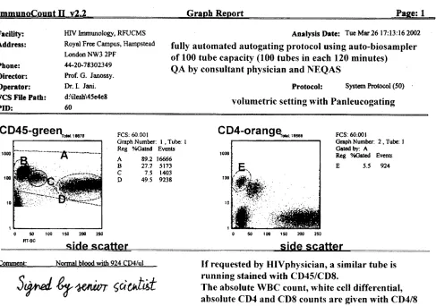
FIG. 2. CD45/CD4 double staining on blood using panleucogating with volumetric absolute counting. The graph report form is printed to document the autogating procedure. First, the CD45 side scatter histogram and gate A (all leukocytes) are established (left). All events of gate A are sent to the second display of CD4 side scatter (right). The CD4⫹and lymphoid cells (in E) are automatically gated to provide the absolute
CD4-T-cell count. E/A⫻100 is the value of the CD4% among all leukocytes. The number of events in gate B (absolute lymphocyte count), gate C (absolute monocyte count), and gate D (absolute granulocyte count) are also defined. E/B⫻100 is the CD4% value among lymphocytes. A parallel tube for CD45/CD8 double staining can also be run to provide the total of 15 parameters listed in Materials and Methods. QA, quality assurance; NEQAS, UK National External Quality Assessment Service.
on August 17, 2020 by guest
http://cvi.asm.org/
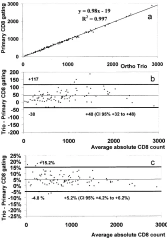
3.
Bland-Altman
plots
to
establish
the
agreements
between
the
volumetric
CD45/CD4
protocol,
single
tube,
and
the
“state-of-the-art
”
single-platform
technology.
The
parameters
were
the
total
absolute
lymphocyte
counts
(a),
CD4-T-cell
percentage
values
among
lymphocytes
(b),
and
absolute
CD4-T-cell
counts
(c
and
d)
.The
standard
technologies
used
were
full
lymphocyte
subset
panel
(three
tubes)
tested
with
the
Ortho
Trio
panel
on
the
Cytoron
A
bsolute
(a,
b,
and
c)
and
the
TruCOUNT
bead-based
method
(one
tube)
performed
on
a
(d).
on August 17, 2020 by guest
http://cvi.asm.org/
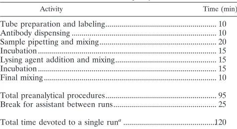
with a month of experience in flow cytometry processed 100 clinical samples using the autobiosampler. The steps of the procedure were monitored for time. A batch of 100 samples was processed in 95 min. The automated acquisition on the flow cytometer lasted for 120 min (Table 2), allowing the op-erator a 25-min break before starting to prepare the following batch. Three batches of 100 samples could be prepared within the normal 8-h working day. An additional batch was also prepared at the end of the day for unattended acquisition during the late hours, to be ready for inspection by the next morning. In total, 300 to 400 clinical samples could be pro-cessed each day.
CD8ⴙ-T-cell enumeration using primary CD45 and CD8
gating.We next evaluated the agreement for CD8
enumera-tion between the CD45-based protocol and the full Trio panel. With the latter protocol, similar to the other currently recom-mended protocols, CD8⫹T cells are counted when they
coex-press both CD3 and CD8 molecules. Using the CD45-based protocol in the absence of a CD3 reagent, a tight gate was placed around lymphoid cells with bright CD8 expression (me-dian, 130⫻103antibody binding capacity per cell), excluding most NK cells (range, 10⫻103to 110⫻103antibody binding capacity per cell; median, 24 ⫻103 ABC per cell) from the CD8 gate. The correlation was excellent throughout the whole CD8 range (Fig. 4a), but a systematic bias was observed (⫹40 CD8 T cells/mm3; 95% confidence interval,⫹32 to⫹48) (Fig. 4b), representing⫹5.2% of CD8⫹lymphocytes (Fig. 4c).
The CD4/CD8 ratios were also evaluated in samples by the volumetric method, using the CD45/CD4 and CD45/CD8 pro-tocols in two parallel tubes, and compared to the CD4/CD8 ratios observed with the full Trio panel (Fig. 5a). The agree-ments were good with virtually no bias (⫺0.05 CD4/CD8; limits of agreement, between⫺0.21 and⫹0.12 CD4/CD8) (Fig. 5a). We extended our study to investigate the agreement be-tween the sum of the CD4⫹and CD8⫹T cells generated with
the volumetric CD45 protocol and the T cells observed using CD3 staining in the full Trio protocol. The CD45-based pro-tocol failed to identify CD3⫹lymphoid cells doubly negative
for CD4 and CD8 antigens (CD3⫹CD4⫺CD8⫺). There was a
bias of⫹125 T cells/mm3(Fig. 5b) in favor of the CD3⫹-T-cell
counts recorded on the full Trio panel. These CD3⫹ CD4⫺
CD8⫺T cells represented a 10.1% bias throughout the whole
range of the T-cell counts (Fig. 5c).
WBC subset enumeration using CD45-based protocols on a
volumetric flow cytometer.The expression of CD45 antigen is
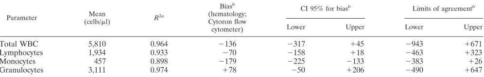
the common feature of all WBCs, and the CD45 staining in-tensity plus SSc distinguishes lymphocytes, monocytes, and granulocytes (Fig. 2) (20). We investigated the agreement be-tween counts generated by this method and those yielded by a hematology analyzer in the routine hematology laboratory of our institution. The agreements for total WBCs, lymphocytes, and granulocytes were good, with a minimal bias of ⫺136 WBCs/mm3, ⫺70 lymphocytes/mm3, and ⫹78 granulocytes/ mm3 (Table 2). However, the agreement for monocyte enu-meration was poor. The hematology analyzer underestimated the monocyte counts with a bias of⫺179 monocytes/mm3. This is a large value, representing 35 to 40% of the total monocyte counts, as already reported for several hematology analyzers (14, 19).
DISCUSSION
The need to improve laboratory services for regions of the world where the HIV epidemic threatens to destroy the fabric of life has revitalized efforts to identify the most efficient tech-niques for monitoring HIV disease. The present changes relate to the common areas of routine immunology and hematology, such as quality assurance, sample processing, and transporta-tion, as well as to the challenges of how to optimally count blood cells (Fig. 1). Immunological methods, based on the specificities and discriminating capacities of MAbs, have re-cently made an impact by recognizing even minor subsets of functionally divergent blood cells (9, 24, 26, 29, 30). By using flow cytometry, the true power of directly identifying cells by antibodies, as opposed to first investigating merely their mor-phological features, is now documented, and the strategy of primary immunological gating is widely used (12, 18, 20, 22). The relevant examples include CD45 for leukocytes and their subpopulations (4, 13, 20), CD3 for T cells (22, 25), CD4 for the major T-cell subset and monocytes (17, 18), and CD8 for the minor T-cell subset and some NK cells (18). Reliable total lymphocyte counts have been achieved by the Immunosum technique (24), providing the sum of the immunogated CD3⫹
(T), CD19⫹(B), and CD16⫹(NK) cells instead of using only
the lymphocytic scatter appearance. The commonly used dis-play on the cytometers is referred to as a heterogeneous, or morphospectral, protocol (23, 27) to show the IF of cells stained with MAb (on one axis) and the side scatter profile of cells (on the other axis) (Fig. 2).
In our study, we combined immunological CD45 gating with volumetric absolute counting on single platforms in order to introduce a robust method for WBC counting and for enumer-ating CD4⫹and CD8⫹T cells. Our four main findings are as
follows.
First, this study confirms our previous work, also performed on volumetric flow cytometers (18), as to the good agreement between the absolute CD4⫹-T-cells counts obtained by direct
CD4 gating and by CD4⫹CD3⫹coexpression (Fig. 3c).
Im-portantly, however, when we previously used CD4 MAb on its own without CD45, reliable CD4%-per-lymphocyte values could be obtained only with the constant vigilance of an
expe-TABLE 2. Timetable of routine operation using a biosampler with a 100-tube capacity
Activity Time (min)
Tube preparation and labeling... 10
Antibody dispensing ... 10
Sample pipetting and mixing... 20
Incubation ... 15
Lysing agent addition and mixing... 15
Incubation ... 15
Final mixing ... 10
Total preanalytical procedures... 95
Break for assistant between runs... 25
Total time devoted to a single runa...120
aOne run for 100 samples is achieved in 2 h (120 min) with a 25-min break for
the assistant. Each day three or four runs can be performed to evaluate 300 to
400 samples. A laboratory with a 5-day-per-week service can handle⬎1,000
tubes (500 to 1,000 patient samples) per week.
on August 17, 2020 by guest
http://cvi.asm.org/
rienced operator, who frequently had to manually modify the lymphocyte gates, a time-wasting procedure (18). We have now added CD45 gating to the protocol and report the excellent agreement between lymphocyte counts determined by CD45-side scatter and by the Immunosum method using the full Trio panel (Fig. 3a). Thus, CD45 staining improves the efficiency of the new autogated protocol (Fig. 2), saving effort and techni-cians’ time. Similarly, this gating strategy shows no bias com-pared to the bead-based CD4 counts (30) but reveals occa-sional differences leading to a wider spread (Fig. 3d). This discrepancy might be a bead-related phenomenon, because
similar results are seen when CD4 counts obtained by panleu-cogating on a double platform are compared to counts ob-tained by the bead-based method (13, 18).
The second conclusion is that the CD45/CD4 protocol on a volumetric cytometer provides an efficient system in which a trained flow cytometrist can run large numbers of tubes per day (⬎300 samples using CD45/CD4 alone [Table 2]). If two parallel tubes are used with CD45/CD4- and CD45/CD8-dou-ble-stained cells,⬎150 blood samples can be studied. Obvi-ously, such intensive diagnostic activity needs to be supple-mented by clerical help and supervisory capacity. Nevertheless,
FIG. 4. Evaluation of the agreement between the volumetric absolute CD8-T-cell counts generated with the CD45/CD8 protocol (CD8⫹⫹
SSc⫹) and the full Trio panel (CD3⫹CD8⫹). The results using linear regression (a) and the Bland-Altman plot (b) and its Pollock modification
(c) are shown. In the Pollock modification, the differences between the two methods of counting CD8 T cells were expressed as a percentage of the total CD8 counts to illustrate the systematic bias at a 5% level.
on August 17, 2020 by guest
http://cvi.asm.org/
this capacity illustrates the high efficiency of flow cytometry compared to that of manual methods such as the Dynabeads system (11), where a single assistant can manually handle only 15 to 20 samples per day. A hugely increased workload for CD4-T-cell enumeration is in line with the expected demand generated by the arrival of generic drugs for antiretroviral therapy. The larger regional centers dedicated to nationwide support with organized sample transportation using TransFix blood stabilizers (17) will require this increased service capac-ity.
The technical efficiency of this technology is directly related to three factors: (i) the fluent operation with a robust autogat-ing process, where only⬍2 to 4% of samples need attention for regating (see above) (Fig. 2), (ii) the use of an efficient autobiosampler (10, 24), and (iii) a convenient system using a Windows environment and a Microsoft Access database for feedback to the clinicians. Flexible reporting, based on consul-tation with clinicians, may include only CD4 counts or any of the 15 parameters listed in Materials and Methods.
The third finding of our study is related to the use of CD3,
FIG. 5. Bland-Altman plots (a and b) and the Pollock modification (c) to establish agreements on the Cytoron between the volumetric CD45/CD4-plus-CD45/CD8 two-tube protocol and the standard volumetric method using Trio reagents. The parameters studied were the CD4/CD8 ratios (a) and the sum of the absolute CD4- plus CD8-T-cell counts versus the CD3⫹-T-cell counts (b and c, respectively). In the Pollock
modification (c), the differences in total T-cell counts were expressed as percentages of T-cell counts to illustrate the regular underestimation of total CD3⫹-T-cell counts, at a 10% level, by the CD45 protocol. This bias is due to the existence of CD3⫹CD4⫺CD8⫺T lymphoid cells.
on August 17, 2020 by guest
http://cvi.asm.org/
the specific T-cell marker. Arguably, CD3 is not required to identify CD4⫹T cells (13, 17, 18). However, the CD8⫹
-lym-phocyte populations are more complex (18) and display CD8 antigen over a wide range (15⫻103to 140⫻103CD8 mol-ecules/cell [5]). The CD8⫹ cells include 80 to 92% proper
CD8⫹CD3⫹T cells that display CD8 at a high level (CD8⫹⫹;
80⫻103to 140⫻103/cell) and 8 to 20% CD8⫹CD3⫺NK cells
that express CD8 at a lower level (CD8⫹;⬍80⫻103/cell). It is therefore logical to place a tight gate around the CD8⫹⫹
population and compare these results with those obtained by counting CD3⫹-gated CD8 T cells (18). The results described
above show that the CD8⫹⫹gate underestimates CD3⫹CD8⫹
counts by 5.2% (Fig. 4c). This bias is apparently too modest to influence the CD4/CD8 ratios (bias,⫺0.05 [Fig. 5a]). An extra advantage of running both CD45/CD4 and CD45/CD8 tubes is the availability of CD4- plus CD8-T-cell counts that disregard the CD3⫹CD4⫺CD8⫺T cells. We have argued elsewhere that
these double-negative T cells represent a functionally different, mostly T-cell receptor␣-negative subset that should not be included in the total T-cell counts (18).
Finally, Loken et al. (20) have documented the differential expression of CD45 antigen on lymphocytes, granulocytes, and CD14⫹monocytes. In our study, the CD45 analysis is
com-bined with volumetric counting in order to generate absolute leukocyte differential counts. These parameters, when defined on hematological counters, can be error prone (3, 19, 33), and the monocyte counts are frequently underestimated (Table 3) (14, 19). On the other hand, the monocyte counts obtained by CD45 gating and carefully confirmed by the CD14 monocytic marker expression (20) are more accurate. Consequently, the methods described above, in combination with the use of sta-bilized blood preparations with long shelf lives (1, 13), will assist the establishment of long-awaited quality assurance schemes for leukocyte differentials and absolute counts, which are required to coordinate the performance of the wide variety of different hematology analyzers.
In conclusion, the present volumetric CD45/CD4 flow cy-tometry, assisted by more affordable sources of MAbs, has wide applicability in the routine laboratories operating in econ-omy-conscious environments. The specification required for the two-color IF plus side scatter used in this study is within the reach of the newly designed, battery-operated, smaller-volu-metric-flow cytometers that carry red diodes or other small light sources as the sole source of light excitation (19, 31) and are also capable of performing bead-based enzyme-linked
im-munosorbent assays with the multiplexing technology (19) in the area of the differential diagnosis of infectious diseases (16).
ACKNOWLEDGMENTS
I.V.J. is an AVERT Ph.D. scholar on a grant provided to G.J. and a receiver of an ORS award from United Kingdom Universities. This study was supported by grants to G.J. (Concerted Action Contract BMH4-97-2611 of the Biomed-2 program and grant QLRI-2000-00436 “Quality of Life and Management of Living Resources”) from the European Commission, Brussels, Belgium, and to D.K.G. (Bristol-Myers Squibb “Secure the Future”).
We are grateful for the generosity of the Royal Free Medical School and the Imperial Cancer Research Fund for the deposition of the RFT4 (CD4), RFT8 (CD8), and 2D1 (CD45) clones at the National Institute for Biological Standards and Control and at the Witwa-tersrand University, Johannesburg, South Africa, for potential general use.
REFERENCES
1.Barnett, D., V. Granger, P. Mayr, I. Storie, G. A. Wilson, and J. T. Reilly.
1996. Evaluation of a novel stable whole blood quality control material for lymphocyte subset analysis: results from the UK NEQAS immune
monitor-ing scheme. Cytometry26:216–222.
2.Barnett, D., V. Granger, A. G. Pockley, J. M. Saxton, L. Storie, L. Whitby, and J. T. Reilly.1998. TransFix: a sample stabilising fluid for use in cellular
haematology and immunology. Cytometry38:88.
3.Bentley, S. A., A. Johnson, and C. A. Bishop.1993. A parallel evaluation of
four automated hematology analyzers. Am. J. Clin. Pathol.100:626–632.
4.Bergeron, M., J. K. Nicholson, S. Phaneuf, T. Ding, N. Soucy, A. D. Badley, N. C. Hawley Foss, and F. Mandy.2002. Selection of lymphocyte gating protocol has an impact on the level of reliability of T-cell subsets in aging
specimens. Cytometry50:53–61.
5.Bikoue, A., F. George, P. Poncelet, M. Mutin, G. Janossy, and J. Sampol.
1996. Quantitative analysis of leukocyte membrane antigen expression:
nor-mal adult values. Cytometry26:137–147.
6.Bikoue, A., G. Janossy, and D. Barnett.2002. Stabilised cellular immmuno-fluorescence assay (SCIFA): CD45 expression as a calibration standard for
human leukocytes. J. Immunol. Methods266:19–32.
7.Bland, J. M., and D. G. Altman. 1986. Statistical methods for assessing
agreement between two methods of clinical measurement. Lanceti:307–310.
8.Bofill, M., G. Janossy, C. A. Lee, D. MacDonald-Burns, A. N. Phillips, C. Sabin, A. Timms, M. A. Johnson, and P. B. Kernoff.1992. Laboratory control values for CD4 and CD8 T lymphocytes. Implications for HIV-1 diagnosis.
Clin. Exp. Immunol.88:243–252.
9.Brando, B., D. Barnett, G. Janossy, F. Mandy, B. Autran, G. Rothe, B. Scarpati, G. D’Avanzo, J. L. D’Hautcourt, R. Lenkei, G. Schmitz, A. Kunkl, R. Chianese, S. Papa, J. W. Gratama, et al.2000. Cytofluorometric methods
for assessing absolute numbers of cell subsets in blood. Cytometry42:327–
346.
10.Connelly, M. C., M. Knight, J. V. Giorgi, J. Kagan, A. L. Landay, J. W. Parker, E. Page, C. Spino, C. Wilkening, and T. J. Mercolino.1995.
Stan-dardization of absolute CD4⫹lymphocyte counts across laboratories: an
evaluation of the Ortho CytoronAbsolute flow cytometry system on normal
donors. Cytometry22:200–210.
11.Didier, J. M., M. D. Kazatchkine, C. Demouchy, C. Moat, S. Diagbouga, C. Sepulveda, A. M. Di Lonardo, and L. Weiss.2001. Comparative assessment
of five alternative methods for CD4⫹T-lymphocyte enumeration for
imple-mentation in developing countries. J. Acquir. Immune Defic. Syndr.26:193–
195.
CytoronAbsolute for absolute WBC enumeration
Parameter (cells/Meanl) R2a
Biasb
(hematology; Cytoron flow cytometer)
CI 95% for biasb Limits of agreementb
Lower Upper Lower Upper
Total WBC 5,810 0.964 ⫺136 ⫺317 ⫹45 ⫺943 ⫹671
Lymphocytes 1,934 0.933 ⫺70 ⫺158 ⫹18 ⫺463 ⫹323
Monocytes 457 0.898 ⫺179 ⫺225 ⫺133 ⫺383 ⫹26
Granulocytes 3,111 0.974 ⫹78 ⫺50 ⫹206 ⫺490 ⫹647
aDefined by linear-regression analysis.
bDefined by Bland-Altman analysis. CI 95%, 95% confidence interval.
on August 17, 2020 by guest
http://cvi.asm.org/
12.Gelman, R., and C. Wilkening.2000. Analyses of quality assessment studies
using CD45 for gating lymphocytes for CD3(⫹)4(⫹)%. Cytometry42:1–4.
13.Glencross, D. K., L. E. Scott, I. V. Jani, D. Barnett, and G. Janossy.2002. CD45-assisted PanLeucogating for accurate, cost-effective dual-platform
CD4⫹T-cell enumeration. Cytometry50:69–77.
14.Goossens, W., L. Van Hove, and R. L. Verwilghen.1991. Monocyte counting: discrepancies in results obtained with different automated instruments.
J. Clin. Pathol.44:224–227.
15.Hoffman, R. A., P. C. Kung, W. P. Hansen, and G. Goldstein.1980. Simple and rapid measurement of human T lymphocytes and their subclasses in
peripheral blood. Proc. Natl. Acad. Sci. USA77:4914–4917.
16.Jani, I. V., G. Janossy, D. W. G. Brown, and F. Mandy.2002. Multiplex immunoassays by flow cytometry for diagnosis and surveillance of infectious
diseases in resource-poor settings. Lancet Infect. Dis.2:243–250.
17.Jani, I. V., G. Janossy, A. Iqbal, F. S. Mhalu, E. F. Lyamuya, G. Biberfeld, D. K. Glencross, L. Scott, J. T. Reilly, V. Granger, and D. Barnett.2001.
Affordable CD4⫹T cell counts by flow cytometry. II. The use of fixed whole
blood in resource-poor settings. J. Immunol. Methods257:145–154.
18.Janossy, G., I. V. Jani, and W. Gohde.2000. Affordable CD4(⫹) T-cell counts on ‘single-platform’ flow cytometers. I. Primary CD4 gating. Br. J.
Haematol.111:1198–1208.
19.Janossy, G., I. V. Jani, M. Kahan, D. Barnett, F. Mandy, and H. Shapiro.
2002. Precise CD4 T cell counting using red diode laser excitation: for richer,
for poorer. Cytometry50:78–85.
20.Loken, M. R., J. M. Brosnan, B. A. Bach, and K. A. Ault.1990. Establishing optimal lymphocyte gates for immunophenotyping by flow cytometry.
Cy-tometry11:453–459.
21.Mandy, F., M. Bergeron, G. Houle, J. Bradley, and J. Fahey.2002. Impact of the international program for quality assessment and standardization for
immunological measures relevant to HIV/AIDS: QASI. Cytometry50:111–
116.
22.Mandy, F., M. Bergeron, D. Recktenwald, and C. A. Izaguirre.1992. A simultaneous three-color T cell subsets analysis with single laser flow cytom-eters using T cell gating protocol. Comparison with conventional two-color
immunophenotyping method. J. Immunol. Methods156:151–162.
23.Mandy, F., J. K. Nicholson, B. Autran, and G. Janossy.2002. T-cell subset counting and the fight against AIDS: reflections over a 20-year struggle.
Cytometry50:39–45.
24.Mercolino, T. J., M. C. Connelly, E. J. Meyer, M. D. Knight, J. W. Parker, G. T. Stelzer, and G. DeChirico.1995. Immunologic differentiation of abso-lute lymphocyte count with an integrated flow cytometric system: a new
concept for absolute T cell subset determinations. Cytometry22:48–59.
25.Nicholson, J. K., M. Hubbard, and B. M. Jones.1996. Use of CD45 fluo-rescence and side scatter characteristics for gating lymphocytes when using
the whole blood lysis procedure and flow cytometry. Cytometry26:16–21.
26.O’Gorman, M. R. G., R. Gelman, Site Investigators, and the NIAID New CD4 Technologies Focus Group.1997. Inter- and intrainstitutional evalua-tion of automated volumetric capillary cytometry for the quantitaevalua-tion of CD4- and CD8-positive T lymphocytes in the peripheral blood of persons infected with human immunodeficiency virus. Clin. Diagn. Lab. Immunol.
4:173–179.
27.O’Gorman, M. R., and J. K. Nicholson.2000. Adoption of single-platform technologies for enumeration of absolute T-lymphocyte subsets in peripheral
blood. Clin. Diagn. Lab. Immunol.7:333–335.
28.Pollock, M. A., S. G. Jefferson, J. W. Kane, K. Lomax, G. MacKinnon, and C. B. Winnard.1992. Method comparison—a different approach. Ann. Clin.
Biochem.29:556–560.
29.Reimann, K. A., M. R. G. O’Gorman, J. Spritzler, C. L. Wilkening, D. E. Sabath, K. Helm, D. E. Campbell, and The NIAID DAIDS New Technologies Evaluation Group.2000. Multisite comparison of CD4 and CD8 T-lympho-cyte counting by single- versus multiple-platform methodologies: evaluation of Beckman Coulter flow-count fluorospheres and the tetraONE system.
Clin. Diagn. Lab. Immunol.7:344–351.
30.Schnizlein-Bick, C. T., J. Spritzler, C. L. Wilkening, J. K. A. Nicholson, M. R. G. O’Gorman, Site Investigators, and The NIAID DAIDS New Tech-nologies Evaluation Group.2000. Evaluation of TruCount absolute-count tubes for determining CD4 and CD8 cell numbers in human
immunodefi-ciency virus-positive adults. Clin. Diagn. Lab. Immunol.7:336–343.
31.Shapiro, H. M.1995. Practical flow cytometry, 3rd ed. Wiley-Liss, Inc., New York, N.Y.
32.Sherman, G. G., J. S. Galpin, J. M. Patel, B. V. Mendelow, and D. K. Glencross.1999. CD4⫹T cell enumeration in HIV infection with limited
resources. J. Immunol. Methods222:209–217.
33.Simson, E., and W. Groner.1995. Variability in absolute lymphocyte counts
obtained by automated cell counters. Cytometry22:26–34.