Drug Design, Development and Therapy
Dove
press
R e v i e w open access to scientific and medical research
Open Access Full Text Article
Current development of biodegradable polymeric
materials for biomedical applications
Richard Song1
Maxwell Murphy1
Chenshuang Li1
Kang Ting1–3
Chia Soo2
Zhong Zheng1
1Division of Growth and Development, Section of
Orthodontics, School of Dentistry, University of California, Los Angeles, Los Angeles, CA, USA; 2UCLA Department of Surgery and Department of Orthopaedic Surgery and The Orthopaedic Hospital Research Center, University of California, Los Angeles, Los Angeles, CA, USA; 3UCLA Department of Bioengineering, School of engineering, University of California, Los Angeles, Los Angeles, CA, USA
Abstract: In the last half-century, the development of biodegradable polymeric materials for biomedical applications has advanced significantly. Biodegradable polymeric materials are favored in the development of therapeutic devices, including temporary implants and three-dimensional scaffolds for tissue engineering. Further advancements have occurred in the utilization of biodegradable polymeric materials for pharmacological applications such as delivery vehicles for controlled/sustained drug release. These applications require particular physicochemical, biological, and degradation properties of the materials to deliver effective therapy. As a result, a wide range of natural or synthetic polymers able to undergo hydrolytic or enzymatic degradation is being studied for biomedical applications. This review outlines the current development of biodegradable natural and synthetic polymeric materials for various biomedical applications, including tissue engineering, temporary implants, wound healing, and drug delivery.
Keywords: tissue engineering, drug delivery, wound healing, natural biomaterials, synthetic biomaterials
Introduction
A biomaterial can be defined as a material intended to interface with biological systems in order to evaluate, treat, augment, or replace any tissue, organ, or func-tion of the body.1 The global market for implantable biomaterials was worth nearly
$75.1 billion in 2013. This market is expected to grow at a compound annual growth rate (CAGR) of 6.7% between 2014 and 2019, resulting in a $79.1 billion global market in 2014 and a $109.5 billion global market in 2019.2 Although biomedical
applications of natural enzymatically degradable polymers date back thousands of years, the application of synthetic biodegradable polymers began only in the second half of the 1960s.3 Considering their advantages over biostable materials in terms of
long-term biocompatibility along with the technical and ethical issues accompanying revision surgeries, investigations into the application of biodegradable biomaterials rather than permanent prosthetic devices for assisting in tissue repair and regen-eration has vigorously increased recently.4–6 As a result, polymeric biomaterials are
quickly replacing other material classes, such as metals, alloys, and ceramics, for use as biomaterials due to their versatility.3–5 Within the global implantable biomaterial
market, the polymeric biomaterials sector is expected to show the highest growth, at a CAGR of 22.1%, because of its promising potential in a wide range of biomedical applications.2 With this in mind, the aim of this review is to highlight the most often
studied polymeric biomaterials and underscore their immense potential in the areas of drug design, development, and therapy.
Correspondence: Chia Soo UCLA Department of Surgery and Department of Orthopaedic Surgery and The Orthopaedic Hospital Research Center, University of California, 675 Charles e Young Drive, South MRL 2641, Los Angeles, CA 90095-1759, USA Tel +1 310 794 5479
Fax +1 310 206 7783 email [email protected]
Zhong Zheng
Division of Growth and Development, Section of Orthodontics, School of Dentistry, University of California, 675 Charles e Young Drive, South MRL 2641, Los Angeles, CA 90095-1759, USA Tel +1 310 206 5646
Fax +1 310 206 7783
email [email protected]
Journal name: Drug Design, Development and Therapy Article Designation: Review
Year: 2018 Volume: 12
Running head verso: Song et al
Running head recto: Biodegradable polymeric materials for biomedical applications DOI: 165440
Drug Design, Development and Therapy downloaded from https://www.dovepress.com/ by 118.70.13.36 on 21-Aug-2020
For personal use only.
Dovepress
Song et al
A critical requirement for a biomaterial is biocompatibility – the ability of a material to function with an appropriate host response in a specific application.1 Many biological and
physicochemical characteristics of an implant material govern the host tissue response to the material. For instance, molecular weight, solubility, hydrophilicity/hydrophobicity, surface energy, material chemistry, mechanism of degrada-tion and/or erosion, lubricity, and shape and structure of the implant can all influence the material’s biocompatibility.7
Importantly, a biodegradable biomaterial requires excellent biocompatibility over time because the physicochemical, mechanical, and biological properties of a biodegradable biomaterial will differ with time and, thus, the resulting degradation products can possess varying levels of tissue compatibility as compared with the initial parent material. An ideal biodegradable biomaterial should have degrada-tion products that are nontoxic and easily metabolized and cleared from the body.
In addition to biocompatibility, several other important properties must be considered when choosing a biodegrad-able biomaterial. First, the degradation time of the biomate-rial should coincide with the regeneration and/or healing process to ensure proper remodeling of the tissue. Second, the biomaterial must maintain suitable permeability and processability for its intended application. Finally, the mechanical properties of the biomaterial should be suffi-cient to promote regeneration during the patient’s everyday activities, and any change in mechanical properties due to degradation should preserve compatibility with the healing or regeneration process.
Given the complexity of the human body and the scope of applications that polymeric biomaterials are currently utilized for, no single polymeric system can be considered the ideal biomaterial for all medical applications. Thus, recent advances in biodegradable biomaterial synthesis have been directed toward developing and synthesizing polymers with properties tailored for specific biomedical applications. Moreover, current developments incorporating multifunc-tional and combinatorial approaches in biomaterial design have accelerated the innovation of novel biodegradable biomaterials. Another hotspot in biomaterials research is the development of therapeutic devices, including temporary prostheses, three-dimensional (3D) porous scaffolds for tissue engineering, and delivery vehicles for pharmacological applications. Most recently, 3D bioprinting has also been acknowledged, and preliminary data collection for biomate-rial use as potential bio-ink for printing of 3D scaffolds have begun. Because biodegradable biomaterials exhibit a variety of biological and physicochemical properties and, therefore,
can replicate the properties of different tissues, these mate-rials are assessed for use as 1) large implants, including bone screws, bone plates, and contraceptive reservoirs; 2) small implants, in the form of sutures and staples; 3) plain membranes for guided tissue regeneration; and 4) porous structures or multifilament meshes for tissue engineering.8
Moreover, by properly engineering the structure and degrada-tion parameters, these biodegradable materials can be used to generate micro- or nanoscale drug-delivery vehicles for controlled drug delivery in an erosive or diffusive manner, or as a combination of both.9
Because of the interest aroused in the areas demanding biodegradable biomaterials, including regenerative medicine, tissue engineering, controlled drug delivery, gene therapy, and nanotechnology, there has been a robust expansion of biomedical applications of synthetic biodegradable polymers and analogous natural polymers, and our review will focus on exploring the most current development of these polymers for biomedical applications in these fields. Although many other reviews have focused on the topic of biodegradable biomaterials for medical use, to our best knowledge, there has not been a review published within the past 5 years that covers as much breadth as our review does about the devel-opment of the most commonly investigated biodegradable polymeric biomaterials in the fields of drug delivery, tissue engineering, and wound application.
Natural biodegradable polymeric
biomaterials
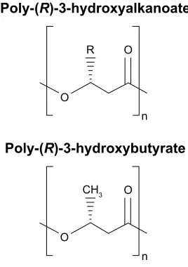
Biodegradable biomaterials can be roughly divided into two categories – natural and synthetic – based on their source and whether they are composed of naturally occurring extracel-lular matrix (ECM). Natural biodegradable polymeric bio-materials generally include proteins (collagen, fibrin, silk, etc.), and polysaccharides (starch, alginate, chitin/chitosan, hyaluronic acid derivatives, etc.).10–12 Furthermore, a family
of native polyesters – polyhydroxyalkanoates (PHA) – has been recognized as natural biodegradable biomaterials and, more recently, sundew adhesives (natural polysaccharide-based hydrogels) and ivy nanoparticles (macromolecular compositions of nanospherical arabinogalactan proteins) have garnered more attention for their ability to create effec-tive nanocomposite adhesives and for their potential use as nano-carriers in drug delivery, respectively.13,14
Collagen
As the most prevalent protein in the human body, collagen offers physical support to tissues by inhabiting the intercel-lular space, acting not only as native structural support for
Drug Design, Development and Therapy downloaded from https://www.dovepress.com/ by 118.70.13.36 on 21-Aug-2020
Dovepress Biodegradable polymeric materials for biomedical applications
organizing cells within connective tissues, but also as a mobile, dynamic, and flexible substance essential to cellular behaviors and tissue function.15 Generally, collagen is a
rod-type polymer approximately 300-nm long with a molecular weight of approximately 300 kDa. Free amino acids in the body are synthesized into subunit chains of collagen, which then undergo transcription, translation, and post-translational modification processes in suitable cells such as osteoblasts and fibroblasts.13 More than 22 different types of collagen
have been identified in the human body, with types I–IV being the most common, and type I collagen being the single greatest abundant protein present in mammals.15 Collagen
undergoes enzymatic degradation within the body by a diverse set of enzymes, such as matrix metalloproteinases and collagenases, to yield its corresponding amino acids. Due to its enzymatic degradability; unique mechanical, biological, and physicochemical properties; nontoxicity; and high tensile strength, collagen has been widely studied for biomedical applications.16,17
Collagen plays a critical role in preserving the biological and structural integrity of the ECM and is highly dynamic, undergoing continuous remodeling for proper physiologic functions. For most soft and hard connective tissues (eg, blood vessels, cornea, skin, tendon, cartilage, and bone), collagen fibrils and their networks function through their highly organized 3D structure. Tissue regeneration attempts to repair both the structural integrity and the intricate remod-eling process of the native ECM, particularly restoring the delicate collagen networks under which normal physiologic regeneration occurs; thus, recent efforts have been focused on replacing native collagen-based ECM by developing novel biomaterials that imitate its intricate fibrillar archi-tecture and function as cell scaffolding. Animal-derived and recombinant collagens, especially type I collagen, are recognized as one of the most valuable biomaterials available and are now extensively used for tissue engineering, drug delivery, and cosmetic surgery. For example, a composite of fibrillar collagen, hydroxyapatite, and tricalcium phosphate (Collagraft®, Angiotech Pharmaceuticals) has been approved
by the United States Food and Drug Administration (FDA) as a biodegradable synthetic bone graft substitute.18
The main sources of collagen presently utilized for bio-medical applications are bovine or porcine skin and bovine or equine Achilles tendons. They are utilized in either their native fibrillar form or after denaturation in various fabricated forms, such as sponges, sheets, plugs, and pellets. Collagen-based materials have been successfully used for skin repair.19
For example, Promogran® (Systagenix) – a spongy collagen
matrix containing oxidized cellulose – is available in the USA
and Europe for treatment of diabetic and ulcer wounds.20
Similarly, an FDA-approved bilayer skin substitute (Integra®
Dermal Regeneration Template, Integra LifeSciences) com-posed of a dermal layer of cross-linked bovine collagen and glycosaminoglycans (GAGs) as well as an epidermal layer of polysiloxane, which deposits ECM components, is in the market for full-thickness or deep partial thickness thermal injury. Moreover, these skin substitutes constructed from cell-seeded collagens have been widely commercialized (eg, Apligraf®, Organogenesis, Inc.) and (OrCel™, Ortec
International Inc.). Interestingly, type I collagen sponges have also been used to engineer patellar tendons in rabbits under different culture conditions.21–23 Using the collagen
sponge combined with bone marrow-derived mesenchymal stem cells, Juncosa-Melvin et al demonstrated that the engi-neered tendon tissue attained almost 75% of the mechanical properties of native tendon.22 In other studies, collagen has
also been successfully used as a scaffold in nerve and bladder engineering.24–26 Additionally, a suture-free, 3D-collagen
matrix graft – DuraGen® (Integra LifeSciences) – has been
developed for spinal dural repair and regeneration and is currently approved by the FDA.27
Collagen is, furthermore, a key initiator of the coagulation cascade and, therefore, it has been successfully developed as a hemostatic agent due to its high thrombogenicity. Multiple collagen-based hemostats are currently available or undergo-ing clinical trials for a variety of surgical indications; these include Sulzer-Spine® Tech’s sealants composed of bovine
collagen and bovine thrombin for cardiovascular and spinal surgical procedures; Floseal® (Baxter Healthcare), a
high-viscosity gel hemostatic agent composed of collagen-derived particles and topical bovine-derived thrombin; and CoStasis®
Surgical Hemostat (Cohesion Technologies), which consists of bovine thrombin and bovine microfibrillar collagen com-bined with autologous plasma.
With regard to drug delivery, collagen has been notably studied for the delivery of low-molecular-weight drugs, proteins, genes, and plasmids. Currently, a few collagen-based gentamicin-delivery vehicles are available in the global market (eg, Sulmycin®-Implant and Collatamp®-G, Innocoll
Pharmaceuticals Ltd). These delivery systems permit a sustained local delivery of antibiotics with limited systemic exposure. Moreover, another product, Septocoll® (Biomet),
achieved prolonged collagen delivery by incorporating two gentamicin salts possessing different solubility, and has been approved for infection prevention.28 More recently, a
new biodegradable, collagen-based chlorhexidine chip has been shown to provide a longer, more sustained release of chlorhexidine in the confines of the periodontal pocket as
Drug Design, Development and Therapy downloaded from https://www.dovepress.com/ by 118.70.13.36 on 21-Aug-2020
Dovepress
Song et al
compared to simple subgingival irrigation of chlorhexidine. The constant outflow of the gingival crevicular fluid from the periodontal pocket (up to 40 times an hour) renders subgingival irrigation of chlorhexidine useless in delivering significant antimicrobial benefits, reduction in probing depths, and clinical attachment level gain to manage chronic periodontitis; however, the use of a collagen-based release vehicle allows for a controlled delivery of chlorhexidine to achieve the intended pharmaceutical effects.29
Further-more, current clinical trials are investigating cross-linked, absorbable collagen sponges as protein-carrier vehicles. Prolonged release of bioactive proteins, such as recombi-nant human bone morphogenic protein-2 (rhBMP-2), have been reported when utilized in conjunction with collagen matrices due to desirable interactions of the collagen matrix with the protein.30 This combination product is approved
by the FDA for simultaneous use with a titanium interbody spine fusion cage for anterior lumbar spinal fusion (InFUSE®
Bone Graft/LT-CAGE® Lumbar Tapered Fusion Device,
Medtronic Spinal and Biologics), and a similar product InductOs® (Medtronic Spinal and Biologics) is approved
in Europe for the treatment of acute tibial fractures in adult patients. Additionally, a recent study reported that the deliv-ery of rhBMP-2 by an absorbable collagen sponge stimulated bone reconstruction in advanced alveolar ridge defects.31
Aside from its use as a protein-delivery vehicle, collagen has been demonstrated to retain gene vector/plasmid DNA whereas simultaneously protecting it from enzymatic or immunological reactions of the body, which underscores its potential in gene and plasmid DNA delivery.32
One disadvantage of these collagen-based biomaterials is their mild immunogenicity due to the antigenic sites in the central helix and the composition of the terminal region, which significantly limits their clinical application.33 The immune
response varies depending on the processing techniques, site of implantation, and species from which the collagen has been isolated.34 Other disadvantages of animal-derived
col-lagen include the varying physicochemical and degradation properties, the high cost of pure collagen, and the allogeneic or xenogeneic sources, which increase the risk of infectious disease transmission. In response to these limitations, pres-ent investigations have directed focus toward recombinant systems that can produce human-based collagen.35,36 Several
potentially useful systems already exist for large-scale pro-duction and purification of recombinant collagen, such as yeasts, transgenic animals, and, most recently, Escherichia coli.37,38 Because the amino acid sequence of recombinant
collagen can be directly modified, it is possible to make
controlled, targeted collagen products for specific applica-tions, thereby diversifying and increasing the potential of collagen-based products.39 However, recombinant collagen
is unable to undergo natural posttranslational modification and, thus, may lack critical biological activities of native tissue.33 This lack of posttranslational modification means
that, despite its flexibility in creating countless different amino acid sequences for different targeted applications, recombinant collagens may not be suitable for many biomedi-cal applications, especially those that require stability and strength such as heart valves. Furthermore, the commercially available recombinant collagens are still expensive to pro-duce and, thus far, only limited amounts are available.39
Collagen as a biomaterial has already seen significant use in several specific applications in skin repair, hemostatic agents, and drug delivery, but its slight immunogenicity, high cost, and varying physicochemical and degradation properties prevent further expansion of collagen-based biomaterials. Although recombinant collagens have the potential to catapult collagen-based biomaterials for widespread use, significant limitations such as their lack of posttranslational modification will need to be continually studied for them to have any noticeable impact for use as a biomaterial.
Gelatin
Gelatin is a natural biopolymer derived from collagen via controlled alkaline, acid, or enzymatic hydrolysis.40 As a
result of its biological origin, it has excellent biodegrad-ability and biocompatibility and because it is widely avail-able, gelatin is a relatively low-cost polymer.40 Gelatin has
been used in medical and pharmaceutical fields as a matrix for implants, and as stabilizers in vaccines such as measles, mumps, and rubella.41 Moreover, gelatin is water permeable
and soluble in water and has multifunctional properties as a drug-delivery carrier.42 Gelatin’s mechanical properties,
swelling behavior, thermal properties, and many other phys-iochemical properties can be dependent upon the collagen source, extraction method, amount of thermal denatured employed, and the degree of cross-linking, thereby making gelatin a very versatile polymer.43 Furthermore, its ability to
produce a thermoreversible gel makes it a very good candi-date as a targeted drug-delivery carrier and, as a result, gelatin can be utilized to develop specific drug-release profiles, allowing for a broad range of applications in drug delivery.43
Gelatin is a versatile biopolymer that traditionally enabled the design of several different drug carrier systems, such as microparticles, nanoparticles, fibers, and hydrogels.44 Each
of these different systems has certain properties that make
Drug Design, Development and Therapy downloaded from https://www.dovepress.com/ by 118.70.13.36 on 21-Aug-2020
Dovepress Biodegradable polymeric materials for biomedical applications
them particularly suitable for drug delivery.44 For example,
gelatin microparticles are popular for serving as vehicles for cell amplification and delivery of large bioactive molecules, and gelatin nanoparticles are better for drug delivery to the brain or in intravenous delivery.44 Recently, a novel and
relatively simple method was discovered for producing gelatin microparticles which allow for very high protein- and drug-loading efficiencies.45 In the experiment, bovine serum
albumin (BSA) was added at a 0.1%–0.4% gelatin concentra-tion, resulting in a gelatin solution.45 The solution was
freeze-dried and the resulting spongy membranes were hardened with liquid nitrogen and subsequently ground to a powder, producing BSA-loaded microparticles in random forms and shapes.45 These microparticles were then incorporated into
porous scaffolds constructed of polylactide (PLLA) and poly-caprolactone (PCL).45 The in vitro release profile of the protein
from the particles alone and from the particle-incorporated scaffolds demonstrated up to 90% protein-loading efficiency, showing that BSA-loaded microparticles in conjunction with particle-incorporated scaffolds for growth factor release may be possible for application in bone tissue engineering.45
Another study studied the in vitro efficacy and toxicity of incorporating polyenes into electrospun gelatin fiber mats as a topical antifungal application.46 The research group
found that polyene-loaded antifungal gelatin mats displayed better antifungal activity than using traditional electrospun gelatin fiber mats.46 Furthermore, they found that polyenes
stabilized the triple helical conformation of gelatin whereas gelatin decreased the hemolytic activity of polyenes, making polyene antifungal-loaded gelatin fiber mats a possibility in managing superficial skin infections in the future.46 Cohen
et al studied novel tissue adhesives based on gelatin, with alginate as a polymeric additive, loaded with bupivacaine or ibuprofen for pain management.47 They found that the release
of drugs from the adhesive matrix was primarily controlled by the gelatin/alginate bioadhesive’s characteristics, such as swelling and hydrophilic group concentrations that can be adjusted by changing the ratio of gelatin to alginate.47
Moreover, they found that the hydrophilicity and electrical interactions between bupivacaine and ibuprofen with the adhesive components had some effect on the release profile of the drug, and that incorporating bupivacaine improved the bonding strength of the adhesive due to its inert nature whereas ibuprofen decreased the bonding strength due to its reactive nature with the alginate/gelatin adhesive.47 Overall,
the study shows that this loading bupivacaine in a gelatin/ alginate bioadhesive can potentially be used for wound-closing applications.47
The primary advantages of gelatin are its biodegrad-ability, availbiodegrad-ability, and cheap cost.44 Porcine skin-derived
gelatin is the most popular source, followed by bovine skin, and bone.48 However, religious issues may arise, as
porcine-derived products and bovine-porcine-derived products are forbidden in Judaism and Hinduism, respectively.48 In addition, there
are health concerns over transmission of pathogenic vectors such as prions.48 Recombinant gelatins, however, can be
utilized to overcome the disadvantages with animal tissue-derived materials.49 Currently, the primary use of gelatin is
in the food, pharmaceutical, and photographic industries.49
In the biomedical field, gelatin is used in drug delivery and in some wound dressing and tissue-regeneration appli-cations, but its use is heavily limited by its poor mechanical properties.49 These mechanical properties can be strengthened
through physical linking as well as chemical cross-linking due to a large number of functional side groups that gelatin possesses; however, agents used to stabilize cross-linked gelatin are often somewhat toxic to the human body.49
Research into improving gelatin’s mechanical properties will need to be investigated in the future for gelatin to have a promising future as a primary biomaterial; but, until then, the most likely incorporation of gelatin will be as a composite with other natural or synthetic biomaterials, or as a carrier for drug delivery.
Fibrin
Fibrin is a 360-kD fibrinogen-derivative biopolymer involved in the natural blood-clotting process, enhancing cell adhe-sion and proliferation.50 In addition to possessing excellent
biodegradability and biocompatibility, fibrin exhibits high elastic and viscous properties; stiffens in response to shear, tension, or compression; and has excellent deformability.51
One of the first developed fibrin-based products was fibrin glue (fibrin sealant). Tissucol/Tisseel™ (Baxter Healthcare) and Beriplast HS/Beriplast P™ (CSL Behring) are the first-generation products of fibrin sealants marketed in Europe. Today, an expansive variety of products possessing different compositions and adhesive properties are available on the market. These products are broadly used for hemostasis and tissue-sealing applications in various surgical procedures, including neurosurgery and plastic and reconstructive surgery.52–54
Furthermore, fibrin has been utilized as a scaffold for the regeneration of numerous tissues, such as adipose tis-sue, bone, cardiac tistis-sue, cartilage, muscle tistis-sue, nervous tissue, ocular tissue, respiratory tissue, skin, tendons and ligaments, and vascular tissue as well as a carrier vehicle
Drug Design, Development and Therapy downloaded from https://www.dovepress.com/ by 118.70.13.36 on 21-Aug-2020
Dovepress
Song et al
for bioactive molecules (drugs, antibiotics, or chemotherapy agents) due to its injectability and biodegradability.55–60 For
instance, several studies have demonstrated that clinical application of fibrin significantly improved the healing of intrabony defects in chronic periodontitis.61,62 Further
evi-dence suggests that proteins interact differently with fibrin clots and, as a result, several cross-linking techniques are presently being studied to control the release profile of bioac-tive molecules from the fibrin matrix.63 Additionally, fibrin
matrices have been utilized as excellent cell-carrier vehicles. One successful example obtained by mixing keratinocytes with fibrin is Bioseed® (DCM Shriram Limited), a
fibrin-based product used to treat chronic cutaneous wounds. Fibrin is natural, highly available, implantable, inexpensive, easy to use, and has low fibrinogen concentrations. Due to its porous morphology, a fibrin scaffold system is adequate for cell attachment, proliferation, differentiation,58,64 and as a
release system of growth factors such as vascular endothelial growth factor and basic fibroblast growth factor (bFGF).60,65
Fibrin-immobilized growth factors have been shown to be continuously released for several days in a controlled manner, making it optimal for numerous tissue-engineering purposes.50 Thus, autologous fibrin could prevent the
com-plications of techniques derived from the use of current commercial available fibrin products and should be further investigated. Fibrin-based scaffolds do have some limita-tions, such as weak mechanical strength and quick degrada-tion rates; however, these properties have been shown to be improved by incorporating stronger natural and synthetic polymers, utilizing various cross-linking methods, and uti-lizing micro/nanospheres.50 Next-generation scaffolds will
likely involve specific cell lines, combined with biomolecules and growth factors to accelerate and increase cell prolif-eration and differentiation on immobilized scaffolds for a variety of tissues, and fibrin-based drug-delivery carriers and tissue-engineered scaffolds, including the relatively recent scaffold development of fibrin microspheres, nanospheres, microfibers, microtubes, and porous sheets, all of which will play a big role in regenerative medicine.50
Hyaluronic acid (HA)
HA is an essential component of the ECM and its structural and biological properties mediate cellular signaling, morpho-genesis, matrix organization, and wound repair.66,67 In 1943,
HA was first isolated from the vitreous humor by Meyer and Palmer.68 HA is a member of the GAG family, which
involves linear polysaccharides consisting of alternating units of N-acetyl-d-glucosamine and glucuronic acid ranging in
size from 5,000 to 20,000,000 Daltons in vivo, and is present in almost every tissue in vertebrates. Native sources of HA include rooster combs, bovine vitreous humor, and synovial fluids.69 HA degradation occurs in the body through free
radicals, such as nitric oxide and matrix metalloproteinases found in the ECM, and then undergoes endocytosis. Further digestion of HA by lysosomal enzymes results in mono- and disaccharides, which are then converted into ammonia, carbon dioxide, and water.69
Recently, HA has become recognized as an important building block for the creation of new biomaterials for use in cell therapy, 3D cell culture, and tissue engineering.70–72 As HA
is secreted at the early stage of wound healing, it has been extensively researched for wound-dressing applications.73,74
HA can be recognized by receptors on a variety of cells associated with tissue repair, and thus presents the capability to stimulate angiogenesis and to regulate injury-induced inflammation as a free radical scavenger.75 Moreover, HA
promotes epithelial and mesenchymal cell migration and differentiation, making it vital for tissue repair.75 These
properties combined with its immunoneutral potency make HA an ideal biomaterial for tissue engineering.75 Moreover,
its aqueous solubility allows for modification of HA into various porous and 3D structures for drug delivery. For example, HYAFF®11 (Anika Therapeutics, Inc.), an
HA-based product, is currently utilized as a carrier vehicle for a variety of growth factors, morphogens, and stem cells.76
In a comparative study, Hunt et al reported an improved healing response to rhBMP-2 delivered by HYAFF®11 than
that delivered by an absorbable collagen sponge.76 A more
recent study indicated that HA-based materials maintain the potential to replace collagen-based materials as injectable soft tissue fillers.77 With regard to tissue engineering, HA has been
successfully incorporated into multiple complex systems. For example, HA-modified poly(D,L-lactic acid-co-glycolic acid) (PLGA) scaffolds were successful in inducing cartilage tissue formation in terms of type II collagen expression and
+\DOXURQLFDFLG
2
2+ 2+
2+
2 2
2
2
2
1+ Q
+2
+2
Figure 1 Structure of hyaluronic acid (HA).
Drug Design, Development and Therapy downloaded from https://www.dovepress.com/ by 118.70.13.36 on 21-Aug-2020
Dovepress Biodegradable polymeric materials for biomedical applications
tissue morphological characteristics.78 Additionally,
high-molecular-weight viscous HA solutions (eg, AMVISC®
and AMVISC® PLUS, Bausch & Lomb) currently function
as vitreous humor substitutes and as protection for sensi-tive eye tissue during glaucoma surgery, cataract extrac-tion, and corneal transplantation. Viscous HA solutions (eg, SYNVISC®, SanofiBiosurgery; and ORTHOVISC®,
Anika Therapeutics, Inc.) are also clinically applied as synovial fluid substitutes to reduce pain and enhance joint mobility in patients with osteoarthritis.79
Moreover, HA derivatives, such as HA esters and cross-linked HA gels, have been thoroughly researched for wound dressing applications. Reports suggest that these chemical modifications significantly reduce the degradation of HA.69,80
For example, in the absence of enzymatic activity, benzyl HA esters undergo hydrolytic degradation via ester bonds, and the degradation time varies from 1 to 2 weeks to 2–3 months depending on the degree of esterification.69,80 Together, these
studies underscore the potential of HA in combined systems for an array of biomedical applications, including tissue engineering, drug delivery, wound healing, and temporary implants.
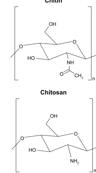
Chitin/chitosan
Chitosan is another naturally derived biodegradable polysac-charide commonly used in tissue engineering.81 Chitosan is
a derivative of chitin – the second most abundant natural polymer commonly found in the exoskeletons of crustacean and insects as well as the cells walls of fungi.81 Chitin is
partially deacetylated to form chitosan, which is composed of glucosamine and N-acetyl glucosamine linked in a β(1–4) manner.82 The molecular weight and the degree of
deacetyla-tion, which are essential in assessing the characteristics of chitosan, are reliant on the source and production process.
Chitin and chitosan are biodegraded by human enzymes, such as lysozymes, which disrupt the linkage between acety-lated units and degrades chitin/chitosan to oligosaccharides.83
The degradability of chitin/chitosan-based materials is essen-tial for scaffold construction because it can influence cell behavior and tissue formation of the engineered construct.84
However, the low mechanical resistance of chitosan makes it disadvantageous to be used as a supporting material in tissue engineering.85 To create a better mechanical profile,
cross-linking agents are used with functional reactive groups to allow for bridges to be made between polymeric chains, optimizing the resistance and elasticity of chitosan mem-branes.85 Further, chitin/chitosan have the ability to chelate
with the Ca2+ or Mg2+ present in the cell wall of bacteria, thus
destroying the entity of bacterial cell wall, and to react with the anionic phosphate groups of phospholipids found on the bacterial cell membrane by using their NH3+ amino group,
thereby leading to changes in the cell membrane permeability and eventual release of the bacteria’s cellular contents.84,86
These functional capabilities make chitin/chitosan-based materials exhibit a bactericidal effect on both Gram-negative and Gram-positive bacteria, giving them a broad medical utility in tissue engineering and biomedical applications.84,86
Specifically, chitin/chitosan-based materials have dem-onstrated potential with regard to connective, nerve, adipose, and vascular tissue-engineering applications.87–89 For
example, a silk fibroin (SF)/chitin-based scaffold was used to repair a musculofascial defect in the abdominal wall, displaying continuous integration with adjacent native tissue and mechanical strength similar to native tissue.88 Similarly,
chitosan/hydroxyapatite-based scaffolds loaded with bFGF for periodontal tissue regeneration promoted vigorous pro-liferation and migration in periodontal ligamental cells and cementoblasts.87 A more recent study produced nanosized
chitosan/hydroxyapatite-based scaffolds via thermally induced phase separation and lyophilization techniques and found that this combination demonstrated greater compressed mechanical properties as compared to the pure chitosan scaffold – a property critical for the success of scaffolds
Figure 2 Structure of chitin and chitosan.
&KLWLQ
2 2
2 2+
+2
1+
Q &+
&KLWRVDQ
2 2
2+
+2
1+
Q
Drug Design, Development and Therapy downloaded from https://www.dovepress.com/ by 118.70.13.36 on 21-Aug-2020
Dovepress
Song et al
to be used as tissue-engineering scaffolds.90 Further tests
will need to be conducted, but it is expected that this scaf-fold will allow cell attachment and tissue growth in vivo as well. Taken together, these results highlight the potential of chitin/chitosan-based scaffolds providing a cell-favorable microenvironment for connective tissue healing.
Though chitosan has been widely used for bone tissue-engineering applications due to its various favorable proper-ties as mentioned earlier, it has a lower tensile strength and modulus range than those of natural bones.91,92 In an effort to
reinforce the mechanical properties of chitosan, Tamburaci et al combined diatomite (diatomaceous earth), a natural silica material, with chitosan to produce a scaffold for bone tissue regeneration; they found that diatomite-reinforced chitosan composite membranes improved the surface area, roughness, swelling properties, and protein adsorption capacities of traditional chitosan membranes, while showing no cytotoxic effect in Saos-2 osteosarcoma cell line with excellent biocompatibility.93 Interestingly, Tamburaci et al
also found that diatomite incorporation into chitosan scaf-folds increased the proliferation and alkaline phosphatase activity of Saos-2 cells remarkably.93 Overall,
diatomite-reinforced chitosan scaffolds showed improved properties while maintaining the high biological activity typical of traditional chitosan scaffolds, illustrating the potential of chitosan for bone tissue-engineering applications.
Moreover, glutaraldehyde-cross-linked collagen-chitosan hydrogels were successfully applied to adipose tissue engineering. A study confirmed the in vitro viability of pre-adipocytes (PAs) on glutaraldehyde-cross-linked collagen– chitosan hydrogel scaffolds. Subsequently, a rat subcutaneous pocket assay was used to evaluate PA-seeded scaffolds in vivo which displayed excellent biocompatibility, formed adipose tissue, and induced vascularization.89 Additionally,
chitin/chitosan-based materials have found success as bioma-terials used in cartilage repair due to their biocompatibility and structural similarity with GAGs found in cartilage.12,94,95
Recently, Lee’s group synthesized an injectable hydrogel consisting of methacrylated glycol chitosan and HA by photo-cross-linking with a riboflavin photo-initiator under visible light.12 This resulted in the formation of a cross-linked
chitosan network in which high-molecular-weight HA was entrapped, forming a semi-interpenetrating network that pro-vided a more chondrogenic-favorable microenvironment.12
Likewise, these photopolymerizable hydrogels exhibit robust capacity as cell carrier vehicles and further emphasize the advantage of chitin/chitosan-based materials for tissue engineering.
Furthermore, chitin/chitosan-based materials have been incorporated successfully into cutaneous wound manage-ment. A number of studies have reported the use of chitin/ chitosan scaffolds and membranes to treat patients with deep burns.96,97 Recently, novel α-chitin/silver nanoparticles
(AgNPs) and β-chitin/AgNP composite scaffolds were tested for wound-healing applications.96,97 These chitin/AgNPs
com-posite scaffolds were found to possess excellent antibacterial activity against Staphylococcus aureus and E. coli, combined with good blood clotting ability.96,97 In addition, β-chitin/
AgNP composite scaffolds functioned as promising matrices capable of providing good cell attachment apart from their antibacterial activity, which suggests that these composite scaffolds are ideal for wound-healing applications.97
Finally, chitin/chitosan-based materials have presented excellent potential in drug delivery systems. For example, in one study, when carboxymethyl chitin nanoparticles were cross-linked with FeCl3 and CaCl2, they were shown to be nontoxic to mouse L929 cells while showing signifi-cant antibacterial activity against Staphylococcus strains.98
Studies indicating that chitin/chitosan-based materials may be used for controlled drug delivery in managing HIV are more exciting. In one study that focused on anti-transferrin and anti-bradykinin B2 antibody-conjugated chitosan nano-particles, the investigators found that chitosan nanoparticles presented the potential to effectively penetrate across the blood–brain barrier and thus enhance the drug delivery in the brain to inhibit HIV replication in the neural system.99
Another study showed that using chitosan nanoparticles to encapsulate conventional antiretroviral drugs targeting HIV led to a more efficient control of the viral proliferation in target T cells.100 Greater cell targeting efficiency was
achieved, mostly due to the fact that chitosan nanoparticles are mildly immunogenic, making them more visible to the immune system, allowing for more efficient uptake by phagocytes.100 A separate investigation studied the effects of
poly(lactic acid) (PLA)/chitosan nanoparticles loaded with lamivudine (a type 1 and type 2 HIV selective inhibitor) in mouse L929 fibroblast cells.101 The in vitro drug-release
studies showed that the drug release rate from PLA/chitosan nanoparticles decreased when the pH of the medium changed from alkaline to acidic and further decreased from acidic to neutral, which could be a result of the repulsion between H+ ions and cationic groups present in the polymeric
nano-particles.101 Because it is ideal that the drug encapsulated in
the delivery system will be protected in the stomach envi-ronment at acidic pH and then provide sustained release in the intestines (neutral pH), these findings suggest that the
Drug Design, Development and Therapy downloaded from https://www.dovepress.com/ by 118.70.13.36 on 21-Aug-2020
Dovepress Biodegradable polymeric materials for biomedical applications
chitosan-based nanoparticles exhibit excellent potential as a carrier system for HIV-controlled drug delivery.
Besides HIV management, chitin/chitosan-based materi-als can be used in cancer therapy as drug-delivery vehicles. For example, a pH-responsive magnetic nanocomposite was wrapped in chitosan for targeted and controlled drug delivery, and the yield product was found to be nontoxic and exhibited a high antitumor activity while maintaining its excellent pH sensitivity at pH ,6.0.102
In addition to improving the targeting and efficiency of cancer and HIV drug-delivery vehicles, chitin/chitosan-based materials have been utilized in various other delivery sys-tems. In a very recent study, Di et al developed an ultrasound-triggered insulin-delivery system which allows for pulsatile insulin release that can provide both long-term, sustained and fast on-demand responses.103 This system incorporated
insulin-loaded PLGA nanocapsules encapsulated within chitosan microgels and, upon ultrasound treatment, the stored insulin can be rapidly released to regulate blood glucose levels.103 The research group found that in a mouse model
of type 1 diabetes, a 30-second ultrasound administration could effectively achieve glycemic control for 1 week and concluded that this delivery system may potentially be used to release other therapeutics in a noninvasive and convenient manner.103 Moreover, Liang et al104 and Jing et al105 have
shown that chitosan derivatives as a delivery system can also enhance oral tablet absorption of bioactive compounds and potentially allow protein and certain peptide drugs to be orally administrable. In addition, hydroxyethyl chitosan – a derivative of chitosan – has shown great potential as a drug-delivery material for the treatment of glaucoma and other ocular diseases due to its great water-solubility and excellent biocompatibility.106 Further investigations must be carried
out to assess any side-effect or instability of chitosan-based materials; however, there seems to be substantial potential for chitosan-derived drug-controlled-release systems.
Chitin/chitosan-based materials are widely studied for several different tissue-engineering applications, regenera-tive medicine, wound healing, and drug delivery for good reason. They have excellent biodegradability and biocom-patibility and has been known to have antiulcer, anti-acid, hypocholesterolemic action, wound-healing, antitumor, and hemostatic properties.107–110 Although chitin/chitosan-based
materials tend to lack mechanical properties, they possess functional reactive side groups that can be cross-linked to make bridges between polymeric chains, optimizing the resistance and elasticity of these materials.85 Due to their
unique combination of physical and chemical properties,
chitin/chitosan can be molded relatively easily into porous scaffolds, and their cationic nature allows them to form polyelectrolyte complexes with many types of anionic GAGs, making them capable of modulating the activity of a variety of growth factors and cytokines for tissue-engineering purposes.85 An important property of chitin/chitosan is its
mucoadhesive nature and its ability to open epithelial tight junctions, making them well-suited for drug delivery across nasal, intestinal, ocular, buccal, and pulmonary systems.85
However, some challenges do exist. Chitosan is insoluble in most organic solvents, making delivery of hydrophobic drugs difficult; moreover, various methodologies to adapt the solubilization of chitosan, such as alkylation, acetylation, and carboxymethylation, all come with certain drawbacks and limitations.111
Overall, chitin/chitosan is a polymer that will have very important applications in both the industrial and biomedical fields of the future. Its unique chemical properties have recently allowed it to be studied as part of a biological functionalization of microelectromechanical systems, which will enable it to perform functions such as biorecognition, enzymatic catalysis, and controlled drug release, all of which is critical to the advancement of drug-delivery and scaffold technology.85
Starch
Starch is the primary energy reserve polysaccharide in plants, and it is present in the form of granules composed of amylose and amylopectin.112 Amylose is a linear polymer composed
of glucose monomers linked through α-D-(1–4) glyco-sidic linkages, whereas amylopectin molecules are huge, branched polymers of glucose known to be one of the highest molecular-weight natural polymers.113 Different plants have
slightly different granule sizes, amylose/amylopectin ratio, mineral contents, and amount of phosphorous and phospho-lipid contents that lead to varying starch properties.112 The
specific characterization of starch is particularly important due to different swelling, solubility, gelatinization, mechanical behavior, enzymatic digestibility, rheological characteristics, and surface characteristics, which affect the way it needs to be processed to convert it to a more usable form such as hydro-gels, pastes, and nanoparticles.114 Generally, the native starch
isolated from different plants tends to have limited shear resistance, thermal resistance, thermal decomposition, and a high tendency toward retrogradation.112 These limitations
have been overcome by combining starch with more stable, synthetic, thermoplastic polymers, or utilizing physical treatment of starch, such as heat or moisture, or utilizing
Drug Design, Development and Therapy downloaded from https://www.dovepress.com/ by 118.70.13.36 on 21-Aug-2020
Dovepress
Song et al
chemical modifications, introducing certain functional groups to remarkably alter its physicochemical properties, making starch vastly more useful in tissue engineering, drug delivery, and delivery of biologically active compounds.112
Among its tissue-engineering applications, starch is most famous for its usefulness in generating scaffolds for bone regeneration due to its bone-bonding behavior when reinforced with hydroxyapatite, good mechanical properties, non-cytotoxic and biocompatible nature, excellent substrate for cell adhesion, and thermoplastic behavior when combined with thermoplastic polymers.115 Lately, further improvements
have been made to these starch-based scaffolds. In one recent study, Mahdieh et al synthesized a nanocomposite biomaterial consisting of a blend of thermoplastic starch and ethylene vinyl alcohol as the polymer matrix, and incorpo-rated nano-structured forsterite and vitamin E as the ceramic reinforcing phase and thermal stabilizer, respectively.116
What they found was that nanofosterite, a newly developed bioceramic, resulted in improved biological and mechanical properties, thereby reducing the degradation rate of the scaf-fold whereas stimulating bone cell proliferation in comparison to the traditional starch–ethylene vinyl alcohol matrix.116
In particular, starch and PCL blends (SPCL) have gar-nered much attention from many research groups for applica-tion as a tissue-engineering construct.117 PCL improves the
processability of starch, reduces its high stiffness, and can overcome the high moisture sensitivity of starch, which is one of the greatest weakness of starch as a biomaterial.118
On the other hand, starch improves the biodegradability of PCL and, as the cheapest biomaterial on the planet, starch can substantially lower the high cost of the final product.119
Through the appropriate blending of starch and PCL, SPCL can overcome limitations of both PCL and starch, whereas additionally allowing for control over mechanical and deg-radative properties by adjusting the component ratio – a sig-nificant advantage that allows it to conform to the numerous differing tissue-regenerative rates.120 SPCL is particularly
effective in bone engineering because one of the greatest biological specifications of SPCL composites is their ability to enhance and stimulate osteoblast cell proliferation.117
In fact, several different research groups have recently experimented with SPCL for this purpose.117 Carvalho et al
studied the bone regeneration potential of undifferentiated human adipose-derived stromal/stem cells loaded in SPCL scaffolds for the regeneration of critical-sized mice calvarial defect.121 They found that SPCL was a suitable scaffold for
bone tissue engineering, allowing for new tissue formation in the calvarial defect to be approximately 20% of the defect size
after 4 weeks and 43% at 8 weeks.121 Link et al did a similar
study of critical-sized cranial defects in male Fisher rats and found that the SPCL fiber mesh proved to be an effective osteoconductive material for bone regeneration.122 Requicha
et al studied the in vivo behavior of a novel, double-layered SPCL scaffold functionalized with silanol groups (SPCL-Si) in a mandibular rodent model and compared the results to a commercial collagen membrane.123 They found that SPCL-Si
scaffolds induced significantly higher new bone formation when compared to the commercial collagen membrane.123
Starch has been proposed as a possible drug-delivery system.124 In its hydrogel form, it can efficiently entrap drugs
in interstitial spaces, thus protecting them from undesirable conditions in the human body.125 In addition, starch
hydro-gels are resistant to gastric juices, allowing for potential oral drug-delivery systems, and they can be modified to be degraded in very specific portions of the gastrointestinal tract, thus allowing for site-specific delivery.126 Moreover,
physical modification of starch by retrogradation leads to the development of high levels of type three resistant starch – a very thermally stable, low solubility form of starch that makes it suitable for colon-specific delivery systems.124 However,
in practice, studies on starch as a possible drug-delivery system have been very limited. Most of the research on starch as part of a drug-delivery system is theoretical, and scientists are still exploring the possible consequences of physical modification of starch on the mechanical and struc-tural properties of hydrogels. To date, there is no conclusive data on this subject.
As the cheapest, most bioavailable, natural polymer, starch is an interesting renewable resource that may have many different biomedical applications.119 Starch is entirely
biodegradable, noncytotoxic, biocompatible, and exhibits a high Young’s modulus with low levels of elongation at break.117 It is relatively easy to modify chemically, and it
has the ability to replace some more expensive, synthetic polymers in the fabrication of composite biopolymers.127
When starch is combined with PCL, SPCL exhibits addi-tional properties, such as better processability, increased mechanical properties, controllable degradability, and enhanced osteoblastic cell proliferation whereas still being relatively cheap to produce.117 As the study by Requicha
et al showed recently, functionalized SPCL-Si polymers induced much higher rates of bone formation as compared to the traditional (30:70) SPCL blend, further showing the potential of starch-based biopolymers as scaffolds.123
However, research outside of bone tissue engineering has largely been limited, and the potential for drug delivery,
Drug Design, Development and Therapy downloaded from https://www.dovepress.com/ by 118.70.13.36 on 21-Aug-2020
Dovepress Biodegradable polymeric materials for biomedical applications
while theoretically studied, has been relatively unexplored as compared to many other natural polymers. Further research needs to be undertaken to exploit the abundance of starch for use in more than just bone tissue engineering.
Alginate
Alginate is a polysaccharide derived from the cell wall of brown seaweed and extracellularly in some bacteria. It is an anionic polymer that is biocompatible, nontoxic, and non-inflammatory – as long as it undergoes multi-purification steps – but it is primarily known for its mild gelation condi-tions, low cost, and relatively simple modifications in making alginate derivatives with new properties.128 In particular,
alginate hydrogels have been prepared by various chemical or physical cross-linking methods for diverse applications in wound healing, delivery of bioactive agents, and tissue engineering.129 The primary drawbacks of alginate are its
generalized lack of strong mechanical properties, poor cell adhesion, and its lack of degradability in mammals.128
How-ever, by combining alginate with other biomaterials such as agarose and chitosan, and by partially oxidizing alginate with molecules like sodium periodate, scientists have managed to enhance its mechanical properties and degradability, confer-ring significant promise on alginate-based biomaterials.129
Lately, there has been a surge of research in the use of alginate for regeneration and engineering of various tissues and organs in the body. One study attempted to create a tissue-engineered skin substitute by developing a fish collagen/alginate (FCA) sponge scaffold that was then functionalized by combining different molecular-weight chitooligosaccharides (COS) with the help of a cross-linking agent.130 What they found was that the excellent biological
and functional properties of collagen, along with the con-trollable porosity of sodium alginate, helped create a matrix for the cellular growth in skin tissue regeneration.130 The
addition of COS to FCA, creating FCA/COS1, resulted in a scaffold with improved cell adhesion and proliferation, ECM compatibility, improved porosity and water uptake, and overall superior physical, mechanical, and biologic properties that could potentially be a candidate for skin tissue-engineering application.130 Another study blended alginate
with poly(ethylene oxide) (PEO), and the resulting product was then modified by acidification of carboxylate groups via trifluoroacetic acid (TFA) to produce poly(alginic acid).131
Whereas electrospun scaffolds of sodium alginate are often limited in use for tissue engineering due to high solubility and uncontrollable degradation dynamics, the poly(alginic acid) exhibited an enhanced stability in the aqueous environment
and controllable degradability by changing the duration of the TFA–alginate reaction, making it attractive in the production of biomedical devices for tissue engineering.131
Alginate – due to its favorable gelation conditions, bio-compatibility, and relatively simple modifications – has also been studied for its use in bone regeneration and myocar-dial tissue regeneration.132 One study showed that utilizing
alginate as a dispersing agent in hydroxyapatite/chitosan composites helps to create more uniform pore structures than using just hydroxyapatite/chitosan composites.133 The
increased pore morphology contributed to an increase in the elastic modulus and compressive strength of the scaf-fold, thus substantially improving osteoblastic differentia-tion for bone regeneradifferentia-tion.133 In a separate study, a novel
nano-biocomposite scaffold combining chitosan, gelatin, alginate, and hydroxyapatite was shown to have mechanical and biological properties mimicking natural bone.134 They
found that they could take advantage of alginate’s anionic nature, coupled with its excellent cross-linking abilities – especially in the presence of multivalent cations – to produce an incredibly stable nanocomposite scaffold with a prolonged degradation time necessary for the formation of neotissue and the ECM.134 When it comes to tissue engineering, treatment
of cardiac tissue is of notable interest, especially in the case of myocardial infarctions (MIs) where large portions of func-tional tissue are often lost. In a study of a rat model of acute MI, Kim et al attempted an immediate post-MI local injection of alginate–chitosan hydrogel into the peri-infarct zone and assessed the results 8 weeks later.135 What they found was
that this treatment promoted greater angiogenesis, increased recruitment of endogenous repair at the infarct zone via recruitment of cardiac stem cells, prevented cell apoptosis, induced cardiomyocyte cell re-entry, and, most importantly, prevented deterioration of cardiac function.135 The
hydrogel-injected animals demonstrated marked improvements after extensive MI and, given the simplicity of manufacturing and the entirely natural makeup of such a hydrogel, alginate-based biomaterials for myocardial regeneration is likely to play a huge role in cardiovascular repair in the future.135
Fur-thermore, sodium alginate has one of the largest applications in the field of wound healing, due in part to not only its excel-lent bioresorbable and biocompatible nature, but also because of its ease of gelation and its physical cross-linking abilities, which is often favored over chemical cross-linking due to the ease with which they can be performed.132,136 One study
formulated freeze-dried wafers combining a 75/25 sodium alginate-to-gelatin ratio and loaded it with silver sulfadiazine (a metal antimicrobial) for application on infected wounds.137
Drug Design, Development and Therapy downloaded from https://www.dovepress.com/ by 118.70.13.36 on 21-Aug-2020
Dovepress
Song et al
Sodium alginate gel was used as the main biomaterial primarily because it allows exchange between ions in the wound exudate and the dressing, thereby creating a moist environment that promotes healing.137 The gelatin is mixed in
to prevent hydrated alginate from losing cation-crosslinkers over time, allowing the wound dressing to release the silver sulfadiazine over a 7-hour period, and severely reducing the bacterial bioburden compared to traditional wound dressings.137 Another study found that combining the use
of alginate and deoxyribonucleic acid (DNA)-based gels resulted in a sustained release of bioactive factors such as outgrowth endothelial cells, as well as neuropeptides and growth factors for treatment of diabetic foot ulcers, leading to a substantially better healing outcome than the delivery of these bioactive factors alone.138 Moreover, in yet a different
study, scientists found that integrating hyaluronic acid in an ionically cross-linked alginate matrix hydrogel promoted significant gap closure on dermal wound injuries compared to using either biomaterial alone.139 The addition of hyaluronic
acid significantly improved the mechanical properties nec-essary for a wound dressing whereas only very minimally affecting alginate gelation time, providing an effective and easy way to improve excisional wound injuries in a clinical setting. That is not to say that alginate-derived wound dressings have been entirely successful.139 In one study, a
500-block randomized prospective study was done to test the effects of a silver-eluting alginate dressing to reduce lower-extremity vascular surgery wound complications compared to a standard dry surgical dressing. They found no significant difference in using this silver alginate dressing in reducing postoperative wound complications.140 However, it is clear
from these novel studies and others that alginate-based wound dressings will continue to play a significant role in the future as the relative ease of modifiability of alginate prop-erties will allow researchers to explore a limitless number of possibilities and solutions to promote better healing.
Alginate is a widely utilized biomaterial, especially in regenerative medicine and in tissue engineering, due to its bio-compatibility, mild and physical gelation process, chemical and physical cross-linking abilities, non-thrombogenic nature, and the resemblance of its hydrogel matrix texture to that of the ECM.132 Moreover, alginate happens to be easily
modified into any form, such as microspheres, sponges, foams, elastomers, fibers, and hydrogels, thereby broadening the scope of application of alginate-based biomaterials, and it can be combined with other natural biomaterials to create and enhance new and existing properties.141 Due to the
abun-dance of algae in water bodies, alginate is one of the most
prevalent natural biomaterials in the world, making it rela-tively a low-cost and feasible biomaterial to use.142 However,
better control of polymer properties and development of its tissue-interactive forms are necessary for breakthroughs in many tissue-engineering applications.132 The introduction
of cell-interactive features to alginate biomaterials will become crucial in the future to properly produce replacement tissues and even organs.132 The type of adhesion ligands and
spatial organizations in the hydrogel of alginates are key for proper functioning of regenerated tissues, and although arginylglycylaspartic acid (RGD) peptides have been used mostly to date as a cell adhesion ligand, multiple ligands in combination – along with solubility factors – will be neces-sary to further our application of alginate-based biomaterials for regenerative purposes.129 For wound-healing applications,
alginate-based gels will need to play a more active role, incor-porating one or more bioactive agents to facilitate wound healing as compared to the rather passive process they play in current clinical applications.129 The future of alginate-based
wound dressings hinges upon establishing more control over the delivery of one or more drugs, as well as their duration and sequence of release while considering external environmental changes.129 Furthering our understanding of the fundamentals
of alginate properties will help researchers take advantage of the remarkable properties and bioavailability of alginate and utilize genetic engineering techniques to control the bacterial synthesis of alginate with new and improved properties, thus revolutionizing the use of this material.129
Silk
Silk fibers are natural biopolymers derived primarily from the silkworm Bombyx mori.143 The silk fiber consists of two
parallel SF proteins, held together by a layer of silk sericin protein glue on the surface.144 Until recently, silk sericin has
been deemed to be immunologically incompatible with the human body and has, therefore, been largely neglected as a biopolymer.145 However, SF has been used as a biomedical
suture material for centuries.143 It is a semi-crystalline
structure that has an incredible combination of mechanical properties, possessing very high tensile strength, coupled with excellent elasticity and flexibility.143 In fact, its
strength-to-density ratio is up to ten times higher than that of steel.146
SF’s unique mechanical properties, tunable biodegradability, diverse side-chain chemistries, and the fact that genetic engi-neering techniques can be used to tailor the protein, allows it to have a variety of novel properties, functions, and applica-tions in the biomedical field.143 Furthermore, SF derived from
other species in the order Lepidoptera often have other unique
Drug Design, Development and Therapy downloaded from https://www.dovepress.com/ by 118.70.13.36 on 21-Aug-2020