CopyrightÓ2006 by the Genetics Society of America DOI: 10.1534/genetics.106.057893
Glial and Neuronal Functions of the Drosophila Homolog of the Human
SWI/SNF Gene
ATR-X
(
DATR-X
) and the
jing
Zinc-Finger Gene Specify
the Lateral Positioning of Longitudinal Glia and Axons
Xuetao Sun, Tatiana Morozova and Margaret Sonnenfeld
1Department of Cellular and Molecular Medicine, University of Ottawa, Ottawa, Ontario K1H 8M5, Canada
Manuscript received March 3, 2006 Accepted for publication April 27, 2006
ABSTRACT
Neuronal–glial communication is essential for constructing the orthogonal axon scaffold in the developing Drosophila central nervous system (CNS). Longitudinal glia (LG) guide extending commissural and longitudinal axons while pioneer and commissural neurons maintain glial survival and positioning. However, the transcriptional regulatory mechanisms controlling these processes are not known. Previous studies showed that the midline function of thejingC2H2-type zinc-finger transcription
factor was only partially required for axon scaffold formation in the Drosophila CNS. We therefore screened for gain-of-function enhancers ofjinggain of function in the eye and identified the Drosophila homolog of the disease gene of human a-thalassemia/mental retardation X-linked (ATR-X) as well as other genes with potential roles in gene expression, translation, synaptic transmission, and cell cycle.jing
andDATR-Xreporter genes are expressed in both CNS neurons and glia, including the LG. Coexpression of jing and DATR-X in embryonic neurons synergistically affects longitudinal connective formation. During embryogenesis, jing and DATR-X have autonomous and nonautonomous roles in the lateral positioning of LG, neurons, and longitudinal axons as shown by cell-specific knockdown of gene expression.jingandDATR-Xare also required autonomously for glial survival.jingandDATR-Xmutations show synergistic effects during longitudinal axon formation suggesting that they are functionally related. These observations support a model in which downstream gene expression controlled by a potential
DATR-X–Jing complex facilitates cellular positioning and axon guidance, ultimately allowing for proper connectivity in the developing Drosophila CNS.
D
URING central nervous system (CNS) develop-ment, axons navigate long distances and are faced with both attractive and repulsive guidance cues that must be properly interpreted (Tessier-Lavigneand Goodman 1996). Many interneurons, whose cell
bodies lie next to the CNS midline, must project their axons across the midline to form the commissural tracts. The ‘‘decision’’ of an axon to cross the midline of the Drosophila ventral nerve cord (VNC) and the vertebrate spinal cord depends on the differential response of axons to the midline repellent Slit and to the attractant Netrins (Seeger et al. 1993; Tessier
-Lavigne 1994; Battye et al. 1999; Kidd et al. 1999;
Longet al.2004; Bhat2005). After commissural axons
cross the midline, they turn to fasciculate with the longitudinal tracts that run parallel to the midline and are repelled from the midline by Slit.
The ligand of Slit is Roundabout (Robo), which is located on the longitudinal glia (LG) and associated pioneer neuron growth cones adjacent to the midline
(Kiddet al.1998a; Kinradeet al.2001). Signaling and
cell–cell contact maintain the ipsilateral positions of both LG and connectives. In fact, Slit–Robo signaling cancels out the attraction of longitudinal axons to the CNS midline by Netrin–Frazzled (Bhat 2005).
Com-missureless (Comm) is a transmembrane protein that prevents the delivery of Robo to the growth cones, specifically in commissural neurons, allowing their axons to cross the midline (Tearet al.1996; Keleman
et al.2002; Kelemanet al.2005). A downregulation of
Robo by genetic means or by overexpression of comm
results in an excess of axons at the CNS midline (Kidd
et al.1998b). Therefore, the differential localization of Comm, Robo, and Slit determines what directions navigating axons of the scaffold will follow. The Slit– Robo system is an important and conserved mechanism to establish cellular positioning and boundaries in the developing vertebrate and invertebrate nervous systems (Kiddet al.1999; Rajagopalanet al.2000a,b; Simpson
et al.2000a,b; Rasbandet al.2003; Barresiet al.2005).
The relationship between neurons and glia and the formation of the Drosophila CNS axon tracts has been extensively studied by genetic and cell ablation methods (Hidalgo and Brand1997, 2000; Boothet al.2000).
1Corresponding author:Department of Cellular and Molecular Medicine,
University of Ottawa, Ottawa, Ontario K1H 8M5, Canada. E-mail: msonnenf@uottawa.ca
The longitudinal axon tracts are constructed by the ex-tensions of four pioneer neurons (Bateand Grunewald
1981; Jacobsand Goodman1989; Hidalgoand Brand
1997). To form a longitudinal fascicle, the dMP2 and MP2 pioneer neurons extend their axons posteriorly to contact the anteriorly projecting growth cones of the vMP2 and pCC neurons ( Jacobsand Goodman1989).
In each hemisegment, LG act as mobile guideposts for the migrating axons (Hidalgoand Booth2000). Early
ablation of LG affects the joining of the descending and ascending pioneer growth cones and the subsequent fasciculation and defasciculation of pioneer and later follower axons (Boothet al.2000; Hidalgoand Booth
2000). Despite the important guidance role of the LG, these cells depend on pioneer axons for their survival (Hidalgo et al. 2001; Kinrade et al. 2001). In
addi-tion, contralateral neuron cell bodies are needed for axon pathfinding onto the longitudinal connective (Whitington et al. 2004). The proper migration of
follower glia in the fly optic lobe requires a preexisting photoreceptor axon scaffold (Hidalgo et al. 2001;
Kinrade et al. 2001; Dearborn and Kunes 2004).
Therefore, neuronal–glial interactions, in addition to guidance molecules, are instrumental during axon patterning (Olandand Tolbert2002).
Thejinggene was originally identified in two genetic screens for regulators of border cell migration in the ovary and for midline cell development during embryo-genesis (Liuand Montell2001; Sedaghatet al.2002).
During embryogenesis, jing transcripts accumulate in the CNS midline, adjacent neuroectoderm, brain, and trachea (Sedaghatet al.2002; Sonnenfeldet al.2004).
In the CNS midline and trachea, Jing functions down-stream of basic helix-loop-helix and PAS (Per–Arnt–Sim)-containing (bHLH-PAS) transcription factors to control tyrosine kinase signaling through the epidermal growth factor receptor (Egfr) and fibroblast growth factor re-ceptor Breathless (Sedaghatet al. 2002; Sonnenfeld
et al.2004). In the CNS midline,jingis required for com-missural and longitudinal axon formation but midline expression of wild-typejingdoes not completely rescue axon defects in mutants, suggesting that other functions of Jing contribute to axon tract formation (Sonnenfeld
et al.2004).
In a search for additional factors important for jing
function, we carried out a genetic screen to identify genes whose function could modify that ofjingin a gain-of-function (GOF) assay in the developing eye-imaginal disc. Seven third chromosome enhancer/promoter-tagged (EP) genes were identified whose GOF en-hanced that of jing. This group of genes specifically interacts with jing and each other during ommatidial formation. Of these, we identified the Drosophila homolog of the disease gene of humana-thalassemia/ mental retardation (MR) X-linked (DATR-X) (Gibbons
et al.1995b). The human ATR-X gene encodes a zinc-finger ATPase that is involved in chromatin remodeling
and is the disease gene of several MR syndromes (Gibbons et al. 1995b; Villardet al. 1996a,b; Abidi
et al.1999; Xueet al.2003; Tanget al., 2004).
To explore the transcriptional mechanisms control-ling axon patterning, we investigated the cell-specific roles ofjingandDATR-Xin regulating axon formation in the embryonic CNS. The roles ofDATR-Xandjingin the CNS were studied by reducing their expression specif-ically in neurons or glia using RNA interference (RNAi) and by analyzing the GOF effects of each gene. We show thatjingandDATR-Xhave (1) autonomous roles in CNS glial survival, (2) autonomous and nonautonomous roles in LG and axon positioning, and (3) autonomous and nonautonomous roles in longitudinal axon out-growth. The phenotypes ofjingandDATR-Xmutations derive from a perturbation in the extensive neuronal– glial communication mechanisms that govern CNS axon scaffold formation. Early ubiquitous expression ofDATR-XmRNA and an enrichment of transcripts and reporter gene expression in embryonic neurons and glia reveal a similarity to the expression pattern ofjing
(Sedaghat et al.2002). The combined effects of jing
andDATR-X knockdown and GOF are synergistic and the phenotypes resulting from single gene knockdown are similar, providing strong evidence that jing and
DATR-Xmay work together. Therefore,jingandDATR-X
function is instrumental in the neuronal–glial commu-nication mechanisms that govern CNS axon scaffold formation.
MATERIALS AND METHODS
Drosophila strains: All flies were raised on standard Drosophila cornmeal medium at 25° (Ashburner 1989). The collection of third chromosome transgenic EP strains, generated and described by Rørth (1996), was obtained from the Hungarian Szeged stock center.GMR-Gal4(second chromosome) was obtained from J. Nambu (Hayet al.1994; Winget al.2002) andpaired(prd)-Gal4(third chromosome) were obtained from S. Crews. The driver,ELAV-Gal4(on X), was used to drive expression in neurons and is a promoter fusion of the embryonic lethal abnormal vision gene (Robinow and White1988; Yaoand White1994); it was obtained from the Bloomington stock center. GCM-Gal4
drives expression in all CNS glia expressing theglial cells missing (GCM) gene and was obtained from M. Freeman (Freemanet al.2003; Hosoyaet al.1995; Joneset al.1995;). btl-Gal4was used to drive expression in the trachea (Shiga et al.1996).
UAS-jingE, jing EMS alleles, and the jing deficiency [Df (2R)ST1] were previously described (Sedaghat et al.2002; Sonnenfeldet al.2004).UAS-jingU is a second chromosome insertion that expresses jing as determined by in situ
hybridization.
Genetic crosses:Balancers were detected using Cyowingless
double RNAi flies carryingELAV-Gal4and UAS-jingRNAi were crossed to flies carrying UAS-DATR-XRNAi.
EP screen:Males from each EP line were crossed to virgin females carryingGMR-Gal4. The eye morphologies of F1male
progeny from this cross were analyzed under a dissecting microscope and male flies with straight wings were then crossed to virgin females carrying UAS-jingE. One copy of
UAS-jingEis not associated with a rough eye phenotype and therefore any rough eye phenotype in combination with a particular EP line andGMR-Gal4represented an interaction. The number of flies with rough eyes was determined under a dissecting microscope where at least 600 progeny were analyzed in each of three trials at 25°. Controls included in each trial included GMR-Gal4/UAS-jingE and GMR-Gal4/1
and did not show a rough eye at 25°. As an additional control, eyes were inspected from flies heterozygous forUAS-jingEand the enhancing EP line. Enhancing lines were subjected to three more genetic trials withGMR-Gal4andUAS-jingEat 25°. The penetrance was determined as the number of rough eyes divided by the 25% that carriedGMR-Gal4,UAS-jingE, and the particular EP strain. A total of 591 individual third chromo-some EP lines were screened. Enhancer EP lines were tested for interactionsinter seand with additional EP lines expressing DNA regulatory genes and random EP lines by the same method as described above.
Microscopy:For scanning electron microscopy (SEM), 1- to 3-day-old adult heads were dissected, fixed, and dehydrated by 15-min incubations in a graded ethanol series. Dehydrated heads were sent to the Mount Sinai Bioimaging Center (Toronto) for sputter coating with gold-palladium and SEM examination. Three eyes from each sample were examined and processed using Adobe Photoshop software. For light microscopy, 1- to 3-day-old adult heads were dissected, analyzed on a Zeiss Axioskop, and images were captured on a Nikon DXM1200 digital camera and processed using Adobe Photoshop software. Confocal microscopy was carried out on a BioRad1024 microscope using 340 water immersion [1.15 numerical aperture (NA)] or320 (1.1 NA) objectives.
Antibodies and immunostaining of Drosophila embryos: The following antibodies were obtained from the Develop-mental Studies Hybridoma Bank (Iowa City, IA): BP102 (1:10), anti-Repo (8D12, 1:10) (Campbellet al.1994), 1D4 (Fasciclin II, 1:10)(VanVactoret al.1993), and anti-Robo (13C9, 1:10) (Kiddet al.1998a). A rabbit antibody againstb-galactosidase (Promega) was used at 1:100 dilution. HRP-conjugated goat anti-mouse IgG was used at 1:100. Texas red and FITC-conjugated secondary antibodies were used for confocal microscopy. Monoclonal antibody 2A12 was used to visualize tracheal tubules (1:3).
Embryos were fixed in PBS containing 4% formaldehyde and embryos were incubated overnight with primary anti-bodies in PTN (13PBS, 0.1% Triton X-100, 5% normal goat serum). Horseradish peroxidase-conjugated secondary anti-bodies were used with H2O2/diaminobenzedine
histochemis-try. Stained embryos were dehydrated through an ethanol series, mounted in methyl salicylate, and examined by No-marski optics on a Zeiss Axioskop microscope. Images were captured on a Nikon DXM1200 digital camera and processed using Adobe Photoshop software.
Molecular genetic analysis of DATR-X and generation of UAS lines: Genomic and cDNA sequences encoding the Dro-sophila DATR-X homolog were identified by searching the Berkeley Drosophila Genome Project (BDGP) database for the EP(3)0635 associated gene (Adamset al.2000; Rubinet al.2000). The location of the EP element in EP(3)0635 was confirmed by PCR using inverted repeat primers and flanking genomicDATR-X
sequence (CGACGGGACCACCTTATGTTATTTCATCATG) fol-lowed by DNA sequence analysis. DATR-X cDNAs [expressed
sequence tags (ESTs) SD07188 and LD28477] were obtained from Open Biosystems andjingcDNA was previously obtained from Research Genetics (Sedaghatet al.2002). DATR-XEST SD07188 contains 2382 bp ofDATR-Xcoding sequence beginning at base pair 1968 with respect to full-length LD28477 cDNA sequence.DATR-Xclones SD07188 and LD28477 were subjected to DNA sequence analysis and compared to that in the Gadfly database (Celera genomics) (Adams et al. 2000). Homology searches were done using BLAST (http://www.ncbi.nlm.nih.gov). Full-lengthDATR-Xsequence (accession AAL13821; FlyBase) was aligned with that of Caenorhabditis elegans(AAD55361), mouse (AAC08741), and human (AAB49970) using Clustal X (1.83). Protein sequences were compared using BL2SEQ (http://www. ncbi.nlm.nih.gov/blast/bl2seq/wblast2.cgi) (Tatusovaand Madden 1999). Protein domains were analyzed using SMART and PROSITE (Huloet al.2004; Letunicet al.2004). Full lengthDATR-XcDNA (LD28477) was subcloned into the pUAST vector (Brandand Perrimon1993) as anEcoRI– KpnI fragment and used to generate germline transformant flies by standard procedures (Genetic Services, Cambridge, MA) (Rubinand Spradling1983). Expression ofDATR-Xwas confirmed in multiple lines byin situhybridization on embryos expressing different DATR-X upstream activating sequence transgenes under control of theprdgene transactivator (prd
-GAL4).
In situ hybridization and reporter gene construction:
Whole-mount in situ hybridization was carried out using digoxigenin-labeled RNA probes according to Janodyet al. (2000). TheDATR-Xriboprobe was generated by linearizing the SD07188 EST withHaeII. Sense probes were synthesized using the T7 promoter and antisense using Sp6 according to manufacturers’ specifications (Roche). Probe specificity was tested in GOF DATR-X embryos carrying the prd-Gal4
driver and EP(3)0635 orUAS-DATR-Xtransgenes. Third instar larval nerve cords were dissected in PBS and fixed in 4% formaldehyde for 20 min. Fixed nerve cords were rinsed three times in fresh PBS and washed for 1 hr. Nerve cords were then subjected to in situ hybridization as described above. Digoxigenin-stained nerve cords and embryos were mounted in 80% glycerol and examined on a Zeiss Axioskop 2.
A total of 1553 bp from the 59region ofjingwere obtained by PCR using Drosophila w1118 genomic DNA as template, sequenced, and cloned into the KpnI and NotI sites of the pCaSper vector forin vivoexpression. A total of 593 bp were obtained by PCR from the 59 DATR-X region surrounding the EP element insertion site. This fragment was cloned into theEcoRI site in pCaSper. To generate transgenic strains, the pCaSper plasmids were co-injected together with a plasmid encoding D2-3 transposase intow1118embryos byP- element-mediated transformation (Genetic Services) (Rubin and Spradling 1983). Multiple strains were analyzed for lacZ expression.
RNA interference: ForDATR-X RNAi, a 697-bp sequence was generated by PCR from base pair positions 3181–3837 using LD28477 cDNA as a template. For RNAi ofjing, a 597-bp sequence was generated by PCR from base pair position 2289 from the ATG on the LD36562 cDNA. The DNA fragments were individually subcloned sequentially, in opposite direc-tions, into theAvrII andNheI sites of the pWIZ plasmid (a gift of R. Carthew) except that the inverted sequence was direc-tionally cloned usingXbaI andNheI restriction enzymes (Lee and Carthew 2003). The final inverted repeat containing vectorsUAS-DATR-XRNAi andUAS-jingRNAi was confirmed by sequence analysis. To generate transgenic strains, theUAS
-DATR-X RNAi or UAS-jing RNAi plasmids, were co-injected together with a plasmid encodingD2-3 transposase intow1118 embryos by P-element-mediated transformation (Genetic Services) (Rubinand Spradling1983).
Several independentUAS-DATR-XRNAi andUAS-jingRNAi transformant lines were bred to homozygosity, mapped, and tested for double-stranded RNAi activity by crossing to CNS-(ELAV-Gal4) and tracheal-specificGal4(btl-Gal4) drivers and immunostaining with monoclonal antibodies 1D4 and 2A12, respectively. To confirm downregulation of mRNA expression, embryos carryingarmadillo-Gal4(Ahmadand Henikoff2001) and UAS-DATR-X RNAi orUAS-jingRNAi were subjected to
in situ hybridization using DATR-X or jing riboprobes, re-spectively. The number of stage 13 embryos with reduced expression in transgene- andGal4driver-carrying embryos was compared with that in control embryos carrying either the driver or the transgene alone.
Quantitative analysis of longitudinal axon and glial cell defects:The number of crossovers per segment was counted in stage 16/17 control embryos and those expressing multiple
jingorDATR-XRNAi transgenes were driven either byELAV
-Gal4or by GCM-Gal4 and stained with 1D4 and anti-Robo. Four independent transgenic lines and controls were ana-lyzed. Neuronal expression of pWiz-DATR-X022, pWiz- DATR-X28-MO1, pWiz-DATR-X26-MO2, and pWiz-DATR-X28-MO2 resulted in 20.9% (n¼200), 22% (n¼178), 27% (n¼187), and 30% (n¼145) of embryos with axon defects, respectively. Neuronal expression of pWiz-jingNC1A, pWiz-jingORO3, pWiz-jingNA2, pWiz-jing006 resulted in 25% (n¼180), 29% (n¼210), 28% (n¼168), and 31% (n¼201) of embryos with axon defects, respectively.
Quantitative analysis of glia was carried out by light microscopy on anti-Repo stained whole-mount embryos from stage 11 to 16. Glia were counted in each nerve cord segment of embryos expressing four independentjingorDATR-XRNAi transgenes as driven either by ELAV-Gal4 or by GCM-Gal4. Controls included embryos heterozygous for each RNAi transgene, the Gal4 driver as well asw1118. Standard deviations in glial numbers in each sample were determined using Microsoft Excel and sample sizes exceeded 250 nerve cord segments. Differences in the number of glia in each sample relative to controls were evaluated for significance using Student’s paired t-test. Any space in the longitudinal con-nectives was considered a longitudinal break.
RESULTS
Identification of the Drosophila homolog of human
ATR-Xand additional genes involved in jing function: Our approach to identify genes interacting withjingwas to use the EP collection of strains including genes tagged by insertion of a transposable element containingUAS
sites (Szeged stock center, Szeged, Hungary) (Brand
and Perrimon1993; Rørth1996). We reasoned that
jingGOF may show dosage effects in the larval eye that could be easily scored in the adult since this neural tissue is sensitive to studies of transcriptional regulatory mechanisms (Rubin 1988; Zipursky and Rubin 1994)
andjinghas a neuronal function (Sedaghatet al.2002).
An enhancer screen was carried out using the third chromosome collection of EP lines (seematerials and methods) (Rørth1996). Of 591 third-chromosome EP
lines screened, 7 were found to repeatedly interact genetically when coexpressed withjingin the eye under control of GMR-Gal4 (Table 1; Figure 1). Eye-specific expression of one copy of a UAS-jingtransgene has no visible effects on ommatidial formation (Figure 1B) as compared with eyes heterozygous forGMR-Gal4(Figure 1A). However, coexpression of jing with EP(3)0635, which controls expression of the Drosophila homolog of human ATR-X (DATR-X), disrupted ommatidial for-mation (Figure 1D). Eye-specific expression of EP(3)0635 alone had no effect (Figure 1C).jingalso interacted with 4 other EP lines, including EP(3)3145, EP(3)3705, EP(3)3354, and EP(3)0473, resulting in rougher eyes (Figure 1, F, K–M). A loss in pigmentation occurred in the case of EP(3)3354. These lines had no effect when expressed alone in the eye (Figure 1N; Table 2).
Two additional EP lines that interacted with jing
caused a rough eye phenotype when expressed alone in the eye, including EP(3)3084 (Figure 1G) and EP(3)3060 (Figure 1I).GMR-Gal4-driven expression of EP(3)3084 was associated with morphological defects, mild reductions in pigmentation, and glossy eyes in 100% of flies (Figure 1, G and N; Figure 2A). However, coexpression of EP(3)3084 with jing resulted in an enhancement of this phenotype in which eye size was reduced, eyes were rougher, and pigmentation was completely lost (Figure 1H; Figure 2B). A total of 35% of eyes with a phenotype were enhanced (n¼345).
Figure1.—Identification of enhancer/promoter (EP) lines that enhancejinggain –of function in the eye. (A–M) Scanning electron micrographs of adult eyes of the following genotypes are shown: (A)GMR-GAL4/1; (B)GMR-GAL4/1;UAS-jingE/1; (C)
GMR-GAL4/1; EP(3)0635/1; (D)GMR-GAL4/1; UAS-jingE /1; EP(3)0635/1; (E)GMR-GAL4/1; EP(3)3145/1; (F)GMR
-GAL4/1; UAS-jingE /1; EP(3)3145/1; (G) GMR-GAL4/1; EP(3)3084/1; (H) GMR-GAL4/1; UAS-jingE /1; EP(3)3084/1; (I)GMR-GAL4/1; EP(3)3060/1; (J) GMR-GAL4/1; UAS-jingE /1; EP(3)3060/1; (K) GMR-GAL4/1; EP(3)3705 /1; UAS
-jingE/1; (L) GMR-GAL4/1; UAS-jingE /1; EP(3)3354/1; (M) GMR-GAL4/1; UAS-jingE /1; EP(3)0473/1. (N) Penetrance values of enhancing phenotypes. Bars, 100mm.
<
TABLE 1
EP lines that enhancedjinggain of function
EP line Gene Predicted function
EP(3)0635 ATR-X/XNP Chromatin remodeling
EP(3)0473 D1 Chromatin remodeling
EP(3)3145 Dataxin-2 Translational regulation
EP(3)3060 lap Receptor-mediated
endocytosis
EP(3)3705 CG32137 Microtubule binding
(cell cycle)
EP(3)3354 CG17383 ( JIGR-1) Transcriptional regulation
EP(3)3084 CG15507 Unknown
Rough eyed flies were scored in a population carryingGMR
Eye-specific expression of EP(3)3060 caused a mild rough eye (Figure 1I) with no loss in pigmentation (Figure 2G). Enhancement of this phenotype was observed after EP(3)3060 andjingcoexpression, which included rougher eyes (Figure 1J) and loss in pigmen-tation (Figure 2H). A total of 38% of eyes with a phenotype were enhanced (n¼301).
Specificity of EP interactions: The genetic synergy between jing and the enhancers suggests that these genes may function in similar processes in neural cells. However, the group of jing-interacting genes are involved in a wide range of functions, including chromatin remodeling (DATR-XandD1) (Rodriguez
-Alfagemeet al.1980; Ashleyet al.1989; Gibbonset al.
1995b; Pickettset al.1996), transcriptional regulation
[EP(3) 3354] (Ewelet al. 1990; Englandet al.1992;
Cutler et al. 1998; Brody et al. 2002; Bhaskar and
Courey2002), translational regulation [EP(3)3145,Datx2]
(Satterfield et al. 2002; Ciosk et al. 2004), synaptic
vesicle transport [EP(3)3060] (Pena-Rangelet al.2002),
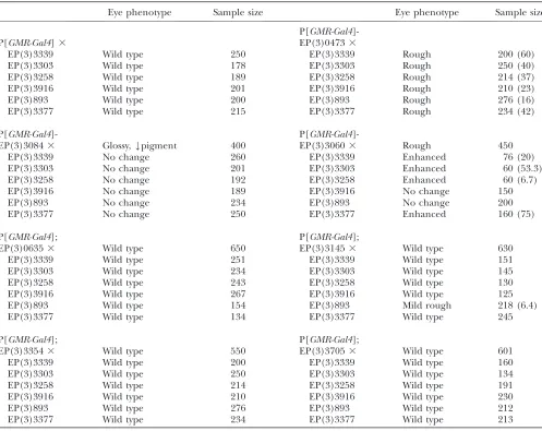
and cell cycle control [EP(3)3705] (BDGP). To test the specificity of the interactions with jing, the enhancers were crossed with six random EP lines, including EP(3) 3339, EP(3)3303, EP(3)3258, EP(3)3916, EP(3)893, and EP(3)3377, and were driven in the eye with GMR-Gal4. Of the enhancers, only EP(3)3060 and EP(3)0473 interacted with multiple EP lines (Table 2; Figure 2, I and J). In addition, EP(3)3145 interacted with EP(3)0893, resulting in mildly rough eyes (Table 2).
Next, we addressed the specificity ofD1andDATR-X
enhancement by testing for interactions with other reg-ulatory genes in the EP collection.DATR-X[EP(3)635] did not interact with other EP elements upstream from genes encoding DNA-binding proteins, including TABLE 2
Random screening ofjinginteracting EP lines
Eye phenotype Sample size Eye phenotype Sample size
P[GMR-Gal4
]-P[GMR-Gal4]3 EP(3)04733
EP(3)3339 Wild type 250 EP(3)3339 Rough 200 (60)
EP(3)3303 Wild type 178 EP(3)3303 Rough 250 (40)
EP(3)3258 Wild type 189 EP(3)3258 Rough 214 (37)
EP(3)3916 Wild type 201 EP(3)3916 Rough 210 (23)
EP(3)893 Wild type 200 EP(3)893 Rough 276 (16)
EP(3)3377 Wild type 215 EP(3)3377 Rough 234 (42)
P[GMR-Gal4]- P[GMR-Gal4
]-EP(3)30843 Glossy,Ypigment 400 EP(3)30603 Rough 450
EP(3)3339 No change 260 EP(3)3339 Enhanced 76 (20)
EP(3)3303 No change 201 EP(3)3303 Enhanced 60 (53.3)
EP(3)3258 No change 192 EP(3)3258 Enhanced 60 (6.7)
EP(3)3916 No change 189 EP(3)3916 No change 150
EP(3)893 No change 234 EP(3)893 No change 200
EP(3)3377 No change 250 EP(3)3377 Enhanced 160 (75)
P[GMR-Gal4]; P[GMR-Gal4];
EP(3)06353 Wild type 650 EP(3)31453 Wild type 630
EP(3)3339 Wild type 251 EP(3)3339 Wild type 151
EP(3)3303 Wild type 234 EP(3)3303 Wild type 145
EP(3)3258 Wild type 243 EP(3)3258 Wild type 130
EP(3)3916 Wild type 267 EP(3)3916 Wild type 125
EP(3)893 Wild type 154 EP(3)893 Mild rough 218 (6.4)
EP(3)3377 Wild type 134 EP(3)3377 Wild type 245
P[GMR-Gal4]; P[GMR-Gal4];
EP(3)33543 Wild type 550 EP(3)37053 Wild type 601
EP(3)3339 Wild type 200 EP(3)3339 Wild type 160
EP(3)3303 Wild type 250 EP(3)3303 Wild type 134
EP(3)3258 Wild type 214 EP(3)3258 Wild type 191
EP(3)3916 Wild type 210 EP(3)3916 Wild type 230
EP(3)893 Wild type 276 EP(3)893 Wild type 212
EP(3)3377 Wild type 234 EP(3)3377 Wild type 213
EP(3)1005 (n¼411) and EP(3)1096 (n¼387). TheP
element in line EP(3)1005 lies upstream from CG15514, which encodes a protein containing a DNA-binding BED finger found in chromatin-boundary-element-binding proteins and transposases (FlyBase) (Aravind
2000). ThePelement in line EP(3)1096 lies upstream of a gene encoding a CXXC zinc finger (FlyBase, CG11033). EP(3)0635 did not interact with EP(3)3058 (n¼200), which lies upstream from thepoly U binding factor 68kD (pUf68) gene (Page-McCaw et al. 1999;
Lasko 2000). However, EP(3)0635 did interact with
EP(3)3205 (35% penetrance, n ¼ 301), which lies upstream of CG7552 encoding a protein with a WW domain and interacts genetically with cyclin E (Tseng
and Hariharan 2002). In contrast, EP(3)0473
inter-acted with EP(3)1005 (48%,n¼205), EP(3)3205 (12%,
n ¼ 150), and EP(3)3058 (83%,n ¼ 256), indicating that it may be a more general transcriptional regulator. Interenhancer interactions: Last, we determined if thejingenhancers interacted with each other in the eye. Eye-specific coexpression of EP(3)3084 with EP(3)0635, EP(3)3354, and EP(3)3145 resulted in more severe Figure2.—Genetic interactions ofjingenhancers. Light level images of adult eyes of the following genotypes are shown: (A) GMR-GAL4 /1; EP(3)3084/1; (B) GMR-GAL4/1; EP(3)3084/1; UAS-jingE/1; (C) GMR-GAL4/1; EP(3)3084/1; (D) EP(3)3377/1; GMR-GAL4/1; EP(3)0635/1; EP(3)3084/1; (E) GMR-GAL4/1; EP(3)3084/1; EP(3)3354/1; (F) GMR
-GAL4/1; EP(3)3084/1; EP(3)3145/1; (G) GMR-GAL4/1; EP(3)3060/1; (H) GMR-GAL4/1; EP(3)3060/1; UAS-jingE/1; (I) GMR-GAL4/1; EP(3)3060/1; EP(3)3377/1; (J) GMR-GAL4/1; EP(3)3060/1; EP(3)3303/1; (K) GMR-GAL4/1; EP(3)3060/1; EP(3)3145/1; (L)GMR-GAL4/1; EP(3)3354/1; (M)GMR-GAL4/1; EP(3)3354/1; EP(3)3303/1; (N)GMR
-GAL4/1; EP(3)0635/1; EP(3)3354/1; (O) GMR-GAL4/1; EP(3)3354/1; EP(3)3145/1; (P) GMR-GAL4/1; EP(3)3145/1; (Q) GMR-GAL4/1; EP(3)3145/1; EP(3)0893/1; (R) GMR-GAL4/1; EP(3)3145/1; EP(3)0635/1; (S) GMR-GAL4/1; EP(3)0635/1. Bars, 50mm.
rough eye phenotypes including losses in pigmentation (Figure 2, D–F; Table 3). Eye-specific expression of EP(3)3060 resulted in rougher eyes when coexpressed with EP(3)3145, EP(3)3354, EP(3)0635, EP(3)0473, and EP(3)3705 (Figure 2K; Table 3). However, the widespread interactions of EP(3)3060 suggest that these results may be considered nonspecific (Table 2; Figure 2, I and J).
Eye-specific coexpression of EP(3)3354 with EP(3)3145 and EP(3)0635 resulted in glossy eyes with severely reduced pigmentation representing synergistic interactions (Table 3; Figure 2, N and O). We have designated EP(3)3354 asjing interacting gene regula-tory 1 ( JIGR1), given its potential role in regulating gene expression (Brodyet al.2002). The EP element in
JIGR1lies upstream of the transcript CG17383, which was identified in a differential head cDNA screen (Brodyet al.2002). JIGR1 contains an MADF domain
shown in the Adf-1 transcription activator to bind DNA specifically to several developmentally regulated Dro-sophila gene promoters (Ewel et al. 1990; England
et al.1992; Cutleret al.1998; Bhaskarand Courey
2002).JIGR1also interacted with EP(3)3705 resulting in a reduced eye size (Table 3).
EP(3)3145 lies upstream ofDrosophila ataxin2(Datx2) and interacted with other jing enhancers from the screen but not with most randomly chosen EP lines. We observed glossy eyes and reduced pigmentation after coexpression of EP(3)3145 with EP(3)635 and
EP(3)3354 (Figure 2, R and O). Collectively, these results identify a group of genes that have related func-tions pertaining specifically tojingand each other dur-ing eye development.
Part of ATR-X is highly conserved: Previous studies showed that ATR-X is involved in gene regulation by chromatin remodeling functions (Cardosoet al.1998;
Xue et al. 2003; Tang et al. 2004). Given the GOF
interactions between DATR-X and jing in the eye, we proposed that there may be a regulatory relationship between DATR-X and Jing in the embryonic CNS and initiated further studies ofDATR-X. Database searches with genomic sequence flanking the EP(3)0635P ele-ment confirmed that the transposon in the strain we had obtained was inserted in the 59-untranslated region of the predicted gene CG4548 encoding the Drosophila homolog of humanXNP/DATR-X(GadFly).
DATR-Xis located at cytological interval 96E1 on the third chromosome (BDGP) and encodes a predicted polypeptide of 1311 amino acids with 49.4% overall similarity to that of human ATR-X by alignment using the Clustal X algorithm (Figure 3A).ATR-Xhomologs are found in human, mouse, C. elegans, planarians, and Drosophila (Figure 3, A and C) (Villardet al.
1999). Most of the sequence conservation between human and DATR-X is confined to the helicase and SNF2 domains. The SNF2 domains of C. elegans, mouse, and human ATR-X show 54.9, 66.4, and 65.8% amino acid identity with that of DATR-X, TABLE 3
Screening ofjinginteracting EP lines
P[GMR-Gal4];
Eye phenotype % penetrance N
P[GMR-Gal4];
Eye phenotype % penetrance N
EP(3)3145 EP(3)0635
EP(3)0635 Glossy,YYpigment 17.9 217 EP(3)3084 YYYpigment 46.1a 256
EP(3)3060 Bumpy,Ypigment 49.0a 300 EP(3)3354 Glossy,YYYpigment 17.2 93
EP(3)3354 Glossy,YYpigment 14.1 85 EP(3)3060 Glossy,YYpigment 88.9a 90
EP(3)3084 Rougher,YYYpigment 30.8a 65 EP(3)3705 Rough 20 200
EP(3)3705 Rough 8.5 94 EP(3)0473 Rough 18.8 223
EP(3)0473 Rough 15.0 167
P[GMR-Gal4];
Eye phenotype % penetrance N
P[GMR-Gal4];
Eye phenotype % penetrance N
EP(3)3084 EP(3)3060
EP(3)3060 No change 0 250 EP(3)3354 Smaller, rougher 40a 70
EP(3)3354 Glossy,YYYpigment 21.4a 234 EP(3)473 Rougher 7.5a 90
EP(3)0473 Rougher 24.9a 201 EP(3)3705 Smaller, rougher 18.7a 75
EP(3)3705 Rougher 20.8a 120
P[GMR-Gal4];
Eye phenotype % penetrance N
EP(3)3354
EP(3)3705 Small 18.4 250
EP(3)0473 Rough 15.6 225
Penetrance is indicated by percentage of the eyes with a phenotype out of the progeny carryingGMR-Gal4and the EP elements.
N, sample sizes. a
respectively (Figure 3A). The ATPase domains of C. elegans, mouse, and human ATR-X show 58.9, 63, and 63% amino acid identity with that ofDATR-X, respec-tively (Figure 3A). ATR-X has nucleosome-stimulated ATPase activity that is used to modify chromatin struc-ture and that is a common site of patient mutations (Gibbons et al. 1995b; Xue et al. 2003; Tang et al.
2004). Mutations associated with MR are in invariant residues and the amino acids at the sites of two human mutations, designated Mu K1600R and Ped23 D2035V, are conserved inDATR-X(Figure 3B).
There are some differences in the amino acid se-quences of vertebrate and invertebrate ATR-X. Human and mouse ATR-X have a zinc-finger DNA-binding do-main consisting of three multicysteine zinc-finger motifs (C2-C2) that is not present in invertebrate ATR-X
pro-teins (Figure 3A) (Cardoso et al. 2000). These zinc
fingers are required for nuclear localization and DNA binding by ATR-X (Cardosoet al.2000). Therefore, it
is possible that invertebrate ATR-X proteins require site-specific DNA targeting to carry out their ATPase activities. The C terminal of ATR-X has a region rich in poly glutamine repeats (Figure 3A) proposed to be involved in protein–protein interactions that also is not present in either D. melanogaster or C. elegans ATR-X (Pickettset al. 1996; data not shown). Sequence
dis-tances betweenATR-Xorthologs in different species are schematized in Figure 3C.
DATR-X andjing-lacZare expressed in CNS glia and neurons:To determine the expression pattern of DATR-X during development, we hybridized whole-mount control embryos with digoxigenin-labeled antisense Figure3.—ATR-X homologs. (A) Comparison ofDATR-Xprotein domain architecture as predicted using SMART and PROSITE. Invertebrate ATR-X is truncated in comparison with mouse and human but shares high identities in the C-terminal amino acids with the latter proteins. Percentage of identity between the SNF2 and helicase C domains ofC. elegans, mouse, and human are in comparison to those ofD. melanogaster. (B) Comparison of amino acid identity over the regions containing mutations found in the ATPase domain of ATR-X from human patients with ATR-X syndrome. The mutations include both Mu K1600R and Ped23 D2035V in highly conserved regions. (C) Phylogenetic analysis of ATR-X orthologs. The distance between any two sequences is the sum of horizontal branch length separating them. Sequences were aligned using CLUSTALW (Tree View). The sequence residues in each column are colored on the basis of an alignment consensus, which is calculated automatically (for detail color information, refer to CLUSTAL W online help). Species designation follows each protein acronym (Dm,Drosophila melanogaster; Ag,Anopheles gambiae; Mm, murine; Hs, human; Ce,Caenorhabditis elegans). Bar indicates the number of substitutions.
and sense DATR-X riboprobes. Ectopic activation of EP(3)0635 was detected inprd-expressing stripes with our antisenseDATR-Xprobe confirming the specificity of the probe and EP line (Figure 4A).DATR-Xtranscripts are present at a low level throughout cellular blastoderm embryos (Figure 4B) and during gastrulation (Figure 4C). In stage 15 embryos,DATR-X transcripts become enriched in the neuroectoderm and supraesophageal ganglion (brain) (Figure 4, D and G). In the mature ventral nerve cord,DATR-XmRNA is expressed in cells along the position of the longitudinal axon tracts (Figure 4G, arrows) and in more laterally located glia (Figure 4G, arrowhead). Similar CNS expression pat-terns were not observed in embryos hybridized with sense DATR-X riboprobes (Figure 4F). DATR-X tran-scripts are also present throughout the brains of third instar wild-type larvae but are predominant in the optic lobe region (Figure 4E).
To better understand the pattern of expression, we costained embryos carrying jing- and DATR-X-lacZ
reporter genes with anti-b-galactosidase and the glial-specific anti-Repo antibody. Analysis by confocal mi-croscopy revealed that jing-lacZ and DATR-X-lacZ are expressed in both glial and neuronal lineages. Many of the DATR-X-lacZ- and jing-lacZ-expressing glia are pre-sent in positions characteristic of the LG (Figure 4, I–R). OtherDATR-X-lacZ- andjing-lacZ-expressing glia occupy more lateral (Figure 4, K and L, arrowheads) or ventral regions (Figure 4, N, O, S).
Pan-neural coexpression of DATR-X and jing syner-gistically disrupts longitudinal axon formation: Since coexpression ofjingandDATR-Xstrongly affected adult neuronal development we wanted to determine if their coexpression had similar affects in embryonic neurons.
jing and DATR-X were expressed in all postmitotic neurons withELAV-GAL4. Expression of either jingor
DATR-Xalone resulted in subtle defects in the CNS axon scaffold as observed with BP102 staining. These defects included thinner longitudinal connectives (Figure 5, B and C, arrowheads) and reduced spacing between the anterior and posterior commissures (Figure 5, B and C, arrows). Additive effects were observed after expression of two copies of either jing (Figure 5E) or DATR-X
(Figure 5F) where commissural axons were not properly separated (Figure 5, E and F, arrows).
Pan-neural coexpression ofjingandDATR-Xresulted in more severe defects in axonal patterning than
expres-sion of one or two copies of either transgene alone. An average of 65% of segments had no longitudinal connectives afterjingandDATR-Xcoexpression (Figure 5D, arrowhead; Figure 5G) in comparison with expression of two copies of UAS-jing(5% of segments,n¼80) and EP(3)0635 (18% of segments, n ¼ 55) (Figure 5G). Therefore, jing and DATR-X coexpression has syn-ergistic effects specifically in embryonic CNS neurons.
Neuronal-specific functions of jing and DATR-X are required for repulsion of longitudinal axons and glia from the CNS midline: We targetedDATR-X and jing
mutations to discern the neuronal and glial contribu-tions of each gene product to axon scaffold formation.
DATR-X and jing expression was knocked down using conditional RNAi (Hammond et al. 2001; Lee and
Carthew2003). A total of 697- and 567-bp sequences
from nonconserved regions of DATR-X and jing were separately subcloned into a P[UAST] derivative plasmid to produce intron-spliced hairpin RNA corresponding to the DATR-X andjing genes, respectively. TheUAS/
Gal4system (Brandand Perrimon1993) was then used
to allow hairpin RNA to conditionally downregulate
DATR-X and jing expression in specific cell lineages, which was confirmed by in situ hybridization. Pene-trance values of CNS axon phenotypes associated with neuronal and glial knockdown of jing and DATR-X
ranged from 24 to 30% (seematerials and methods).
Longitudinal connective formation was analyzed in homozygous jing mutant embryos and those with neuronal-specific knockdown of jing and DATR-X
stained with an antibody to Fasciclin II (1D4). Neuronal specificity was directed by theELAV-Gal4driver. In these mutant embryos, FasII-positive longitudinal axons ab-errantly cross the CNS midline in stage 16 embryos (Figure 6, B–D, arrowheads; Figure 6M). In addition, there are breaks in the longitudinal connectives sug-gesting axon outgrowth defects (Figure 6, C and D, arrows). In embryos with simultaneous neuronal knock-down of jing and DATR-X all FasII-positive lateral fascicles fuse into a single tract at the CNS midline (Figure 6E). These results establish an autonomous neuronal requirement forjingandDATR-Xfunction in the outgrowth and lateral positioning of longitudinal axons and a genetic synergy during this process.
Consistent with these results, pan-neural knockdown ofjingorDATR-Xwas associated with high levels of Robo at the CNS midline during both stage 12 (Figure 6, G
(arrowhead).DATR-Xis expressed most strongly in the ventral region of the ganglion (vg, arrowhead) and optic lobe (ol, arrow). (F)In situhybridization using aDATR-Xnegative control sense probe. (G)DATR-Xexpression in a stage 15 ventral nerve cord (ventral view). Strong expression is observed in cells lining the longitudinal connectives (arrows) and in lateral cells (arrowhead). (H–R) Focal planes ofDATR-Xandjing-lacZexpression in the longitudinal glia (LG) (arrows). (N and O) Sagittal views to show thatDATR-X-lacZis expressed in ventral glia (arrowhead) and the dorsal longitudinal glia (arrow).DATR-X-lacZis also expressed in neurons (double arrowhead). (P–R)jing-lacZis strongly expressed in the two rows of longitudinal glial cells. (S)jing-lacZis also expressed in a bilateral glial lineage (arrow) and in a laterally located lineage (arrowhead) in the ventral focal plane. Bars: (A–F) 200mm; (G) 30mm; (H–S) 50mm.
and H) and stage 16 (Figure 6, J and K). The phenotypes were variable (Figure 6K), as in the most severe case all Robo-positive axons were present at the midline (Figure 6K) and in less severe cases midline Robo was observed in fewer hemisegments (Figure 6L, arrowhead). We also observed breaks in Robo-positive connectives (Figure 6L, arrow). The high Robo levels injing and DATR-X
pan-neural mutants suggest that these genes do not regulate robo expression. However, the medial axon displacement suggests that these mutations may affect
the expression of other genes required for Robo to read the Slit cue. Alternatively,jingandDATR-Xmay regulate longitudinal positioning in a Robo-independent man-ner (Kinradeet al., 2001).
Glial functions of Jing andDATR-Xare required for ipsilateral positioning of longitudinal glia, glial sur-vival, and patterning of longitudinal axons: Genetic and cell ablation experiments have shown the critical role that neuronal–glial communication plays during pioneering of the longitudinal axon tracts (Boothet al.
2000; Hidalgoand Booth2000; Hidalgoet al.2001;
Kinrade et al. 2001; Whitington et al. 2004). We
therefore thought it might be informative to examine whetherjingandDATR-Xplay a role in glial guidance of longitudinal axons. Glial development was examined after pan-glial expression of jing and DATR-X RNAi transgenes under the control ofglial cells missing(gcm
)-Gal4(Hosoyaet al.1995; Joneset al.1995) and using
anti-Repo as a marker to assess glial fates (Campbell
et al.1994; Xionget al.1994; Halteret al.1995).
Robo-positive and Repo-positive LG were already misplaced medially during stage 12 after pan-glial knockdown ofjingandDATR-X(Figure 7, C–F) and in
jing hemizygotes (Figure 7B). Wild-type numbers of glia in these mutants at this stage reveal thatjing and
DATR-Xare involved in glial differentiation and not in the division or specification of these cells (Figure 7O). Later during wild-type embryogenesis Robo is present only in axons (see Figure 6I) and therefore glial move-ment is restricted by Robo-independent mechanisms, including axon contact and trophic support (Kinrade
et al., 2001). In the mature cord, we found that both LG and longitudinal axons were medially misplaced to the CNS midline injinghemizygotes and after glial knock-down ofjingandDATR-X(Figure 7, H–J, M, N). Despite the maintenance of glial–axonal contact, the numbers of total glia steadily decreased during embryogenesis in
jing andDATR-X glial mutants (Figure 7O). However, glial numbers were unaffected after neuronal-specific
Figure 5.—Pan-neural expression ofDATR-X and jing in embryonic CNS neurons synergistically affects axon pattern-ing. Ventral views of the ventral nerve cord (VNC) of stage 15 embryos stained with BP102 (A–F) and shown with ante-rior up. (A) Control embryo heterozygous forelav-Gal4 and
Figure6.—DATR-Xandjingfunctions are required specifically in CNS neurons for longitudinal connective formation.DATR-X andjingRNAi transgene expression was driven in neurons byELAV-Gal4. (A–E and F–L) Shown are close-up views of the VNC of whole-mount stage16 embryos stained with anti-Fasciclin II monoclonal antibody (1D4) (A–E) and anti-Robo (F–L). Stage 12 embryos stained with anti-Robo (F–H). (A) In wild-type embryos, three longitudinal fascicles are clearly delineated. (B–D) In
jing(B and C) andDATR-X(D) mutants, longitudinal fascicles misroute across the midline (arrowheads) and are broken (arrows). (E) After coexpression ofjingandDATR-XRNAi transgenes in neurons, longitudinal fascicles fuse into one tract at the midline. (continued)
jingandDATR-Xknockdown despite a medial misplace-ment and mispositioning of LG (Figure 7, K, L, O). Therefore, jing and DATR-X glial-specific mutations perturb an autonomous survival function that cannot be compensated for by axon contact.
DISCUSSION
A conserved role for ATR-X in the embryonic CNS? Of the candidates from the screen, we chose to further studyDATR-Xdue to a possible involvement in Jing CNS function and disease relevance. Mutations in the human
ATR-X gene are associated with several X-linked MR phenotypes that lead to cognitive delay, facial dysmor-phism, microcephaly, skeletal and genital abnormali-ties, and neonatal hypotonia (Gibbonset al. 1995a,b;
Villardet al. 1996a,b; Gibbons and Higgs 2000). A
total of 87% of MR genes have a fruit fly homolog and 76% have a candidate functional ortholog revealing a remarkable conservation between humans andD. mela-nogaster(Inlowand Restifo2004). Some orthologs of
human MR genes have cellular phenotypes involving neurons, glia, and neural precursor cells and arise from defects in proliferation, migration, and process exten-sion or arborization (Inlow and Restifo 2004). For
example, targeted mutation of ATR-X to the early forebrain in mice leads to cortical progenitor cell death and reduced forebrain size (Be´ rube´ et al. 2005). In
addition, mutations in genes controlling the identity of forebrain neuronal precursors can result in holopro-sencephaly in which the brain hemispheres do not separate (Wallis and Muenke 2000). An increased
understanding of the molecular and cellular bases for hereditary MR is critical for the generation of drug treatments.
ATR-X belongs to the SWI/SNF group of chromatin remodeling proteins, which use the energy provided by ATP hydrolysis to disrupt histone–DNA associations and move nucleosomes to different positions (Kingston
and Narlikar 1999; Whitehouse et al. 1999). This
chromatin modulation allows for the access of activators or repressors to their DNA binding sites in their target genes. The helicase C and SNF2N domains of ATR-X have been shown to have DNA translocase and nucleo-some-remodeling activities (Xueet al.2003; Tanget al.
2004). Accordingly, mutations in ATR-X have been mapped to the helicase C and SNF2N domains, which show60% homology with those inDATR-Xand have been conserved fromC. elegansto humans. This conser-vation supports a conserved role for Drosophila ATR-X in chromatin remodeling (Tanget al.2004; this work).
Vertebrate ATR-X has a C2C2zinc-finger motif in the
amino terminus that is similar to a plant homeodomain finger previously identified in proteins involved in chromatin-mediated transcriptional regulation (Aasland
et al. 1995; Gibbons et al. 1997). Interestingly, D.
mela-nogasterandC. elegansATR-X proteins do not contain the zinc-finger domain, suggesting that these structures may have been acquired through evolution due to a necessity in vertebrate chromatin remodeling mechanisms. Pa-tients have been identified with mutations in the ATPase and zinc-finger domains of ATR-X, confirming that these are essential functional regions of the protein (Villard
et al.1996b,c; Gibbonsand Higgs2000).
Given the absence of the zinc-finger domains in
DATR-X, we postulate that invertebrateDATR-Xproteins may be complexed with proteins containing a nuclear targeting and DNA-binding motif to regulate gene expression at the proper regulatory sites. This may be one role of Jing since it has an embryonic expression pattern as well as mutant and overexpression pheno-types very similar to those of DATR-X. Therefore, it seems that the ATPase domain of DATR-X has been conserved through evolution and that the other regions of the protein may have evolved to suit the specific needs of the cell. In summary, different mechanisms of ATR-X function and different binding partners across species may account for the divergence of sequence with respect to the amino terminal and Q-rich repeats while the main chromatin remodeling aspects of ATR-X remain similar.
Jing encodes a nuclear protein with putative DNA-binding and transcriptional regulatory domains (Liu
and Montell2001; Sedaghatet al.2002). The C2H2
zinc fingers of Jing are most similar (50% identical) to those of the mouse adipocyte enhancer binding protein 2 (AEBP2) and also show 25% homology to those of the Kru¨ppel family of transcription factors, including those encodinggliandZIC2(Liuand Montell2001). AEBP2
function is implicated in chromatin remodeling events (Cardosoet al. 1998; Cao and Zhang 2004) and has
strong expression in the brain (Heet al.1999).
Genetic screening identifies a related group of jing -interacting genes: We have utilized a background sensitive to jing function to conduct a genetic screen in the eye. For the GOF screen, we hypothesized that misexpression of jing in the eye in combination with other genes involved in jing transcriptional regula-tion would lead to alteraregula-tions in gene expression and consequently disrupt ommatidial formation. The genetic relationship between DATR-X and jing in em-bryonic neurons and glia shows that the screen was
successful in identifying genes whose function in adult neuronal cells is relevant to jing function in the embryonic CNS. This is consistent with previous find-ings that the fly eye is a valid system for targeting genes that function in other tissues (Raymondet al.2004).
EP(3)3084 contains a transposon in proximity to a novel gene known by its FlyBase transcript identifier as CG15507. Despite strong effects of EP(3)3084 expres-sion in the eye these were specifically strongly enhanced after coexpression withjing,DAtx2, andJIGR1. Further-more, each gene specifically interacted with each other but not with randomly chosen EP lines, suggesting a functional relationship between the four genes. The EP elements in these lines are located in the 59untranslated region of the downstream genes, suggesting they may result in overexpression (BDGP). Given the regulatory role of MADF domains, it is possible that JIGR1 regu-lates gene expression with Jing andDATR-X(England
et al.1992; Bhaskarand Courey 2002). Alternatively,
JIGR1 may regulate the expression of a Jing/DATR-X target gene. Likewise, DAtx2 may be involved in regu-lating the translation of a protein that is an essential component of a Jing/DATR-X/JIGR1 complex or a down-stream target of these genes (Satterfield et al.2002;
Ciosket al. 2004). A role for the orthologs of
transla-tional regulators in MR has been shown for the Dro-sophila ortholog of fragile-X MR 1 (Dfmr1).Dfmr1regulates the MAP1B homolog of Futsch to control synaptic structure and function in the embryonic Drosophila CNS (Zhanget al.2001). Therefore, genetic screening
and phenotypic analysis in Drosophila have the power to decipher pathways and the cellular bases of MR genes.
Neuronal and glial functions of jing and DATR-X
dictate axon tract formation: In wild-type Drosophila embryos, LG assume characteristic positions and do not cross the midline or into adjacent VNC segments (Ito
et al.1995). This is due to multiple mechanisms at dif-ferent stages of development, including response to re-pulsive and attractive molecules, cell–cell contact, trophic support, and axon contact (Kinradeet al., 2001). A
dis-ruption in any of these processes will perturb formation of the glial and axonal scaffolds. The expression ofjing
andDATR-Xreporter genes in LG correlates with the LG phenotypes associated with mutations in these genes.
During stage 12, Robo present on the LG responds to repulsive midline Slit molecules to maintain lateral positioning (Kinradeet al.2001). The medial
misplace-ment of Robo- and Repo-positive LG during stage 12 after
jingandDATR-X glial-specific knockdown suggests that there may have been a breakdown in Robo-dependent repulsive mechanisms. However, the fact that Robo protein was present after jing and DATR-X glial and neuronal knockdown suggests thatroboexpression may not be regulated by Jing andDATR-X. Alternatively, Jing andDATR-Xmay regulate the expression of a factor that controls how Robo reads the Slit signal. In support, misrouting of axons across the midline in the presence of Robo occurs in calmodulin andSon of sevenless mutants where these proteins are required to process the Sli signal (Fritz and VanBerkum2000). It is also possible
thatjingandDATR-Xregulate the expression of factors controlling glial and neuronal positioning in a Robo-independent fashion (Kinradeet al.2001).
jingandDATR-X mutations clearly affect more than Robo-mediated LG positioning. First, glial survival is not affected in robomutant embryos, whereas glia die despite continuous axonal contact injingandDATR-X
glial-specific mutants. Therefore, the loss of CNS glia may reflect a breakdown in an intrinsic survival pathway mediated by jing and DATR-X. The expression of jing
and DATR-X reporter genes in glia is consistent with such a role. Furthermore, bothjingandDATR-X/ATR-X
have been implicated in cell survival processes in the CNS midline and tracheal cells and in cortical progen-itors, respectively (Sedaghatet al. 2002; Sonnenfeld
et al.2004; Be´ rube´et al.2005).
Second, in robomutants only the central pCC/MP2 fascicle, but not the outer two longitudinal fascicles, is affected. However, injingandDATR-Xglial and neuronal mutants the outer fascicles are fused, often broken, and can be seen crossing the midline. These defects are simi-lar to those after ablation of neurons or glia and after genetic loss of glia as ingcmmutants (Hosoyaet al.1995;
Joneset al.1995; Vincentet al.1996; Hidalgoand Brand
1997). These observations suggest that multiple biological processes require the proper function of these genes and are consistent with an important upstream role for jing
andDATR-Xin glial and neuronal differentiation.
whole-mount embryos stained with anti-Repo. (E, F, M, and N) Embryos stained with anti-Robo. Shown are close-up ventral views of the nerve cord of stage 12/3 (A–F) and stage 15 (G–N) embryos. (A) In wild-type stage 12/3 embryos, longitudinal glia (LG) have already migrated medially to their positions to guide pioneer longitudinal axons. (B–D) In contrast, LG inappropriately occupy positions at the CNS midline in hemizygousjingembryos (B) and in those with glial-specific knockdown of jing(C) andDATR-X (D) as driven by gcm-Gal4. (E and F) Robo-positive glia are misplaced medially after expression ofjing(E) and
DATR-X(F) RNAi transgenes in CNS glia. (G) In wild-type embryos, the LG occupy a two-cell wide row lining the longitudinal connectives (arrows). The LG are observed only in the dorsal plane of view. (H–J) Injinghemizygotes (H) and injing(I) and
Evidence is accumulating that chromatin accessibility plays a key role in the transcriptional regulation of cell-type-specific gene expression in the CNS (Hsieh et al.
2004). The conservation in ATPase domains along with the similar phenotype ofDATR-X andjing muta-tions and in their expression patterns raises the possi-bility that Jing is involved in the targeting of a chromatin remodeling complex containing DATR-X to transcrip-tional target genes whose products are required for the response of longitudinal growth cones and glia to guidance cues.
In summary, we have identified a group of genes that pertain to jing function and specifically genetically interact in adult neuronal cells. Our results show that specific neural and glial developmental defects underlie the problems in axon guidance associated with muta-tions inDATR-X andjing. More studies using targeted mutations of MR genes will alleviate the view that brain phenotypes result from generic effects due to a height-ened sensitivity of the brain.
We thank Marc Freeman forgcm-gal4 and S. Hayashi forbtl-gal4. We thank Doug Holmyard at the Bioimaging Centre at Mount Sinai Hos-pital for performing SEM. We thank Helga Agah for help with immu-nohistochemistry. We thank Andrew Ridsdale and the OGHRI for use of the confocal microscope and training. This work was supported by a grant from the Canadian Institutes of Health Research to M.S.
LITERATURE CITED
Aasland, R., T. J. Gibsonand A. F. Stewart, 1995 The PHD finger:
implications for chromatin-mediated transcriptional regulation. TIBS20:56–59.
Abidi, F., N. J. Carpenter, L. Villard, M. Curtis, M. Fonteset al.,
1999 The Carpenter-Waziri syndrome results from a mutation inDATR-X. Am. J. Med. Genet.85:249–251.
Adams, M. D., S. E. Celniker, R. A. Holt, C. A. Evans, J. D. Gocayne
et al., 2000 The genome sequence ofDrosophila melanogaster.
Science5461:2185–2195.
Ahmad, K., and S. Henikoff, 2001 Modulation of a transcription
factor counteracts heterochromatic gene silencing inDrosophila.
Cell104:839–847.
Aravind, L., 2000 The BED finger, a novel DNA-binding domain
in chromatin- boundary-element-binding proteins and transpo-sases. Trends Biochem. Sci.25:421–423.
Ashburner, M., 1989 Drosophila:A Laboratory Manual.Cold Spring
Harbor Laboratory Press, Cold Spring Harbor, NY.
Ashley, C. T., C. G. Pendleton, W. W. Jennings, A. Saxenaand
C. V. Glover, 1989 Isolation and sequencing of cDNA clones
en-codingDrosophilachromosomal protein D1. A repeating motif in proteins which recognize at DNA. J. Biol. Chem.264:8394–8401. Bate, C. M., and E. B. Grunewald, 1981 Embryogenesis of an
in-sect nervous system II: a second class of neuron precursor cells and the origin of the intersegmental connectives. J. Embryol. Exp. Morphol.61:317–330.
Barresi, M. J. F., L. D. Hutson, C-B. Chienand R. O. Karlstrom,
2005 Hedgehog regulated Slit expression determines commis-sure and glial cell position in the zebrafish forebrain. Develop-ment132:3643–3656.
Battye, R., A. Stevensand J. R. Jacobs, 1999 Axon repulsion from
the midline of theDrosophilaCNS requires slit function. Develop-ment126:2475–2481.
Be´ rube´, N. G., M. Mangelsdorf, M. Jagla, J. Vanderluit, D. Garrick
et al., 2005 The chromatin-remodeling protein ATRX is critical for neuronal survival during corticogenesis. J. Clin. Invest.115:258–267. Bhaskar, V., and A. J. Courey, 2002 The MADF-BESS domain
fac-tor Dip3 potentiates synergistic activation by Dorsal and Twist. Gene299:173–184.
Bhat, K. M., 2005 Slit-Roundabout signaling neutralizes
netrin--frazzled-mediated attractant cue to specify the lateral positioning of longitudinal axon pathways. Genetics170:149–159. Booth, G. E., E. F. Kinradeand A. Hidalgo, 2000 Glia maintain
follower neuron survival during Drosophila CNS development. Development127:237–244.
Brand, A. H., and N. Perrimon, 1993 Targeted gene expression as a
means of altering cell fates and generating dominant pheno-types. Development118:401–415.
Brody, T., C. Stivers, J. Nagleand W. F. Odenwald, 2002 Iden
tification of novelDrosophilaneural precursor genes using a dif-ferential embryonic head cDNA screen. Mech. Dev.113:41–59. Campbell, G., H. Goring, T. Lin, E. Spana, S. Anderson et al.,
1994 RK2, a glila-specific homeodomain protein required for embryonic nerve cord condensation and viability inDrosophila.
Development120:2957–2966.
Cao, R., and Y. Zhang, 2004 SUZ12 is required for both the histone
methyltransferase activity and the silencing function of the EED-EZH2 complex. Mol. Cell15(1): 57–67.
Cardoso, C., S. Timsit, L. Villard, M. Khrestchatisky, M. Fontes
et al., 1998 Specific interaction between the ATR-X gene product and the SET domain of the human EZH2 protein. Hum. Mol. Genet.7:679–684.
Cardoso, C., Y. Lutz, C. Mignon, E. Compe, D. Depetris et al.,
2000 ATR-X mutations cause impaired nuclear location and altered DNA binding properties of the ATR-X protein. J. Med. Genet.37:746–751.
Ciosk, R., M. DePalmaand J. R. Priess, 2004 ATX-2, theC. elegans
ortholog of ataxin 2, functions in translational regulation in the germline. Development131:4831–4841.
Cutler, G., K. M. Perryand R. Tjian, 1998 Adf-1 is a nonmodular
transcription factor that contains a TAF-binding Myb-like motif. Mol. Cell Biol.18:2252–2261.
Dearborn, R., Jr., and S. Kunes, 2004 An axon scaffold induced by
retinal axons directs glia to destinations in theDrosophilaoptic lobe. Development131:2291–2303.
England, B. P., A. Admonand R. Tjian, 1992 Cloning of the
Dro-sophilatranscription factor Adf-1 reveals homology to Myb onco-proteins. Proc. Natl. Acad. Sci. USA89:683–687.
Ewel, A., J. R. Jacksonand C. Benyajati, 1990 Alternative
DNA-protein interactions in variable-length internucleosomal regions associated withDrosophila Adhdistal promoter expression. Nucleic Acids Res.18:1771–1781.
Freeman, M. R., J. Delrow, J. Kim, E. Johnson and C. Q. Doe,
2003 Unwrapping glial biology: Gcm target genes regulating glial development, diversification and function. Neuron38:567–580. Fritz, J. L., and M. F. VanBerkum, 2000 Calmodulin and son of
sev-enless dependent signaling pathways regulate midline crossing of axons in the Drosophila CNS. Development27(9): 1991–2000. Gibbons, R. J., and D. R. Higgs, 2000 Molecular-clinical spectrum
of the ATR-X syndrome. Am. J. Med. Genet.97:204–212. Gibbons, R. J., L. Brueton, V. J. J. BuckleBurn, J. Clayton-Smith,
B. C. C. Davisonet al., 1995a The clinical and hematological
features of the X-linked a-thalassemia/mental retardation syn-drome (ATR-X). Am. J. Med. Genet.55:288–299.
Gibbons, R. J., D. J. Picketts, L. Villard and D. R. Higgs,
1995b Mutations in a putative global transcriptional regulator cause X-linked mental retardation with a-thalassemia (ATR-X syndrome). Cell80:837–845.
Gibbons, R. J., S. Bachoo, D. J. Picketts, S. Aftimos, B. Asenbauer
et al., 1997 Mutations in transcriptional regulator ATRX estab-lish the functional significance of a PHD-like domain. Nat. Genet.
17:146–148.
Halter, D. A., J. Urban, C. Rickert, S. S. Ner, K. Ito et al.,
1995 The homeobox generepois required for the differentia-tion and maintenance of glia funcdifferentia-tion in the embryonic nervous system ofDrosophila.Development121:317–332.
Hammond, S. M., A. A. Caudyand G. J. Hannon, 2001
Post-tran-scriptional gene silencing by double-stranded RNA. Nat. Rev. Genet. 2(2): 110–119.
Hay, B. A., T. Wolffand G.M. Rubin, 1994 Expression of baculovirus
P35 prevents cell death inDrosophila.Development120:2121–2129. He, G.-P., S. Kimand H.-S. Ro, 1999 Cloning and characterization of
a novel zinc finger transcriptional repressor. J. Biol. Chem.274:
14678–14684.
Hidalgo, A., and A. H. Brand, 1997 Targeted neuronal ablation:
the role of pioneer neurons in guidance and fasciculation in the CNS ofDrosophila.Development124:3253–3262.
Hidalgo, A., and G. Booth, 2000 Glia dictate trajectories of
pio-neer neurons in the Drosophilaembryonic CNS. Development
127:393–402.
Hidalgo, A., E. F Kinradeand M. Georgiou, 2001 TheDrosophila
neuregulin vein maintains glial survival during axon guidance in the CNS. Dev. Cell1:679–690.
Hosoya, T., K. Takizawa, K. Nittaand Y. Hotta, 1995 Glial cells
missing: a binary switch between neuronal and glial determina-tion inDrosophila.Cell82:1025–1036.
Hsieh, J., K. Nakashima, T. Kuwabara, E. Mejiaand F. H. Gage,
2004 Histone deacetylase inhibition-mediated neuronal differ-entiation of multipotent adult neural progenitor cells. Proc. Natl. Acad. Sci. USA101:16659–16664.
Huang, Y., S. J. Myersand R. Dingledine, 1999 Transcriptional
re-pression by REST: recruitment of Sin3A and histone deacetylase to neuronal genes. Nat. Neurosci.2(10): 867–872.
Hulo, N., C. J. A. Sigrist, V. LeSaux, P. S. Langendijk-Genevaux, L.
Bordoliet al., 2004 Recent improvements to the PROSITE
database. Nucleic Acids Res.32:D134–D137.
Inlow, J. K., and L. L. Restifo, 2004 Molecular and comparative
genetics of mental retardation. Genetics166:835–881. Ito, K., J. Urbanand G. M. Technau, 1995 Distribution,
classifica-tion and development ofDrosophilaglial cells in the late embry-onic and early larval ventral nerve cord. Roux’s Arch. Dev. Biol.
204:284–307.
Jacobs, J. R., and C. S. Goodman, 1989 Embryonic development of
axon pathways in the DrosophilaCNS. II. Behavior of pioneer growth cones. J. Neurosci.9:2402–2411.
Janody, F., J. Reischl and N. Dostatni, 2000 Persistence of
Hunchback in the terminal region of theDrosophilablastoderm embryo impairs anterior development. Dev.127:1573–1582. Jones, B. W., R. D. Fetter, G. Tearand C. S. Goodman, 1995 Glial
cells missing: a genetic switch that controls glial versus neuronal fate. Cell82:1013–1023.
Keleman, K., S. Rajagopalan, D. Cleppien, D. Teis, K. Palhaet al.,
2002 Comm sorts Robo to control axon guidance at the Dro-sophilamidline. Cell110:415–427.
Keleman, K., C. Ribeiroand B. J. Dickson, 2005 Comm function in
commissural axon guidance: cell-autonomous sorting of Robo in vivo. Nat. Neurosci.8:156–163.
Kidd, T., K. Brose, K. J. Mitchell, R. D. Fetter, M. Tessier-Lavigne
et al., 1998a Roundabout controls axon crossing of the CNS midline and defines a novel subfamily of evolutionary conserved guidance receptors. Cell92:205–215.
Kidd, T., C. Russell, C. S. Goodmanand G. Tear, 1998b
Dosage-sensitive and complementary functions of roundabout and commis-sureless control axon crossing of the CNS midline. Neuron20:25–33. Kidd, T., K. S. Blandand C. S. Goodman, 1999 Slit is the midline
repellent for the Robo receptor inDrosophila.Cell96:785–794. Kingston, R. E., and G. J. Narlikar, 1999 ATP-dependent
remod-eling and acetylation as regulators of chromatin fluidity. Genes Dev.13:2339–2352.
Kinrade, E. F. V., T. Brates, G. Tear, and A. Hidalgo, 2001 Round
about signaling, cell contact and trophic support confine longi-tudinal glia and axons in theDrosophilaCNS. Development128:
207–216.
Lasko, P., 2000 TheDrosophilamelanogaster genome: translation
factors and RNA binding proteins. J. Cell Biol.150:F51–F56. Lee, Y. S., and R. W. Carthew, 2003 Making a better RNAi vector for
Drosophila: use of intron spacers. Methods30:322–329. Letunic, I., R. R. Copley, S. Schmidt, F. D. Ciccarelli, T. Doerks
et al., 2004 SMART 4.0: towards genomic data integration. Nu-cleic Acids Res.32:D142–D144.
Liu, Y., and D. J. Montell, 2001 jing: a downstream target ofslbo
required for developmental control of border cell migration. Development128:321–330.
Long, H., C. Sabatier, L. Ma, A. Plump, W. Yuanet al., 2004
Con-served roles for Slit and Robo proteins in midline commissural axon guidance. Neuron42:213–223.
Oland, L. A., and L. P. Tolbert, 2002 Key interactions between
neurons and glial cells during neural development in insects. Annu. Rev. Entomol.48:89–110.
Page-McCaw, P. S., K. Amonlirdvimanand P. A. Sharp, 1999 PUF60:
a novel U2AF65-related splicing activity. RNA5:1548–1560. Pena-Rangel, M. T., I. Rodriguez and J. R. Riesgo-Escovar,
2002 A misexpression study examining dorsal thorax formation inDrosophila melanogaster.Genetics160:1035–1050.
Picketts, D. J., D. R. Higgs, S. Bachoo, D. J. Blake, O. W. J.
Quarrellet al., 1996 ATRXencodes a novel member of the
SNF2 family of proteins: mutations point to a common mecha-nism underlying the ATR-X syndrome. Hum. Mol. Genet. 5:
1899–1907.
Rajagopalan, S., E. Nicolas, V. Vivancos, J. Bergerand B. J. D ick-son, 2000a Crossing the midline. Roles and regulation of Robo
receptors. Neuron28:767–777.
Rajagopalan, S., V. Vivancos, E. Nicolas and B. J. Dickson,
2000b Selecting a longitudinal pathway. Robo receptors specify the lateral position of axons in theDrosophilaCNS. Cell 103:
1033–1045.
Rasband, K., M. Hardyand C. B. Chien, 2003 Generating X:
for-mation of the optic chiasm. Neuron39:885–888.
Raymond, K., E. Bergeret, A. Avet-Rochex, R. Griffin-Sheaand
M.-O. Fauvarque,2004 A screen for modifiers of RacGAP(84C)
gain-of-function in theDrosophilaeye revealed the LIM kinase Cdi/TESK1 as a downstream effector of Rac1 during spermato-genesis. J. Cell Sci.117:2777–2789.
Robinow, S., and K. White, 1988 The locusELAVofDrosophila
mel-anogasteris expressed in neurons at all developmental stages. Dev. Biol.126:294–303.
Rodriguez-Alfageme, G. T., G. T. Rudkinand L. Cohen, 1980 Isolation,
properties and cellular distribution of D1, a chromosomal protein of
Drosophila.Chromosoma78:1–31.
Rørth, P., 1996 A modular misexpression screen in Drosophila
detecting tissue- specific phenotypes. Proc. Natl. Acad. Sci. USA93:12418–12422.
Rubin, G. M., 1988 Drosophila melanogasteras an experimental
organ-ism. Science240:1453–1459.
Rubin, G. M., and A. C. Spradling, 1983 Vectors for P element-mediated
gene transfer inDrosophila.Nucleic Acids Res.11:6341–6351. Rubin, G. M., L. Hong, P. Brokstein, M. Evans-Holm, E. Friseet al.,
2000 ADrosophilacomplementary DNA resource. Science5461:
2222–2224.
Satterfield, T. F., S. M. Jacksonand L. J. Pallanck, 2002 A
Dro-sophila homolog of the polyglutamine disease geneSCA2 is a dosage-sensitive regulator of actin filament formation. Genetics
162:1687–1702.
Sedaghat, Y., W. Mirandaand M. Sonnenfeld, 2002 The jing Zn
finger transcription factor is a mediator of cellular differentiation in theDrosophilaCNS midline and trachea. Development 129:
2591–2606.
Seeger, M., G. Tear, D. Ferres-Marco and C. S. Goodman,
1993 Mutations affecting growth cone guidance inDrosophila: genes necessary for guidance toward or away from the midline. Neuron10:409–426.
Shiga, Y., M. Tanada-Matakatsuand S. A. Hayashi, 1996 Nuclear
b-galactosidase fusion protein as a marker of morphogenesis in livingDrosophila.Dev. Growth Differ.38:99–106.
Simpson, J. H., K. S. Bland, R. D. Fetter and C. S. Goodman,
2000a Short-range and long-range guidance by Slit and its Robo receptors: a combinatorial code of Robo receptors controls lateral position. Cell103(7): 1019–1032.
Simpson, J. H., T. Kidd, K. S. Blandand C. S. Goodman, 2000b
Short-range and long-Short-range guidance by Slit and its Robo receptors: Robo and Robo2 play distinct roles in midline guidance. Neuron28:
753–766.
Sonnenfeld, M. J., N. Barazesh, Y. Sedaghatand C. Fan, 2004 The
jingandras1pathways are functionally related during CNS midline and tracheal development. Mech. Dev.121:1531–1547. Tang, J., S. Wu, H. Liu, R. Stratt, O. G. Baraket al., 2004 A novel
transcription regulatory complex containing death domain-associated protein and the ATR-X syndrome protein. J. Biol. Chem.279:20369–20377.
Tatusova, T. A., and T. L. Madden, 1999 Blast 2 sequences-a new
tool for comparing protein and nucleotide sequences. FEMS Microbiol. Lett.174:247–250.
Tear, G., R. Harris, S. Sutaria, K. Kilomanski, C. S. Goodmanet al.,