©
DOI: 10.1534/genetics.104.039727
Identification of Zebrafish Insertional Mutants With Defects in
Visual System Development and Function
Jeffrey M. Gross,*
,1Brian D. Perkins,*
,2Adam Amsterdam,
†Ana Egan
˜a,* Tristan Darland,*
Jonathan I. Matsui,* Salvatore Sciascia,* Nancy Hopkins
†and John E. Dowling*
*Department of Molecular and Cellular Biology, Harvard University, Cambridge, Massachusetts 02138 and†Center for Cancer Research and Department of Biology, Massachusetts Institute of Technology, Cambridge, Massachusetts 02139
Manuscript received December 13, 2004 Accepted for publication January 21, 2005
ABSTRACT
Genetic analysis in zebrafish has been instrumental in identifying genes necessary for visual system develop-ment and function. Recently, a large-scale retroviral insertional mutagenesis screen, in which 315 different genes were mutated, that resulted in obvious phenotypic defects by 5 days postfertilization was completed. That the disrupted gene has been identified in each of these mutants provides unique resource through which the formation, function, or physiology of individual organ systems can be studied. To that end, a screen for visual system mutants was performed on 250 of the mutants in this collection, examining each of them histologically for morphological defects in the eye and behaviorally for overall visual system function. Forty loci whose disruption resulted in defects in eye development and/or visual function were identified. The mutants have been divided into the following phenotypic classes that show defects in: (1) morphogenesis, (2) growth and central retinal development, (3) the peripheral marginal zone, (4) retinal lamination, (5) the photoreceptor cell layer, (6) the retinal pigment epithelium, (7) the lens, (8) retinal containment, and (9) behavior. The affected genes in these mutants highlight a diverse set of proteins necessary for the development, maintenance, and function of the vertebrate visual system.
T
HE zebrafish has been an important model through apparent by the 18–19 SS. The first postmitotic neurons of the retina are generated at 28 hr postfertilization which genes necessary for visual systemdevelop-ment and function have been identified (reviewed in (hpf) and by 72 hpf the retina is functional (Easter andNicola1996;HuandEaster1999;Schmittand EasterandMalicki2002 andNeuhauss2003).
Zebra-fish eyes are large, easily accessible, and structurally Dowling 1999). Retinas of many fish and amphibians also possess a specialized region at their margins, termed similar to the human eye. Eye formation in zebrafish is
analogous to that observed in other vertebrate embryos, peripheral or ciliary marginal zones, that perpetually adds cells to the retina during the lifetime of the animal thus providing an excellent model system with which
the understanding of vertebrate eye development can (Johns1977).
Several generations of chemically based forward ge-be advanced. Additionally, many disrupted genes and
pathways identified as integral to the formation of the netic screens have been undertaken in zebrafish (Driever
et al.1996;Haffteret al.1996;MatsudaandMishina
zebrafish eye produce phenotypes that resemble
disor-ders of the human visual system. Thus, characterization 2004), some of which have focused on eye development and function (Malicki et al.1996;Fadoolet al.1997; of the molecular mechanisms of eye development in
zebra-fish should facilitate a better understanding of these hu- Neuhauss et al. 1999). While these chemically based screens have been instrumental in generating interesting man pathologies (GoldsmithandHarris2003).
Eye development in zebrafish first becomes morpho- mutant phenotypes, positional cloning of these mutations is still quite laborious, despite the genomic advances of logically obvious at the 6 somite stage (SS), the time at
which the optic lobes evaginate from the diencephalon the last few years. Retrovirus-mediated insertional muta-(Schmitt andDowling 1994). Thereafter, eye devel- genesis provides an attractive alternative to chemical opment proceeds rapidly with lens induction occurring mutagenesis techniques since the affected gene can be around the 14–15 SS and morphological distinction be- rapidly identified using the proviral insert as a molecular tween the retina and retinal pigment epithelium (RPE) tag to localize the site of insertion in the genome and thereby to identify the mutated gene (Gaiano et al. 1996; Amsterdam et al. 1999; Amsterdam 2003).
In-1Corresponding author:Department of Molecular and Cellular
Biol-deed, a large-scale insertional mutagenesis screen
per-ogy, Harvard University, 16 Divinity Ave., Cambridge, MA 02138.
formed over the last 6 years has generated⬎500
inser-E-mail: [email protected]
tional mutants of which 315 different affected loci have
2Present address:Department of Biology, Texas A&M University,
Col-lege Station, TX 77843. been identified (Gollinget al.2002;Amsterdamet al.
1⫻30 min and incubated in secondary antibody, diluted in 2004). This collection of mutants presents a wealth of
5% NGS, for 90 min at RT. Slides were washed 3⫻15 min possible analyses since studies are not limited to gross
in PBS at RT and mounted in Vectashield mounting medium phenotypic characterizations ; one can target for study containing DAPI (Vector Laboratories, Burlingame, CA). The specific physiological processes and biochemical path- following antibodies and dilutions were used: 1d1 (1:30), 5e11 (1:100), zn5 (1:100), and goat anti-mouse Cy3 secondary ways in which the mutated gene’s protein product would
(1:500). Imaging was performed in a Zeiss 510 laser scanning normally function.
confocal microscope. A total of 3–5 optical sections (1m in It is estimated that this collection of 315 insertional
thickness) were collected and projected using Zeiss confocal mutants representsⵑ22% of the vertebrate gene com- software.
plement that can be mutated to result in a visible embry- Optokinetic response assay :Optokinetic response (OKR) assays were performed afterBrockerhoff et al.(1995). For onic phenotype (Amsterdam et al. 2004). With that
testing, two to four embryos at a time were immobilized in a utility in mind, a shelf screen was performed on 250
small Petri dish containing 5% methylcellulose. The Petri dish different insertional loci for which the affected genes
was positioned in the center of a drum lined with vertical have been cloned to identify those possessing defects black and white stripes, each 1 cm wide. The drum was illumi-in the development and function of the visual system. nated with a tungsten light source attenuated by up to 3.5 log units with neutral density filters. The drum rotated atⵑ8 rpm Forty mutations were identified that affected the visual
and, during 30-sec trials, the direction of rotation was changed system and this article reports the identification and
four to six times. Beginning at full light intensity, embryos initial characterization of each of these mutants.
were tested to see if they could respond to the moving stripes. A response was defined as the demonstration of either a smooth pursuit-saccade cycle or eye tracking movements in both the MATERIALS AND METHODS counterclockwise and the clockwise directions depending on
the rotation of the drum.
Animals:The methods for the generation and identification
Electroretinography :Isolated whole-eye electroretinograms of insertional mutants have been reported previously in
Gai-(ERGs) were obtained using methods described inKainzet
anoet al.(1996) andAmsterdamet al.(1999). Embryos were
al.(2003). Briefly, 5 days postfertilization (dpf), light-adapted obtained from the natural spawning of heterozygous carriers
larvae were placed onto filter paper moistened with Mangel’s setup in pairwise crosses. Embryos were collected and raised
Ringer solution with 20 mm dextrose (pH 7.8). A pair of at 28.5⬚afterWesterfield(1995) and were staged according
forceps held the animal and a small loop made of tungsten toKimmelet al.(1995).
wire gently removed the eye, which was placed onto the filter
Histology:Mutants and wild-type siblings were collected and
paper, cornea side up. A pulled glass micropipette (tip 10m fixed overnight at 4⬚in a solution of 1% (w/v)
paraformalde-in diameter) contaparaformalde-inparaformalde-ing Rparaformalde-inger solution and a chloride-coated hyde, 2.5% glutaraldehyde, and 3% sucrose in PBS. They were
silver wire was inserted into the eye at the border of the washed 3⫻5 min in PBS and refixed for 90 min at 4⬚in a
marginal zone and the lens. A ground wire was placed under 2% OsO4 solution, washed 3 ⫻ 5 min in PBS at RT, and
the moistened filter paper within the recording chamber. The dehydrated through a graded ethanol series (50, 70, 80, 90,
eye was bathed in Ringers throughout the course of a re-2⫻100%). Embryos were further dehydrated 2⫻10 min in
cording session, which lasted 30–75 min. ERGs were recorded propylene oxide and infiltrated 1–2 hr in a 50% propylene
at 24⬚–25⬚. Responses were amplified by a Dagan Cornerstone oxide/50% Epon/Araldite mixture (Polysciences). Embryos
amplifier bandpass filtered (0.1–100 Hz), total gainⵑ10,000, were then incubated overnight at RT in 100% Epon/Araldite
and collected using PClamp software (Axon Instruments, Bur-resin with caps open to allow for propylene oxide evaporation
lingame, CA). A 1409 W/cm2background light attenuated by and resin infiltration, embedded and baked at 60⬚ for 2–3
a⫺1.6 log unit ND filter was used. The stimulus was produced days. Sections 1–1.25m were cut, mounted on glass slides,
by a tungsten halogen light, 9503W/cm2unattenuated inten-and stained in a 1% methylene blue/1% borax solution.
Sec-sity and was adjusted with neutral denSec-sity filters. The duration tions were mounted in DPX (Electron Microscopy Sciences,
of the stimulus was 1000 msec, while the interstimulus time Fort Washington, PA) and photographed on a Leica DMRB
was 15 sec. microscope mounted with a QImaging Retiga EXi digital
cam-era. Images were subsequently processed using Adobe Pho-toshop 5.0.
RESULTS
Acridine orange staining:Acridine orange (Molecular Probes,
Eugene, OR) was diluted in fish water to a final concentration Insertional mutagenesis screen:The generation, screen-of 1g/ml. Embryos were placed in this solution for 10 min,
ing, and cataloging of zebrafish insertional mutants have washed briefly five times in fish water, and immediately
ob-been described elsewhere (Gaianoet al.1996; Amster-served under GFP optics on a Leica dissecting scope.
Immunohistochemistry:Mutants and wild-type siblings were damet al.1999, 2004;Gollinget al.2002). In this study, collected and fixed overnight at 4⬚in a solution of 4% para- 250 different insertional mutants were screened for de-formaldehyde in PBS. Embryos were washed at RT 3⫻5 min fects in eye morphology and for deficits in visual behav-in PBS and then behav-infiltrated by 35% sucrose for 1–2 hr at RT.
ior. Figure 1 provides a general summary of the screen Embryos were then arranged and embedded in plastic molds
and its results. The morphology screen sought to iden-containing TBS cryopreservation media (Triangle Biomedical
Sciences, Durham, NC). Cryosections 12m in thickness were tify mutants with abnormal development or mainte-cut on a Leica CM1900 cryostat and adhered to gelatin-coated nance of eye structures at 5 dpf. Of the initial 250 lines, slides. After drying for 1–2 hr at RT, slides were lined with a 192 survived until 5 dpf and these were further studied. hydrophobic marker (PAP pen), rehydrated briefly in PBS,
To assay eye development, several mutant embryos from and blocked for 1–2 hr in 5% NGS. Primary antibodies, diluted
each of these 192 lines were fixed, processed, and sec-in 5% NGS, were added and slides were sec-incubated overnight
Figure 1.—Summary of the screen. The af-fected loci in 315 insertional mutants have been identified (Amsterdamet al. 2004). This screen studied 250 of these, 192 of which survived until 5 dpf and whose histology was examined. Of these 192 mutants, 81 showed a nonspecific morpholog-ical phenotype in the eye likely as a secondary result of more general systemic defects; 38 showed a morphological phenotype in the eye that ap-peared direct,i.e., not a secondary result of gen-eral systemic defects; 40 mutants showed small eyes with normal morphology; and 33 total mu-tants had wild-type eye size and morphology, 2 of which, however, failed behavioral testing for visual function.
Obvious phenotypic defects in eye development were 1). Eighty-one mutant loci displayed eye phenotypes that were deemed likely to be secondary to more general observed in⬎60% of the mutant lines sectioned. These
phenotypes mostly manifest as severe retinal degenera- system-wide defects (see supplementary Table 1 at http:// www.genetics.org/supplemental/). In addition, 33 other tion evident by the presence of many pyknotic nuclei
scattered through all retinal cell layers as well as large mutants displayed wild-type eye size and morphology (see supplementary Table 2 at http://www.genetics.org/sup regions of acellular holes, indicative of prior cell death.
In addition, many mutants exhibited severe lens degen- plemental/; Figure 10), whereas 40 mutants displayed smaller eyes with no other apparent morphological de-eration in conjunction with the above retinal
degenera-tion. Thus, it became necessary to separate those pheno- fects in eye development (see supplementary Table 3 at http://www.genetics.org/supplemental/). In the latter types likely resulting from a direct role of the affected
gene product in the eye from those resulting from mutants, all cell layers had formed and were properly patterned but the size of the eye was smaller overall. multisystem defects and, therefore, presumably
second-ary to a more general set of physiological problems. A These mutants were not pursued further from the mor-phological standpoint of the screen.
set of criteria was established to score the overall health
of the mutant embryos up to 5 dpf and to correlate For each of the 38 morphological mutants described herein, the phenotypes were observed in multiple em-these with the nature of the eye phenotype observed
after histological analysis. These criteria included daily bryos from a minimum of two independent crosses. The histology presented is representative of that observed observations of overall embryo health as well as a variety
of physical features such as the amount of unconsumed in all insertional mutants at this locus. Because of the large number of mutants screened, this report describes yolk, overall head and body size, presence of cardiac
edema, blood circulation, general or localized necrosis, only the preliminary phenotypic characterization of their eye defects. A more detailed molecular and ultra-and locomotion. Analysis of the histological data in
con-junction with these physical observations identified a structural analysis of each will be required to fully char-acterize their defects. Additionally, detailed information subset of mutant lines that might represent an eye
phe-notype directly related to the mutated gene’s normal about defects in other organ systems in each of these mutants can be found inAmsterdamet al.(2004). The cellular role rather than those phenotypes resulting
from general physiological causes. In addition, roughly mutants have been grouped into eight phenotypic cate-gories on the basis of defects observed in: (1) morpho-one-fifth of the mutants that presented severe eye
de-generation phenotypes at 5 dpf showed a more subtle genesis, (2) growth and central retinal development, (3) the peripheral marginal zone, (4) retinal lamination, (5) phenotype at earlier stages. These mutants were
re-screened at 3 dpf since it was possible that relevant eye the photoreceptor cell layer, (6) the retinal pigment epi-thelium, (7) the lens, and (8) retinal containment. defects were obscured by overall eye degeneration by 5
dpf. All rescreens entailed a second round of histology A second screen performed on the insertional mutants assayed the function of their visual systems using the OKR, for phenotypic verification, as well as acridine orange
staining at 3 dpf to provide an indication of the amount a robust assay of visual behavior (reviewed in Neuhauss 2003). This assay has proven useful in identifying many of cell death occurring in the mutants. Acridine orange
staining enabled a distinction between generalized CNS visually deficient zebrafish mutants (Brockerhoffet al. 1995, 1998;Neuhausset al. 1999). As detailed above, degeneration and cell death localized to the eye,
possi-bly in conjunction with other limited CNS structures. many of the insertional mutants presented system-wide defects that would obviously prevent them from behav-Of the 192 mutant loci histologically examined, 38
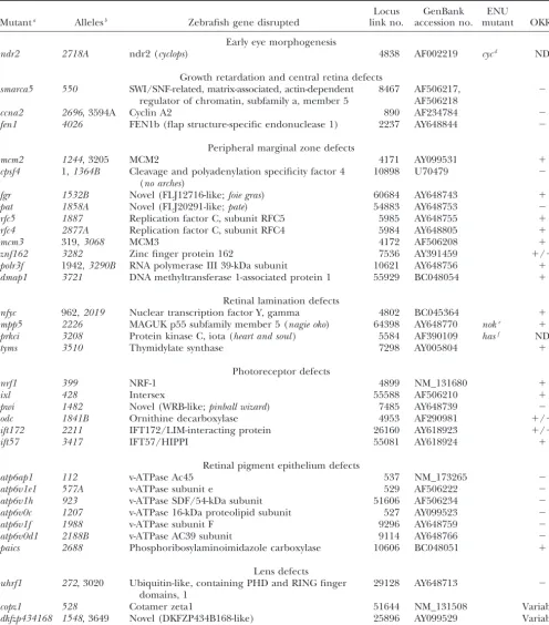
TABLE 1
Gene identities of zebrafish insertional mutations
Locus GenBank ENU
Mutanta Allelesb Zebrafish gene disrupted link no. accession no. mutant OKRc
Early eye morphogenesis
ndr2 2718A ndr2 (cyclops) 4838 AF002219 cycd ND
Growth retardation and central retina defects
smarca5 550 SWI/SNF-related, matrix-associated, actin-dependent 8467 AF506217, ⫺ regulator of chromatin, subfamily a, member 5 AF506218
ccna2 2696, 3594A Cyclin A2 890 AF234784 ⫺
fen1 4026 FEN1b (flap structure-specific endonuclease 1) 2237 AY648844 ⫺
Peripheral marginal zone defects
mcm2 1244, 3205 MCM2 4171 AY099531 ⫹
cpsf4 1,1364B Cleavage and polyadenylation specificity factor 4 10898 U70479 ⫺ (no arches)
fgr 1532B Novel (FLJ12716-like;foie gras) 60684 AY648743 ⫹
pat 1858A Novel (FLJ20291-like;pate) 54883 AY648753 ⫺
rfc5 1887 Replication factor C, subunit RFC5 5985 AY648755 ⫹
rfc4 2877A Replication factor C, subunit RFC4 5984 AY648805 ⫹
mcm3 319,3068 MCM3 4172 AF506208 ⫹
znf162 3282 Zinc finger protein 162 7536 AY391459 ⫹/⫺
polr3f 1942,3290B RNA polymerase III 39-kDa subunit 10621 AY648756 ⫹ dmap1 3721 DNA methyltransferase 1-associated protein 1 55929 BC048054 ⫹
Retinal lamination defects
nfyc 962,2019 Nuclear transcription factor Y, gamma 4802 BC045364 ⫹
mpp5 2226 MAGUK p55 subfamily member 5 (nagie oko) 64398 AY648770 noke ⫹
prkci 3208 Protein kinase C, iota (heart and soul) 5584 AF390109 hasf ND
tyms 3510 Thymidylate synthase 7298 AY005804 ⫹
Photoreceptor defects
nrf1 399 NRF-1 4899 NM_131680 ⫹
ixl 428 Intersex 55588 AF506210 ⫹
pwi 1482 Novel (WRB-like;pinball wizard) 7485 AY648739 ⫺
odc 1841B Ornithine decarboxylase 4953 AF290981 ⫹/⫺
ift172 2211 IFT172/LIM-interacting protein 26160 AY618923 ⫹/⫺
ift57 3417 IFT57/HIPPI 55081 AY618924 ⫹
Retinal pigment epithelium defects
atp6ap1 112 v-ATPase Ac45 537 NM_173265 ⫺
atp6v1e1 577A v-ATPase subunit e 529 AF506222 ⫺
atp6v1h 923 v-ATPase SDF/54-kDa subunit 51606 AF506234 ⫺
atp6v0c 1207 v-ATPase 16-kDa proteolipid subunit 527 AY099523 ⫺
atp6v1f 1988 v-ATPase subunit F 9296 AY648759 ⫺
atp6v0d1 2188B v-ATPase AC39 subunit 9114 AY648766 ⫺
paics 2688 Phosphoribosylaminoimidazole carboxylase 10606 BC048051 ⫹
Lens defects
uhrf1 272, 3020 Ubiquitin-like, containing PHD and RING finger 29128 AY648713 ⫺ domains, 1
copz1 528 Cotamer zeta1 51644 NM_131508 Variableg
dkfzp434168 1548, 3649 Novel (DKFZP434B168-like) 25896 AY099529 Variableg
mgc10433l 2735A Novel (MGC10433-like) 79171 AY648798 ⫺
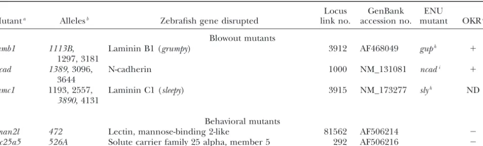
TABLE 1
(Continued)
Locus GenBank ENU
Mutanta Allelesb Zebrafish gene disrupted link no. accession no. mutant OKRc
Blowout mutants
lamb1 1113B, Laminin B1 (grumpy) 3912 AF468049 guph ⫹
1297, 3181
ncad 1389, 3096, N-cadherin 1000 NM_131081 ncadi ⫹
3644
lamc1 1193, 2557, Laminin C1 (sleepy) 3915 NM_173277 slyh ND
3890, 4131
Behavioral mutants
lman2l 472 Lectin, mannose-binding 2-like 81562 AF506214 ⫺ slc25a5 526A Solute carrier family 25 alpha, member 5 292 AF506216 ⫺
aImages of mutants can be found at http://web.mit.edu/ccr/pnas_zebrafish_mutant_images/ and information on other
pheno-types in these mutants can be found in supplemental data fromAmsterdamet al. (2004).
bHopkins allele designation (hi number). Italic type indicates allele from which histology is pictured. cOKR results were as follows:⫺, no response;⫹, normal response;⫹/⫺, weak response; ND, not tested. dRebagliatiet al. (1998);Sampathet al. (1998).
eWeiandMalicki(2002).
fHorne-Badovinacet al. (2001);Petersonet al. (2001).
gVariable OKR results were dependent on the severity of lens defects. Those embryos with severe lens defects did not respond
while those with less severe defects responded weakly to normally.
hParsonset al. (2002).
iLeleet al. (2002);Malickiet al. (2003).
secondary to more general defects. Additionally, many of photoreceptors with well-defined inner and outer segments at 5 dpf (Figure 2D). The most mature and of the morphological mutants also showed deficits in
visual function (Table 1). For the behavioral mutant prominent photoreceptors in this image are the short UV-sensitive cones lying in the innermost region of the category, however, behaviorally deficient fish with
mor-phologically normal eyes were specifically being sought. ONL (asterisks in Figure 2D), whereas the more distal photoreceptors are blue cones, red/green double cones, Thus, screening of the 33 mutant lines with wild-type
eye size/morphology and the 40 small eyed/normal and a few rods (arrows in Figure 2D).
Mutations affecting early eye morphogenesis: One morphology lines resulted in the identification of two
behavioral mutants (Table 1). mutant was identified, ndr2, that displayed abnormal morphogenesis of the eye field (Table 1). This mutant Histology of wild-type 3-dpf and 5-dpf zebrafish eyes
is presented in Figure 2 for comparison with that of the is an allele ofcyclops(cyc), which encodes a nodal related signaling factor (Rebagliatiet al.1998;Sampathet al. mutants described below. At 3 dpf, wild-type embryos
have formed the five principal laminae in the retina: 1998).ndr2mutants, likecycb16, develop two partial eyes,
fused at the ventral midline (data not shown;Hattaet the three cellular laminae [the ganglion cell (GCL),
the inner nuclear (INL), and outer nuclear (ONL) lay- al.1991;Fulwileret al.1997).
Mutations resulting in growth retardation and central ers] and the two plexiform layers [the inner (IPL) and
the outer (OPL)] (Figure 2A). By 5 dpf these laminae retinal defects:Three mutants were identified that dis-played a small eye phenotype at 5 dpf, the retinas of are more fully formed such that only small, proliferative
regions at the retinal margin remain nonlaminated (Fig- which appeared to lack most retinal cell types (Table 1). Histological sections of eyes from these mutants are ure 2, B and C). The GCL is composed mainly of the
retinal ganglion cells, the output neurons of the retina, presented in Figure 3, B–D. Each of these mutants was easily identifiable at 3 dpf by their much smaller eyes along with so-called “displaced” amacrine cells. The INL
is composed of amacrine, bipolar, and horizontal cells, relative to sibling control embryos. By 5 dpf, their eyes were roughly half the size of their wild-type siblings. By whereas the ONL is composed of the photoreceptor
cells. Zebrafish possess four types of photoreceptors that histology, retinas from these lines show evidence of some lamination and have likely generated retinal gan-mature in a specific order: UV cones first, blue cones
next, red/green double cones next, and finally rods. A glion cells (RGCs) as a rudimentary cell layer has formed in the inner retina where the GCL normally resides. higher-magnification view of the ONL, dorsal to the
to sibling controls and are highly disorganized. For ex-ample, relative to the wild-type retina at 5 dpf and nor-malized to their smaller size, these mutants, on average, have 48% less 5e11-stained amacrine cells in their reti-nas (data not shown). Localized regions of cell death in the brains of these mutants were obvious at 2 dpf and acridine orange staining at 3 dpf identified apo-ptotic nuclei scattered throughout their retinas. At later days, however, continuing cell death in the CNS was no longer obvious and the mutants appeared to develop normally (data not shown). By 5 dpf, they were vigorous swimmers and displayed robust touch responses and, other than having much smaller heads and eyes, they appeared generally healthy. That the eyes in these mu-tants are small and appear to lack large numbers of retinal neurons suggests that a defect in progenitor cell maintenance might have occurred. While more detailed experiments will be necessary to ascertain the nature of their phenotype, that these loci encode proteins in-volved in transcriptional and cell cycle regulation sup-ports this hypothesis (Table 1).
Mutations affecting the peripheral marginal zone:As noted above, the teleost retina possesses a peripheral marginal zone (MZ) that in many cold-blooded species perpetually adds cells to the retina during the animal’s life (Figure 2C). Cells of the marginal zone retain stem cell-like characteristics and can continually generate all types of retinal cells (Johns1977; WettsandFraser
Figure2.—Wild-type development of the zebrafish retina.
1988). Some higher vertebrates, such as birds and mice, (A) A 3-dpf retina with well-formed laminae in the central
retina and large regions of undifferentiated cells at the retinal may also possess a similar region between the neural periphery. (B) A 5-dpf retina exhibiting nuclear laminae and retina and the ciliary epithelium at their retinal periph-cell types characteristic of the mature retina. (C) High-magni- ery (FischerandReh2000;Tropepeet al.2000). Several fication view of the dorsal peripheral retina. Note the change
mutations that disrupt the formation or maintenance of in cell shape as epithelial progenitor cells divide at the
periph-the peripheral marginal zone have been reported in ze-eral-most dMZ (arrow) and are subsequently displaced into
the central retina to differentiate into more rounded ap- brafish, but the affected loci have not yet been identified pearing retinal neurons (arrowhead). (D) High-magnification (Fadoolet al.1997;LinkandDarland2001). Ten inser-view of the central retina and optic nerve. Note the prominent tional mutations were identified in this screen that affect short single cones in the innermost region of the ONL
(aster-loci involved in marginal zone formation or mainte-isks) and the elongated rod outer segments in the outer ONL,
nance (Table 1). Retinal histology from each of these protruding into the RPE (arrows). Dorsal is up in all panels.
Bar, 100m. RPE, retinal pigment epithelium; ONL, outer mutants is presented in Figure 4.
nuclear layer; OPL, outer plexiform layer; INL, inner nuclear Mutants in this category appear quite similar in that layer; IPL, inner plexiform layer; GCL, ganglion cell layer; central retinal development and patterning is normal dMZ, dorsal marginal zone; vMZ, ventral marginal zone.
whereas peripheral marginal zones are greatly reduced both dorsally and ventrally (Figure 4). Differing levels of phenotypic severity are observed between the lines in nerve, although it is possible this is a remnant of the
optic stalk. The INL and ONL of these mutants are this group. For example, therfc4,rfc5, andmcm2mutants show abnormal RPE invasion into regions of the retinal poorly formed. It is difficult to morphologically identify
many of the cells in these layers as specific neuronal periphery, resulting in the retinal tissue there being nearly encased in RPE (Figure 4, A–D). On the basis of their subtypes purely by histological means.
To begin to understand what cell types are present morphology, the cells in these regions appear to be largely undifferentiated (Figure 4B) although the significance of in retinas of these mutants, immunohistochemistry was
performed on 5-dpf retinal sections using markers for the RPE protrusions is currently unknown. The marginal zones of one mutant,znf162, are filled with dense, co-RGCs (zn5; Figure 3, E–H), amacrine cells (5e11; Figure
3, I–L), and rods (1d1; Figure 3, M–P). It is apparent lumnar cells possibly reflecting a large block of cells that have failed to differentiate (Figure 4, E and F). The from this marker staining that neurons composing each
Figure3.—Mutants with growth retardation and reti-nal degeneration. (A) Wild type, (B)smarca5, (C)ccna2, and (D)fen1have drastically smaller eyes with defects in the number and organiza-tion of retinal neurons at 5 dpf. Retinal patterning is se-verely affected with the most obvious defects in the outer retina. (E–P) Immunohis-tochemical analysis of wild-type (E, I, and M),smarca5 (F, J, and N),ccna2 (G, K, and O), andfen1(H, L, and P) mutants at 5 dpf. (E–H) zn5 staining of RGCs. Each of the mutants differentiated a population of RGCs, the ax-ons of which form an optic nerve of significantly less di-ameter than that of wild-type siblings. (I–L) 5e11 staining of amacrine cells and their processes. Each of the mu-tants has differentiated am-acrine cells, although their number is much reduced and their distribution is chaotic relative to wild-type siblings. (M–P) 1d1 staining of rod photoreceptors. Each of the mutants has formed rods but in numbers reduced rel-ative to wild-type siblings. Dorsal is up. Bars, 100m in A–E, I, and M and 70m in F –H, J–L, and N–P.
an abrupt termination between the cells of the outer proteins of unknown biochemical function, while the other eight loci encode proteins involved in various retina and the region normally occupied by the MZ
(Figure 4K). A high-magnification view of this region aspects of DNA replication and mRNA modification (Table 1).
highlights the abrupt outer retina termination at the
retinal periphery (Figure 4L). Here, the INL, OPL, and Mutations affecting retinal lamination:Lamination in the zebrafish retina becomes evident by 3 dpf when the ONL extend to the retinal margin with no intervening
cells (compare Figure 4L with Figure 2C). The two re- three principal cellular laminae can be readily observed (Figure 2A). As the retina grows and new neurons are maining mutants,cpsf4andfgr, have even more reduced
marginal zone regions (Figure 4, M–O). In addition, generated by the MZs, the new cells maintain appro-priate laminar positions for their cell types such that fgrmutants also frequently have malformed lenses (data
not shown). Several of the marginal zone mutants show only the MZs remain nonlaminated at later days of devel-opment (Figure 2, B and C). Four insertional mutants localized regions of cell death in their MZs when viewed
at earlier days (data not shown). In general, this class of with defects in retinal lamination were identified (Table 1). Retinal histology from each of these mutants is pre-mutants frequently showed phenotypic defects in other
proliferative regions of the body, including the brain, sented in Figure 5.
prkciandmpp5mutants both present defects in retinal liver, and gut (Amsterdamet al.2004; data not shown).
Thus, it is possible that these mutants represent a more lamination within their outer retinas (Figure 5, A and
B).prkcimutants also showed aberrant RPE formation.
general growth inhibition at later stages of development
Figure4.—Mutants with defects in the formation and/or maintenance of the peripheral marginal zones. Each of these mutants shows reduced or abnormal marginal zones normally present at the retinal periphery. (A, C, and D) Several of the mutants exhibit RPE protrusions invading the region normally solely occupied by the marginal zone cells. (B) A high-magnification view of the boxed region in A. (E and F) The marginal zones ofznf162are filled with dense, columnar tissue possibly reflecting a large block of cells that have failed to differentiate. (G–N) Mutants with decreased marginal zones. (K and L)patmutants exhibit an abrupt termination of the outer retina prior to reaching the retinal periphery. (L) A high-magnification view of the boxed region in K illustrating this region. Note that the ONL terminates prematurely and the INL and IPL abut the RPE. (M and N) Mutants with nearly absent marginal zones. (O) High-magnification view of N showing that only the peripheral-most marginal zone cells remain in thefgrmutant. Dorsal is up. Bar, 100m.
where ectopic plexiform layers can be readily observed phenotype is manifest as a lack of photoreceptor forma-tion peripherally with either a breakdown in laminaforma-tion (arrows in Figure 5E).mpp5is allelic to the ENU-induced
nagie oko (nok) locus that encodes a membrane-associ- between the ONL and INL or a lack of OPL formation
in these peripheral regions (Figure 5C). The central ated guanylate kinase (MAGUK) scaffolding factor (Wei
andMalicki2002).prkciis allelic toheart and soul(has), retina appears unaffected in nfyc mutants. A higher-magnification view of the dorsal retina highlights their an ENU-induced locus encoding an atypical protein
kinase C (Horne-Badovinac et al.2001; Peterson et defect, as regions of the INL can be seen intermingling with the ONL in conjunction with a lack of OPL forma-al.2001). The defects described here for the insertional
alleles are consistent with those reported for nok and tion (Figure 5F). Likenfyc,tymsmutants also show either regions where a breakdown in lamination between cells has, respectively, although mpp5 (nok) insertional
mu-tants are not as severely affected as those reported for of the INL and ONL has occurred or a lack of OPL formation (Figure 5D). In tyms mutants, INL regions nokm227andnokm520(WeiandMalicki2002).
Figure5.—Mutants with retinal lami-nation defects. (A)prkci(has) and (B) mpp5(nok) mutants. (E) A high-magni-fication view of the boxed region in B illustrating the ectopic IPL regions in the outer retina of this mutant (arrows). (C)nfycand (D)tymsmutants show more subtle defects in the lamination of the outer retina. (F) A high-magnification view of the boxed region in C illustrating the outer retina lamination defect in nfyc. Lamination is normal in the central-most retina and breaks down toward the periphery. It does not appear that photo-receptors have formed in the peripheral retina. (G) A high-magnification view of the boxed region in D illustrating the intermingling of cells between the INL and ONL in this mutant with some re-gions showing no OPL. Differentiated photoreceptors are present in this mu-tant (arrow). Dorsal is up. Bar, 100m.
The gene disrupted innfycis NFY-␥, part of a trimeric ceptor-specific retinal degeneration: pwi, nrf1, ift172, andift57(Figure 6, C–F).pwimutants show ONL defects transcription factor complex (Table 1;Maity andde
Crombrugghe1998). The mutation in tyms is in the both centrally and peripherally (Figure 6C). A magnification view of the retina from this mutant high-thymidylate synthase gene that encodes an enzyme
in-volved in metabolism, DNA synthesis, and DNA repair lights the acellular regions observed in the central retina (Figure 6C⬘). In these mutants there are also a few (Table 1).
Mutations affecting photoreceptor development and acellular holes in the INL (Figure 6C) but otherwise their inner retinas are relatively normal. Since both the survival: Six mutants were identified with phenotypic
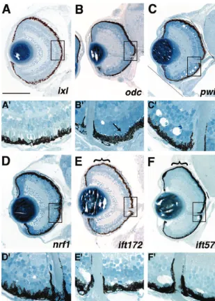
defects in the formation and/or maintenance of the central and the peripheral retinas are affected in pwi mutants, it is unknown whether PRs initially form and photoreceptor cell layer. Two mutants appear to have
defects in ONL formation,ixlandodc.ixlmutants pres- subsequently degenerate or whether defects are present at the onset of PR formation. The pwilocus encodes a ent an interesting ONL phenotype in which the
photo-receptors have formed but are morphologically abnor- protein of unknown biochemical function (Table 1). nrf1mutants exhibit extensive photoreceptor degenera-mal, resulting in a distortion of overall retinal shape to
a more oval appearance in mutant eyes (Figure 6, A tion by 5 dpf (Figure 6 D and D⬘). This locus encodes an ortholog of human nuclear respiratory factor 1 (nrf1), and A⬘). In this mutant, it appears as if the
photorecep-tors (PRs) might have precociously developed such that which has been previously characterized (Beckeret al. 1998). ift172andift 57 also exhibit photoreceptor de-their outer segments are much more robust in
appear-ance (Figure 6A⬘) when compared with a wild-type con- generation throughout the ONL (Figure 6 E, E⬘, F, and F⬘). That the PRs of the peripheral retina are unaffected trol (Figure 2D). Morphologically, the ONL inixl
mu-tants appears to be composed of all photoreceptor in these mutants implies that the oldest PRs in the cen-tral retina are formed normally but degenerate at later subtypes but conclusive identification of these cells will
require a molecular or electron microscopic character- days of development.ift172andift 57mutants are partic-ularly interesting as the affected loci encode proteins ization.ixlencodes a zebrafish ortholog of Intersex, a
transcriptional cofactor (Table 1).odcmutants display that are involved in intraflagellar transport (IFT) a pro-cess required for the assembly and maintenance of cilia a well-defined ONL but the photoreceptors in these
mutants extend little to no outer segment material (Fig- and flagella in vertebrates (reviewed in Pazour and Rosenbaum 2002). Indeed, Tsujikawa and Malicki ure 6, B and B⬘). ONL development otherwise appears
grossly normal (Figure 6B⬘). This locus encodes orni- (2004) have recently reported a role for IFT proteins in photoreceptor maintenance in zebrafish. In their thine decarboxylase, an enzyme involved in polyamine
photore-Figure6.—Mutants with defects in the forma-tion and/or maintenance of the photoreceptor layer. (A and A⬘)ixlmutants exhibit abnormal photoreceptor development and morphology. When compared with wild-type morphology in Figure 2D, the outer segments in this mutant appear larger and more robust (A⬘). (B and B⬘) odcphotoreceptors, while clearly present, form little outer segment material (arrow in B⬘ point-ing to small outer segment). (C and C⬘) pwi mutants show acellular holes in both the central and the peripheral retina. (D and D⬘)nrf1 mu-tants have fewer photoreceptors likely as a result of photoreceptor-specific degeneration (D⬘). (E and E⬘)ift172and (F and F⬘)ift57mutants ex-hibit acellular holes in the central ONL, likely representing photoreceptor degeneration as photoreceptors are clearly observed in the reti-nal periphery (PR regions in the periphery are highlighted by brackets in E and F). Note that below the low-magnification image of each sec-tion (A–F), a high-magnificasec-tion image is pre-sented to highlight the boxed region of the ONL (A⬘–F⬘) compared to that of a wild-type sibling in Figure 2D. In A–F, dorsal is up. For A⬘–F⬘, the images are rotated 90⬚clockwise relative to their respective retinal sections. Bar, 100m.
basis of morpholino antisense oligonucleotide targeting ogy from seven of these lines is presented in Figure 7. The eighth mutant line, cop z1, is discussed below in a of its transcript.
Mutations affecting the retinal pigment epithelium: category devoted to lens mutants since embryos from this line display prominent defects in lens formation A variety of functions in the eye are served by the RPE
(reviewed inMarmorstein2001). Much of the blood in conjunction with RPE defects. Six lines with RPE de-fects, atp6ap1, atp6v1e1, atp6v1h, atp6v0c, atp6v1f, and supply for the retina is provided by the choroid and
cells of the RPE function as a blood-retina barrier to atp6v0d1exhibited pigmentation defects and abnormal RPE structure (Figure 7, A–I). These mutants also dis-regulate the bidirectional flow of materials into and out
of the photoreceptor cells. RPE cells also carry out the played differing degrees of retinal degeneration. The loci affected in each of these mutant lines encode sub-isomerization of all-transretinal to 11-cisretinol during
the visual cycle. Additionally, photoreceptors shed roughly units or accessory proteins of the lysosomal vacuolar ATPase complex (Table 1). Each of these mutants also 10% of their outer segment material daily and it is the
cells of the RPE that phagocytose this material and de- has decreased melanophore pigmentation along the length of its body axis (Gollinget al.2002;Amsterdam grade it. A number of human pathologies that result
in visual impairment affect the RPE, underscoring the et al.2004). RPE morphology is abnormal in these mu-tants, and thus the defect is not solely manifest as a lack importance of this cell layer in photoreceptor function
and maintenance. of RPE pigmentation (Figure 7F).atp6v1handatp6v1f mutants show a severe retinal degeneration at 5 dpf Eight loci that affect the formation and/or
Figure7.—Mutants with defects in the forma-tion and/or maintenance of the retinal pigment epithelium. All mutants in this class exhibit patchy RPE pigmentation and abnormal RPE morphology and most exhibit RPE degenera-tion. Additional defects are as follows: (A) atp-6ap1and (D) atp6v0cmutants show abnormal retinal morphology with the dorsal-central ret-ina extending into the forebrain. The retret-ina has not expelled through the RPE or Bruch’s mem-brane, however. In several mutants (A, D, and G) pyknotic nuclei are observed at the transition between the MZ and the central retina. (B) A high-magnification view of the top boxed region in A. (C) Photoreceptor outer segments are present where pigmented epithelium is present but their morphology appears abnormal. (E) The ONL ofatp6v1e1has significantly degener-ated as pyknotic nuclei and acellular holes are easily observed. (F) A high-magnification view of the boxed region in E. Note the abnormal RPE morphology. (H and I) RPE mutants with extensive retinal degeneration. ( J)paicsdefects are mainly limited to RPE pigmentation and morphology. Dorsal is up. Bar, 100m.
scattered throughout the INL and GCL (Figure 7, H eral retina appears grossly normal. Finally, the seventh mutant in this category,paics, exhibits patchy pigmenta-and I). The brains of these mutants also frequently show
signs of cell death (data not shown). Retinas ofatp6ap1, tion of the RPE and abnormal RPE morphology but unlike the v-ATPase mutants, this mutant shows no obvi-atp6v0c, and atp6v0d1 contain pyknotic nuclei but, in
these mutant retinas, the foci are mainly limited to the ous degeneration in the adjacent retina (Figure 7J).
Thepaicslocus encodes an enzyme involved in purine
transition region between newly generated neurons of
the peripheral marginal zone and the retina proper biosynthesis, phosphoribosylaminoimidazole carboxyl-ase (Table 1).
(Figure 7, A, D, and G; high-magnification view of
atp-6ap1 in Figure 7B). Additionally,atp6ap1 and atp6v0c Mutations affecting the lens:The development and maintenance of the lens in zebrafish has not yet been mutants also have distorted retinal shapes with dorsal
regions extending farther back into the adjacent fore- well characterized although several mutants with lens defects have been reported (Heisenberg et al. 1996; brain (Figure 7, A and D). Interestingly, in these
mu-tants, where there is pigmented RPE, photoreceptor Vihtelicet al.2001). Lens formation in zebrafish initi-ates with a thickening of the surface ectoderm overlying outer segments are readily observed (Figure 7C). These
outer segments, however, are morphologically abnor- the optic vesicle at the 14–15 SS. By the 18 SS, the lens placode has thickened and its cells adopt a radial orienta-mal in length and appearance (high-magnification view
ofatp6ap1in Figure 7C). Inatp6v1e1mutants, the ONL tion relative to the adjacent neural retina (Schmittand
Dowling1994). Subsequently, cell differentiation com-has degenerated significantly centrally (Figure 7, E and
periph-(Figure 8B). The locus disrupted in dkfzp434168 en-codes a protein of unknown biochemical function while that of uhrf1encodes a zebrafish ortholog of UHRF1, a nuclear phosphoprotein involved in cell cycle progres-sion (Table 1;Mutoet al.1995). The other two mutants in this category,copz1andmgc104331, both show normal formation of the central lens nucleus with apparent defects in the surrounding regions of the lens cortex (Figure 8, C and D). The cells of the lens nucleus are the oldest cells in the lens, with growth proceeding circumferentially around the lens perimeter in the cor-tex. Thus, these mutations possibly reflect defects in the formation or differentiation of newly generated lens cells. Like uhrf1, the MZs ofmgc104331 are also com-posed mainly of columnar cells that appear to be an extension of the abnormal cells originating in the lens cortex (Figure 8D, asterisk).mgc104331encodes a pro-tein of unknown biochemical function (Table 1).
“Blowout” mutants:Mutations in three loci resulted in a retinal blowout: the expulsion of retinal cells through the RPE into the adjacent forebrain (Figure 9). These loci, lamb1, ncad, and lambc1, each exhibit retinal blowout by 3 dpf. All three of these mutants are alleles of known ENU-induced loci:lamb1:grumpy(gup; Parsons et al. 2002), ncad:ncad (Lele et al. 2002; Malicki et al. 2003) and lambc1:sleepy(sly;Parsons et
Figure 8.—Mutants with lens defects. (A) uhrf1mutants
al.2002). Two mutant alleles of thencadlocus [parachute have severe lens disorganization as well as expansion of
epithe-(pac) andglass onion(glo)] have been characterized in lial-like tissue into the marginal zones (asterisk). (B)dkfzp434168
mutants exhibit gross lens disorganization and lack both dorsal detail and shown to encode the N-cadherin protein and ventral marginal zones. (C)copz1mutants show abnormal (PujicandMalicki2001;Leleet al.2002;Erdmannet formation of the peripheral lens cortex. These mutants also al.2003;Malickiet al.2003). Histologically, insertional exhibit patchy RPE pigmentation. (D) mgc104331 mutants
ncadmutant retinas look similar to these reported alleles exhibit defects in the lens cortex and lens tissue expands and
in that, in addition to the retinal blowout, there is some fills the anterior chamber space between the lens and cornea
as well as forming a continuum with the peripheral marginal disruption of regular lamination (Figure 9B). Thelamb1 zones (asterisk). Dorsal is up. Bar, 100m. mutant, an allele of gup, encodes laminin 1 while
lambc1, an allele ofsly, encodes laminin␥1 (Table 1). gupand slymutants have not been reported to have a fiber cells compose the lens proper while the lens epi- blowout phenotype; however, they were identified in a thelial cells form a monolayer and surround the lens at behavioral screen for zebrafish mutants with defective its distal and lateral perimeters. visual function and shown to form disorganized optic Four mutations that affected the development of the nerves (Neuhausset al.1999). Beyond the blowout in lens were identified in our screen (Table 1). Two muta- lamb1, gross retinal morphology looks normal at 5 dpf tions,uhrf1anddkfzp434168, result in severe disorgani- (Figure 9A) while that oflambc1is disrupted. At 5 dpf, zation of lens structure (Figure 8, A and B). Theuhrf1 lambc1mutants show minor lamination defects and sig-mutant lenses are composed of a disordered mass of nificant lens and corneal malformations (Figure 9C). cells and possibly reflect lens degeneration rather than Several other insertional alleles of the laminin␥1 locus a defect in lens morphogenesis (Figure 8A). The lens have also been isolated (Table 1;Amsterdamet al.2004).
in dkfzp434168 mutants is of normal size but displays While the phenotypes are similar between these alleles,
loosely packed, disorganized cells (Figure 8B). In addi- one of them, hi2557, presents a more severe retinal tion, the distal-most region of the lens, underlying the phenotype than that of hi3890 (data not shown). corneal epithelium, appears to have degenerated as only Behavioral mutants:As discussed above, many mutant a mass of tissue remains. Defects in marginal zone for- loci were identified that were behaviorally blind or ex-mation are also frequently observed at these loci. In hibited severely attenuated responses when tested by
uhrf1, the MZ territory is occupied by columnar cells OKR assays (data not shown). In total, 33 of the 250
Figure 9.—Blowout mutants. (A) lamb1(gup) retinas expel through the RPE and into the forebrain. Retinal lami-nation and pattern are grossly normal, however. (B)ncadmutants also exhibit retinal blowout into the forebrain and these mutants show some defects in reti-nal lamination. (C)lambc1(sly) retinas expel through the RPE and these mu-tants have clear lens and cornea malfor-mations. Dorsal is up. Bar, 90m.
DISCUSSION When tested by OKR, 2 of these 73 loci showed distinct
behavioral defects, both of the wild-type eye size group, Insertional mutagenesis has emerged as a powerful and these mutants were further examined (Figure 10). technique to rapidly identify genes important in verte-Of 21lman2lmutants tested, only 6 responded to OKR brate development (Gollinget al.2002;Zambrowicz stimuli at 0 log unit light intensity (full light), while at et al.2003;Amsterdamet al.2004). To date, insertional ⫺1 log unit intensity, none of the 21 mutants responded.
The mutants showed no eye movements to track the moving OKR stripe while wild-type siblings displayed normal tracking behaviors down to⫺3 log unit intensity in these assays (data not shown).slc25a5 mutants also did not track properly in OKR assays at all light levels tested;i.e., they showed weak to no eye movements to track the moving stripes (n ⫽19). That both of these mutants did not track properly in the OKR assay indi-cates that they are behaviorally deficient but does not assess whether their retinal circuitry functions properly. The underlying defects in these mutants could reflect a motor or tectal defect rather than one in retinal physi-ology. To begin to differentiate between these possibili-ties and to assay outer retina function, ERGs were per-formed. ERGs measure the summed field potential of the outer retina in response to a pulse of light and are extensively used in zebrafish to assess retinal function (e.g.,Van Eppset al.2001;Kainzet al.2003). ERGs from lman2lmutants were normal at all light levels (data not shown).lman2lmutants are touch insensitive (Golling et al.2002) and thus, this mutant possibly reflects a more general sensory/motor defect rather than one based in retinal physiology. It is also possible, however, that de-fects inlman2lmutants lie in the inner retina or the optic tectum, possibilities not addressed by ERG recordings. Further experiments will be necessary to ascertain the cellular basis of their functional deficits. The locus af-fected inlman2lencodes a vip36-like mannose-binding lectin that is thought to be involved in trafficking of glycoproteins between the Golgi and the cell surface (Table 1;Fiedleret al.1994;FiedlerandSimons1996).
Figure10.—Behavioral mutants with normal eye histology. Conversely, ERGs from slc25a5 show a clear deficit in (A)lman2l mutants do not move their eyes in response to outer retinal function (Figure 10). At all light levels OKR stimuli but behave normally in ERG testing. (B)slc25a5 mutants respond weakly to OKR stimuli and exhibit defective tested,slc25a5ERG recordings were significantly
attenu-ERG responses. attenu-ERG recordings from a wild-type 5-dpf sibling ated relative to sibling controls. The locus affected in
(control) and recordings from an slc25a5 mutant are pre-slc25a5encodes a mitochondrial ADP/ATP carrier pro- sented. Even at the brightest light levels (0,⫺1 log units), tein (Table 1) and the ERG deficits therefore may possi- slc25a5embryos respond much less robustly to visual stimuli
mutagenesis in zebrafish has identified 315 loci that pic nature of their phenotype did not fit the criteria of our screen as possessing eye-specific defects and, thus, when disrupted, produce visible embryonic phenotypes
they were not included in this report. The CCT complex by 5 dpf and for which the disrupted genes have been
is composed of eight subunits (reviewed inDunn et al. identified (Gollinget al.2002;Amsterdamet al.2004).
2001) and in addition to the␥-subunit, insertional mu-Of 250 insertional loci screened, 40 were identified that
tants at four other CCT subunit-encoding loci have been directly affected the development and function of the
identified (Gollinget al.2002;Amsterdamet al.2004). visual system. Of these loci, only 8 have been previously
Much likecct3, each of these mutants has small eyes and ascribed a role in vertebrate eye development, nearly
overall CNS degeneration ( J. M. Gross, unpublished all from mutational and functional studies in zebrafish.
observations). By our screening criteria, these did not In addition to these 40 mutants, 40 additional mutants
represent eye-specific phenotypes and so these mutants were identified that had smaller eyes but were
morpho-were not included in this report. These examples are logically normal (see supplementary Table 3 at http://
meant to highlight the importance of specifically defin-www.genetics.org/supplemental/). These mutants likely
ing the screening criteria utilized herein. Other screen-reflect defects in eye growth, possibly in the context of
ing parameters might identify as eye mutants some of overall CNS growth defects. MZs in these mutants were
the insertional mutants excluded here and proceed in proportional for the reduced eye size. Since we were
characterizing them as such. seeking mutants with clear morphological defects, and
From this screen, a diverse set of genes has been to limit the scope of the screen, these mutants were not
identified as playing important roles in eye development further studied. Morphological defects were observed
and visual function. Many of these have not yet been in 81 other insertional mutants in the collection (see
implicated in or well studied during eye development supplementary Table 1 at http://www.genetics.org/sup
and therefore their molecular characterization should plemental/). These mutants were not included in this
greatly increase our understanding of these processes report, however, as their eye phenotypes were in the
as well as human pathologies that affect the eye. For context of massive CNS degeneration and/or
accompa-example, two mutants,ift172andift57, show significant nied by multiple defects in other organ systems. Eye
degrees of photoreceptor degeneration in their central phenotypes observed in these lines could not be
sepa-retinas (Figure 6, E, E⬘, F, and F⬘). These loci encode rated from these more general system-wide mutation
members of the IFT protein family that are required effects. The morphological portion of this screen sought
in cilia for anterograde transport of proteins from the to identify eye mutations that were generally healthy at 5
cytoplasm, along the ciliary axoneme to the distal cilia dpf and thus likely reflecting the loss of a direct cellular
(reviewed in PazourandRosenbaum2002). The dis-function for the affected locus in the eye. Changing this
ruption of several IFT proteins has been associated with parameter and thereby the threshold for inclusion in
retinitis pigmentosa-like pathologies, both in mouse and the screen would certainly increase the number of
mu-in zebrafish (Pazouret al.2002;TsujikawaandMalicki tants identified on the basis of these new criteria.
2004). Additionally, insertional mutations at these two Indeed, several zebrafish mutants with reported eye
loci also result in polycystic kidneys, implicating a common defects were excluded from our screen. For example,
pathway in the zebrafish embryo for the generation and/ one such insertional mutation, that at thedead eyelocus or maintenance of ciliated cell types (Sun et al. 2004). (dye), results in severe necrosis in the eyes and tectum Thus, these mutants should provide an excellentin vivo visible at 2 dpf (Allende et al.1996). dye mutants die model for understanding intraflagellar transport and at 5 dpf and show severe necrosis in all regions of the its potential role in PR degeneration.
brain in addition to lacking most of their pharyngeal We identified six insertional loci with abnormal RPE skeleton. The pleiotropic nature of thedye phenotype morphology and pigmentation and varying degrees of made it too severe to be included in this screen for retinal degeneration that encode components of the relatively visual system-specific mutants. Another zebra- vacuolar ATPase (v-ATPase) protein complex or v-ATP fish mutant with eye defects,no tectal neuron(ntn), gener- ase-associated proteins (Table 1). The v-ATPase is com-ated in a trimethylpsoralen mutagenesis screen (Mat- posed of two domains: the peripheral V
1complex con-suda and Mishina 2004), affects the cct3 locus that sisting of eight subunits and the integral V
pleiotro-Age-related macular degeneration (AMD) affects approxi- opment are thereby normal, but upon depletion of these maternal stores, a phenotype is manifest in the continu-mately one-fourth of the population over age 65,
result-ally proliferative MZs. Indeed, a significant role for ma-ing in varyma-ing degrees of visual impairment. The
molec-ternal factors in morphogenesis and establishment of ular mechanisms underlying AMD, however, are largely
the embryonic body plan after the zygotic transition has unknown. A hallmark of AMD progression is the
accum-recently been reported (Wagner et al. 2004). These ulation of drusen, storage bodies composed mainly of
phenotypes therefore might represent a continuum, not undegraded photoreceptor outer segment lipids
(Feeney-of specific gene product function but actually (Feeney-of mater-Burnset al.1980;Crabbet al.2002). A major
compo-nal load. Many mutants died or showed pleiotropic de-nent of drusen, A2E, accumulates in lysosomes and
fects casting doubt on the specificity of their retinal blocks their ability to degrade outer segment material
phenotype and were not included in this report. Many (Finnemannet al.2002). The v-ATPase pump is
inhib-of these mutants might represent one end inhib-of this contin-ited by high concentrations of A2E, suggesting a possible
uum: low maternal load. The three mutants that dis-link between v-ATPase function and AMD progression
played growth retardation and central retinal defects (Bergmannet al.2004). The six v-ATPase mutants
iden-likely due to progenitor cell death might reside some-tified in this screen show differing degrees of retinal
where in the middle of the continuum. These mutants degeneration, ranging from minor degeneration in
may have possessed enough maternal mRNA or protein atp6v0d1(V0subunit d; Figure 7G) to severe
degenera-to develop generally well but they manifest defects in tion inatp6v1h(V1subunit H; Figure 7H) andatp6v1f
slightly later aspects of retinal development when these (V1subunit F; Figure 7I). It will be interesting to look
maternal loads were depleted. Finally, the MZ mutants for adult phenotypes in heterozygous animals at these
would reside at the other end of the continuum where loci to determine if these are applicable animal models
a high maternal complement enabled them to develop for the study of AMD and AMD-related pathologies in
quite normally and phenotypic defects were obvious humans, several of which are autosomal dominant
disor-only in the latest aspects of retinal development,i.e., in ders.
MZ maintenance. Thus, are these proteins specifically We identified several mutations that affected the
de-involved in retinal development or the maintenance of velopment of the central retina or the development of
MZ cells in the retina or are they actually required in the peripheral MZs and that resulted from the
disrup-all proliferating cells, a function masked to differing tion of genes encoding proteins that function in aspects
degrees by maternal stores? A survey of gene expression of transcription, translation, and cell cycle regulation
in mammalian neural progenitors relative to differenti-(Table 1). It is interesting to speculate on the nature
ated neurons has pointed to a significant enrichment of these mutations with respect to early retinal
develop-of transcripts in progenitors that encode proteins in-ment as well as stem cell maintenance in the retinal
volved in DNA replication, protein synthesis, protein periphery. It is possible that the central retinal defects
turnover, and chromatin remodeling (Livesey et al. manifest in thesmarca5,ccna2, andfen1mutants result
2004). This is not surprising given the high transcrip-from specific roles of these proteins in early events of
tional activity and unique developmental functions of retinal development, i.e., in the formation of specific
neural progenitor cells, suggesting that the latter of the retinal cell types. It is also possible, however, that these above scenarios might be the case. Further characteriza-phenotypes result simply from a block in retinoblast tion of these mutants will clearly be necessary however, proliferation such that most retinal cell types are miss- to differentiate between these possibilities.
ing, since their progenitors die at earlier phases of devel- In summary, in this screen we have identified a diverse opment. Thus, in these mutants, the role for the disrupted set of genes that are involved in visual system development protein may not be specifically in retinal patterning, but and function. Many of these have not yet been implicated rather may be in maintaining progenitor populations such in, or studied during, eye development and therefore their that an appropriate number of progenitors are present molecular characterization should greatly increase the to generate all retinal cell neurons. This logic can be understanding of these processes as well as human pa-applied to the mutants that displayed reduced MZs as thologies that affect the eye. Additionally, multiple well. Most of these resulted from disruption of a gene screens of this collection of insertional mutants, similar encoding a factor involved in DNA replication or mRNA in scope to the one reported here, are currently under-modification (Table 1). Are these factors specifically way to assay a variety of developmental, physiological, required only for maintenance of the MZ stem cell pop- and behavioral parameters. As more of these screens ulation? It is doubtful that such proteins are not utilized are completed, an integrated picture should emerge as in early retinal development. This suggests that a sig- to the roles that each of these loci play during embryonic nificant maternal complement of mRNA or protein must development. The synthesis of data from several such be present, and/or maternally supplied proteins must screens will likely reveal previously unidentified similari-have unusually high stability such that they persist through ties between the formation and maintenance of multiple