DOI: 10.1534/genetics.105.047100
Mutational Analysis of
Stubble-stubbloid
Gene Structure and Function in
Drosophila Leg and Bristle Morphogenesis
Ann S. Hammonds and James W. Fristrom
1Department of Molecular and Cell Biology, University of California, Berkeley, California 94720
Manuscript received June 23, 2005 Accepted for publication November 11, 2005
ABSTRACT
TheStubble-stubbloid(Sb-sbd) gene is required for ecdysone-regulated epithelial morphogenesis of imag-inal tissues during Drosophila metamorphosis. Mutations inSb-sbdare associated with defects in apical cell shape changes critical for the evagination of the leg imaginal disc and with defects in assembly and extension of parallel actin bundles in growing mechanosensory bristles. TheSb-sbd gene encodes a type II trans-membrane serine protease (TTSP). Here we use aSb-sbdtransgenic construct to rescue both bristle and leg morphogenesis defects inSb-sbdmutations. Molecular characterization ofSb-sbdmutations and rescue ex-periments with wild-type and modified Sb-sbd transgenic constructs show that the protease domain is required for both leg and bristle functions. Truncated proteins that express the noncatalytic domains without the protease have dominant effects in bristles but not in legs. Leg morphogenesis, but not bristle growth, is sensitive toSb-sbdoverexpression. Antibody localization of the Sb-sbd protein shows apical ex-pression in elongating legs. Sb-sbd protein is found in the base and shaft in budding bristles and then concentrates at the growing tip when bristles are elongating rapidly. We propose a model wherebySb-sbd
helps coordinate proteolytic modification of extracellular matrix attachments with cytoskeletal changes in both legs and bristles.
E
LABORATE changes in the dimensions and to-pology of epithelial sheets are required for the normal development of multicellular organisms; devel-opmental events as basic as gastrulation and as specialized as the formation of the stereocilia of the mammalian inner ear are examples of epithelial morphogenesis. Cell division and death, cell rearrangement, and cell-shape change all contribute to different types of epithelial morphogenesis (reviewed in Fristrom1988). These cellbehaviors depend in part on changes in the cytoskel-eton but occur in the context of neighboring cells, extracellular matrices (ECM), and hormonal milieus. Drosophila imaginal discs provide an attractive exper-imental system to study the complex interrelationships of the cytoskeleton, ECM, cell junctions, and extracel-lular signals during epithelial morphogenesis in a genet-ically tractable model organism.
During metamorphosis in Drosophila, the adult epi-dermis is pieced together from a collection of anlagen, the imaginal (adult) discs. Imaginal discs are simple, folded epithelial sacs which, in response to the meta-morphic steroid hormone 20-hydroxyedcysone (ecdy-sone), undergo rapid and radical tissue reorganization to form specific structures of the adult integument. The thoracic imaginal discs give rise to the adult thoracic
appendages (legs, wings, and halteres); their proximal parts fuse to form the epidermis of the thorax. The ini-tial transformation from folded, undifferentiated ima-ginal discs to appendages with the basic shape of the adult structures takes place in the prepupal period, the first 12 hr after pupariation (AP). Following the ecdysone-triggered transition to the 84-hr pupal period, the appendage morphology is further refined, bristles and hairs form, and finally the adult cuticle is deposited. Significant progress has been made in understanding how the ecdysone receptor and its partner ultraspirical interact with nuclear receptor cofactors and ecdysone-induced transcription factors to confer temporal and tissue specificity onto signals from this single hormone (reviewed in Thummel 1997, 2002). Less is known
about products of the effector genes, molecules that have a direct function in cell and tissue morphogenesis. Genetic interaction screens, pioneered in our labora-tory, have identified some of the genes that act in imag-inal disc morphogenesis (Beatonet al.1988; Gotwals
and Fristrom 1991; Clark et al. 1995; Edwards and
Kiehart1996; Gatesand Thummel2000; Bayeret al.
2003; Wardet al.2003; Chenet al.2004). A role for the Stubble-stubbloid(Sb-sbd) gene in leg and wing morphogen-esis was discovered in the first of these studies (Beaton et al.1988).
Sb-sbd transcription is induced by ecdysone and is required both in prepupae, for the initial elongation of the leg disc to form a tubular leg, and in pupae (32 hr 1Corresponding author:Department of Molecular and Cell Biology, 569
Life Science Addition, University of California, Berkeley, CA 94720-3200. E-mail: [email protected]
AP), for the apical extension of a single cell to form the mechanosensory bristle shaft (Appelet al.1993). Bristle
phenotypes are distinct in dominant (Sb) and recessive (sbd) mutations. Defects in leg morphogenesis are re-cessive in allSb-sbdmutants (Table 1).
In prepupae the leg disc telescopes out of the con-centrically folded epithelium to form a cylinder and everts to the outside of the developing imago. This change in tissue shape results primarily from changes in cell shape (Condicet al.1991; Fristromand Fristrom
1993). At the end of the third instar, cells that will form the basitarsis and distal tibia maintain an anisometric shape with the proximal-distal axis compressed and the circumferential axis elongated. By 6 hr AP, the leg has become tubular and the elongated cells have be-come isometric, the change in cell shape, longer in the proximal-distal direction and narrower in width, mediat-ing the change in tissue shape. InSb-sbdmutants, these cell-shape changes are limited and the legs of the adult exhibit the malformed (mlf) phenotype with leg segments that are short, thick, and often kinked or gnarled (see Beatonet al.1988, Figure 1).
In pupae, each bristle develops as an apical cyto-plasmic extension of a tricogen cell, one of the four cells that make up the mechanosensory bristle organ (Leesand Waddington1942; Leesand Picken1945;
Hartensteinand Posakony1989; Tilneyet al.1995).
Within the extending bristle cell, a scaffold is formed by a core of microtubules surrounded by modules of membrane-associated actin microfilament bundles joined end to end (Overton1967; Appelet al.1993; Tilney et al. 1995, 1996, 1998; Wulfkuhle et al. 1998). The
bristle grows as individual actin filaments continuously form at the bristle tip on the cytoplasmic side of the plasma membrane and are gathered together to form progressively larger and more tightly crosslinked bun-dles by the sequential action of two actin-binding proteins, forked and singed (Lees and Picken 1945;
Tilneyet al.1996). Once the bristle is fully elongated,
a chitinous cuticle is secreted and the actin bundle scaffolding is dismantled (Tilney et al. 1996, 2003;
Guild et al. 2002). Bristles of Sb mutants are short,
thick, and blunt ended. In these developing bristles the number of actin bundles is increased and in the most severe alleles, the bundles are also disorganized, with some bundles in the center of the bristle rather than regularly distributed around the cell perimeter (Lees
and Picken1945; Appelet al.1993). Developing bristles
in recessivesbdmutants start out with the normal num-ber and distribution of actin bundles, but the bundles become deranged at the tip and stop prematurely and asynchronously (Appelet al.1993). Reflecting this
de-velopmental defect, bristles of the homozygoussbdadults are short but with slightly tapered and frayed ends (see Appelet al.1993, Figure 5).
The Sb-sbd protein (which for convenience we will refer to as stubblin) is the first identified invertebrate
member of the type II transmembrane serine prote-ases (TTSP), each containing a C-terminal extracellular serine protease domain, a short N-terminal intracellular domain, and a variety of structural motifs connecting the transmembrane domain with the catalytic domain (Hooperet al.2001; Netzel-Arnettet al. 2003). The
modular structure of these proteases suggests a possible role linking proteolysis of the ECM to cytoskeletal re-arrangements. Like other members of the TTSP fam-ily, the predicted 786-aa stubblin (Figure 2) includes an N-terminal cytoplasmic domain, followed by a trans-membrane signal/anchor domain and an extracellular stem region with a C-terminal serine protease domain (Appelet al.1993; Hooperet al.2001). The 244-aa
pro-tease domain shares extensive sequence similarity to the S1 family (clan SA) of trypsin-like proteases (Furie
and Furie1988; Rawlingset al.2004) in the conserved
regions of the substrate-binding pocket, cleavage acti-vation site, and catalytic triad, indicating a preference for cleavage after arginine or lysine. Commonly, these proteases are zymogens that are activated by cleavage at a characteristic activation site motif. In some proteases with a long prodomain, the noncatalytic domain re-mains tethered via a disulfide bond to the catalytic do-main after cleavage (Rawlingset al.2004). In stubblin,
Cys-531 and Cys-659 are appropriately placed to provide this function. In the extracellular region just beyond the transmembrane domain, a conserved pattern of three pairs of cysteines defines a disuflide knot domain (or CLIP domain) like that found in several other arthro-pod proteases, including Limulus proclotting enzyme and the Drosophila snake and easter proteins, both se-creted proteases involved in embryonic dorsal/ventral patterning (Gayand Keith1992; Smithand DeLotto
1992; Mutaet al.1993; Jiangand Kanost2000). The
disulfide knot has been proposed to act as a binding site for a protease activator (Mutaet al.1990). For
success-ful transgenic rescue ofsnakemutants, thesnake trans-genic construct must include an intact knot domain (Smithet al.1994). A long serine/threonine-rich stem
(aa 260–480) connects the stubblin knot and the pro-tease. A similar, although shorter, region is essential for snake function (Smithet al.1994).
Initial molecular studies of the Sb-sbd gene showed that the most severe dominant mutants were character-ized by DNA insertions between the stem and the pro-tease domain (Appelet al.1993). The dominant bristle
surrounding apical ECM. The malformed leg pheno-type, but not the bristle phenopheno-type, inSb-sbdmutants is enhanced in the presence of mutations in genes in-volved in myosin II-driven apical cell-shape change, includingzipper (zip), the gene coding for nonmuscle myosin II heavy chain (Gotwals and Fristrom1991;
Bayer et al. 2003), reflecting the difference between
apical cytoskeletal contraction in the elongating leg disc and formation of a long apical cytoplasmic extension by assembly and extension of parallel actin bundles in the developing bristle.
We took advantage of the differences in the bristle and leg roles forSb-sbdto begin to connect biological functions to specific domains of stubblin. We have iden-tified the molecular defects in three dominantSb mu-tants and found that the most severe entirely lack a protease domain, while a mild allele is a frameshift mutation that adds a C-terminal hydrophobic sequence to the protease domain, arguably disabling the protease by interfering with folding and transmembrane process-ing. A severe recessive sbd mutant has a single base change likely to reduce but not eliminate protease func-tion. Using transgenic lines expressing wild-typeSb-sbd, protease-disabledSb-sbd, and a series of truncations, we show that a functional stubblin protease domain is re-quired for both leg and bristle morphogenesis. Expres-sion of truncated transgenes in a wild-type background generatesSb-like bristles, but not malformed legs. An-tibody localization of stubblin in developing bristles of wild-type andSbmutants indicates that the stubblin knot and/or stem domains help localize protease activity to the growing bristle tip. Surprisingly, overexpression of wild-typeSb-sbdhas dominant effects on leg but not bris-tle morphogenesis during specific sensitive periods in both prepupal and pupal development. These results, taken in light of recently reported genetic interaction studies of Sb-sbdand Rho-signaling pathway mutations (Halsellet al.2000; Bayeret al.2003; Wardet al.2003;
Chen et al. 2004), suggest that Sb-sbd may coordinate
ECM remodeling and cytoskeletal reorganization in both legs and bristles.
MATERIALS AND METHODS
Drosophila stocks: Drosophila Sb-sbd stocks used in this study are described in Beatonet al.(1988) and Table 1.
Wild-type strains used were Oregon-R and Canton-S lines that have been maintained in our laboratory since 1964 and 1979, re-spectively. Transgenic stocks are described below. Stocks were maintained on standard cornmeal–agar medium at either 25°
or 18°.
Transgenic constructs:See Figure 3. Generation of the wild-type Sb-sbd transgene (hs-Sb-sbd1
) was described previously (Hammonds2002; Bayeret al.2003). Briefly, full-lengthSb-sbd
cDNA (Appelet al.1993) was cloned into a pLitmus 29 Vector
(New England Biolabs, Beverly, MA) and then into the heat-shock-inducibleP-element transformation vector pCaSpeR-hs (Thummeland Pirrotta1992) to enable expression of the
transgene under heat-shock control. The pLitmus29 Sb-sbd
cDNA clone was used as a mutagenesis template to construct modified Sb-sbdtransgenes, which were subsequently cloned into pCaSpeR-hs and injected intow1118embryos using
stan-dard methods (Rubinand Spradling1982). The
protease-disabled S737A construct, which substitutes an alanine for the catalytic serine at amino acid 737, was made from the wild-type template using QuikChange mutagenesis (Stratagene, La Jolla, CA) to change Ser (TCA) to Ala (GCA) using the primer
59-TGTCAGGGCGATGCAGGAGGTCC-39. The truncated Sb
transgene hs-Sb-Dprotease deletes the wild-type Sb-sbd cDNA coding for the protein sequence C-terminal to amino acid 517 and so lacks the entire protease domain. The truncated
Sb-transgene hs-SbCD-TM-knot deletes the DNA coding for
the protein sequence C-terminal of residue 257, so that only the cytoplasmic domain, the transmembrane domain, and the
disulfide knot remain. The hs-SbCD and hs-SbCD-TM stocks
were gifts from C. Bayer. Thehs-SbCDconstruct includes only the cytoplasmic domain whilehs-SbCD-TMretains the cytoplas-mic domain with the transmembrane domain and some extra-cellular sequence but excludes the disulfide knot. Expression of transgenic protein products was confirmed by Western blotting for thehs-Sb-sbd1
,hs-Sb-Dprotease, andhs-Sb-S737A con-structs. The shorter truncations do not contain the epitope against which our antibody was generated so their expression cannot be verified and results with these constructs must be considered preliminary.
Heat-shock protocol:Animals were staged from white pre-pupae (WPP) and heat-shocked immediately for the 0-hr time point or kept at 25°until the desired age, described in hours AP. The collected WPP were placed onto the walls of standard plastic food vials. For heat shocks the vials were immersed in a 37°water bath for 60 min. After heat shock the vials were re-turned to 25°until the adults eclosed. Adults were then scored for bristle or leg phenotype.
Sequence analysis ofSb-sbdmutants:To identify sequence
defects in Sb-sbdmutants, genomic DNA from each
homozy-gote mutant (Sbspike,sbd2,sbd201, and thesbd201progenitorbr1)
was amplified usingSb-sbdexon-specific primers. All sequences were determined in both directions. Mutations and polymor-phisms were verified by sequencing independent PCR prod-ucts using at least two different primer pairs. The 39-ends of
Sb63bandSb70cDNA were isolated from homozygote WPP total
RNA by 39-RACE PCR (Frohman1993) using reagents from
the Invitrogen (Carlsbad, CA) 39-RACE kit. First-strand cDNA synthesis was primed with an oligo(dT) primer containing an adaptor sequence at its 59-end. The PCR step used a 59primer from exon 5 in theSb-sbdstem region (nt 1558–1773) and the adaptor sequence as the reverse primer. This PCR product was then cloned into a PCRII vector (Invitrogen) and sequenced. Manual double-stranded sequencing was done with the di-deoxy chain termination method using Sequenase version 2 kit (USB, Cleveland). Automated sequencing was done at the University of California DNA sequencing facility. PCR and sequencing primers were made by GIBCO–BRL/Invitrogen or the University of California Cancer Research Laboratory Micro-chemical Facility. DNA sequence analysis was facilitated by the use of MacVector sequence analysis software (Oxford Molec-ular Group/Accelrys, San Diego) and the NCBI BLAST server.
Preparation of antibodies:A 26-amino-acid synthetic pep-tide from a nonrepetitive region in the stubblin stem was used to generate polyclonal antisera in guinea pigs (see Figure 3). The stem peptide was a generous gift of D. King. Antisera were prepared by Covance Research Services (Richmond, CA).
Dissection and antibody staining of imaginal discs:White prepupae were collected and either dissected immediately or aged for 1–4 hr at 25°. The disc–brain complex was dissected in Drosophila Ringers (130 mmNaCl, 5 mmKCl, 1.5 mmCaCl2)
(20 mmTris, 137 mmNaCl) at room temperature on a rocker.
The fixed tissue was washed three times in TBS and then transferred to blocking solution (2% goat serum, 1% BSA in TBS). In the block, the discs were separated from the brain and any other adhering tissues. The cleaned discs were trans-ferred to a fresh block and incubated, rocking, at room temperature for a total of 2 hr. Discs were then incubated overnight at 4°in stubblin antibody diluted 1:400 in blocking solution. Primary antibody was washed out with five washes of TBS. After a 15-min blocking step, discs were incubated for 1.5 hr in secondary antibody, fluorescein-conjugated anti-guinea
pig IgG (H 1 L), affinity purified (Vector Laboratories,
Burlingame, CA), and used at 20mg/ml. Discs were then washed five times with TBS. After the last wash, TBS was replaced with 25% glycerol in TBS and the discs were allowed to equilibrate for 5 min. Discs were mounted on slides with Vectashield mounting medium (Vector Laboratories, Burlingame, CA).
Dissection and antibody staining of developing bristles:
Pupae were staged from WPP and aged at 25°for 32–48 hr or at
18° to an equivalent developmental stage. Thoraces were
dissected using a modification of the method described by Tilneyet al.(1996). Pupae were placed on double-stick tape
and pupal cases were removed. Dorsal thoraces were collected as described (Tilneyet al.1996) except that cleaning of the
tissue was delayed until after a longer initial fixation step:
30 min at room temperature and then 2 hr to overnight at 4°. Following fixation, the large tracheoles and fat body were removed, and the pupal cuticle was gently peeled away from the epidermis. The fixed, cleaned thoraces were incubated in 0.1% Triton X-100, 4% formaldehyde in TBS for 20 min and then washed five times in TBS, 0.1% Triton. For antibody staining, thoraces were incubated in blocking solution (1% BSA, 2% goat serum in TBS) for 1.5 hr and then in antibody solution overnight at 4°at the same dilutions as used for disc staining. Secondary antibody incubations and subsequent washes were done as for disc staining. For mounting, a final 40% glycerol equilibration step was added before mounting in Vectashield mounting medium diluted 1:1 in TBS. For phal-loidin staining of actin, fixed and permeablized thoraces were incubated 2 hr at room temperature or overnight at 4°in phal-loidin conjugated to either Texas Red or fluorescein (Molecular Probes, Eugene, OR) and diluted into 4% formaldehyde in TBS and then washed and mounted as for antibody-stained tissues.
Conventional fluorescence and confocal microscopy:Slides were examined with both conventional fluorescence micros-copy on a Zeiss Axiophot microscope and scanning laser confocal microscopy using a Bio-Rad (Hercules, CA) 1024 confocal microscope and COMOS software. Confocal images were processed and analyzed using either NIH Image 1.62 or Photoshop 3.0.5 (Adobe, San Jose, CA).
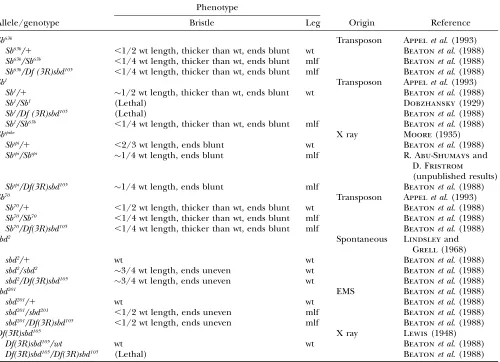
TABLE 1
Leg and bristle phenotypes ofSb-sbdmutant alleles
Phenotype
Allele/genotype Bristle Leg Origin Reference
Sb63b Transposon Appelet al. (1993)
Sb63b/1 ,1/2 wt length, thicker than wt, ends blunt wt Beatonet al. (1988)
Sb63b/Sb63b ,1/4 wt length, thicker than wt, ends blunt mlf Beatonet al. (1988)
Sb63b/Df (3R)sbd105 ,1/4 wt length, thicker than wt, ends blunt mlf Beatonet al. (1988)
Sb1 Transposon Appelet al. (1993)
Sb1/1 1/2 wt length, thicker than wt, ends blunt wt Beatonet al. (1988)
Sb1/Sb1 (Lethal) Dobzhansky(1929)
Sb1/Df (3R)sbd105 (Lethal) Beatonet al. (1988)
Sb1/Sb63b ,1/4 wt length, thicker than wt, ends blunt mlf Beatonet al. (1988)
Sbspike X ray Moore(1935)
Sbspi/1 ,2/3 wt length, ends blunt wt Beatonet al. (1988)
Sbspi/Sbspi 1/4 wt length, ends blunt mlf R. Abu-Shumaysand
D. Fristrom
(unpublished results)
Sbspi/Df(3R)sbd105 1/4 wt length, ends blunt mlf Beatonet al. (1988)
Sb70 Transposon Appelet al. (1993)
Sb70/1 ,1/2 wt length, thicker than wt, ends blunt wt Beatonet al. (1988)
Sb70/Sb70 ,1/4 wt length, thicker than wt, ends blunt mlf Beatonet al. (1988)
Sb70/Df(3R)sbd105 ,1/4 wt length, thicker than wt, ends blunt mlf Beatonet al. (1988)
sbd2 Spontaneous Lindsleyand
Grell(1968)
sbd2/1 wt wt Beatonet al. (1988)
sbd2/sbd2 3/4 wt length, ends uneven wt Beatonet al. (1988)
sbd2/Df(3R)sbd105 3/4 wt length, ends uneven wt Beatonet al. (1988)
sbd201 EMS Beatonet al. (1988)
sbd201/1 wt wt Beatonet al. (1988)
sbd201/sbd201 ,1/2 wt length, ends uneven mlf Beatonet al. (1988)
sbd201/Df(3R)sbd105 ,1/2 wt length, ends uneven mlf Beatonet al. (1988)
Df(3R)sbd105 X ray Lewis(1948)
Df(3R)sbd105/wt wt wt Beatonet al. (1988)
Df(3R)sbd105/Df(3R)sbd105 (Lethal) Beatonet al. (1988)
RESULTS
Sb-sbdgenomic structure and polymorphisms:The
se-quence of theSb-sbdcDNA and the preliminary genomic structure were published previously (Appelet al.1993).
These results have been revised and extended (Figure 1). Several polymorphisms were found when the cDNA sequence (Appelet al.1993; gi 158511) from a wild-type
Oregon-R strain was compared to the published geno-mic sequence from ay; cn bw spstrain (Adamset al.2000;
gi 7300108). Four changes, all in exons 6 or 7 of the repetitive stem region, result in amino acid sequence differences (Figure 1). The concentration of polymor-phisms in the stem exons, including nonconservative substitutions (e.g., a proline to serine at aa 349), suggests that the function of the stem tolerates sequence and secondary structure variation.
Molecular characterization of Sb-sbd mutations: To
address specifically the basis of the differences between
the effects of dominant and recessive mutants on leg morphogenesis and bristle development, we sequenced dominant (Sb) and recessive (sbd) alleles (Figure 2). The dominant alleles form an allelic series with respect to both bristle length and leg morphology. Sb63band
Sb70 are equivalent and stronger than Sb1, which in turn is more severe thanSbspike(Beaton
et al.1988). Deficien-cies for the locus (e.g.,sbd105) have no dominant pheno-type and have sbd bristles but are not malformed in heteroallelic combination with the mildest sbd allele,
sbd2. A more severesbdallele,sbd201, isolated on the basis of its malfomed leg phenotype in combination withbr1, is both sbd and malformed when heterozygous with a
Sb-sbddeficiency (Beatonet al.1988). Although it is a
severe recessive allele, several genetic observations sup-port the conclusion thatsbd201
is not a null but an anti-morph. First, compared to deficiencies for the Sb-sbd
locus, sbd201
interacts with (broad) br and zip mutants
Figure 1.—Sb-sbdgenomic structure. Sizes of
exons and introns are represented to scale and indicated in kilobases at the bottom. Exons are shown as solid bars with exon number below. Features from the cDNA (59- and 39-UTR, ATG translation start) and amino acid sequence are in-dicated above the exons in which they are found. The protease domain is split among the last four exons. Each of the amino acids forming the cat-alytic triad (H, D, S) is encoded in a separate exon, indicated by marks above the appropriate exon. The transmembrane signal/ anchor (TMD), disulfide knot (knot), and the first part of the stem are in exon 4. See text and Figure 2 for further description of protein domains. Four polymorphisms that result in a change in amino acid sequence are found in exons 6 and 7 and are indicated by marks above the line. In exon 6, an in-frame deletion of CAG in the Oregon-R strain reduces a string of seven glu-tamines to six between amino acids 287 and 293. An A-to-T change at nucleotide 1910 (cDNA numbering) results in a serine-for-threonine substitution at amino acid 349 within a serine/threonine (S/T)-rich region. At nucleotide 1924, a C-to-T change replaces a proline with a serine at amino acid 354. In exon 7, a third wild-type strain (the progenitor for thesbd201mutant) deletes
nine nucleotides inframe, resulting in the deletion of one unit of a tandem repeat of threonine, threonine, serine (TTS), between amino acids 410 and 416 in an S/T-rich region. Nucleotide differences between the Oregon-R andy; cn bw spstrains that produce no amino acid changes are found in exon 4 (nt 1150), exon 6 (nt1859), and exon 10 (nt 2198) and are indicated by marks below the exons.
Figure 2.—Sequence changes in Sb-sbd
mutants. Amino acid residue number is shown in the scale at the bottom. The top line shows protein domains of wild-type stubblin (wt): CD (cytoplasmic domain) aa 1–58, TM (trans-membrane domain) aa 59–81, knot (disulfide-knotted domain) aa 138–173, S/T-rich (serine and threonine rich region of the stem) aa 260–480, and serine protease from activation cleavage site at aa 542 to the C terminus at aa 786. Position of the cleavage site is shown by a solid vertical bar and the catalytic serine at aa 737 is indicated by an open vertical bar. The truncatedSb63bcDNA sequence contains
sequence from the whiteblood transposable
ele-ment after the end of exon 6 in the stem atSb-sbdcDNA nucleotide 2028 (corresponding to aa 386). A stop occurs 22 codons into thewhitebloodsequence. The amino acid residues from thewhitebloodsequence are indicated by a solid bar.Sb70cDNA is similarly
truncated (see text). InSbspike, a frameshift mutation occurs in the C-terminal arginine codon (AGA to GAT), resulting in the
substitution of an aspartic acid for the final arginine and the addition of 23 residues [(R786D)DDQKILTTADRLLLFVLIYQLYL]. The additional sequence extending the protein length to 809 residues is indicated by an open bar.sbd201has a single base change
with greater penetrance to produce severely mlf legs ( J. Fristrom, unpublished results). If it were a null, sbd201would behave similarly to the deficiency. Similarly, a nullsbd201homozygote would be expected to have the same phenotype assbd201in heteroallelic combination with aSb-sbddeficiency. Both of these combinations have a severe mlf leg phenotype like that of Sb dominant mutants either homozygous or in combination with a deficiency (Beatonet al.1988). The bristle phenotype,
however, is more severe in thesbd201
homozygote than in the deficiencytrans-heterozygote (our unpublished ob-servation), and unlikeSb-sbddeficiencies,sbd201
does not enhance the bristle phenotype of the mild hypomorph
sbd2. Thus, although the bristle phenotype in
sbd201 mu-tants is recessive,sbd201effects on bristles are different from those of asbddeficiency and, with respect to the leg phe-notype,sbd201behaves more like the
Sballeles. For this rea-son,sbd201was chosen for further sequence analysis along withsbd2and the dominant mutantsSb63b,Sb70, andSbspike.
sbd201: Genomic DNA from sbd201 homozygotes and the progenitor stock forsbd201was sequenced in both directions through the entire protein-coding region and the two 59untranslated exons. Two differences from the published wild-typeSb-sbdsequence were found. In exon 7, in an ST-rich region of the stem,sbd201
DNA contains an in-frame 9-bp deletion, which eliminates one STT repeat. This variation is also in the progenitor stock and so does not itself cause thesbdphenotype. The second and more significant difference is a single base change (A to G) at cDNA nucleotide 2583, resulting in an amino acid substitution of arginine (CGC) for histidine (CAC) at aa 572 (Figure 2). The progenitor stock forsbd201has the wild-type sequence at this position. This amino acid is within a conserved region of the catalytic domain that forms the side of the P19 pocket. This residue is equivalent to chymotrypsinogen His40, which has been proposed to have a role in stabilization of the zymogen by forming a hydrogen bond with Asp194 (Bodeet al.
1978; Madisonet al.1993). Cleavage of the prodomain
at the activation cleavage site disrupts this bond and leads to a conformational change that completes forma-tion of the oxyanion hole and substrate-binding pockets. Alignment of all 29 mammalian TTSPs characterized to date (reviewed in Netzel-Arnettet al.2003) shows that
histidine 40 is conserved in 25 of these. Mouse and human matriptase substitute alanine and the enterop-eptidases substitute leucine at this position. The histi-dine-to-arginine substitution insbd201 may prevent the formation of a stable zymogen, resulting in nonspecific activation of thesbd201protease, degradation of the pro-tease, or both, or may disrupt prime-side substrate inter-actions to reduce effective substrate binding. Therefore, this mutation is likely to severely reduce but not com-pletely eliminate stubblin protease activity.
sbd2
:All 11 exons of theSb-sbdcDNA, including the 9 protein-coding exons and both 59untranslated exons, were sequenced fromsbd2
/sbd2
genomic DNA. The only
detected coding sequence difference betweensbd2and the two published wild-type sequences (Oregon-R andy; cn bw sp) is the same STT deletion in exon 7 seen with
sbd201and its wild-type progenitor. Because it also occurs in wild-type flies, this deletion is not the cause of the mutant phenotype of sbd2. No other alterations were found in the coding sequence. In the 697-bp intron between exons 6 and 7 in the stem, there is a tandem duplication of ggttctg not found in any sequenced wild-type strain; it is conceivable that this duplication may have a regulatory effect, such as disruption of an in-tronic enhancer binding site, although no specific reg-ulatory function in this intron has been established. The possibility remains that there are changes in an as-yet-unidentified promoter region, splice junction, or other regulatory region. Considering thatSb-sbddeficiencies have a sbdphenotype, a regulatory mutation, particu-larly one that results inSb-sbdunderexpression, would be consistent with the mild hypomorphic phenotype of
sbd2/sbd2.
Sb dominant mutations:ThreeSb-sbdmutations with
dominant bristle phenotypes, Sb1, Sb70, and Sb63b, are associated with insertions in the 39region of theSb-sbd
gene, in front of the catalytic domain (Appel 1992;
Appelet al.1993). Northern blots show that all three
mutations produce truncated transcripts of 2.8 kb instead of the wild-type 3.7-kb transcript (Abu-Shumays
1995). To identify the specific defect in the most severe mutation,Sb63b, the 39-end of the cDNA, isolated by RT– PCR from homozygoteSb63b/
Sb63bwhite prepupal RNA, was sequenced. The shortened transcript matches Sb
cDNA up to nucleotide 2028 (the end of exon 6, stem region) with 647 nucleotides from awhiteblood retroviral-like transposable element (Bingham and Chapman
1986) joined to the 39-end. The combinedSb:whiteblood transcript is 2.7 kb, consistent with the 2.8 kb-band seen on Northern blots (Appelet al.1993; Abu-Shumays
1995). A stop codon occurs after 66 nucleotides of
whiteblood
sequence so that the protein product from this transcript could include up to 22 amino acids derived from the whiteblood
insertion (Figure 2). The transition fromSb-sbdto whiteblood
occurs at a splice junction, indi-cating that the transposable element is likely to be inserted in the intron preceding exon 7. Sequencing of
Sb70, which has a severe bristle phenotype like that of Sb63b, identified a whiteblood insertion producing a similarly truncated Sb:whiteblood transcript. Unlike Sb63b, however, Sb70does not have a second insertion at the 59-end of the gene (Appel 1992). The second insertion in Sb63b has
not been identified. It is successfully spliced out in white prepupal RNA detectable by Northern or RT– PCR (Abu-Shumays1995). It should be noted that the Sb:whiteblood
transcript is overexpressed approximately four-fold compared to the wild-type transcript in bothSb63
/1 andSb70
/1white prepupae (Abu-Shumays1995). Sb1
1993).Sb1
/1flies have a milder bristle phenotype than
Sb63bor
Sb70heterozygotes and a weaker mlf leg pheno-type intrans-heterozygote combinations.Sb1/Sb1 homo-zygotes are lethal as early first instar larvae (data not shown). There is evidence thatSb1lethality may be caused by closely linked but functionally independent loci (Appel 1992; Nelsonand Szauter 1992; Hammonds
2002). Because the lethality ofSb1
homozygotes and the poor viability of the deficiencytrans-heterozygotes make isolation of Sb1
RNA problematical, cDNA sequence analysis was not pursued for this mutant. However, the
trans-heterozygoteSb1 /Sb63b
is weakly viable. Amplification of genomic DNA from these flies adjacent to and across the insertion site of theSb63b
whitebloodelement between exons 6 and 7 indicates that theSb1insertion also dis-rupts this region of the gene (data not shown), consis-tent with Southern analysis (Appelet al.1993) and the
2.8-kb transcript size (Abu-Shumays1995).
Sbspike produces the wild-type-size 3.7-kb transcript (Abu-Shumays 1995). Both Sbspike/Df (3R)sbd105 trans
-heterozygotes andSbspikehomozygotes are viable and are mlf with a more severe bristle phenotype thanSbspike/1. Exon-specific primers were used to amplify the Sb-sbd
coding exons and the 39 and 59 untranslated regions fromSbspike
/Sbspike
genomic DNA . Three differences be-tween theSbspike
/Sbspike
DNA and the published wild-type sequences were detected. In exon 11, the Sbspike
/Sbspike DNA sequence drops the A in the first position of the C-terminal arginine codon, resulting in a frameshift with the substitution of aspartic acid (GAT) for the arginine (AGA), substitution of another aspartic acid for the stop codon, and addition of 23 amino acids to the highly conserved protease domain before a new stop codon occurs (Figure 2). This appended sequence seems unlikely to be benign and may result in a complete loss of normal stubblin function. Of the first five residues, four are highly charged (DDDQK) and 11 of 12 of the last residues are hydrophobic (LLLFVLIYQLYL), suggesting that this sequence may interfere with processing of the protein through the membrane. In addition, the extra residues in the Sbspike
protease domain could disrupt effective folding, resulting in a change in the structure that alters or abolishes the protease function.Sbspike
DNA also contains an A-to-T substitution in the repetitive ST-rich region of exon 7 (stem), resulting in a threonine (ACA) being replaced by isoleucine (ATA) at aa 428. Because there are several differences in this region of the stem among the sequences of three wild-type strains, the additional residues at the end of the protease do-main are more likely to be significant to the function of the protein. As well as these changes in the coding se-quence, the intron between exons 6 and 7 contains the same 7-bp tandem repeat that is found insbd2
.We have not ruled out the possibility that this duplication con-tributes to the hypomorphic phenotype of sbd2
and therefore also could be a factor inSbspike
, for example, by reducing expression levels of the Sbspike
transcript and
contributing to the observed milder (compared toSb63b andSb70
) phenotype.
Rescue of Sb-sbd phenotypes with
heat-shock-inducibleSb-sbd transgenes: Although the abundance of
genomic mutational evidence supports the view that we have identified the structural Sb-sbd gene (Appel et al.
1993), the ultimate demonstration of gene identity is rescue of mutant phenotypes with the wild-type gene. Rescue of hypomorphic mutations and antimorphic mu-tations should be achievable with sufficient quantities of the wild-type gene product. Neomorphic mutations, with a qualitatively different function, should be resistant to rescue by the wild-type gene. To confirm by transgenic rescue that the phenotypes of the Sb-sbd recessive muta-tions result from insufficient wild-typeSb-sbdgene product, to replicate the bristle phenotypes of the Sb dominant mutations, and to initiate a structure/function analysis of the Sb-sbd gene, we constructed a series of Sb-sbd trans-genes, shown in Figure 3. Wild-type Sb-sbd cDNA and a set of modified Sb-sbd cDNAs were cloned into a heat-shock-inducible transformation vector (seematerials and methods) to allow expression of the transgenes during
either leg morphogenesis (0–6 hr AP) or bristle formation (24–48 hr AP).
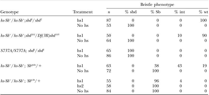
Rescue of bristle phenotypes: Table 2 shows rescue
with a wild-type transgene of the bristle phenotype of the mild hypomorphsbd2
and the more severe mutant
sbd201
with a single 1-hr heat shock at 27.5 hr AP, shortly before bristle nubs appear. With two copies of the trans-gene, 100% of the treatedsbd2
flies had wild-type bris-tles. A single copy of the hs-Sb-sbd1
transgene typically
Figure 3.—Sb-sbd constructs for transgenic lines. Also
shown is the location of the epitope for the stubblin stem (stem Ab) antiserum. The wild-typeSb-sbd(Sb1
produced bristles intermediate betweensbd2
and wild-type length (data not shown). Two copies of thehs-Sb1
transgene induced at 27.5 hr AP also rescued the more severe recessive bristle mutation,sbd201, with 90% rescue of bristles to wild type and 10% to bristles intermediate betweensbd201and wild-type length. Because of the high incidence of lethality in thesbd201homozygotes,sbd201/ Df sbd105animals were used for these experiments. Bris-tles of the dominant mutantSb63/1were not rescued by the wild-type transgene even with multiple heat shocks throughout early bristle development (24–30 hr AP). A single heat shock at 27.5 hr AP rescued bristles of the milder dominant mutantSbspike
/1 to wild type in 19% of the treated flies, while another 27% had bristles intermediate betweenSbspike
and wild type. No rescue was evident in 38% of theSbspike
/1heat-shocked flies. These results confirm the identity of theSb-sbdgene and are consistent with genetic predictions thatsbd2
is a hypo-morph, thatsbd201, although recessive, is an antimorph, and thatSbdominant mutants are neomorphs (Sb63b) or antimorphs (Sbspike).
Rescue of leg phenotypes:To rescue mlf legs inSb-sbd
mutants, the wild-typeSb-sbdtransgene was induced by a 1 hr 37° heat shock at the white prepupal stage (0 hr AP), corresponding to the time when theSb-sbdgene is normally expressed in prepupal development. Results from these experiments are summarized in Table 3. Induction of the wild-type transgene increased the occurrence of wild-type legs in bothsbd201
homozygotes and deficiency heterozygotes [sbd201
/Df(3R)sbd105 ]
car-rying two copies of the wild-type transgene. A single copy of the wild-typeSb-sbdtransgene insbd201
deficiency heterozygotes [hs-Sb-sbd1/1;
sbd201
/Df(3R)sbd105] res-cued mlf legs more effectively than two copies did, suggesting that leg morphogenesis is sensitive to over-expression of wild-type Sb-sbd. In preliminary experi-ments no rescue of malformed legs in the dominantSb
mutants Sb1/Df(3R)sbd105 or Sbspike/Df(3R)sbd105 was pro-duced by induction of the wild-typeSb-sbdtransgene at 0 hr AP using two copies of the transgene or in Sb63b/ Df(3R)sbd105 with either one or two copies (data not shown). This was unexpected, given that the mlf syn-drome is recessive, and indicates a functional difference between the defect in thesbd201
stubblin and theSb mu-tant stubblins despite similar leg phenotypes in these mutants. It is possible that some endogenous Sb-sbd
transcription precedes the 0-hr-AP induction of the wild-type transgene, so that mutant stubblin is already pres-ent and the heat-shock-induced wild-type stubblin is too late to rescue mlf legs in theSbmutants. Alternatively, or additionally, ‘‘collateral damage’’ of overexpression of
Sb-sbdcould mask rescue of the specificSbleg morpho-genesis defect. The improved rescue ofsbd201/Df(3R)sbd105 with one compared to two copies of the wild-type trans-gene supports the argument that there are deleterious effects of overexpression ofSb-sbd.
Overexpression of hs-Sb-sbd1
in wild-type flies: To
investigate the effects of overexpression of wild-type
Sb-sbd, the hs-Sb-sbd1
transgene was induced at other times throughout prepupal and pupal development in TABLE 2
Rescue of bristle phenotype with heat-shock induction of wild-typeSb-sbdtransgene
Bristle phenotype
Genotype Treatment n % sbd % Sb % int % wt
hs-Sb1 /hs-Sb1
;sbd2/sbd2 hs1 87 0 0 0 100
No hs 53 100 0 0 0
hs-Sb1 /hs-Sb1
;sbd201/Df(3R)sbd105 hs1 50 0 0 10 90
No hs 64 100 0 0 0
S737A/S737A;sbd2/sbd2 hs1 65 100 0 0 0
No hs 86 100 0 0 0
hs-Sb1 /hs-Sb1
;Sbspike/1 hs1 63 0 38 43 19
No hs 72 0 100 0 0
hs-Sb1/hs-Sb1;Sb63b/1 hs1 55 0 96 4 0
hs2 58 0 100 0 0
No hs 84 0 100 0 0
Homozygote sbd2/sbd2, deficiency trans-heterozygotesbd201/Df(3R)sbd105 or heterozygote Sb63b/1, and Sbspike/1
stocks carrying two copies of the wild-typeSb-sbdtransgene (hs-Sb1 /hs-Sb1
), andsbd2/sbd2carrying two copies
of the protease-disabled S737A transgene were collected as white prepupae, aged at 25°until 27.5 hr AP, and then either heat shocked at 37°for 1 hr (hs1) or maintained at 25°(no hs). Another set ofSb63bpupae carrying
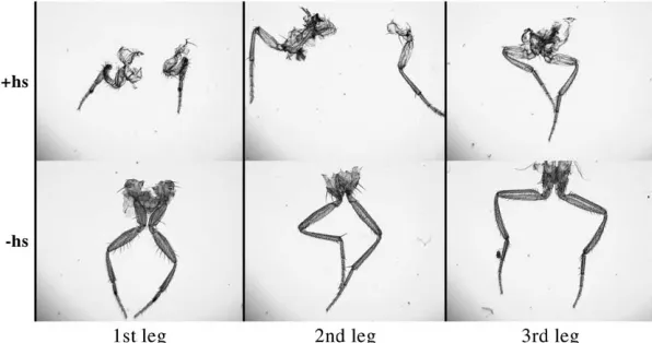
wild-type animals (Table 4). Induction ofhs-Sb-sbd1
at 0 hr AP or 3 hr AP produced malformed legs. After a 3-hr-AP heat shock, only 50% of the treated flies developed wild-type legs (the other 50% developed mlf) compared to 96% of the no-transgene control animals. With a 0-hr-AP-only heat shock, 41% of the treated flies had wild-type legs compared to 93% of the no-transgene controls. These observations are similar to those made by Bayer et al. (2003). This effect was not seen in prepupal heat shocks after 4 hr AP. Induction of hs-Sb-sbd1
during pupal development with a double heat shock at 30 hr AP and 33 hr AP produced a different and unanticipated phenotype (Figure 4). All segments of all legs in these
flies are shortened, with severity increasing on a distal-to-proximal axis. First and third legs are more severely affected than second legs. In addition to the leg phe-notype, there are defects in cuticle on the dorsal thorax (not shown), including a mild cleft and disorientation of the bristles similar to the phenotypes of mutations that affect thoracic closure (Usui-Ishiharaet al.2000;
Pena-Rangelet al.2002). The bristle morphology and
length are normal. A single heat shock at 27.5 hr AP, which rescuedsbdbristles, produced a slight shortening of prothoracic femurs, but otherwise wild-type flies. Heat shocks after 42 hr AP, after secretion of the adult cuticle, produced no leg or thoracic phenotype.
Expression of the protease-disabled transgene in
wild-type and Sb-sbd mutant flies: To determine if an
intact protease domain is required for rescue ofSb-sbd
loss-of-function mutations or overexpression effects, we
Figure4.—Leg effects ofSb-sbdoverexpression
in pupae in the first (1st), second (2nd), and third (3rd) legs of adults from pupae carrying two copies of thehs-Sb-sbd1
transgene either with (1hs) or without (ÿhs) a double heat shock at 30 and 33 hr AP. Note short proximal segments de-creasing in severity from femur to basitarsis in the legs from heat-shocked pupae. First legs show the most extreme effect.
TABLE 3
Rescue of mlf legs by heat-shock induction of the wild-type Sb-sbdtransgene
Leg phenotypes
Genotype Treatment n % mlf % wt
hs-Sb1
/hs-Sb1;
sbd201/sbd201 1hs 63 68 32
ÿhs 85 89 11
hs-Sb1/
hs-Sb1;
sbd201/Df(3R)sbd105 1hs 72 67 33
ÿhs 58 93 7
hs-Sb1/1;
sbd201/Df(3R)sbd105 1hs 117 54 46
ÿhs 118 91 9
1/1;sbd201/sbd201 1hs 52 94 6
ÿhs 88 96 4
S737A/1;sbd201/Df(3R)sbd105 1hs 121 100 0
ÿhs 122 93 7
White prepupae fromsbd201/sbd201orsbd201/Df (3R) sbd105stocks
carrying zero (1/1), one (hs-Sb1
/1), or two (hs-Sb1
/hs-Sb1 ) copies of the wild-type Sb-sbd transgene or one copy of the S737A protease-disabled transgene were either heat shocked for 1 hr at 37°immediately after collection at 0 hr AP (1hs) or maintained at 25°(ÿhs). Adults were scored for the mlf leg phenotype. The number scored (n) and percentage with mlf (% mlf) and wt (% wt) phenotypes are shown.
TABLE 4
Overexpression of wild-typeSb-sbdcauses defects in leg morphogenesis
Genotype Treatment n % wt
hs-Sb1/hs-Sb1;1/1 0 hr AP hs 118 54
3 hr AP hs 58 50
30 and 33 hr AP hs 55 0
No hs 120 100
No transgene 0 hr AP hs 88 96
3 hr AP hs 56 93
30 and 33 hr AP hs 70 100
No hs 70 100
White prepupae from wild-type flies carrying two copies of the wild-typeSb-sbdtransgene (hs-Sb1
repeated the rescue and overexpression experiments using a transgene with alanine replacing the critical cat-alytic serine at aa 737 (S737A) to disable the proteolytic function (see Figure 3). This transgene (hs-Sb-sbdS737A) was unable to rescuesbd2bristles (Table 2) orsbd201mlf legs (Table 3). Induction ofhs-Sb-sbdS737A in wild-type prepupae at 0 hr AP, 3 hr AP, or with a double heat shock at 0 and 3 hr produced no effect on leg morphogenesis (data not shown). Similarly, the leg and cuticle defects seen with the wild-typeSb-sbdtransgene induced at 30 hr and 33 hr AP were not seen with the protease-disabled transgene, implying a requirement for a functional pro-tease for the overexpression effects as well as for normal leg and bristle morphogenesis.
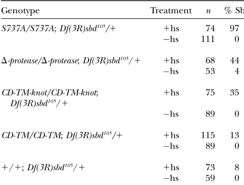
Generation of Sb bristles by expression the Sb-sbd
truncation and S737A transgenes: To generate
domi-nant bristle phenotypes similar to those seen with the dominant mutations that make truncated transcripts, we made a series of truncatedSb-sbdtransgenes (Figure 3). Three transgenic constructs—the truncation delet-ing the protease domain (D-protease) and the trunca-tion deleting the protease and most of the stem, but including the disulfide knot (CD–TM–knot), as well as the S737A mutation—all produced at least some short, thickSb-like bristles when expressed throughout early bristle development. A single heat shock at 27.5 hr did not produce a detectable phenotype. Table 5 shows the results of a series of four heat shocks at 25, 27, 29, and 31
hr AP with two copies of the transgene and a single copy of the endogenous Sb-sbdgene. In all cases there was some variation in the bristle phenotype, with the dorso-central bristles most consistently short. The hs-Sb-sbd
S737A transgene produced short-bristle phenotypes with highest penetrance and severity; 97% of the heat-shocked pupae produced adults with predominantly short bristles, many shorter than those of Sb1
. While some of these bristles are both short and thick as inSb63b, some have thick bases with thin extensions, a combina-tion of theSbandsbdbristle phenotypes. Figure 5 shows a confocal image of an actin-stained bristle dissected at 37 hr AP from a pupa carrying the S737A transgene induced by heat shock at 25, 27, 29, and 31 hr AP. Also shown is a bristle dissected from a wild-type pupa (no transgene) heat shocked in parallel. Although there is a dramatic difference in bristle length between these two 37-hr-AP bristles, the short bristle does not show the consistent actin bundle mislocalization and increase in the number of bundles seen in aSb63b
bristle,i.e., 25–30 bundles not restricted to the cell periphery inSb63b
homo-zyogtes compared to 12–15 solely peripheral bundles in wild-type bristles (Appelet al.1993). The more
consis-tent and severe phenotype may require the constant and abundant presence of the truncated protein in bristles as suggested by the difference in bristle phenotype be-tween Sb1, in which the truncated
Sb1 transcript is ex-pressed in amounts comparable to the wild-type transcript, andSb63b, in which the similarly truncated transcript is overexpressed compared to the wild-type transcript.Sb1 bristles are longer than those ofSb63band whileSb1 bris-tles often have extra actin bundles, the number of bundles (11–18) overlaps with that of wild type (Leesand Picken
1945; Overton1967).
The bristle effects of transgenes expressing only the cytoplasmic domain with the transmembrane domain (CD–TM) were indistinguishable from heat-shocked
Figure 5.—Sb-like bristle generated by expression of
the S737A transgene. Confocal image (negative image) of phalloidin-stained bristles dissected from pupae at 37 hr AP following heat shocks at 25, 27, 29, and 31 hr AP. (Left) An Sb-like bristle from an S737A transgene-carrying pupa [S737A/S737A; Df(3R)sbd105/1]. (Right) A normal bristle
from a wild-type control pupa (wt) also subjected to the same heat-shock regimen. The tip of the much longer wild-type bristle cannot be seen in this view. The entire S737A bristle is shown. Bars, 5mm.
TABLE 5
Transgenic expression ofSb-Sbdtruncations or a disabled protease in pupae produces Sb-like bristles
Genotype Treatment n % Sb
S737A/S737A;Df(3R)sbd105/1 1hs 74 97
ÿhs 111 0
D-protease/D-protease;Df(3R)sbd105/1 1hs 68 44
ÿhs 53 4
CD-TM-knot/CD-TM-knot;
Df(3R)sbd105/1
1hs 75 35
ÿhs 89 0
CD-TM/CD-TM;Df(3R)sbd105/1 1hs 115 13
ÿhs 89 0
1/1;Df(3R)sbd105/1 1hs 73 8
ÿhs 59 0
Sb-sbddeficiencytrans-heterozygotes [Df(3R)sbd105/1]
carry-ing two copies of the S737A transgene or two copies of one of the truncation transgenes [D-protease, CD-TM-knot, CD-TM] or no transgene [1/1; Df(3R)sbd105/1] were collected and
lines without any transgene (w; sbd105/1), even when the copy number of the transgene was increased to 3 ( w/hs-Sb CD-TM;hs-Sb CD-TM/hs-Sb CD-TM: sbd105/1). How-ever, because we cannot confirm expression of these shorter constructs with our antibody to the stubblin stem, these results must be considered preliminary. Pupal heat shocks with the wild-type transgene resulted in adults with wild-type bristle length but with severe leg and thorax malformations (Table 4 and data not shown). No leg or thorax effects were seen with any of the truncations or the S737A substitution.
Localization of stubblin in discs: Cell-shape change
occurring at the apical surface during prepupal morpho-genesis is driven by actin–myosin contractility (Condic et al.1991). Previously, we reported that an antibody to the juxtamembrane region of stubblin localized to the apical surface in prepupal discs, consistent with a direct role for stubblin in apical cell-shape change (vonKalm et al.1995). We repeated this localization using a new antiserum made against a nonrepetitive region of the stem (see Figure 3) with similar results (Figure 6, A, B, and E). This antiserum also stains prepupal discs from
Sb63b
homozygotes, but more intensely, as predictable from the overexpression of theSb63b
transcript (Figure 6D). At 3.5 hr AP, theSb63bdiscs already are clearly shorter than their wild-type counterparts, although elongation has started. Leg discs from heat-shocked third instar wild-type larvae carrying two copies of the wild-wild-type Sb-sbd
transgene show precocious apical localization, visible as concentric rings in the unevaginated disc (Figure 6F). Non-heat-shocked controls show no stubblin expression as expected for larval discs prior to the rise in ecdysone titer (Figure 6C).
Localization of stubblin in developing bristles:
Be-causeSb-sbdmutants affect the actin bundle scaffolding in growing bristles, it was of interest to determine the localization of stubblin during bristle development when actin bundles are forming. We used the stubblin stem
antiserum for immunolocalization studies in bristles from 32 hr AP, when bristle buds first appear, through 38 hr AP, about midway through bristle extrusion. Actin staining of early bristles shows the beginning of actin bundles as punctate staining on the surface of the bristle bud with discrete bundles extending to the base of the shaft (Tilneyet al.1996; Wulfkuhleet al.1998; Figure
7B). In contrast, stubblin staining at 34–35 hr AP shows cytoplasmic staining of the cell body of the bristle shaft cell with diffuse staining of the budding shaft and tip (Figure 7, A and C). By 37 hr AP, stubblin staining is more concentrated at the tips, indicating that stubblin is transferred to the tip (Figure 7D). From our micro-graphs, we cannot rule out that the early staining seen at the bristle base is in the socket cell, but because ex-truding bristle shafts clearly show stubblin staining, we argue by parsimony that expression is restricted to the shaft cell.Sb63bbristles at 37 hr AP are shorter than their wild-type counterparts but stain in a similar pattern ex-cept the staining is more intense, with the appearance of heavily staining cap-like extensions on the tips (Figure 8). These localization patterns of stubblin in growing bris-tles, distinct from those of actin, particularly in sprout-ing bristles when actin bundles are first detectable, indicate that the actin bundle defects seen inSb-sbd mu-tants do not result from a direct interaction of stubblin with actin bundles. The cap of stubblin that appears at the ends ofSb63bbristles suggests the possibility that an accumulation of defective stubblin at the bristle tips may block bristle extension, consistent with the Sb bristle phenotype of abruptly terminated bristles.
DISCUSSION
The ecdysone-induced Sb-sbd gene is required for normal morphogenesis of imaginal discs and formation of bristle shafts, both processes involving cytoskeletal
Figure6.—Larval and prepupal leg discs
stained with stubblin antistem antiserum. (A and E) Wild-type leg discs dissected at 3.5 hr AP show staining at the apical sur-face. (E) A confocal image; (A–D and F) conventional epifluorescence. (B) Nega-tive control (no primary antibody). (D)
Sb63b/Sb63bleg discs dissected at 3.5 hr AP
show a similar pattern but more intense staining, consistent with the overexpres-sion of theSb63btranscript.Sb63b/Sb63bdiscs
have not elongated as far as their wild-type counterparts. (C and F) Larval discs dis-sected prior to normal endogenousSb-sbd
changes and extracellular proteolytic activity. The mor-phogenesis of the leg imaginal disc to form a tubular leg depends on myosin-driven contraction leading to apical cell-shape change. In contrast, formation of bristles is driven by polymerization of membrane-associated, par-allel actin filaments. We discuss here the possible roles of Sb-sbd in imaginal disc morphogenesis and bristle extrusion and speculate that the stubblin protease mod-ifies apical ECM and contributes, directly or indirectly, to activation of a Rho-signaling pathway.
The role of Sb-sbdin leg disc morphogenesis:
Mor-phogenesis of leg imaginal discs requires both contrac-tion of the apical actin–myosin contractile ring and proteolysis. In rapid response to the ecdysone spike that triggers metamorphosis, the shapes of cells in the distal femur, tibia, and basitarsis change from elongated to isometric (Condic et al. 1991). If these changes are
blocked, the resulting legs in the adult fly are mal-formed with short and thick proximal segments. Genet-ical and cell biologGenet-ical observations lead to the view that leg morphogenesis depends on myosin-driven contrac-tility. Both actin and nonmuscle myosin II localize to the apical belt in leg discs (vonKalmet al.1995).
Cytocha-lasins, which disrupt actin filaments, reversibly inhibit leg disc elongation (Fristrom and Fristrom 1975).
Mutations in the zip gene, which encodes the non-muscle myosin II heavy chain, cause the mlf phenotype as do those of the myosin regulatory light chain gene,
spaghetti squash(sqh) (Edwardsand Kiehart1996).
Contractility alone is insufficient for leg morpho-genesis. The leg disc epithelium is covered by a basal ECM similar to those of vertebrates, containing collagen IV, laminin, and sulfated proteoglycans (Fessler and
Fessler1989; Fristromand Fristrom1993). The disc
apical surface secretes a chitinous cuticle (exoskeleton) during metamorphosis. Epidermal morphogenesis, in-cluding leg elongation during prepupal morphogenesis and bristle formation during pupal morphogenesis, must occur when the epidermis is unconstrained by the exoskeleton. Prepupal leg elongation occurs between pupariation and the formation of the pupal chitinous exoskeleton (Fristrom and Fristrom1993). Apolysis
of the pupal cuticle at 18 hr AP allows further leg morphogenesis and formation of bristles and hairs before the adult exoskeleton is deposited between 36 to 70 hr AP. After apolysis, the apical surface of the disc secretes a poorly characterized, fibrous, nonchitinous ECM (Brower et al. 1987; Fristrom and Fristrom
1993). A role for proteolysis in disc morphogenesis is well documented. In addition toSb-sbd, other proteases known to be regulated by ecdysone and potentially involved in disc morphogenesis include an uniden-tified extracellular protease secreted by cultured discs in response to ecdysone (Pino-Heissand Schubiger
Figure 8.—Wild-type and Sb63b/1 bristles from 36-hr-AP
pupae stained with stubblin stem antiserum. Both show stub-blin protein localized to bristle cell base, shaft, and tip, but
Sb63bstaining is more intense, with heavily staining tip
exten-sions (arrow) not seen in the wild-type bristles. Bars, 20mm.
Figure7.—Stubblin localization in
1989) and a collagenase that cleaves type IV collagen (Birret al.1990; Fessleret al.1993). Disc
morphogen-esis is restricted by protease inhibitors (Pino-Heissand
Schubiger1989). It is acceleratedin vitroby exogenous
trypsin or chymotrypsin (Poodryand Schneiderman
1971; Feketeet al.1975). Addition of 0.1% trypsin to the in vitromedium decreases the time of leg elongation of dissected discs from 2–3 hr to 10 min, but only in discs that have been exposed to ecdysone (Feketeet al.1975).
Protease treatment does not passively lead to leg elonga-tion; trypsin acceleration of leg elongation is inhibited under conditions that reduce ATP levels and does not overcome cytochalasin B inhibition of disc elongation (Feketeet al.1975). Thus, contractility and proteolysis
must be coordinated for normal morphogenesis to oc-cur. Stubblin, the product of ecdysone-dependentSb-sbd
and a TTSP that localizes to the apical surface of leg discs, is a candidate for one of the coordinators.
A role forSb-sbd in leg morphogenesis was first de-scribed by Dobzhansky (1929) and rediscovered
be-cause of the genetic interaction betweenSb-sbdand the metamorphic transcriptional regulator, br, to produce mlf legs (Beaton et al.1988). The legs of some Sb-sbd
homozyogtes,Sb-sbd trans-hetereozygotes, orbr; Sb-sbd/1 double mutants are mlf. Leg morphogenesis in these mutants can be rescuedin vitroby exposure to exoge-nous trypsin in prepupal discs (Appelet al.1993). Our
current studies show that induction of the wild-type Sb-sbdtransgene early in prepupal development (0 hr AP) partially rescued severe sbd201/
sbd201 mlf legs, but not alleles with dominant bristle phenotypes (Sb1,
Sb63b, Sbspike). Rescue depends on the presence of a functional pro-tease catalytic domain; the catalytically disabled S737A transgene did not rescue mlf legs. One copy of the wild-type transgene rescued more animals than two copies (50% rescuevs. 30%), demonstrating that overexpres-sion of wild-type stubblin has deleterious effects on leg development. Expression of the wild-type transgene in wild-type animals during prepupal development re-sulted in adults with mlf legs, confirming the sensitivity of leg morphogenesis to overexpression of stubblin. Overexpression of proteasein vivomay result in excess detachment of epidermis from the apical ECM, com-promising the structural integrity of the elongated leg and causing malformations even if cell-shape changes are completed. Relevant here is our observation that some stubblin overexpression phenotypes resulting from induction of the wild-typeSb-sbdtransgene in wild-type prepupae (0 and 3 hr AP) were not identical to ‘‘classic’’ mlf. While ‘‘classic’’ mlf legs associated with limited cell-shape changes have short, kinked femurs, primarily on the metathoracic legs (see Beatonet al.1988, Figure 1),
prepupal overexpression of stubblin also produced some longer, fragile mesothoracic and metathoracic legs that arguably could result from excessive leg elongation like that seenin vitrowith protease exposure (Fekete et al.
1975).
Myosin II contractility depends on the phosphoryla-tion state of myosin regulatory light chain (MLC) (Tan et al.1992; Amanoet al.1996). Numerous studies point
to the Rho subfamily of small GTPases as upstream regulators of the actin cytoskeletal rearrangements and control of MLC phosphorylation (Tapon and Hall
1997; VanAelstand D’Souza-Schorey 1997; Ridley
2001). Recent genetic interaction studies have identi-fied members of a potential Rho-signaling pathway that may lead to the contraction of the actin–myosin apical belt and the cell-shape changes that drive prepupal leg disc morphogenesis. Rho-signaling pathway com-ponents implicated in Drosophila disc morphogenesis includeRhoGEF2andRhoA(Halsellet al.2000; Ward et al. 2003) and DrosophilaRho kinase(drok) (Winter et al. 2001). Mutations in these genes interact withzip
mutations to produce mlf legs (Gotwalsand Fristrom
1991; Halsellet al.2000; Bayeret al.2003). Activators
upstream ofRhoAin leg morphogenesis have not been identified. The possibility thatSb-sbdis involved in this or a parallel pathway is suggested by the mlf leg phenotype of Sb-sbdmutations in combination withzipmutations
(Gotwals and Fristrom 1991) or with mutations of
RhoA,RhoGEF2, ordrokgenes (Bayeret al.2003; Ward et al.2003).
Any consideration of the mechanism of action of the
Sb-sbdgene in imaginal disc morphogenesis must take into account the nature of the morphogenetic defects associated with Sb-sbd mutations. The proteolytic do-main of stubblin is essential for normal morphogenesis. Stubblin insbd201
/sbd201, which has an arguably impaired catalytic domain, and stubblin molecules lacking the proteolytic domain (inSb63b,Sb70, andSb1) are associated with abnormalities in disc morphogenesis. Models for roles of the proteolytic domain are all speculative be-cause the stubblin substrate is unknown. The possibility that stubblin may directly or indirectly modify the apical ECM has been suggested often (Beaton et al. 1988;
Appelet al. 1993;vonKalmet al. 1995; Hooperet al.
2001; Bayeret al.2003) and is supported by the
accel-eration of leg elongation caused by exogenous trypsin, the in vitro rescue of partially elongated Sb63b
/Sb63b leg discs by trypsin, and the apical localization of stubblin in leg discs. However, stubblin may have a dual role both in modifying the ECM to permit cell-shape change and in stimulating the contractility of the apical contractile belt, for example, by activating a Rho-signaling pathway. Because morphogenesis begins inSb mutants that are missing the protease domain, stubblin cannot be acting alone to initiate contractility, but may be required to amplify a signal to complete morphogenesis.
is unlikely because the stubblin cytoplasmic domain lacks identified protein interaction motifs, and our pre-liminary studies suggest that overexpression of the SbCD or SbCD-TM transgenes has no effect on leg mor-phogenesis. Regarding the second possibility, serine proteases that, like stubblin, use the Ser-His-Asp cata-lytic triad can be classified according to highly con-served evolutionary markers on the basis of codon usage at Ser195 and Ser214 and the presence of Provs. Tyr at residue 225 (chymotrypsin numbering) (Kremand
DiCera2001). According to this classification system,
stubblin belongs to the oldest lineage and therefore is more likely, on the basis of analysis of other proteolytic cascades, to be the terminal or penultimate protease in an activation cascade and less likely to be an initiator (Krem and Di Cera 2001). To speculate briefly, a
protease-activated receptor (PAR) could be a stubblin substrate. PARs belong to a family of G-protein-coupled receptors that are activated by site-specific proteol-ysis (Dery et al. 1998). Several other TTSP proteases
have been demonstrated to activate protease-activated receptor-2 (PAR-2) (Linet al.1999; Takeuchiet al.2000;
Friedrichet al.2002; Iwakiriet al.2004; Wilsonet al.
2005). Proteolytic modification of ECM attachments not only may be permissive, but also may actively con-tribute to Rho-mediated signaling. Future studies must focus on the identification of stubblin substrates.
The role of Sb-sbd in bristle morphogenesis: Bristle
formation involves actin polymerization but not myosin II contractility. The first bristle nubs appear at32 hr AP, during a period of high ecdysone titer, and extend to their full length by 48 hr AP (Leesand Waddington
1942; Fristrom and Fristrom 1993). Bristles grow
from the tip (Leesand Picken1945). The bristle
elon-gates by the assembly of membrane-associated, cross-linked actin filament bundles formed in short modules, new bundles being joined end to end with the pre-ceding more basal bundle (Tilneyet al.1996; Guild et al. 2003). From observations of bristle growth in cultured thoraces in the presence of compounds that effect actin polymerization or microtubule dynamics, Tilney et al. (2000, 2003) concluded that actin
poly-merization drives bristle elongation. Unlike leg disc morphogenesis, bristle development is not affected by mutations inzip and these mutations do not interact withSb-sbdmutations to affect bristle elongation (Bayer et al.2003). In the bristles of recessivesbdanimals, the actin bundles are normal at first, but stop prematurely and asynchronously, so that some bundles continue to extend, while others stop. The resulting bristles are shorter than wild-type bristles and have ragged ends in-stead of smooth, tapered ends. This defect was rescued by the wild-typeSb-sbdtransgene in both the mild allele
sbd2
and the more severe allele sbd201
. The molecular defect insbd201
, a histidine-to-arginine substitution at aa 571 in the catalytic domain, is likely to compromise but not abolish protease activity (seeresults). In contrast
to the results with mlf leg rescue, complete rescue of
sbd201orsbd2bristles required two copies of thehs-Sb-sbd1
transgene, a single copy resulting in partial rescue (intermediate length bristles). Only the transgene with an intact protease domain rescued bristles. Unlike legs, bristles are not sensitive to overexpression of wild-type stubblin.
Identification of molecular defects in Sb dominant mutations, Sb63b
and Sb70
, showed that truncated, pro-tease-absent stubblin causes the severe dominant bristle phenotypes. The bristles in these mutants and those in
Sb1
, another dominant mutant associated with a trans-posable element eliminating the protease domain, were not rescued by the wild-type transgene, and so these three mutations are likely to be neomorphs, acting in some novel way not characteristic of the wild-type pro-tein. The bristle phenotype of the milder allele Sbspike, a frameshift mutation that adds sequence to the C terminus of the protease domain and likely disrupts normal stubblin function, was ameliorated by the wild-type transgene, although not rescued completely to wild-type length. Induction of transgenes truncated to remove the protease domain and then progressively larger portions of the noncatalytic region showed dom-inant effects from expression of truncated protein as short as the CD–TM–knot construct. The most pene-trant and severe phenotypes were produced by the S737A construct, which changes only the catalytic ser-ine. Consistent with the difference in the dominant and recessive character ofSbandsbdmutations in bristle and legs, none of the truncation transgenes had any effects on leg morphogenesis. Taken together, these obser-vations indicate a role for the stubblin noncatalytic domains that is specific to bristle elongation. The lo-calization pattern of stubblin in bristles brings up the possibility that the stem or knot may be involved in localization of the protease to the bristle tip, although this is not directly testable with our antiserum to the C-terminal region of the stem. Stubblin appears first in the budding bristle cell body (and perhaps in the socket cell) and then along the shaft to the bristle tip where it concentrates. Immunolocalization with the stubblin stem antiserum in Sb63b
/1 developing bristles showed excessive accumulation of protein at the bristle tips compared to the normal protein in wild-type bristles. If the noncatalytic domains direct stubblin localization, expression of a truncatedSb63borSb1protein may block wild-type stubblin proteolytic function. The observation that overexpression of the wild-type Sb-sbd transgene fails to rescue the bristle phenotype of these mutants suggests that either the transgenic stubblin is ineffi-ciently transported or the interference is irreversible. The requirement for two copies of the wild-type trans-gene for bristle rescue of even the mildestsbdmutation indicates that bristle elongation depends on a sufficient concentration of protease. This extremely long