University of Windsor University of Windsor
Scholarship at UWindsor
Scholarship at UWindsor
Electronic Theses and Dissertations Theses, Dissertations, and Major Papers
7-11-2015
Ontogeny of glomerular territory patterning in the olfactory bulb of
Ontogeny of glomerular territory patterning in the olfactory bulb of
juvenile Chinook salmon (Oncorhynchus tshawytscha) and
juvenile Chinook salmon (Oncorhynchus tshawytscha) and
exploring for potential effects of sensory experience on
exploring for potential effects of sensory experience on
glomerular development
glomerular development
Courtney Lucy Ochs University of Windsor
Follow this and additional works at: https://scholar.uwindsor.ca/etd
Recommended Citation Recommended Citation
Ochs, Courtney Lucy, "Ontogeny of glomerular territory patterning in the olfactory bulb of juvenile Chinook salmon (Oncorhynchus tshawytscha) and exploring for potential effects of sensory experience on glomerular development" (2015). Electronic Theses and Dissertations. 5319.
https://scholar.uwindsor.ca/etd/5319
This online database contains the full-text of PhD dissertations and Masters’ theses of University of Windsor students from 1954 forward. These documents are made available for personal study and research purposes only, in accordance with the Canadian Copyright Act and the Creative Commons license—CC BY-NC-ND (Attribution, Non-Commercial, No Derivative Works). Under this license, works must always be attributed to the copyright holder (original author), cannot be used for any commercial purposes, and may not be altered. Any other use would require the permission of the copyright holder. Students may inquire about withdrawing their dissertation and/or thesis from this database. For additional inquiries, please contact the repository administrator via email
Ontogeny of glomerular territory patterning in the olfactory bulb of juvenile Chinook salmon (Oncorhynchus tshawytscha) and exploring for potential effects of
sensory experience on glomerular development
By
Cory L. Ochs
A Thesis
Submitted to the Faculty of Graduate Studies through the Department of Biological Sciences
in Partial Fulfillment of the Requirements for the Degree of Master of Science
at the University of Windsor
Windsor, Ontario, Canada
2015
Ontogeny of glomerular territory patterning in the olfactory bulb of juvenile Chinook salmon (Oncorhynchus tshawytscha) and exploring for potential effect of
sensory experience on glomerular development
By
Cory L. Ochs
APPROVED BY:
__________________________________________________ C. Semeniuk
Great Lakes Institute for Environmental Research
__________________________________________________ D. Higgs
Department of Biological Sciences
_________________________________________________ B. Zielinski, Advisor
Department of Biological Sciences
_________________________________________________ T. Pitcher, Advisor
Department of Biological Sciences
iii
Declaration of Co-Authorship
I hereby certify that this thesis incorporates material that is the result of joint research as
follows: My second chapter was co-authored with my supervisors, Dr. Barbara Zielinski
and Dr. Trevor Pitcher, as well as Tina Suntres. My third chapter was co-authored with
my supervisors, Dr. Barbara Zielinski and Dr. Trevor Pitcher. My collaborators
contributed to experimental design, provided technical support that directly contributed to
the dataset, and provided editorial input. I am the primary contributor to each chapter of
this thesis. No part of this thesis has been published or submitted for publication.
I am aware of the University of Windsor Senate policy on authorship and I certify that I
have properly acknowledged the contribution of other researchers to my thesis, and have
obtained written permission from my co-authors to include the above materials in my
thesis.
I certify that, with the above qualification, this thesis, and the research to which it refers,
is the product of my own work, completed during my registration as a graduate student at
the University of Windsor.
I certify that, to the best of my knowledge, my thesis does not infringe upon anyone’s
copyright nor violate any proprietary rights and that any ideas, techniques, quotations, or
any other material from the work of other people included in my thesis, published or
otherwise, are fully acknowledged in accordance with the standard referencing practices.
iv
bounds of fair dealing within the meaning of the Canada Copyright Act, I certify that I
have obtained a written permission from the copyright owner(s) to include such
material(s) in my thesis and have included copies of such copyright clearances to my
appendix.
I declare that this is a true copy of my thesis, including any final revisions, as approved
by my thesis committee and the Graduate Studies office, and that this thesis has not been
v
Abstract
Axon terminals of olfactory sensory neurons (OSNs) aggregate into glomeruli, functional
units of odour discrimination within the olfactory bulb. Glomerular patterning facilitates
assessment of olfactory stimulation-induced changes to neural circuitry. Contemporary
studies indicate Chinook salmon (Oncorhynchus tshawytscha) alevin imprint to olfactory
cues, purportedly amino acids, prior to emergence. In this study, OSNs were labelled
against keyhole limpet hemocyanin to characterize the development of glomerular
territories from hatch to emergence, focusing on calretinin-immunoreactive OSNs.
Glomerular territories were distinguishable at hatch, and showed expansion and moderate
refinement with maturation into emergent fry. Calretinin-immunoreactive OSNs
innervated the dorsolateral and lateral glomerular territories, and four lateral glomeruli,
lG1,lG2,lG3/4,and lG6. Neither amino acid exposure nor somatic growth influenced
glomerular volume of one glomerulus, lG1. The establishment of glomerular territories at
hatch infers a functional olfactory system, but a sensitive stage for visible effects of
vi
Dedicated to Pierre-Paul and Sawyer Bitton, Karen Ochs, and Cameron Beck
“A simple intuition, a single observation, can open vistas of unimagined potential. Once caught in the web of an idea, the researcher is happily doomed, for the outcome is always uncertain, and the resolution of the mystery may take years to unfold.”
vii
Acknowledgements
The sciences are increasingly integrated, and as such, a team contributes to the
completion and success of any project. I thank my supervisors, Dr. Barbara Zielinski and
Dr. Trevor Pitcher for time and resources they invested in this project. Substantial
freedom was granted in the development of this research project. Dr. Zielinski
approached my research with a meticulousness I was unprepared for, yet appreciated. She
patiently integrated the neuroscience component into my research, and indeed, it became
the primary analytical approach. Her mentorship has expanded my interests to the
dynamic field of sensory ecology, and specifically chemical signalling. Dr. Trevor
Pitcher provided an outlet to the ecological field, and introduced me to fisheries. I did not
anticipate that I would enjoy this cold, damp system as much as I have, and I value the
applicability of this new skillset.
My committee members, Dr. Christina Semeniuk and Dr. Dennis Higgs, invested
hours in my research over the course of my Master’s degree. They contributed valuable
feedback and encouragement, improving the project and manuscripts.
I am grateful to past members of the Pitcher lab, notably Craig Black, Katelyn
Johnson, and Michaela Haring, for providing support in the field and with animal care. I
enjoyed working with Ontario Ministry of Natural Resources and Forestry and
Normendale Fish Culture Station employees during three consecutive field seasons, and
appreciate the knowledge they passed down. The Normendale Fish Culture Station also
donated the fertilized eggs that made my third chapter possible. Current Pitcher lab
viii
lab, conversation with Dr. Warren Green inspired my entry into sensory ecology.
Pierre-Paul Bitton, Jennifer Smith and Dr. Michelle Nevett provided field support on the most
difficult days. Dr. Nevett was further integral in the final collation of this thesis. Tina
Suntres and Alex Zygowska provided invaluable contributions to this project as talented
undergraduate researchers. Thank-you to the past and current members of the Zielinski
lab for your friendship, candid banter, stimulating discussion and substantial support:
Karl Boyes, Dr. Eric Clelland, Gianfranco Grande, Dr. Warren Green, Jenna Jones,
Charrie McFadden, Dr. Michelle Nevett, Jennifer Smith, and Tina Suntres. Our many
skilled undergraduate students provided valuable assistance.
Chirag Patel, Lena Jamal and Sehrish Butt provided sound advice, technical
assistance, and eased my entry into neuroscience. The Swan lab was always available to
provide friendly assistance with the confocal microscope. The Department of Biology
staff, particularly Nancy Barkley, Ingrid Churchill, Bob Hodge, and Rodica Leu create a
fantastic work environment, and make research possible. Marc St. Pierre and Steve
Budinsky provided technical assistance. Past and present members of the Ciborowski,
Doucet, Mennill and Vanlaerhoven labs have made Windsor home.
Support from family allowed me to dedicate more than two years to this thesis.
Thank-you Guy, Franҫoise, Marie-France, and Carmine for unyielding support.
Pierre-Paul Bitton provided technical and analytical contributions in conjunction with
camaraderie for the duration of this project. Your patience, encouragement, and solid
presence on the home front enabled the completion of this thesis. Thank you for jumping
in at quitting points by retrieving viable eggs, assisting in post-hurricane fieldwork, and
ix
been one of my greatest motivators. My intelligent parents, Karen and Cameron, instilled
the importance of dedication and quality workwomanship. Your feedback keeps me
honest and grounded. Thank-you also for being here for Sawyer. Peter, Helen, and Jessie,
x
Table of Contents
Declaration of Co-Authorship ____________________________________________ iii Abstract ______________________________________________________________ v Acknowledgements ____________________________________________________ vii List of Tables _________________________________________________________ xii List of Figures ________________________________________________________ xiii List of Abbreviations __________________________________________________ xv
Chapter 1: General Introduction _________________________________________ 1 Ecological significance of olfaction in Pacific salmonids _____________________ 1 Olfactory development from embryo to emergence in salmonids _______________ 1 Olfactory glomerular patterning as a measure of olfactory experience ___________ 5 Potential impact of hatchery environment on olfaction _______________________ 7 Chinook salmon as a model species _____________________________________ 7 Olfactory imprinting paradigms ________________________________________ 9 Thesis Objectives ____________________________________________________ 11 References __________________________________________________________ 13 Figures _____________________________________________________________ 21
Chapter 2: Ontogeny of glomerular territory patterning in the olfactory bulb of juvenile Chinook salmon Oncorhynchus tshawytscha ________________________ 22
Introduction _________________________________________________________ 23 Materials and Methods ________________________________________________ 28 Fertilization and rearing conditions of lacustrine Chinook salmon _____________ 28 Sample collection and tissue preparation ________________________________ 29 Immunocytochemistry techniques ______________________________________ 30 Results _____________________________________________________________ 34 Dorsal glomerular territories __________________________________________ 35 Lateral glomerular territory ___________________________________________ 36 Ventral glomerular territories _________________________________________ 37 Ontogeny of glomerular territories _____________________________________ 38 Discussion __________________________________________________________ 40 Ontogeny of glomerular patterning and functional implications _______________ 40 Glomerular patterning across teleosts ___________________________________ 43 References __________________________________________________________ 47 Tables and Figures ___________________________________________________ 55
Chapter 3: Exploring for effects of olfactory enrichment on volume of lateral
xi
Experimental animals _______________________________________________ 83 Experimental design ________________________________________________ 83 Sample collection and preparation______________________________________ 84 Immunocytochemistry and microscopy __________________________________ 85 Volumetric and statistical analyses _____________________________________ 86 Results _____________________________________________________________ 88 Discussion __________________________________________________________ 90 References __________________________________________________________ 96 Tables and Figures __________________________________________________ 104
Chapter 4: General Discussion _________________________________________ 116 Summary __________________________________________________________ 116 Overview of Chapter 2 _______________________________________________ 117 Overview of Chapter 3 _______________________________________________ 122 Conclusions ________________________________________________________ 124 References _________________________________________________________ 126
xii
List of Tables
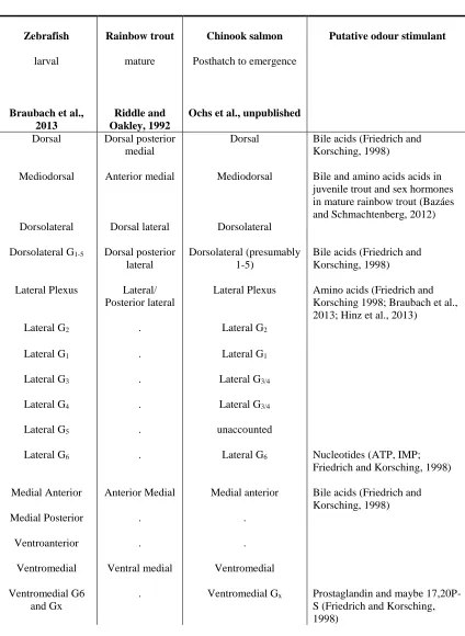
Table 2.1. Nomenclature of terminal fields of olfactory sensory neuron (OSN) axonal projections in the teleost olfactory bulb in across-species comparison of olfactory
glomerular patterning. ... 55
Table 2.2. Glomerular territories and glomeruli identified in the Chinook salmon alevin olfactory bulb analogous to those identified in zebrafish larvae and mature rainbow trout. ... 57
Table 3.1. Effects of sensory experience on neural circuitry of corresponding glomeruli in the antennal lobe of insects in a laboratory setting. ... 104
Table 3.2. Effects of passive and associative olfactory sensory experience on neural circuitry of corresponding glomeruli in the laboratory mouse olfactory bulb. . ... 105
Table 3.3. Influence of passive olfactory sensory experience on neural circuitry of corresponding glomeruli in the zebrafish olfactory bulb. ... 106
Table 3.4. L-amino acids and concentrations used in previous studies to investigate the effects of amino acid exposure on the olfactory responses of teleosts. ... 107
Table 3.5. Properties of stock solution amino acids to test whether odours affect the early olfactory development of juvenile Chinook salmon. ... 108
Table 3.6. Mass of test L-amino acids required for 500 mL stock solution to inject into the amino acid incubation tray for a duration of two days ((2 mL * 4 injections /
hr)*24*2). ... 109
xiii
List of Figures
Figure 1.1. Olfactory sensory neuron (OSN) pathway from the olfactory epithelium to olfactory bulb. ... 21
Figure 2.1. Comparison of glomerular patterning at three increments in Chinook salmon to assess ontogeny of these structures from posthatch to emergence.. ... 59
Figure 2.2. Chinook salmon emergent fry olfactory bulb situated dorsal to the olfactory nerve and olfactory epithelium. ... 60
Figure 2.3. KLH and Calretinin immunolabeled horizontal serial sections of the olfactory epithelium, olfactory nerve and olfactory bulb of Chinook salmon emergent fry. ... 61
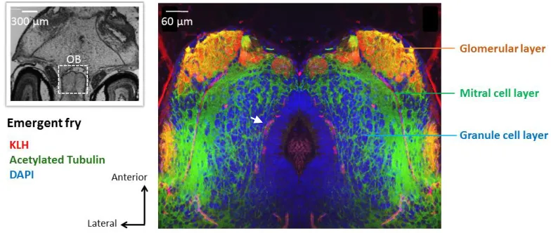
Figure 2.4. Horizontal section of an emergent Chinook salmon olfactory bulb triple labelled against KLH, DAPI, and AT to identify the different layers. ... 62
Figure 2.5. Organization of glomerular territories in an emergent fry, innervated by KLH- and calretinin-immunoreactive OSN axons. ... 63
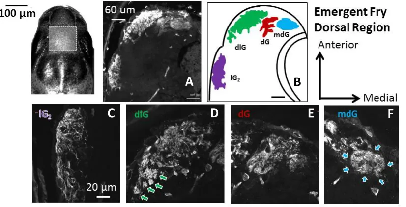
Figure 2.6. A,B: Confocal acquired micrographs of KLH-immunoreactive glomerular territories in the dorsal region of the emergent Chinook salmon olfactory bulb (horizontal sections). ... 64
Figure 2.7. Dorsal glomerular territories (108 µm from the dorsal-most section) in emergent Chinook salmon double-labelled against KLH (A) and calretinin (B). ... 65
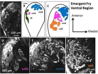
Figure 2.8. A, D, E, F: Confocal acquired micrographs of KLH-immunoreactive glomerular territories in the ventral region of the emergent Chinook salmon olfactory bulb (502 µm from dorsal-most section). ... 66
Figure 2.9. Ventral glomerular territories in emergent Chinook salmon double-labelled against KLH (A, D), calretinin (B, E) and merged labelling (C, F). ... 67
Figure 2.10. A-F: Confocal projections of calretinin-IR lateral glomeruli in horizontal sections of the posthatch Chinook salmon olfactory bulb (olfactory bulb depth from dorsal-most section noted) ... 68
Figure 2.11. Coarse patterning of glomerular territories innervated by
KLH-immunoreactive (left) and calretinin-KLH-immunoreactive (right) olfactory sensory neurons in emergent Chinook salmon. ... 69
xiv
Figure 2.13. Horizontal sections showing glomerular patterning in mid-alevin (5-week-old) Chinook salmon. ... 71
Figure 2.14. Low power fluorescence microscopy of horizontal sections of the dorsal olfactory bulb of Chinook salmon emergent fry, distinctly occupied by
KLH-immunoreactive dorsolateral (dlG), dorsal (dG), mediodorsal (mdG), and lateral (lG) glomerular territories. ... 72
Figure 2.15. Low power fluorescence microscopy of horizontal sections of the ventral olfactory bulb of Chinook salmon emergent fry labelled against KLH. ... 73
Figure 2.16. Distribution of KLH-immunoreactive glomerular territories in the
glomerular layer of the olfactory bulb depicted with horizontal sections of the olfactory bulb from the dorsal- to ventral-most regions. ... 74
Figure 2.17. Calretinin-immunoreactive lateral glomeruli are visible from hatch to emergence in Chinook salmon alevin. ... 75
Figure 3.1. Somatotopic organization of lateral glomeruli in Chinook salmon posthatch alevin. ... 111
Figure 3.2. Growth charts (mass and length) for Chinook salmon alevin collected from hatch to emergence... 112
Figure 3.3. Micrographs of serial sections of a single posthatch Chinook salmon alevin lateral glomerulus (lG1a and lG1b) labelled against calretinin allowed for calculation of area using ImageJ with colour threshold adjustment to outline glomerulus. ... 113
Figure 3.4. Glomerular volume of lG1 in 29 paired samples from hatch to emergence. 114
xv
List of Abbreviations
Abbreviation Description
OB Olfactory bulb OE Olfactory epithelium ON Olfactory nerve OR Olfactory receptor OSN Olfactory sensory neuron
dd Degree days
CAL Calretinin
ICC Immunocytochemistry
IgG Immunoglobulin G
IR Immunoreactive
KLH Keyhole limpet hemocyanin
dG Dorsal glomerular territory
dlG Dorsolateral glomerular territory
lG Lateral glomerular territory
maG Medial anterior glomerular territory
mdG Mediodorsal glomerular territory
vmG Ventromedial glomerular territory
vpG Ventroposterior glomerular territory
Ala Alanine
Glu Glutamic acid
His Histidine
Pro Proline
Ser Serine
1
Chapter 1: General Introduction
Ecological significance of olfaction in Pacific salmonids
Chemosensation, specifically olfaction, is key to inter- and intraspecific interactions
among fishes. Teleosts utilize olfactory cues for breeding (Stacey et al., 2003), feeding
(Løkkeborg, 1998), kin recognition (Olsen and Winberg, 1996), predator avoidance
(Chivers and Smith, 1998), and homing (Hasler and Wisby, 1951); ultimately, these
factors are vital to the fitness of the fish (i. e. survival and reproductive success). Of the
multi-modal sensory mechanisms used to map and orient to natal waters by Pacific
salmonids (Oncorhynchus spp), olfaction appears to be the most critical navigational
mechanism, supported by the successful navigation of sockeye salmon to natal streams
despite impaired visual and magnetic sensory systems (Ueda et al., 1998). Behavioural
evidence supports salmonids respond to olfactory cues early in development,
discriminating odours as embryos (Bodznick, 1978), and responding to feeding-specific
chemosensory cues as emergent fry (Mearns, 1986). A contemporary review encourages
fisheries managers to focus on the life history stage prior to exogenous feeding
(emergence) as a probable developmental stage for imprinting to natal stream odours
(Dittman et al., 2015), a passive and permanent learning process in which an organism
learns to recognize a cue within a specific timeframe (Lorenz, 1935). A better
understanding of olfactory system ontogeny from hatch to emergence may help elucidate
the olfactory discriminatory abilities present at this early life stage.
Olfactory development from embryo to emergence in salmonids
2
temperature is highly correlated with development in salmon (Crisp, 1981), degree days
(dd), the sum of the average daily water temperature from fertilization to each collection
date, reflects a broadly-accepted assessment of age. Prior to the eyed-egg stage and to the
first heartbeat, salmonid olfactory epithelia, consisting of a rosette-shaped
pseudostratified epithelium that supports OSNs, are observable (150-200 dd; Ballard,
1973). Olfactory nerves projecting from the olfactory placodes to the olfactory bulbs
develop shortly afterwards, at around 220 and 280 dd, commencing the development of
the olfactory bulb. Specific olfactory sensory neuron (OSN) morphotypes, such as the
ciliated OSNs, are identifiable shortly after the eyed-egg stage (340-430 dd; Yanagi et al.,
2004). Spontaneous firing of these OSNs is detectable well before hatch and prior to the
development of the embryo’s fins (Zielinski and Hara 1988).
Corresponding with the period close to hatching, the olfactory pits fully develop
into the two nares that facilitate the intake and output of water through the olfactory pit
(Kunz, 2004), as the connective tissue around the olfactory pits thicken (460-580 dd;
Yanagi et al., 2004). The pseudostratified epithelia within the olfactory pits reflect those
of adult olfactory epithelia (Yanagi et al., 2004). The development of the olfactory
system continues after hatch as the larval fish grows and experiences sensory induced
activity. After hatch, the olfactory epithelial cell-types are distinct, and can be classified
into four types (olfactory sensory neurons, basal cells, sustentacular cells, and goblet
cells; Yanagi et al., 2004). Although the olfactory pits and nasal cavities are developed,
nostril formation continues during this post-hatch stage (Yanagi et al., 2004). Indeed,
alevin and fry Chinook possess only 4-11 olfactory lamellae, whereas about 18 lamellae
3
allometric with fork length, and rather than increasing in density, appear to increase in
number as the development of additional olfactory lamellae increase surface area in the
olfactory rosette (Kudo et al., 2009).
Detection and distinction of odours molecules
In teleosts, detection of water-soluble odours including amino acids, bile acids,
prostaglandins, steroids, and nucleotides is initiated by the binding of an odour molecule
to the G-protein coupled receptor located on the apical surface of OSNs (Hino et al.,
2009) found within the olfactory epithelium (OE) of the nares. The depolarization and
subsequent excitatory response is conducted by OSN axons along the olfactory nerve
(cranial nerve 1). The OSN axons terminate in the olfactory bulb (OB; Friedrich and
Korsching, 1997) the most rostral structure in the teleost brain (Fig. 1.1). OSN axons
synapse onto output neurons (mitral cells), further transmitting odour responses to higher
brain centres including the dorsal-posterior region and ventral nucleus of the
telencephalon, in addition to the habenula and hypothalamus (Zebrafish, Danio rerio,
reviewed in Kermen et al., 2013). In teleosts, the olfactory bulb is divided into four layers
in accordance to the characteristic cellular organization. The outermost layer of the
olfactory bulb, the glomerular layer, houses the axonal endings of OSNs, which terminate
on highly organized neuropil called glomeruli, which are distinguishable by olfactory
receptor (OR) type (Mombaerts, 1996). Glomeruli vary neuroanatomically (Gayoso et al.,
2011), physiologically (Friedrich and Korsching, 1997), and according to outputs
4
The coarse organization of glomerular patterning is facilitated by the targeted
migration of OSN axons from the OE to the OB, which is controlled by a number of
factors including guidance by pioneer axons, intercellular signaling systems, axon
guidance receptors, and OR expression (Miyasaka et al., 2013; Mombaerts, 2006; Miller
et al., 2010). Pioneer axons originate from the olfactory placode, and extend from the OE
to the OB, creating a framework for following OSN axons. The pioneer axons are guided
by a combination of chemokine signaling from the olfactory placode and expression of
axon guidance receptors from the Robo family. OSN axons extend from the OE to a
targeted glomerulus in a direct, uninterrupted manner assisted by Slit/Robo signalling
(Dynes and Ngai, 1998).
Odour discrimination is facilitated, in part, by OSN morphotype, the OR
expressed by the OSN and the organization of the OSN axons in the OB. OSNs are
represented by three morphotypes, and are highly discriminative, where one of the
approximately three hundred ORs is stimulated by a particular odour molecule
(Zebrafish, Shi and Zhang, 2009), thus following the one neuron- one receptor rule. The
subfamily Olfactory Receptor (OR) is expressed in ciliated OSNs, which are broadly
responsive to bile acids (Miyasaka, 2013). Crypt OSNs, responsive to steroids, express
Vomeronasal 1 Receptors (V1Rs), whereas microvillous OSNs, responsive to amino
acids, express both V1R and V2R subfamilies (Miyasaka, 2013). Olfactory
discrimination is further facilitated by the organization of the OSN axons in the
glomerular layer of the OB, where axons from OSNs expressing the same OR converge
upon one glomerulus. Crypt OSN axons project into the dorsomedial region of the
5
OSNs terminate predominantly in the dorsomedial region of the OB. Microvillous OSN
axonal projections terminate in the lateral region of the OB. The clusters of axons
expressing a specific OR converge upon a single glomerulus, respecting the one receptor-
one glomerulus rule evident in adults (Mice: Mori and Sakano, 2011; Zebrafish: Sato et
al., 2007). Glomerular patterning in the teleost olfactory bulb is highly stereotyped, where
groups of glomeruli congregate into distinctive glomerular territories that are predictably
situated throughout the glomerular layer (Friedrich and Korsching, 1997; Gayoso et al.,
2011; Braubach et al., 2012, 2013).
Olfactory glomerular patterning as a measure of olfactory experience
The consistent organization of the aforementioned olfactory glomeruli within species has
led to the generation of anatomical maps depicting glomerular patterning, which are
strengthened by physiological studies that match the stimulation of particular glomerular
regions to the corresponding odour classes. Odotopic maps have been generated for
invertebrates and vertebrates, including fishes (Zebrafish: Baier and Korsching, 1994;
Braubach et al., 2012), and provide a tool to determine factors that may influence an
animal’s ability to learn to detect and discern odourants, especially when combined with
behavioural or physiological assays.
Genetic and environmental influences both appear to contribute to glomerular
maturation. Because the coarse organization of olfactory glomeruli is apparent at hatch in
some teleosts, it is surmised to be genetically predetermined (Zebrafish: Braubach et al.,
2013). However, a particular olfactory glomerulus may initially be innervated by more
6
(Zebrafish, Braubach et al., 2013; Mice, Zou et al., 2004). Olfactory glomerular
refinement may reflect the developmental patterning of neurons observed in the human
cerebral cortex, where synaptic connectivity and synaptic number peaks at an early
developmental stage before declining due to the pruning of non-stimulated connections
(reviewed in Huttenlocher, 1985). Exposure to odourants, and the subsequent excitation
of the corresponding OSNs, can refine the organization of axons within glomeruli,
leading to changes in either glomerular volume, the number of supernumerary glomeruli,
or the number of OSN axons projecting to a specific glomerulus (Braubach et al., 2013).
Thus, sensory experience may be a second factor that further refines glomerular
development (Zebrafish: Braubach et al., 2013; Mice: Todrank et al., 2011; Drosophila:
Devaud et al., 2003). Furthermore, there appears to be a sensitive period for the
refinement of glomerular development which varies with olfactory receptor type (Zou et
al., 2004). Glomeruli are responsive to olfactory enrichment very early in development;
an increase in glomerular volume was found in mice exposed to odours in utero and as
pups (Todrank et al., 2011), but a decrease in lateral glomerular volume was found when
zebrafish were exposed to amino acids during the larval developmental stage (Braubach
et al., 2013). The neuroanatomical plasticity of olfactory glomeruli in response to sensory
stimuli may improve the animal’s ability to detect and respond to olfactory cues it
frequently encounters in its environment, inferring neural plasticity is an adaptive
mechanism. Although the patterning of glomerular territories has been described in adult
salmonid species, rainbow trout, Oncorhynchus mykiss (Riddle and Oakley, 1992),
individual glomeruli have not been specified, specifically in the amino acid-stimulated
7
Potential impact of hatchery environment on olfaction
Supportive breeding programs using hatchery-reared fish to supplement salmon
populations are of high socio-economic value, yet there is little understanding of the
effects of rearing environment on development and behaviour of released salmon. Studies
exploring the effects of environmental enrichment on the development of juvenile fishes
have focused primarily on the structural component of the captive environment. Physical
complexity of the rearing environment has been positively correlated with
neurodevelopment in fishes (Salvanes et al., 2013; Kihslinger and Nevitt, 2006). Sensory
experience, however, further impacts the development of an animal. Behaviourally,
foraging success is inhibited in hatchery reared fish (Orlov et al., 2006; Larsson et al.,
2011), while brain structures, including the olfactory bulb, telencephalon and cerebellum,
are larger in salmonids raised in rivers compared to those raised in hatcheries (Kihslinger
et al., 2006; Kihslinger and Nevitt, 2006). Together, these findings suggest early rearing
environment is important for behavioural and neural development. The question whether
the olfactory system of a younger salmon (alevin) is sufficiently mature to detect and
discern odours, and whether the system is plastic to neuro-stimuli caused by olfaction,
can be evaluated using neuro-anatomical metrics.
Chinook salmon as a model species
Relationships between neuroanatomy and environment and/or behaviour are more evident
in teleosts than in other taxa (Ito et al., 2006). Chinook salmon (O. tshawytscha) are one
8
open waters. Originally sourced from a Pacific population in Washington State, they were
introduced to the Laurentian Great Lakes in the 1960’s (Crawford, 2001). Natural
populations of Chinook salmon are now well established in the Great Lakes system,
including Lake Ontario (Smith et al., 2006), Lake Huron, and Lake Michigan (Dettmers
et al., 2012), all of which continue to be augmented by hatchery-reared parr that exhibit
homing behaviour. The socioeconomic and ecological importance of both the Pacific and
Laurentian Great Lakes populations of Chinook salmon warrants a better understanding
of the mechanisms that are involved in their homeward migration to spawn.
Lake Ontario Chinook salmon spawn after upstream migration from open waters
to their natal streams in October. The eggs hatch during the winter months, and alevin
remain in redds until the yolk-sac is absorbed between April and May, at which point the
fry emerge and disperse. Juvenile dispersal rate varies with differences in population
density, food availability, and habitat quality (Grant and Noakes, 1987; Gowan et al.,
1994; Achord et al., 2003), and isotopic analysis of otoliths have revealed that juvenile
Chinook salmon may occupy a number of different streams prior to smoltification
(Shrimpton et al., 2014). Smoltification occurs in the early summer, and Chinook salmon
mature from smolts into adults after migrating from the river to open waters (Quinn,
2005). Individuals from the Lake Ontario populations exhibit ocean-type migratory
behaviour, migrating downstream after inhabiting estuaries for at least a few weeks
Crawford, 2001). Once in open waters, they feed for three to four years where they gain
more than 90% of their biomass (Quinn, 2005). Once they reach sexual maturity, most
populations migrate upstream in the fall, returning to their natal streams to spawn. All
9
spawning (Quinn, 2005).
American waters alone account for the introduction of hundreds of millions of
salmon raised in hatcheries to supplement natural or naturalized populations (Rand et al.,
2012), and current management guidelines suggest straying rates, where the adult salmon
fails to return to its natal stream to spawn (Dittman et al., 2015), should not exceed 10%
(Paquet et al., 2011). The benefit of natal stream fidelity is two-fold: to mitigate
potentially detrimental interactions between hatchery-reared fish and natural populations,
and to re-establish threatened or extirpated populations. Consequentially, this
socioeconomic importance of both the Pacific and Laurentian Great Lakes populations of
Chinook salmon, in addition to their ecological relevance as top predators, warrants a
better understanding of the mechanisms involved in their homeward migration to spawn.
Olfactory imprinting paradigms
Two explanations for salmonid homing behaviour have been proposed: the pheromone
hypothesis (Nordeng, 1971) and the olfactory hypothesis (Hasler and Scholz, 1983). The
pheromone hypothesis suggests juvenile salmon imprint to pheromones, species-specific
chemical signals (Wyatt, 2003). Released by conspecifics upstream, these pheromones
may serve as migratory cues for spawning adults during upstream migration (Nordeng,
1971). However, during the timeframe in which Pacific salmon migrate to their home
streams to spawn, there are not necessarily juvenile salmon upstream releasing
pheromones to guide the migrating spawning adults.
The olfactory hypothesis suggests salmon imprint to the chemical cues that are
10
hypothesis stems from a preceding study showing salmonids can discriminate between
stream odours (Hasler and Wisby, 1951). Additionally, behavioural experiments
demonstrated the imprinting and homing abilities of salmonids when exposed to low
concentrations of the artificial odours phenylethyl alcohol and morpholine during
parr-to-smolt transformation (Scholz et al., 1976; Dittman et al. 1996). Support of the
broadly-accepted olfactory hypothesis was further garnered by electro-physiological and
behavioural evidence supporting the responsiveness of Oncorhynchus sp. to dissolved
free amino acids (DFAA), naturally occurring odours released by a stream’s unique
biofilm composition (reviewed in Ueda, 2012). Application of artificial stream water
reflecting the DFAA composition of the natural waters to the olfactory epithelium elicited
a larger electro-physiological response than bile acids, and yields similar response levels
to those stimulated by natural water application (Shoji et al., 2000). Additionally, chum
salmon, O. keta, introduced to a y-maze showed higher attraction to artificial water
reflecting the DFAA composition of their natal stream than artificial water reflecting the
DFAA composition of an alternate stream (Yamamoto and Ueda, 2009). Together, these
results introduce DFAA as a possible homing odour candidate recognized by Pacific
salmonids.
Studies support that olfaction appears to be a major driver of successful homing
behaviour in Pacific salmonids (Ueda, 1998), yet there is little understanding of the
ontogeny of the olfactory system in salmonids from hatch to emergence. This
developmental stage is potentially important for olfactory imprinting to natal water
odourants (Tilson et al., 1994), which largely facilitates the spawning migration
11
2015) is again re-averting attention to this possible critical stage for olfactory learning in
Pacific salmonids. The ability to discern and recognize requires a well-developed
olfactory system, where the organization of the sensory cells responsive to olfactory cues
is presumably established. Thus, a description of the organization of OSN axonal
projections in the olfactory bulb from hatch to emergence may provide an inference of
the odour discriminatory abilities of these juvenile salmon.
Thesis Objectives
The establishment of a map characterizing glomerular patterning in Chinook salmon
alevin could be used to assess whether this glomerular patterning persists into adulthood
(Adult rainbow trout, Riddle and Oakley, 1992), as it does in zebrafish (Braubach et al.,
2013). Well-developed glomerular patterning early in development would imply the
salmon is able to discern odours, an imperative function to allow the fish to learn to
respond appropriately to olfactory cues prior to emergence and exogenous feeding. As
amino acid-derived olfactory cues are particularly important to stimulate feeding
behaviours and potentially for imprinting, the lateral region of the olfactory bulb where
the microvillous OSNs project is of particular interest.
In the first data chapter of this thesis, I will describe the immunocytochemical
techniques that were applied to generate maps of the ontogenic progression of glomerular
patterning in Chinook salmon ranging from posthatch (1- to 2-weeks-posthatch) to
emergence (9- to 13-weeks-posthatch), a potentially sensitive period for imprinting.
Because DFAAs have been implicated as possible olfactory imprinting cues in salmonids,
12
distinguishable glomeruli. Secondly, glomerular patterning was compared across
different families to establish the stability of the system. Lastly, the glomerular patterning
observed in Chinook salmon alevin was compared to that of alternate teleosts (Rainbow
trout, O. mykiss: Riddle and Oakley, 1992; Brown trout, Salmo trutta: Castro et al., 2008;
Zebrafish: Baier and Korsching, 1994; Braubach et al., 2012) to determine the extent to
which glomerular patterning is evolutionarily conserved.
My second data chapter experimentally tests whether olfactory stimulation refines
neural circuitry in Chinook salmon alevin glomeruli, measured as a change in glomerular
volume. Chinook salmon eggs from one family were equally subdivided and assigned to
one of two environments, olfactory-enriched with amino acid odours or the control
environment. Throughout the duration of the treatment, from hatch to emergence, alevin
from both groups were sacrificed weekly, facilitating a two-by-two factor design with
treatment as the between factor and age as the within factor. Amino acid-responsive
OSNs were labelled in a pair-wise comparison (amino acid-enriched versus control) for
each age group, allowing for a direct comparison of whether amino acid exposure
influenced glomerular size. Glomerular volume was expected to increase with somatic
growth of the alevin and olfactory bulb, and were expected to decrease in volume in
response to olfactory enrichment as observed in a previous teleost study (Zebrafish;
13
References
Ahuja G, Ivandic I, Saltuerk M, Oka Y, Nadler W, Korsching SI. 2013. Zebrafish
crypt neurons project to a single, identified mediodorsal glomerulus. Scientific
Reports 3:2063.
Achord S, Levin PS, Zabel RW. 2003. Density‐dependent mortality in Pacific
salmon: the ghost of impacts past? Ecology Letters 6:335-342.
Baier H, Korsching S. 1994. Olfactory glomeruli in the zebrafish form an invariant
pattern and are identifiable across animals. Journal of Neuroscience
14:219-230.
Ballard WW 1973. Normal embryonic stages for salmonid fishes, based on Salmo
gairdneri Richardson and Salvelinus fontinalis (Mitchill). Journal of Experimental
Zoology 184:7-26.
Braubach OR, Fine A, Croll RP. 2012. Distribution and functional organization of
glomeruli in the olfactory bulbs of zebrafish (Danio rerio). Journal of
Comparative Neurology 520:2317-2339.
Braubach OR, Miyasaka N, Koide T, Yoshihara Y, Croll RP, Fine A. 2013.
Experience-dependent versus experience-independent postembryonic
development of distinct groups of zebrafish olfactory glomeruli.Journal of
Neuroscience 33:6905-6916.
Castro A, Becerra M, Anadón R, Manso MJ. 2008. Distribution of calretinin during
development of the olfactory system in the brown trout, Salmo trutta fario:
14
Neuroanatomy 35:306-316.
Chivers DP, Smith RJF. 1998. Chemical alarm signalling in aquatic predator-prey
systems: a review and prospectus. Ecoscience 5:338-352.
Crawford SS. 2001. Salmonine introductions to the Laurentian Great Lakes: an
historical review and evaluation of ecological effects. Canadian Special
Publication of Fisheries and Aquatic Sciences 132:205.
Crisp DT. 1981. A desk study of the relationship between temperature and hatching time
for the eggs of five species of salmonid fishes. Freshwater Biology 11:361-368.
Dettmers JM, Goddard CI, Smith KD. 2012. Management of alewife using Pacific
salmon in the Great Lakes: whether to manage for economics or the
ecosystem? Fisheries 37:495-501.
Devaud JM, Acebes A, Ramaswami M, Ferrús A. 2003. Structural and functional
changes in the olfactory pathway of adult Drosophila take place at a critical
age. Journal of Neurobiology 56:13-23.
Dittman AH, Pearsons TN, May D, Couture RB, Noakes DLG. 2015. Imprinting of
hatchery-reared salmon to targeted spawning locations: A new embryonic
imprinting paradigm for hatchery programs. Fisheries 40:114-123.
Dynes JL, Ngai J. 1998. Pathfinding of olfactory neuron axons to stereotyped glomerular
targets revealed by dynamic imaging in living zebrafish embryos. Neuron
20:1081-1091.
Friedrich RW, Korsching SI. 1997. Combinatorial and chemotopic odorant coding
in the zebrafish olfactory bulb visualized by optical imaging. Neuron 18:737-752.
15
extrabulbar projections of diverse olfactory receptor neuron populations in
the adult zebrafish (Danio rerio). Journal of Comparative Neurology
519:247-276.
Gowan C, Young MK, Fausch KD, Riley SC. 1994. Restricted movement in
resident stream salmonids: a paradigm lost? Canadian Journal of Fisheries and
Aquatic Sciences 51:2626-2637.
Grant JW, Noakes DL. 1987. A simple model of optimal territory size for drift-feeding
fish. Canadian Journal of Zoology 65:270-276.
Hasler AD, Scholtz AT. 1983. Olfactory imprinting and homing in salmon. Berlin:
Springer-Verlag.
Hasler AD, Wisby WJ. 1951. Discrimination of stream odors by fishes and relation to
parent stream behavior. American Naturalist 85:223-238.
Hino H, Miles NG, Bandoh H, Ueda H. 2009. Molecular biological research on
olfactory chemoreception in fishes. Journal of Fish Biology 75:945-959.
Huttenlocher PR. 1985. Synapse elimination and plasticity in developing human cerebral
cortex. American Journal of Mental Deficiency 88:488-496.
Kermen F, Franco LM, Wyatt C, Yaksi E. 2013. Neural circuits mediating
olfactory-driven behavior in fish. Frontiers in neural circuits 7.
Kihslinger RL, Lema SC, Nevitt GA. 2006. Environmental rearing conditions
produce forebrain differences in wild Chinook salmon (Oncorhychus
tshawytscha). Comparative Biochemistry and Physiolology, Part A 145:145-151.
Kihslinger RL, Nevitt GA. 2006. Early rearing environment impacts cerebellar growth
16
Kudo H, Shinto M, Sakurai Y, Kaeriyama M. 2009. Morphometry of olfactory
lamellae and olfactory receptor neurons during the life history of chum salmon
(Oncorhychus keta). Chemical Senses 34:617-624.
Kunz YW. 2004. Developmental biology of teleost fishes (Vol. 28). Springer Science &
Business Media.
Ito H, Ishikawa Y, Yoshimoto M, Yamamoto N. 2006. Diversity of brain morphology in
teleosts: brain and ecological niche. Brain, Behavior and Evolution 69:76-86.
Løkkeborg S. 1998. Feeding behaviour of cod, Gadus morhua: activity rhythm and
chemically mediated food search. Animal Behaviour 56:371-378.
Larsson S, Linnansaari T, Vatanen S, Serrano I, Haikonen, A. 2011. Feeding of wild and
hatchery reared Atlantic salmon (Salmo salar L.) smolts during downstream
migration. Environmental biology of fishes 92:361-369.
Lorenz K. 1935. Der Kumpan in der Umwelt des Vogels. Journal of Ornithology.
83:137-213.
Mearns KJ, 1986. Sensitivity of brown tour (Salmo trutta L.) and Atlantic salmon (Salmo
salar L.) fry to amino acids at the start of exogenous feeding. Aquaculture
55:191-200.
Miller AM, Treloar HB, Greer CA. 2010. Composition of the migratory mass during
development of the olfactory nerve. Journal of Comparative Neurology
518:4825-4841.
Miyasaka N, Morimoto K, Tsubokawa T, Higashijima SI, Okamoto H, Yoshihara Y.
2009. From the olfactory bulb to higher brain centers: genetic visualization of
:4756-17
4767.
Miyasaka N, Wanner AA, Li J, Mack-Bucher J, Genoud C, Yoshihara Y, Friedrich RW.
2013. Functional development of the olfactory system in zebrafish. Mechanisms
of Development 130:336-346.
Mombaerts P, Wang F, Dulac C, Chao SK, Nemes A, Mendelsohn M, Edmondson J,
Axel R. 1996. Visualizing an olfactory sensory map. Cell 87:675-686.
Mombaerts P. 2006. Axonal wiring in the mouse olfactory system. Annual Review of
Cell and Developmental Biology 22:713-737.
Mori K, Sakano H. 2011. How is the olfactory map formed and interpreted in the
mammalian brain? Annual review of neuroscience 34:467-499.
Nordeng H. 1971. Is the local orientation of anadromous fishes determined by
pheromones? Nature 233:411-413.
Olsen KH, Winberg S. 1996. Learning and sibling odor preference in juvenile arctic
char, Salvelinus alpinus (L.). Journal of Chemical Ecology 22:773-786.
Orlov AV, Gerasimov YV, Lapshin OM. 2006. The feeding behaviour of cultured and
wild Atlantic salmon, Salmo salar L., in the Louvenga River, Kola Peninsula,
Russia. ICES Journal of Marine Science: Journal du Conseil 63:1297-1303.
Paquet PJ, Flagg T, Appleby A, Barr J, Blankenship L, Campton D, Delarm M, Evelyn T,
Fast D, Gislason J, Kline P, Maynard D, Mobrand L, Nandor G, Seidel P, Smith
S. 2011. Hatcheries, conservation, and sustainable fisheries—achieving multiple
goals: results of the Hatchery Scientific Review Group's Columbia River basin
18
Quinn TP. 2005. The behavior and ecology of Pacific salmon and trout. American
Fisheries Society.
Riddle DR, Oakley B. 1992. Immunocytochemical identification of primary olfactory
afferents in rainbow trout. Journal of Comparative Neurology 324:575-589.
Salvanes, AGV, Moberg O, Ebbesson LO, Nilsen TO, Jensen KH, Braithwaite VA. 2013.
Environmental enrichment promotes neural plasticity and cognitive ability in
fish. Proceedings of the Royal Society B: Biological Sciences 280(1767):
20131331.
Sato Y, Miyasaka N, Yoshihara Y. 2007. Hierarchical regulation of odorant receptor
gene choice and subsequent axonal projection of olfactory sensory neurons in
zebrafish. Journal of Neuroscience 27:1606-1615.
Scholz AT, Horrall RM, Cooper JC, Hasler AD. 1976. Imprinting to chemical cues: The
basis for home stream selection in salmon. Science. 192:1247-1248.
Shi P, Zhang J. 2009. Extraordinary diversity of chemosensory receptor gene
repertoires among vertebrates. In Chemosensory Systems in Mammals, Fishes,
and Insects. pp. 57-75. Springer Berlin Heiderlberg.
Shoji T, Ueda H, Ohgami T, Sakamoto T, Katsuragi Y, Yamauchi K, Kurihara K. 2000.
Amino acids dissolved in stream water as possible home stream odorants for masu
salmon. Chemical Senses 25:533-540.
Shrimpton JM, Warren KD, Todd NL, McRae CJ, Glova GJ, Telmer KH, Clarke AD.
2014. Freshwater movement patterns by juvenile Pacific salmon Oncorhynchus
spp. before they migrate to the ocean: Oh the places you'll go! Journal of Fish
19
Smith NG, Sullivan PJ, Rudstam LG. 2006. Using otolith microstructure to
determine natal origin of Lake Ontario Chinook salmon. Transaction of the
American Fisheries Society 135:908-914.
Stacey N, Chojnacki A, Narayanan A, Cole T, Murphy C. 2003. Hormonally derived sex
pheromones in fish: endogenous cues from gonads to brain. Canadian Journal of
Physiology and Pharmacology 81:329-341.
Tilson MB, Scholz AT, White RJ, Galloway H. 1994. Thyroid-induced chemical
imprinting in early life stages and assessment of smoltification in kokanee salmon
hatcheries. 1993 Annual report. Prepared for Bonneville Power Administration,
Portland, Oregon.
Todrank J, Heth G, Restrepo D. 2011. Effects of in utero odorant exposure on
neuroanatomical development of the olfactory bulb and odour
preferences. Proceedings of the Royal Society B: Biological Sciences
278:1949-1955.
Ueda H, Kaeriyama M, Mukasa K, Urano A, Kudo H, Shoji T, Tokumitsu Y,
Ymauchi K, Kurihara K. 1998. Lacustrince sockeye salmon return straight to
their natal area from open water using both visual and olfactory cues. Chemical
Senses 23:207-212.
Ueda H. 2012. Physiological mechanisms of imprinting and homing migration in Pacific
salmon Oncorhynchus spp. Journal of Fish Biology 81:543-558.
Yamamoto Y, Hino H, Ueda H. 2010. Olfactory imprinting of amino acids in lacustrine
sockeye salmon. PLoS One 5(1):e8633.
20
demonstration of salmon olfactory glutathione S-transferase class pi (N24) in the
olfactory system of lacustrine sockeye salmon during ontogenesis and cell
proliferation. Anatomical Embryology 208:231-238.
Zielinski B, Hara TJ. 1988. Morphological and physiological development of olfactory
receptor cells in the rainbow trout (Salmo gairdneri) embryos. Journal of
Comparative Neurology 271:300-311.
Zou DJ, Feinstein P, Rivers AL, Mathews GA, Kim A, Greer CA, Mombaerts P,
Firestein S. 2004. Postnatal refinement of peripheral olfactory projections.
21
Figures
22
Chapter 2: Ontogeny of glomerular territory patterning in the olfactory bulb of juvenile Chinook salmon Oncorhynchus tshawytscha
Cory L. Ochsa, Tina Suntresa, Trevor Pitchera,b, Barbara S. Zielinskia,b*
a Department of Biological Sciences,University of Windsor, 401 Sunset Avenue, Windsor, Ontario N9B 3P4 Canada
bGreat Lakes Institute for Environmental Research, University of Windsor, 401 Sunset Avenue, Windsor, Ontario N9B 3P4 Canada
*Corresponding author. Phone: 519-253-3000 x 2726; Fax: 519-971-3609; E-mail: [email protected]
Ochs & Suntres: Phone: 519-253-3000 x4770; Fax: 519-971-3609; E-mail: [email protected], [email protected]
Pitcher: Phone: 519-253-3000 x2710; Fax: 519-971-3609; E-mail: [email protected]
23
Introduction
Olfaction is induced by the binding of an odour molecule to a corresponding receptor on
olfactory sensory neurons (OSNs) in the peripheral organ. Fish utilize olfaction for
assessing mate quality (Stacey et al., 2003), kin recognition (Olsen and Winberg, 1996),
predator avoidance (Chivers and Smith, 1998), foraging (Løkkeborg, 1998), and homing
(Hasler and Wisby, 1951). The precocial development of the teleost olfactory system is
exhibited by salmonid species which show responsiveness to olfactory cues by
distinguishing different water sources as embryos (Bodznick, 1978), and behaviourally
responding to feeding-specific chemosensory cues at emergence (Mearns, 1986).
The sensory pathway for olfaction is well-documented in fishes (reviewed in
Laberge and Hara, 2001; Miyasaka et al., 2013). Three main features of the olfactory
system contribute to odour discrimination in fishes: olfactory sensory neuron (OSN)
morphotype, the receptor type expressed by the OSN, and the organization of OSN
axonal endings in the olfactory bulb. Approximately 300 genes code for different receptor
classes (Zebrafish, Danio rerio, Shi and Zhang, 2009) and allow teleosts to detect
water-soluble odours including amino acids, bile acids, prostaglandins, steroids, and nucleotides
(Sorenson and Caprio, 1998).
Across taxa, only one receptor type is found on a single OSN (one neuron – one
receptor rule). These receptors are found on one of three distinct OSN morphotypes.
Microvillous OSNs that bind amino acids predominantly express V2R-like olfcs olfactory
receptors (Sato et al., 2005). Ciliated OSNs are generalists that bind bile acids, steroids,
24
trace amine associated receptors (TAARs; Hussain et al., 2009; Korsching, 2009). Crypt
OSNs, responsive to steroid odours (Rainbow trout, Oncorhynchus mykiss, Bazaes and
Schmachtenberg, 2012), express V1R-likes oras odourant receptors (Zebrafish, Oka et
al., 2011). Once the odour molecule binds to its corresponding receptor, the subsequent
neuron depolarization and excitatory response is conducted via the OSN axon towards the
olfactory bulb. OSN axons terminate in the glomerular layer of the bilaterally
symmetrical olfactory bulbs (Friedrich and Korsching, 1997), situated rostroventrally to
the telencephalon.
Functional units of odour discrimination are formed by the coalescence of OSN
axon endings within the glomerular layer of the olfactory bulb. These OSN axon endings
terminate in discrete clusters of axons defined as olfactory glomeruli, regions of high
synaptic connectivity with mitral cells (Shepherd, 2004). Olfactory glomeruli were
initially identified by Ramon y Cajal (1891), and have since been described in insects
(Rospars and Chambille, 1981; Rospars, 1983; Stocker et al., 1983; Rospars and
Hildebrand, 1992), fish (Baier and Korsching, 1994; Hamdani and Doving, 2007), and
mammals (Vasser et al., 1994; Mombaerts et al., 1996).
Generally comprised of OSN axons from a single OSN morphotype, glomeruli are
consequently activated by chemically similar odours (Mori et al., 2006; Friedrich and
Korsching, 1997). Glomeruli responsive to similar odours aggregate, forming glomerular
territories patterned consistently throughout the olfactory bulb glomerular layer. In
zebrafish, axons of ciliated OSNs, responsive to bile acid odours, terminate in the anterior
medial region of the olfactory bulb (Sato et al., 2005), and to a lesser extent, the posterior
25
acids stimulate neurons in the medial dorsal region of the olfactory bulb in salmonids
(Doving et al., 1980; Hara and Zhang, 1998; Laberge and Hara, 2003). Axons of crypt
OSNs, responsive to sex pheromones (Rainbow trout, Bazaes and Schmachtenberg,
2012), project to the dorsomedial region in the zebrafish olfactory bulb (Gayoso et al.,
2011; Ahuja et al., 2013). Physiological recordings indicate the responsiveness of ventral
medial glomeruli to prostaglandin and saponin extract in zebrafish (Friedrich and
Korsching, 1998), but not in salmonids (Laberge and Hara, 2003). Microvillous OSNs are
specialists in rainbow trout, responding almost exclusively to amino acids (Sato and
Suzuki, 2001), and terminate in the lateral region of the olfactory bulb in zebrafish
(Hamdani and Doving, 2007; Sato et al., 2005). Amino acid odours stimulate the anterior
lateral region of the OB in zebrafish (Friedrich and Korsching, 1998) and the lateral OB
region in salmonids (Doving et al., 1980; Hara and Zhang, 1998). Alternatively, amino
acid odours stimulate the lateral posterior region of the mid-bulb in brown and rainbow
trout (Laberge and Hara, 2003). MHC peptides are comprised of amino acids, and have
been implicated as kin odours in zebrafish (Hinz et al., 2013). These odours also
stimulate the lateral region of the olfactory bulb. Nucleotides are odours that may indicate
the freshness of food, and stimulate glomeruli in the lateral posterior OB (Zebrafish,
Friedrich and Korsching, 1998).
Socio-economic initiatives rely on the release of millions of juvenile salmon to
supplement natural populations (Rand et al., 2012). These juvenile salmon depend on a
functional olfactory system for survival, but little is known about the ontogeny of the
glomerular territories that largely contribute to odour discrimination. Pacific salmonids,
26
waters, guided by natal stream-specific olfactory cues, purportedly composed of
dissolved free amino acids (Shoji et al., 2000; Ueda, 2012). The identification of a critical
period for olfactory imprinting in salmonids remains ambiguous, but can occur as early as
the alevin developmental stage prior to emergence from the redd (Sockeye salmon, O.
nerka, Tilson et al., 1994). Early imprinting is further supported by isotopic analysis of
otoliths that suggests parr occupy an average of four chemically distinct streams prior to
smoltification (Shrimpton et al., 2014). This redirection of focus on embryonic imprinting
as a fish management strategy (Dittman et al., 2015) warrants characterization of
glomerular patterning from hatch to emergence in a Pacific salmon species to infer the
olfactory-discriminatory abilities of this age group.
Immunolabelling OSNs against the metalloprotein keyhole limpet hemocyanin
(KLH) revealed distinct glomerular territories in the lateral region of the olfactory bulb of
juvenile Chinook salmon O. tshawytscha at 0.1, 2 and 4-months-posthatch (Jarrard,
1997). These territories morphologically correspond with KLH-immunoreactive
glomerular territories identified in adult rainbow trout(Riddle and Oakley, 1992; Porteros
et al., 1997), and unspecified lateral glomerular territories stained with cobalt-lysine in
emergent Chinook salmon (Bazer et al., 1987). As KLH binds to an unknown epitope
general to all OSN morphotypes, this immunocytochemical (ICC) technique has also
been applied to the zebrafish olfactory bulb to comprehensively map glomerular
patterning (Baier and Korsching, 1994; Braubach et al., 2012, 2013, White et al., 2015).
Because of the responsiveness of microvillous OSNs to amino acids, the putative
homing cue for salmonids, the identification of the corresponding glomeruli in alevin
27
labelling for identification. Calretinin, a calcium binding protein, successfully targets
microvillous OSNs in zebrafish adults (Castro et al., 2006, Germana et al., 2007; Duggan
et al. 2008, Braubach et al., 2012); zebrafish larvae (Braubach et al., 2013); and embryo,
larval, and adult salmonids (Rainbow trout, Porteros et al., 1997; Brown trout, Castro et
al., 2008). The general description of calretinin-immunoreactive OSN axons in juvenile
teleosts (Rainbow trout, Porteros et al., 1997; Brown trout, Castro et al., 2008; Zebrafish,
Braubach et al., 2013) in conjunction with a description of glomerular territories in adults
(Rainbow trout, Riddle and Oakley, 1992, Porteros et al. 1997; Chinook salmon, Jarrard,
1997; Zebrafish, Baier and Korsching, 1994; Braubach et al., 2012) provide a base to
develop a comprehensive map of glomerular pattering in developing Chinook salmon
(Table 2.1).
ICC techniques were modified and applied to map the patterning of OSN axonal
endings in the olfactory bulb of juvenile Chinook salmon. Specifically, patterning of
glomerular territories was described comprehensively by labelling against KLH.
Additional labelling against calretinin facilitated the identification of glomerular
territories and individual glomeruli innervated by amino acid-responsive microvillous
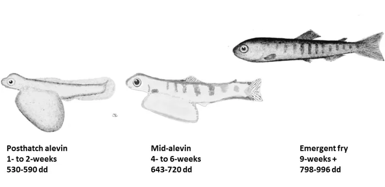
OSNs. The ontogeny of glomerular patterning at three incremental stages from hatch to
emergence was described and included posthatch alevin (one- to two-weeks posthatch;
539-590 dd), midalevin (four- to six-weeks posthatch; 643-720 dd), and emergent fry
(nine-weeks plus posthatch, and absorption of yolk-sac; 798-996 dd). The structural
maturity of this system was further inferred by comparing the glomerular patterning to
that in adult salmonids (Chinook salmon, Jarrard, 1997; Rainbow trout, Riddle and
28
across-family and across-species comparisons.
I anticipated consistent glomerular patterning throughout the alevin
developmental stage from hatch to emergence for two reasons: the coarse organization of
glomerular territories is determined during larval development in zebrafish (Braubach et
al., 2013), and important behaviours exhibited by juvenile teleosts are
olfactory-mediated, including those used for kin recognition (Zebrafish: Hinz et al., 2013) and
feeding (Salmonids: Mearns, 1986). Amino acid-responsive lateral
calretinin-immunoreactive glomeruli should be consistently identifiable no later than emergence, as
predicted by the sensitivity of this stage to this odour class (Mearns, 1986; Tilson et al.,
1994). Physiological studies support the functional role of glomerular pattering for odour
discrimination across taxa (Fish: Friedrich and Korsching, 1998; Mice: Marks, 2006;
Insects: Lei et al., 2004). Because the stimulation of specific regions of the OB with
specific odour classes yields similar results within the subclass teleostei, consistency in
glomerular patterning should also be observed. Additionally, there are observable
consistencies in patterning of glomerular territory patterning between phylogenetically
distantly related rainbow trout (Riddle and Oakley, 1992) and zebrafish (Baier and
Korsching, 1994; Gayoso et al., 2011; Braubach et al., 2012; Braubach et al., 2013).
Materials and Methods
Fertilization and rearing conditions of lacustrine Chinook salmon
All animal handling and care was conducted with approval by the University of Windsor
29
Spawning Chinook salmon were electro-shocked from the Credit River, Mississauga,
Ontario (43° N, 79° W), during the upstream migration from Lake Ontario in October
2012 and 2013. Adult fish were euthanized by percussive stunning prior to gamete
collection.
Eggs were collected from females and milt from males within a one-hour
timeframe. The eggs and milt were transferred to the University of Windsor Animal Care
Facilities, where the eggs were fertilized using Detroit River water to activate the milt. To
accommodate across-family comparison of glomerular patterning in alevin, eggs from
each female were fertilized using milt from a unique male, creating seven different family
groups. Eggs and alevin were reared in incubation trays sourced by a flow-through of
dechlorinated municipal water. Water temperature is an important determinant of
development in salmonids, and was monitored throughout this period using HOBO
temperature loggers to record temperature every 15 minutes. Thus degree days (dd), the
sum of the average daily water temperature from fertilization to each collection date
(Crisp, 1981), were presented with developmental stage to provide a more accurate
assessment of age.
Sample collection and tissue preparation
The ontogeny of glomerular patterning in alevin was determined by sampling alevin at
three increments (Fig. 2.1). Glomerular patterning was formulated for 18 posthatch alevin
(2013 animals, one family), six mid-alevin individuals (2013 animals, one family), and
more than 25 emergent fry (2012 animals, six families, 996 dd; 2013 animals, one
30
prior to application to the 2013 group.
Alevin were euthanized by anaesthetic overdose (1g/L MS-222; pH 7.4),
decapitated over the gills, and heads dropped-fixed in 4% paraformaldehyde (PFA) in
0.1M PBS. A few days prior to sectioning, heads were further dissected by removing the
mandible, tissue caudal to the eyes and dorsal skin to expose neural tissue, and post-fixed
in fresh 4% PFA in 0.1 M phosphate buffer saline (PBS). Tissue was cryoprotected by
immersion in a 20% and 30% sucrose gradient in 0.1 M PBS overnight. Horizontal 16-30
µm thick serial sections were sectioned from the olfactory bulb to the olfactory
epithelium using a cryotome (Leica CM 3050A) and collected onto Fisherbrand
Superfrost Plus microscope slides (Fisher Scientific, Waltham MA, 12-550-15). Sections
were left to dry at room temperature before storing at -20 °C.
Immunocytochemistry techniques
The application of several immunocytochemical labels to the OSN pathway from the
origin in the olfactory epithelium to the termination in the olfactory bulb (Fig. 2.2),
facilitated the adaptation of several ICC protocols. Serial tissue sections from the
olfactory epithelium to the olfactory bulb were double-labelled against KLH produced in
rabbit (Sigma-Aldrich, Oakville ON, H0892), and monoclonal acetylated tubulin
produced in mouse (AT; Sigma-Aldrich, Oakville ON, T7451), a marker for an α-tubulin
epitope on microtubules that is used as a probe for vertebrate neurons. OSN axons are
KLH-immunoreactive, and are easily traced from the OE to the OB, where they
fasciculate onto olfactory glomeruli, using fluorescence microscopy. The KLH antibody,