1
Moderate Effect of Flavonoids on Vascular and Renal Function
In Spontaneously Hypertensive Rats
María D. Paredes1, Paola Romecín1, Noemí M. Atucha1, Francisco O’Valle2,
Julián Castillo3, M. Clara Ortiz1, Joaquín García-Estañ1*
1, Departamento de Fisiología, Facultad de Medicina & Instituto Murciano de
Investigaciones Biosanitarias, Universidad de Murcia, Murcia, Spain
2, Departamento de Anatomía Patológica, Facultad de Medicina, IBIMER
(CIBM) & Ibs.GRANADA, Universidad de Granada, Granada, Spain
3, Instituto Universitario de Envejecimiento & Research and Development
Department, Nutrafur SA-FRUTAROM Group, Alcantarilla (Murcia), Spain
* Corresponding author: jgestan@um.es (JG-E)
ABSTRACT
We have evaluated the antihypertensive effect of several flavonoid
extracts in the spontaneous hypertensive rat model (SHR). Treatments were
carried out for 6 and 12 weeks in two groups of SHR rats which received
Apigenin, Lemon Extract, Grapefruit + Bitter Orange (GBO) extracts and Cocoa
extract. Captopril was used as a positive control in the SHR group treated for 6
weeks (SHR6) and Diosmin was used as the industry reference in the SHR
group treated for 12 weeks (SHR12). Captopril and GBO extracts significantly
reduced the elevated blood pressure of the SHR6 animals, but none of the
2
extracts was effective in the SHR12 group. Apigenin, LE, GBO and captopril
also ameliorated nitric oxide-dependent and independent aortic vascular
relaxation and elevated plasma and urinary excretion of nitrites, only in the
SHR6 group. Kidney and urinary TBARS were also significantly reduced by GBO
in the SHR6 rats. Apigenin also improved vascular relaxation in the SHR12
group and all the flavonoids studied reduced urinary TBARS excretion and
proteinuria. Vascular abnormalities such as lumen/wall ratio in coronary
arteries and thoracic aorta were moderately improved by these treatments in
the SHR6 group. In conclusion, the flavonoids included in this study, especially
apigenin, LE and GBO improved vascular vasodilatory function of young adult
SHRs but only the GBO-treated rats benefited from a reduction in BP. These
extracts may be used as functional food ingredients with a moderate
therapeutic benefit, especially in the early phases of arterial hypertension.
Keywords: flavonoids, nitric oxide, heart, kidney, sodium balance,
phenylephrine, acetylcholine
Introduction
Animal studies using flavonoid-rich foods are a valid alternative to
advance on the comprehension of the mechanisms underlying their blood
pressure lowering effects during arterial hypertension [1]. The intake of
polyphenols has been inversely related with the reduced risk of this disease
[2]. In fact, epidemiological studies associate an increased consumption of
foods and beverages rich in flavonoids with a reduced risk of cardiovascular
3
cause scarce side-effects is an attractive possibility to be considered when
treating several pathologies [5]. Several studies have correlated the
consumption of flavonoid-rich food, as well as isolated compounds, with the
beneficial effects on several cardiovascular parameters such as flow-mediated
dilation, blood pressure, and cardiovascular risk biomarkers [6-7]. Additionally,
many flavonoids induce the release of endothelium derived vasodilator factors
such as nitric oxide (NO) or endothelium-derived hyperpolarizing factor (EDHF)
and decrease the release of pro-inflammatory substances with a consequent
improvement of endothelial function [8-9].
In a previous study in the L-NAME model of arterial hypertension (10),
we showed that some flavonoids, especially apigenin, were effective to reduce
the elevated blood pressure associated with the chronic deficiency of nitric
oxide (NO). An important feature of that study (10) is that we used a low dose,
far from those usually applied in human therapy or experimental animals
(11-12), a dose that responded to an objective criterion of a possible later
commercial use in humans. As in other studies (13-14), the blood
pressure-lowering effect of these flavonoids were related to a combination of vasodilator
and antioxidant actions (10).
In the present study we have carried out a similar study in another
model of arterial hypertension, the spontaneously hypertensive rat (SHR),
thought to be the most comparable with the human form of hypertension
(15-16). Thus, SHR rats have increased activity of fluid retention mechanisms,
sodium reabsorption and increased vascular resistance. This increase in
vascular resistance being produced first by neurogenic mechanisms and later
4
to evaluate the vascular and renal effects of several flavonoid extracts in SHR
rats. We have also examined some of the possible mechanisms involved in
their beneficial effects such as an improvement in NO bioavalaibility and
endothelial and vascular function, the reduction in oxidative stress markers
and the effects on cardiovascular morphological changes.
Material and methods
Animals
All the experiments were performed in male SHR and WKY rats (Harlan
Lab, Barcelona, Spain) housed in a temperature controlled environment, with
12:12-h light-dark cycle in the Animal Care Facility of the University of Murcia
(REGAES300305440012). The animals were kept and treated according to the
guidelines established by the European Union for the protection of animals
used in experiments (86/609/EEC). All procedures were approved by the
Animal Care and Use Committee of the University of Murcia (C1310050303).
Experimental groups
There were two treatment groups of male SHR, one treated for 6 weeks
and another group treated for 12 weeks, along with their respective controls
(WKY) rats.
1. SHR6. The first treatment group (6 weeks) was composed of 56 SHR (8-9
week old, initial weight, 185-271 g) and 6 WKY rats (initial weight, 213-220 g).
This group was composed of the following experimental groups:
1) Control (n = 6), WKY rats without any treatment;
5
3) Apigenin (A, n = 6), SHR rats treated with A (1.44 mg/Kg/day);
4) Lemon Extract (LE, n = 7), SHR rats treated with LBC (2.84
mg/Kg/day);
5) Grapefruit + Bitter Orange Extracts (GBO, n = 7), SHR rats treated
with GBO extract (9.28 mg/Kg/day);
6) Cocoa Extract (COE, n = 6), SHR rats treated with COE (2.52
mg/Kg/day);
7) Captopril or CPT (n = 6), SHR rats treated with CPT (100 mg/Kg/day).
This angiotensin converting enzyme inhibitor was used as a positive
control, since it has been shown to be very effective for lowering blood
pressure of the SHR model (18-19).
2. SHR12. The second treatment group (12 weeks) was composed of 38
SHR (8-9 week old male, initial weight, 210-267 g) and normotensive
Wistar-Kyoto (WKY) rats (initial weight, 212-230 g). This was composed of 7 different
experimental groups:
1) Control (n = 6), WKY rats without any treatment;
2) SHR (n = 9), SHR rats without any treatment;
3) Apigenin (A, n = 6), SHR rats treated with A (1.44 mg/Kg/day);
4) Lemon Extract (LE, n = 6), SHR rats treated with LBC (2.84
mg/Kg/day);
5) Grapefruit + Bitter Orange Extracts (GBO, n = 6), SHR rats treated
with GBO extract (9.28 mg/Kg/day);
6) Cocoa Extract (COE, n = 6), SHR rats treated with COE (2.52
mg/Kg/day);
6
The doses and composition of the different extracts have been previously
published (10). Briefly, the doses were selected as those being economically
competitive in case of a future commercial use. All treatments were
administered during the required time, 6 or 12 weeks, in the drinking water,
except for Diosmin that was given mixed with the powdered food, in powder
feeders (Tecniplast, USA). All animals had free access to a standard rat diet
with a 0.5% of sodium content (104 mEq/Kg) and tap water, with or without
treatments. The concentrations of the drugs were adjusted daily according to
the body weight and water and food intake. All products, except L-NAME and
captopril (Sigma), were kindly provided by Nutrafur SA-FRUTAROM Group.
Experimental procedures
Rats were maintained in their cages and they were progressively
accustomed to individual metabolic cages (Tecniplast, USA) three days a week.
Then, in the treatment week 6th or 12th, after two days of adaptation, we
measured food and water intake and urinary volume (diuresis) in 24 hours.
The urine samples were collected and centrifuged (1000 g, 10 min) to remove
solid matter and then kept at -80º C for posterior analysis. The urinary sodium
concentration was determined using a sodium electrode (Thermo Scientific
Orion, USA). Sodium balance (mEq/day/100 g) was calculated as the
difference between sodium intake and excretion, and factored by body weight.
Sodium intake (mEq/day) was obtained by multiplying the consumption of food
per day (g/day) by sodium content of the diet (0.104 mEq/g). Urinary sodium
excretion (mEq/day) was determined as the product of sodium concentration
7
Measurement of blood pressure and samples extraction
After the metabolic study was completed, the animals were anesthetized
with sodium pentobarbital (5 mg/Kg, ip) and placed on a heated table to
maintain body temperature at 37º C. A polyethylene catheter (PE-50) was
placed in the right femoral artery to measure mean arterial pressure (MAP;
Hewlett Packard 1280 pressure transducer and amplifier 8805D, Andover, MA)
and to collect blood samples (20-21). Then, blood was collected into
heparinized tubes and plasma was obtained by centrifugation (1,000 g, 10
min, 4º C). Thereafter, the animal was euthanized by opening the thorax. We
extracted the descending thoracic aorta and placed it in a Petri dish containing
oxygenated and pre-warmed Krebs solution for the vascular reactivity study.
Finally, kidneys, heart and abdominal aorta were also removed. All samples
were frozen (- 80º C) and a small portion was also fixed with a 10%-formalin
solution for pathology studies.
Vascular reactivity study
The thoracic aorta was cleaned of adhering fat and connective tissue;
care was taken not to disrupt vascular endothelium, as previously described
[21]. Then, the aorta was cut into four rings (3-4 mm) and mounted in 10 ml
organ baths (organ bath system LE 01004, Panlab, Barcelona, Spain)
containing a physiological Krebs solution with the following composition (mM):
NaCl, 118; KCl, 4.7; CaCl2, 2.5; MgSO4, 1.2; NaHCO3, 25; KH2PO4, 1.2;
edetate calcium disodium, 0.026; and glucose, 5.6. The Krebs solution was
maintained at 37º C and continuously bubbled with a mixture of 95% O2 and
8
Panlab) to detect tension changes that were acquired and analyzed with a data
acquisition system (AD Instrument, Oxford, UK) consisting of a bridge amplifier
(FE228), a data acquisition hardware (PowerLab 8/30) and a software
(LabChart 6.0). Aortic rings were equilibrated for at least 45 min at a resting
tension of 2 g before any specific experimental protocol was initiated. During
this period, the bathing solution was replaced every 15 min and, if needed, the
basal tone readjusted to 2 g.
After the stabilization period, the aortic rings were constricted using a
cumulative dose–response curve to phenylephrine (PHE, 10−9-10−4 M),
administered in 0.1 ml bolus. Then, the rings were frequently washed until the
resting tension was reached again and a second stabilization period of 30 min
was allowed. To evaluate the vasodilator responses to acetylcholine (ACH), the
aortic rings were pre-contracted with a maximal dose of PHE (10−4 mol/l).
Once a stable plateau was reached, a cumulative dose–response curve to the
ACH (10−9–10−4 mol/l) was performed to assess the endothelium-dependent
vasodilatation. Thereafter, the rings were frequently washed once again and a
third stabilization period of 30 min was permitted and followed by an
incubation period of 30 min with the NOS-inhibitor L-NAME (10-4 M) to inhibit
NO synthesis. Next, a cumulative concentration-response curve to ACH was
again performed, to evaluate the role of NO in the endothelium-dependent
vasodilatation. Finally, we added a single dose of SNP (10-4 M) to test the
independent vasodilator responses and the functionality of the smooth muscle.
The responses to PHE are expressed in grams and the relaxation to ACH and
SNP as percentage of the maximal PHE effect. Stock solutions of these drugs
9
solutions were prepared daily in Krebs solution. Drug concentrations are
expressed as final bath concentrations. All reagents and vasoactive compounds
were purchased from Sigma-Aldrich and Panreac (Spain).
Analytical procedures
TBARS (thiobarbituric acid reactive substances) in plasma and kidney
tissue were determined as a measure of lipid peroxidation by using a
colorimetric method, as described previously [22]. Briefly, 0.5 ml of potassium
phosphate buffer (0.1 M, pH 7.4) was added to 100 µl of plasma sample mixed
or 50 μl of kidney tissue lysate. After mixing, 1 ml of reagent solution [1
mmol/l deferoxamine mesylate, 7.5% (w/v) trichloroacetic acid, 0.25 mol/lHCl
and 0.37%thiobarbituric acid] was added and the mixture was vortex-mixed,
covered with aluminium foil to avoid evaporation and heated at 90º C for 15
minutes in a dry block heater (Heatblock II, VWR, Thorofare, NJ USA). After
the mixture had returned to room temperature, TBARS from standards
(prepared from 1,1,3,3-tetraethoxypropane) and samples were extracted into
1 ml of butanol. After a vigorous vortex-mixing and a brief centrifugation
(1000 g for 5 min), the absorbance of the butanol layer was read at 532 nm in
a spectrophotometer (Eppendorf Biophotometer Plus, Hamburgo, Germany),
and the value was expressed as nmol/mL of plasma or nmol/mg of kidney
protein.
The protein concentration was measured in the urine and lysates using
the bicinchoninic acid based-method (Sigma).
The plasma and urinary excretion of nitrite was determined by using the
10
1% Sulfanilamide in 5% Potassium Phosphate. Then 50 µL of 0.1%
N-(1-Naphthyl) Ethyl-Enediamine dihydrochloride was added and incubated for 15
minutes. The nitrite concentration was quantified in a spectrophotometer at
540 nm against the standards and subtracting a blank from each individual
sample. The final concentration was expressed in µg/mL for plasma or µg/day
for urine samples.
Histological Analysis
Aortic, cardiac and renal tissue samples were fixed in 10% buffered
formaldehyde, and then processed, embedded in paraffin and sectioned (4 µm)
as previously reported [10, 20]. Transversal kidney, ventricular heart and
thoracic and abdominal aorta sections were stained with hematoxylin-eosin
and periodic acid-Schiff stain. The morphological study was done by a
pathologist in blinded randomized sections of the tissues, with light microscopy
and using the most appropriate stain for each lesion. The histo-morphometric
measurements were performed with the software ImageJ 1.47 (NIH,
http://rsb.info.nih.gov/ij/).
In the aorta, wall thickness was measured in three different, randomly
selected regions, and also three times in each region. In the heart, we
analyzed the inter-ventricular septum thickness, assessed in the middle central
region of the cardiac cavities as well as the relation between luminal diameter
and wall thickness in main and intramural coronary arteries, obtained from 5
measurements of each artery.
In the kidney, we evaluated the main alterations observed in transversal
11
presence of hyaline arteriopathy in all the arteries seen in the whole section;
2) the relation between luminal diameter and wall thickness in the main renal
artery or principal branches (if the first is missing); and 3) the absence (0) or
presence (1) of tubular cast/cylinders in the cortical and medullary region from
the entire kidney section.
Statistical Methods
Data are presented as the mean ± standard error. Differences between
groups were compared mainly by one-way analysis of variance (ANOVA). In
the vascular reactivity experiments, the values of EC50 were calculated from
the individual dose-response curves and expressed as the negative logarithm
(pEC50). Differences were considered statistically significant at a p level lower
than 0.05.
3. Results
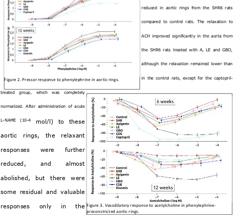
Blood pressure and urinary variables
Mean arterial pressure (MAP)
data are shown in figure 1 and table
1. In the SHR6 group, most of the
treated groups showed slightly
lower MAP values than the group
with untreated hypertension (SHR),
but a significant difference was
12
treatment with GBO and CPT. In the SHR12 group, no statistically significant
differences were observed in the treatment groups as compared to the
SHR-untreated rats. Heart rate was significantly decreased by captopril in the SHR6
group and GBO, COE and D-treated groups showed lower heart rate values in
the SHR 12 group.
Table 1. Final blood pressure and heart rate in the experimental groups.
SHR6 MAP (mmHg) Heart rate (bpm) SHR12 MAP (mmHg) Heart rate (bpm)
WKY 112,0 ± 3,7 363,6 ± 7,5 WKY 97,0 ± 7,1 313,6 ± 17,1
SHR 162,3 ± 5,7* 384,9 ± 12,8 SHR 175,1 ± 5,4 * 395,3 ± 10,1*
A 151,0 ± 6,9* 379,2 ± 16,7 A 157,5 ± 9,2 * 399,8 ± 8,3*
LE 152,7 ± 2,1* 358,9 ± 10,8 LE 163,3 ± 5,9 * 369,2 ± 11,8*
GBO 136,7 ± 6,3*† 392,4 ± 12,9 GBO 167,8 ± 11,3 * 367,0 ± 5,6 *†
COE 153,3 ± 6,4* 378,6 ± 12,9 COE 163,7 ± 9,4 * 363,8 ± 12,9 †
CPT 125,0 ± 1,8 *† 314,3 ± 9,1*† D 162,5 ± 5,8 * 356,4 ± 12,6 †
Body weight and hematocrit of all the experimental groups are listed in
table 2. In the SHR6 group, there were no significant difference in body weight
among the experimental groups. In the SHR12 group, the group that received
GBO showed a significantly lower body weight compared with the untreated
rats. Diuresis was not statistically different in the experimental group SHR6,
except for the group treated with captopril. In the SHR12 group, diuresis was
significantly lower only in COE and D-treated groups, as compared with the
untreated SHR group (table 2). Regarding sodium balance, there were no
differences between untreated WKY and SHR rats, but the SHR6 group treated
13
the SHR12 groups, a greater sodium balance was observed in the GBO and
COE-treated groups (table 2).
Vascular function
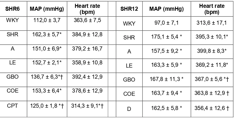
Dose-response curve to PHE was significantly shifted downwards in the
arteries from all treated and untreated groups (figure 2), reducing the
maximum contractile responses in all the SHR6 groups, as compared to the
controls. The pEC50 was significantly enhanced also in all SHR6 groups and
Table 2. Body weight and hematocrit values in the experimental groups.
SHR6 Body weight (g) Hematocrit (%) Food intake (g/24h)
Water intake (ml/24h)
Diuresis
(ml/24h) Natriuresis (mEq/24h)
Sodium balance (mEq/24h/100
g)
WKY 317,2 ± 12,4 47,5 ± 0,6 20,9 ± 1,1 41,6 ± 4,4 11,2 ± 1,9 0,36 ± 0,03 0,57 ± 0,03 SHR 305,5 ± 4,7 55 ± 1,06* 19,4 ± 0,4 28,4 ± 0,8* 11,7 ± 0,9 0,41 ± 0,03 0,53 ± 0,02 A 304,8 ± 4,4 53 ± 0,5* 22,4 ± 0 7† 30,0 ± 1,7 10,6 ± 1,8 0,51 ± 0,06 0,6 ± 0,04
LE 309,2 ± 4,3 54,1 ± 0,8* 18,2 ± 0,4*† 28,8 ± 1,8* 13,3 ± 1,3 0,71 ± 0,09*† 0,38 ± 0,03*†
GBO 302,4 ± 4,6 54,7 ± 1,3* 19,8 ± 1,4 26,9 ± 0,9* 9,7 ± 0,5 0,46 ± 0,05 0,53 ± 0,06 COE 302,9 ± 6,9 51,5 ± 0,7*† 19,9 ± 0,7 33,1 ± 1,8 14,8 ± 1,7 0,57 ± 0,04 0,5 ± 0,03
CPT 322,2 ± 9,2 49,7 ± 1,9† 18,5 ± 0,9 39,8 ± 4,2 20,5 ± 3,4* 0,7 ± 0,14*† 0,38 ± 0,04*†
SHR12
WKY 354,5 ± 7,7 45,5 ± 0,6 17,7 ± 0,8 33,9 ± 1,8 9,0± 1,2 0,15 ± 0,02 0,48 ± 0,02 SHR 359,1 ± 5,9 50,5 ± 0,9* 18,2 ± 0,8 29,0 ± 1,6 11,8 ± 1,1 0,34 ± 0,04 0,41 ± 0,02 A 351,2 ± 8,7 52,2 ± 1,1* 19,4 ± 0,8 28,3 ± 1,5 11,7 ± 0,9 0,53 ± 0,08 0,42 ± 0,03 LE 330,8 ± 11,8 49,5 ± 0,4* 18,7 ± 0,6 28,9 ± 4,5 13,2 ± 3,4 0,35 ± 0,04 0,49 ± 0,04 GBO 342,4 ± 4,6† 48,4 ± 0,4* 20,1 ± 0,6* 33,7 ± 2,0* 12,6 ± 1,1 0,21 ± 0,04 0,55 ± 0,02*†
COE 320,7 ± 16,3 50,4 ± 1,0* 18,6 ± 0,7 25,7 ± 1,1 7,2 ± 1,0† 0,19 ± 0,03 0,54 ± 0,02*†
D 351,8 ± 8,0 50,6 ± 0,8* 17,4 ± 0,5 20,6 ± 1,0 8,2 ± 0,8† 0,36 ± 0,05 0,41 ± 0,02
Abbreviations: WKY (wistar kyoto rats), SHR (spontaneously hypertensive rats), A (Apigenin), LE
14
none of the treatments changed them (table 3). In the SHR12 groups, the
maximum contractile responses were also reduced compared with the controls
response, but the pEC50 did not change significantly (table 3).
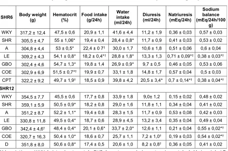
Maximal ACH-induced vasodilatation
(figure 3 and table 3) was significantly
reduced in aortic rings from the SHR6 rats
compared to control rats. The relaxation to
ACH improved significantly in the aorta from
the SHR6 rats treated with A, LE and GBO,
although the relaxation remained lower than
in the control rats, except for the
captopril-treated group, which was completely
normalized. After administration of acute
L-NAME (10-4 mol/l) to these
aortic rings, the relaxant
responses were further
reduced, and almost
abolished, but there were
some residual and valuable
responses only in the
captopril-treated group. Regarding the SHR12 group, a similar decrease was
observed in all the groups, and only the captopril-treated group showed a
significantly greater relaxation.
Vasorelaxation in response to SNP was significantly reduced in the
flavonoid-untreated SHR6 and SHR12 groups when compared with the control Figure 2. Pressor response to phenylephrine in aortic rings.
15
rats. SNP-induced vasorelaxation improved significantly in all flavonoid-treated
groups (table 3).
Effect of flavonoid extracts on Oxidative Stress Status
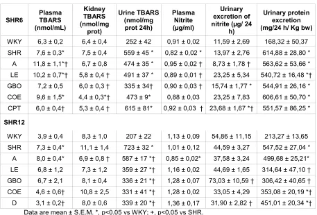
Values of TBARS, nitrite and urinary protein excretion are shown in table
4. Regarding TBARS, a significant increase was found only in the plasma and
urine of the untreated animals as compared with controls. In the SHR6 group,
only captopril reduced plasma TBARS, whereas captopril, LE, GBO and COE
reduced renal levels compared to the untreated SHR group. In the SHR12, only
apigenin reduced renal TBARS values although the urinary values were
significantly lower in all the flavonoid-treated SHR12 rats.
Regarding nitrite urinary excretion, only treatment with captopril in the
SHR6 and GBO in the SHR12 groups showed a significantly greater value as
compared to the untreated group. Also, urinary protein excretion was
significantly higher in the untreated group but proteinuria was reduced in the
LE-treated group (in SHR6 and SHR12) and in the groups treated with GBO,
COE, D (in SHR12).
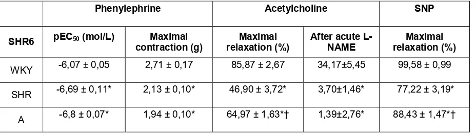
Table 3. Contractile response to phenylephrine and maximal relaxation to
acetylcholine and sodium nitroprusside in the experimental groups.
Phenylephrine Acetylcholine SNP
SHR6 pEC50 (mol/L) contraction (g)Maximal relaxation (%)Maximal After acute L-NAME relaxation (%)Maximal
WKY -6,07 ± 0,05 2,71 ± 0,17 85,87 ± 2,67 34,17±5,45 99,58 ± 0,99
SHR -6,69 ± 0,11* 2,13 ± 0,10* 46,90 ± 3,72* 3,70±1,46* 77,22 ± 3,19*
16
Phenylephrine Acetylcholine SNP
SHR6 pEC50 (mol/L) Maximal contraction (g)
Maximal relaxation (%)
After acute L-NAME
Maximal relaxation (%)
LE -6,6 ± 0,05* 2,13 ± 0,11* 55,97 ± 2,38*† 3,47±1,43* 88,54 ± 1,86*†
GBO -6,6 ± 0,06* 2,17 ± 0,13* 60,56 ± 2,72*† 1,56±0,65* 90,09 ± 2,18*†
COE -6,77 ± 0,03* 2,13 ± 0,13* 49,86 ± 4,86* 0,86±0,61*† 91,07 ± 1,49*†
CPT -6,7 ± 0,07* 1,46 ± 0,04*† 95,57 ± 0,77*† 19,43±4,47† 101,89 ± 1,34†
SHR12
WKY -6,84 ± 0,06 2,62 ± 0,11 68,41 ± 2,80 11,26±2,14 93,94±2,21
SHR -6,66 ± 0,05 2,03 ± 0,08* 24,00 ± 2,68* 1,47±0,94* 64,66±3,04*
A -6,74 ± 0,05 2,25 ± 0,09* 43,13 ± 3,45*† 0,22±0,23* 80,28±2,80*†
LE -6,75 ± 0,13 2,33 ± 0,15 26,50 ± 2,58* 2,49±1,07* 70,64±2,60*†
GBO -6,73 ± 0,1 2,27 ± 0,10* 35,00 ± 8,44* 2,45±0,65* 80,63±5,48*†
COE -6,6 ± 0,11 1,94 ± 0,16* 32,74 ± 6,39* 3,07±1,32* 77,69±5,82*†
D -6,68 ± 0,09 2,11 ± 0,14* 21,38 ± 2,02* 6,22±2,24*† 79,44±3,78*† Data are mean ± S.E.M. Abbreviations as in Table 1. pEC50 is the negative logarithm of the half
maximal effective concentration (EC50). *, p< 0.05 vs. WKY; †, p < 0.05 vs. SHR.
Histopathology results
The analysis of the heart revealed that both SHR untreated and treated
groups (both SHR6 and SHR20) showed no infarct zones, hyaline arteriopathy
or fibrinoid necrosis (data not shown), features that were observed in the
study performed in the L-NAME hypertension (10). Wall-lumen ratio of
coronary arteries was decreased in the SHR6 when compared with control and
treatments with A and COE increased them. In the SHR12 group, there were
no changes. Interventricular heart septum thickness was significantly higher in
the SHR6 untreated group compared to the control rats and only captopril
17
groups were very similar without significant differences between them. With
respect to the thickness of the abdominal and thoracic aorta (table 5), there
were significant reductions in the thickness of the abdominal aorta in all the
SHR6-treated groups only. In the SHR12 group, the tendency of the
flavonoid-treated groups was to show an increase in the thickness in both vessels.
Regarding the kidney (table 5), significant increases were found in the LWR
parameter in the LE and GBO-treated SHR6 groups. Also, the absence of HA
and tubular cylinders was evident in all the treated SHR6 groups, except in the
A group. In the SHR12 groups, HA and TC were also found in the untreated
18
Table 4. Measurements of TBARS, nitrite and proteinuria in the experimental groups.
SHR6 Plasma TBARS (nmol/mL) Kidney TBARS (nmol/mg prot) Urine TBARS (nmol/mg prot 24h) Plasma Nitrite (µg/ml) Urinary excretion of nitrite (µg/ 24
h)
Urinary protein excretion (mg/24 h/ Kg bw)
WKY 6,3 ± 0,2 6,4 ± 0,4 252 ± 42 0,91 ± 0,02 11,59 ± 2,69 168,32 ± 50,37 SHR 7,6 ± 0,3* 7,5 ± 0,4 559 ± 45 * 0,82 ± 0,02 * 13,97 ± 2,76 614,88 ± 28,80 *
A 11,8 ± 1,1*† 6,7 ± 0,8 474 ± 35 * 0,95 ± 0,02 † 8,73 ± 1,78 † 563,62 ± 53,66 * LE 10,2 ± 0,7*† 5,8 ± 0,4 † 491 ± 37 * 0,89 ± 0,01 † 23,25 ± 5,34 540,72 ± 16,48 *† GBO 7,2 ± 0,5 6,0 ± 0,3 † 335 ± 34† 0,90 ± 0,03 † 15,74 ± 1,77 * 544,91 ± 26,16 * COE 9,6 ± 1,5* 4,4 ± 0,3*† 473 ± 9* 0,88 ± 0,03 23,25 ± 7,83 606,61 ± 50,70 * CPT 6,0 ± 0,4† 5,3 ± 0,4 † 615 ± 81* 0,92 ± 0,03 † 23,68 ± 1,67 *† 551,57 ± 86,25 *
SHR12
WKY 3,9 ± 0,4 8,3 ± 1,0 207 ± 22 1,13 ± 0,09 54,86 ± 11,15 213,27 ± 13,65 SHR 7,3 ± 0,4* 11,1 ± 1,4 723 ± 32 * 1,01 ± 0,12 44,59 ± 3,27 547,52 ± 27,04 *
A 8,0 ± 0,4* 6,9 ± 0,8 † 587 ± 17 *† 0,85 ± 0,02* 37,58 ± 3,24 499,68 ± 25,21* LE 6,8 ± 1,2 7,3 ± 1,2 359 ± 27 *† 1,16 ± 0,02 44,69 ± 1,65 314,64 ± 47,10 † GBO 6,7 ± 2,1 8,1 ± 0,4 336 ± 21 *† 1,28 ± 0,07 73,03 ± 10,59 † 306,42 ± 40,65 † COE 4,6 ± 0,6† 10,8 ± 2,5 331 ± 41 *† 1,28 ± 0,02 33,05 ± 4,29 353,08 ± 20,19 *†
D 3,1 ± 0,2† 8,0 ± 0,6 339 ± 20 *† 1,36 ± 0,17 31,90 ± 2,82 † 451,01 ± 20,34 *† Data are mean ± S.E.M. *, p<0.05 vs WKY; +, p<0,05 vs SHR.
Discussion
The results of the present study show that some of the flavonoids studied
have a very modest effect on blood pressure and renal and vascular function in
the SHR model of arterial hypertension. Moreover, these effects can be
observed only in the younger animals, treated for 6 weeks. Doubling the
treatment time (12 weeks) almost eliminated these beneficial effects.
In the SHR6 groups, only GBO had a significant effect on blood pressure
and this was accompanied by a significant improvement of the vascular
vasodilator response, likely related to an increased production of vascular NO.
A reduction in renal oxidative status with beneficial changes in the
histopathological parameters in heart and kidney was also observed in this
19
extract-treated groups, similar vascular and renal changes were observed in
these animals when compared to the GBO-treated group.
As we explained in our previous article in nitric oxide-deficient
hypertensive animals (10), the dose chosen of each of the treatments
responds to an objective criterion of potential subsequent application in
humans. Therefore, the doses ingested daily by our animals are very low
compared to those used in other studies with similar compounds, since
generally, in animal studies, doses much higher are used, proportionally, than
those applied in a human therapy (5, 14).
It is clear that both types of arterial hypertension models are very
different. It seems that the flavonoids that we have used are more useful in
the nitric oxide-deficient hypertensive animals. In fact, one of the most
universal effects of flavonoids is through the augmentation of NO synthesis and
action (23-24). In this way, they can be of interest in diseases of endothelial
dysfunction (25). However, the SHR model is a completely different model,
with an important genetic component (19). Hypertension in the SHR model
develops as a result of increased peripheral resistance, first produced by
neurogenic and renal factors and later structural vascular changes associated
with increased vascular protein synthesis because of the chronic elevation in
blood pressure. As observed, our results clearly confirm that the inhibition of
the renin-angiotensin system is one of the treatments of choice for this model
and this has been shown previously (19).
20 SHR6 Coronary
LWR
Heart IVS Thoracic AT Abdominal AT
Renal LWR Renal HA Renal TC
WKY 2,27 ± 0,21 2,36 ± 0,14 107,2 ± 5,9 110,3 ± 7,5 1,69 ± 0,10 0,0 0,0 SHR 1,59 ± 0,03* 2,95 ± 0,18* 131,0 ± 5,6* 138,8 ± 3,4* 1,18 ± 0,08* 0,0 0,0 A 2,84 ± 0,22† 2,91 ± 0,13* 125,3 ± 2,9* 100,5 ± 2,1† 1,32 ± 0,31 0,0 0,25 LE 1,80 ± 0,21 2,73 ± 0,05* 150,4 ± 4,5* 127,0 ± 3,6 † 1,51 ± 0,04† 0,0 0,0 GBO 1,48 ± 0,13* 2,50 ± 0,07 134,6 ± 2,3* 117,5 ± 5,2† 1,69 ± 0,10† 0,0 0,0 COE 2,80 ± 0,58† 2,83 ± 0,07* 134,5 ± 3,8* 87,1 ± 4,6† 1,10 ± 0,24 0,0 0,0 CPT 1,78 ± 0,19 2,39 ± 0,12† 133,3 ± 7,3* 101,7 ± 4,0† 1,26 ± 0,10* 0,0 0,0
SHR12
WKY 3,08 ± 0,13 3,22 ± 0,15 104,8 ± 4,4 86,7 ± 5,8 1,76 ± 0,19 0,0 0,0 SHR 3,00 ± 0,80 3,43 ± 0,09 116,3 ± 2,2 90,5 ± 5,3 1,27 ± 0,26 0,25 ± 0,25 0,25 ± 0,25
A 1,97 ± 0,41 2,84 ± 0,23 126,8 ± 3,2*† 94,6 ± 2,7 1,61 ± 0,30 0,0 0,75 ± 0,25 LE 3,43 ± 0,28 3,04 ± 0,29 137,5 ± 3,5*† 114,8 ± 3,7*† 1,67 ± 0,44 0,0 0,0 GBO 2,20 ± 0,44 3,01 ± 0,09 131,4 ± 3,6*† 107,6 ± 4,0*† 1,79 ± 0,32 0,0 0,0 COE 3,06 ± 0,40 3,40 ± 0,15 142,2 ± 3,0*† 118,8 ± 3,7*† 1,39 ± 0,11 0,0 0,25 ± 0,25
D 3,15 ± 0,79 3,61 ± 0,09 130,8 ± 2,9*† 104,6 ± 3,3*† 1,43 ± 0,20 0,0 0,0
Data are mean ± S.E.M. Abbreviations as in table 2. LWR, lumen to wall ratio; IVS,
Interventricular septum width (mm); AT, Aorta Thickness (mm); HA, Hyaline Arteriopathy; TC,
tubular cylinders. *, p<0.05 vs WKY; +, p<0,05 vs SHR.
Many studies have reported a reduction in blood pressure following the
consumption of flavonoid-rich products. In vitro studies have reported that
flavonoids such as genistein, quercetin, and (-)-epicatechin regulated (directly
or indirectly) NO production in isolated vessels or cultured endothelial cells [8,
26-29]. In our data, A, LE, GBO and of course, captopril, improved the
acetylcholine relaxation in the SHR6 rats, the dependent on NO production
(after acetylcholine administration). Moreover, in these SHR6 animals, the
administration of sodium nitroprusside also improved the reduced relaxation in
the animals receiving all the flavonoids, indicative of an action close to the NO
21
in favor of a reduced production of vascular NO in the SHR model, as
suggested previously (19).
Other mechanisms have been suggested to explain the increased
endothelial NO bioavailability promoted by flavonoids. Several studies have
shown that a regular consumption of flavonoids or flavonoid-rich foods can
significantly improve the oxidative status as well the endothelial function [8,
10, 24-25, 30]. In the present study, we detected a significant increase in ROS
levels, as measured as TBARS, in plasma and urine in the SHR untreated
animals. It is likely that the reduction in kidney TBARS, observed in some of
the flavonoid-treated groups (Table 4), is also contributing to the normalization
of BP. It may be interesting to consider that the treatments with a greater
specific antioxidant efficacy are those having flavonoids with B-ring catechol
structure (3', 4'-dihydroxy), LE (eriocitrin) and COE (catechin compounds).
It is known that the chronic elevation of systemic blood pressure is
associated with the presence of proteinuria and the development of
glomerulosclerosis [32], as our data show (table 4). The flavonoids treatments
showed a reduction in proteinuria, but it only reached a significant difference in
the LE-treated SHR6 group. Interestingly, proteinuria was also reduced
significantly in the SHR 12 groups, and this is a result that merits further
study.
The metabolic and hemodynamic changes of hypertension are also
associated with the development of structural abnormalities, such as left
ventricular hypertrophy, cardiac fibrosis, necrosis and protein remodeling, as
well as with vascular wall hypertrophy [33-34], some of them shown in the
22
the hypertensive load on vascular tissues, increased monocyte and platelet
adhesion with the release of growth factors would contribute to the thickening
of the vascular wall. The proliferation was limited to the media, which is in
agreement with the findings of others [30-32]. All the flavonoids treatments
were effective to reduce the abdominal aortic thickness in the SHR 6 group,
but only captopril reduced cardiac hypertrophy. Regarding the renal structural
changes, only in the SHR 12 group, flavonoids seem to exert some beneficial
effect, since hyaline arteriopathy was present only in the SHR untreated group,
and tubule cylinders were also not observed in the LE, GBO and
diosmin-treated groups.
Our results suggest that the flavonoids included in this study, and
already present in the market as nutritional supplements, may be used as a
functional food ingredients but the beneficial effect on hypertension is
moderate, specially in the younger SHR group. Further studies are necessary
to elucidate the mechanisms involved in their effects, including an evaluation
of the dose-activity relationship in order to determine the molecular structures
most active. In any case, our results suggest that the effects of these
flavonoids may be related to a combination of vasodilator and antioxidant
actions.
Clinical Perspectives.
Flavonoids are important substances with biological actions of interest in
arterial hypertension. We aimed to analyze the role of some flavonoids in
a model of arterial hypertension, the spontaneously hypertensive rat
23 hypertension.
Grapefruit extract significantly reduced the elevated blood pressure of
the younger SHR animals (12 weeks), but none of the extracts was
effective in the older SHR group (18 weeks). Vascular reactivity was also
ameliorated with some of these treatments.
These extracts may be used as functional food ingredients with a
moderate therapeutic benefit, especially in the early phases of arterial
hypertension.
Funding sources.
This report was supported by a grant from the National Spanish R&D
Program CENIT of the Spanish Ministry of Science and Technology
denominated ‘‘Industrial research diets and food with specific features for the
elderly’’, CEN-20091006; Acronym: SENIFOOD.
References
1.Duarte J, Francisco V, Perez-Vizcaino F. Modulation of nitric oxide by flavonoids.
Food Funct. 2014; 5: 1653-1668.
2.Miranda AM, Steluti J, Fisberg RM, Marchioni DM. Association between Polyphenol
Intake and Hypertension in Adults and Older Adults: A Population-Based Study in
Brazil. PLoS One. 2016; 11: e0165791.
3.Wallace TC. Anthocyanins in Cardiovascular Disease. Adv Nutr. 2011 2: 1–7.
4.Basu A, Rhone M, Lyons TJ. Berries: emerging impact on cardiovascular health.
Nutr Rev. 2010; 68: 168–177.
5.Quiñones M, Miguel M, Muguerza B, Aleixandre A. Effect of cocoa polyphenol
24
6.Habauzit V, Morand C. Evidence for a protective effect of polyphenols-containing
foods on cardiovascular health: an update for clinicians. Ther Adv Chronic Dis.
2012; 3: 87–106.
7.Khurana S, Venkataraman K, Hollingsworth A, Piche M, Tai TC. Polyphenols:
Benefits to the Cardiovascular System in Health and in Aging. Nutrients. 2013; 5:
3779–3827.
8.Almeida Rezende B, Pereira AC, Cortes SF, Lemos VS. Vascular effects of
flavonoids. Curr Med Chem 2016; 23: 87-102.
9.López-Sepúlveda R, Jiménez R, Romero M, Zarzuelo MJ, Sánchez M,
Gómez-Guzmán M, Vargas F, O'Valle F, Zarzuelo A, Pérez-Vizcaíno F, Duarte J. Wine
Polyphenols Improve Endothelial Function in Large Vessels of Female
Spontaneously Hypertensive Rats. Hypertension. 2008; 51: 1088-1095.
10.Paredes MD, Romecín P, Atucha NM, O’Valle F, Castillo J, Ortiz MC, García-Estañ
J. Beneficial Effects of Different Flavonoids on Vascular and Renal Function in
L-NAME Hypertensive Rats. Nutrients 2018; 10(4), 484; doi:10.3390/nu10040484
11.Pechánová O, Rezzani R, Babál P, Bernátová I, Andriantsitohaina R. Beneficial
effects of Provinols: cardiovascular system and kidney. Physiol Res 2006; Suppl 1:
S17-30.
12.Fu JY, Qian LB, Zhu LG, Liang HT, Tan YN, Lu HT, Lu JF, Wang HP, Xia Q.
Betulinic acid ameliorates endothelium-dependent relaxation in L-NAME-induced
hypertensive rats by reducing oxidative stress. Eur J Pharm Sci 2011; 44:
385-391.
13.Duarte, J.; Jiménez, R.; O’Valle, F.; Galisteo, M.; Pérez-Palencia, R.; Vargas, F.;
Pérez-Vizcaíno, F.; Zarzuelo, A.; Tamargo, J. Protective effects of the flavonoid
quercetin in chronic nitric oxide deficient rats. J. Hypertens. 2002, 20, 1843–1854.
14.Jiménez, R.; Duarte, J.; Pérez-Vizcaíno, F. Epicatechin: Endothelial Function and
25
15.Trippodo NC, Frohlich ED. Similarities of genetic (spontaneous) hypertension.
Man and rat. Circ Res. 1981; 48(3):309-19.
16.Zhou X, Frohlich ED. Analogy of Cardiac and Renal Complications in Essential
Hypertension and Aged SHR or L-NAME/SHR. Medicinal Chemistry 2007; 3, 61-65.
17.Grisk O., Kloting I., Exner J., Spiess S., Schmidt R., Junghans D., Lorenz G.,
Rettig R. Long-term arterial pressure in spontaneously hypertensive rats is set by
the kidney. J. Hypertens. 2002; 20, 131–138.
18.Giudicelli JF, Freslon JL, Glasson S, Richer C. Captopril and hypertension
development in the SHR. Clin Exp Hypertens. 1980; 2, 1083-96.
19.Ahmeda AF, Alzoghaibi M. Factors regulating the renal circulation in
spontaneously hypertensive rats. Saudi Journal of Biological Sciences 2016; 23,
441–451.
20.García-Estañ J, Ortiz MC, O´Valle F, Alcaraz A, Navarro EG, Vargas F, Evangelista
S, Atucha NM. Effects of angiotensin-converting-enzyme inhibitors in combination
with diuretics on blood pressure and renal injury in nitric oxide-deficiency-induced
hypertension in rats. Clin Sci (Lond) 2006; 110: 227-233.
21.Fortepiani LA, Rodrigo E, Ortíz MC, Cachofeiro V, Atucha NM, Ruilope LM, Lahera
V, García-Estañ J. Pressure natriuresis in nitric oxide-deficient hypertensive rats:
effect of antihypertensive treatments. J Am Soc Nephrol. 1999;10: 21-27.
22.Alcaraz A, Hernández D, Iyú D, Mota R, Atucha NM, Ortiz AJ, García-Estañ J,
Ortiz MC. Effects of chronic L-NAME on nitrotyrosine expression and renal vascular
reactivity in rats with chronic bile-duct ligation. Clin Sci 2008; 115: 57-68.
23.Duarte J, Jiménez R, O’Valle F, Galisteo M, Palencia R, Vargas F,
Pérez-Vizcaíno F, Zarzuelo A, Tamargo J. Protective effects of the flavonoid quercetin in
chronic nitric oxide deficient rats. J. Hyperten 2002; 20: 1843–1854.
24.Jiménez R, Duarte J, Pérez-Vizcaíno, F. Epicatechin: Endothelial Function and
26
25.Vargas F, Romecín P, García-Guillén AI, Wangesteen R, Vargas-Tendero P,
Paredes MD, Atucha NM, García-Estañ J. Flavonoids in Kidney Health and Disease.
Front Physiol. 2018; 24; 9, 394.
26.Litterio MC, Jaggers G, Celep GS, Adamo AM, Costa MA, Oteiza PI, Fraga CG,
Galleano M. Blood pressure-lowering effect of dietary (-)-epicatechin administration
in L-NAME-treated rats is associated with restored nitric oxide levels. Free Radical
Biology and Medicine 2012; 53: 1894–1902.
27.Zhang YH, Park YS, Kim TJ, Fang LH, Ahn HY, Hong JT, Kim Y, Lee CK, Yun YP.
Endothelium-dependent vasorelaxant and antiproliferative effects of apigenin. Gen
Pharmacol. 2000; 35: 341-347.
28.Ajay M, Gilani AU, Mustafa MR. Effects of flavonoids on vascular smooth muscle
of the isolated rat thoracic aorta. Life Sci. 2003;74: 603-612.
29.Martínez-Fernández L, Pons Z, Margalef M, Arola-Arnal A, Muguerza B. Regulation
of vascular endothelial genes by dietary flavonoids: structure-expression
relationship studies and the role of the transcription factor KLF-2. J Nutr Biochem.
2015; 26: 277-284.
30.Benavente-Garcı́a O, Castillo J, Lorente J, Ortuño A, Del Rio JA. Antioxidant
activity of phenolics extracted fromOlea europaea L. leaves. Food Chem 2000; 68:
457-462.
31.Mali VR, Mohan V, Bodhankar SL. Antihypertensive and cardioprotective effects of
the Lagenaria siceraria fruit in NG-nitro-L-arginine methyl ester (L-NAME) induced
hypertensive rats. Pharmaceutical Biology, 2012; 50: 1428–1435.
32.Klahr S. The role of nitric oxide in hypertension and renal disease progression.
Nephrol Dial Transplant. 2001; 16: 60-62.
33.Kristek F, Gerova M, Devat L, Varga I. Cardiac hypertrophy and vascular
remodelling in NO-deficient hypertension. Endothelium 1995; 3: s94.
27
Takeshita A. Chronic inhibition of nitric oxide synthesis causes coronary