| FLYBOOK REPAIR, RECOMBINATION, AND CELL DIVISION
DNA Replication Control During
Drosophila
Development: Insights into the Onset of S Phase,
Replication Initiation, and Fork Progression
Brian L. Hua1and Terry L. Orr-Weaver2
Whitehead Institute and Department of Biology, Massachusetts Institute of Technology, Cambridge, Massachusetts 02142 ORCID IDs: 0000-0002-7580-3399 (B.L.H.); 0000-0002-7934-111X (T.L.O.-W.)
ABSTRACTProper control of DNA replication is critical to ensure genomic integrity during cell proliferation. In addition, differential regulation of the DNA replication program during development can change gene copy number to influence cell size and gene expression.Drosophila melanogasterserves as a powerful organism to study the developmental control of DNA replication in various cell cycle contexts in a variety of differentiated cell and tissue types. Additionally,Drosophilahas provided several developmentally regulated replication models to dissect the molecular mechanisms that underlie replication-based copy number changes in the genome, which include differential underreplication and gene amplification. Here, we review key findings and our current standing of the developmental control of DNA replication in the contexts of the archetypal replication program as well as of under-replication and differential gene amplification. We focus on the use of these latter two replication systems to delineate many of the molecular mechanisms that underlie the developmental control of replication initiation and fork elongation.
KEYWORDSFlyBook;Drosophila melanogaster; origin activation; endocycle; differential replication; underreplication; gene amplification; rereplication
TABLE OF CONTENTS
Abstract 29
DNA Replication Overview 30
Protein Players at the Origin of Replication 30
Hurdles for the Molecular Study of Metazoan DNA Replication 30
Fundamentals of Drosophila DNA Replication and Insights Contributed to the DNA Replication Field 31
Identification of replication proteins 31
Analysis of replication origins in Drosophila 32
Developmental regulation of DNA replication in Drosophila 33
Developmentally regulated S phase changes 33
Tissue specificity of Drosophila origins 35
Insights into Regulation of DNA Replication from Localized Changes in DNA Copy Number 35 Continued
Copyright © 2017 by the Genetics Society of America doi:https://doi.org/10.1534/genetics.115.186627
Manuscript received August 30, 2016; accepted for publication May 19, 2017 Available freely online through the author-supported open access option.
1Current address: Centers for Disease Control and Prevention, 1600 Clifton Rd., Atlanta, GA 30329.
CONTENTS,continued
Underreplication and local copy number reduction 35
Inhibition of fork progression by SUUR 36
Fork instability and DNA damage in UR regions 37
Potential biological functions of underreplication 37
Underreplication as a model for common chromosomal fragile sites 38
Developmentally programmed follicle cell gene amplification to increase local copy number 38
Control of origin activation during gene amplification 39
Drosophila gene amplification as a tool to study fork progression 40
Mutations that enhance fork elongation 41
Fork instability and DNA damage during rereplication 41
Conclusions, Implications, and Future Directions 42
Differential regulation of origin activation 42
Developmental control of replication timing and fork progression 42
DNA Replication Overview
B
efore cell division, the genome must be completely and accurately replicated to maintain the integrity of genetic information across cell generations. DNA replication initiates from thousands of DNA elements within the genome called origins of replication. Origins of replication direct the assem-bly of a large group of proteins and protein complexes to the site that ultimately allow for DNA unwinding and the estab-lishment of two, bidirectional replication forks. DNA ahead of the fork is progressively unwound, generating single-stranded DNA that serves as a template for the synthesis of new DNA (Bleichertet al.2017; Parkeret al.2017). Through the molecular study of DNA replication initiation and elon-gation, it is clear that the mechanisms that regulate origin activity and replication fork progression are diverse and com-plex, particularly in the context of development. Drosophilahas provided powerful developmental systems to study both replication initiation and elongation at the cellular and molec-ular levels (Nordman and Orr-Weaver 2012). Here, we sum-marize important insights that theDrosophilasystem has shed upon the regulation of metazoan DNA replication. We then detail seminal studies that have led to critical understanding of the developmental control of replication origin activation and fork elongation. Finally, we address prevailing questions in DNA replication control and the outlook for thefield.
Protein Players at the Origin of Replication
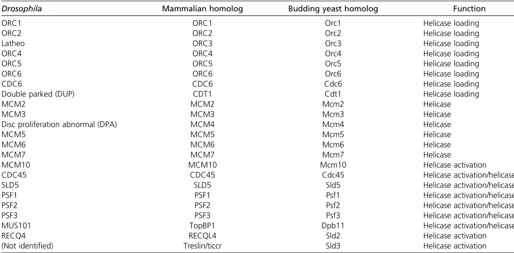
DNA replication initiation requires the sequential recruitment and activation of a large number of replication protein com-ponents. Unlike in budding yeast, metazoan origins of repli-cation are not defined by any known consensus sequence (Parker et al. 2017). However, protein factors required to establish the replication initiation complex and the replica-tion fork are highly conserved in eukaryotes (Table 1). Rep-lication initiationfirst requires that origins of replication are bound by the origin recognition complex (ORC) (composed
of the six proteins ORC1–6) in late M and G1 phases of the cell cycle (Figure 1). The replication initiation factor cell di-vision cycle 6 (Cdc6) is then recruited to the origin to form a complex with ORC. ORC and Cdc6 work cooperatively to recruit the initiation factor Cdt1 [Double Parked (DUP) in
Drosophila] and the six-membered Minichromosome Mainte-nance (MCM)2–7 replicative helicase complex. In budding yeast, Cdt1 and MCM2–7 form a stable complex in cell lysates and are recruited to origins of replication together (Tanaka and Diffley 2002; Kawasakiet al.2006; Remuset al.2009). In
Xenopusextracts, however, Cdt1 and MCM2–7 do not copre-cipitate, suggesting that Cdt1 and the MCM2–7 complex may be recruited sequentially to replication origins in metazoans (Maioranoet al.2000).
Two hexamers of the MCM2–7 complex are loaded onto origin DNA in an inactive state before the onset of S phase. Under the regulation of two kinases, S phase Cyclin-Dependent Kinase (CDK) and Dbf4-Dependent Kinase (DDK), the MCM2–7 complex is joined by CDC45 and the Go-Ichi-Ni-San (GINS) complex, a four-membered complex composed of Sld5, Psf1, Psf2, and Psf3. Together, the CDC45/MCM2–7/GINS (CMG) complex forms the functional replicative helicase (Bleichert
et al. 2017; Parker et al.2017). As two MCM2–7 hexamers are loaded onto a single origin of replication, two CMG com-plexes establish the independent, bidirectional replication forks after origin activation (Figure 1).
Hurdles for the Molecular Study of Metazoan DNA Replication
metazoans than in budding yeast. Although in both only a subset of origins are activated at a given time point in S phase (Aparicio 2013), this effect becomes more pronounced during the prolonged period of S phase occurring in most metazoan cells. Furthermore, origins of replication are not uniformly distributed throughout the metazoan genome, resulting in large genomic regions that require the activity of replication forks emanating from distant origins for their replication (Debatisseet al.2012). It also has been difficult to examine replication forks emanating from a single origin of replication. Finally, how developmental signals modu-late the activity of replication origins and forks remains to be elucidated.
Fundamentals of Drosophila DNA Replication and Insights Contributed to the DNA Replication Field
Identification of replication proteins
Elegant genetic and biochemical studies initially performed in budding yeast allowed for a comprehensive identification of the key protein factors that are involved in origin activation and fork elongation (Bell and Labib 2016). Significantly, the minimal set of protein factors required for DNA replica-tion in budding yeastin vitrohas been described (Yeeleset al.
2015). The establishment of a cell-free replication system fromXenopuseggs allowed for powerful biochemical dissection of DNA replication in a metazoan system (Lohka and Masui 1983; Blow and Laskey 1986; Blow and Watson 1987; Hutchisonet al.1987; Almouzni and Mechali 1988). Semi-nal studies using this system led to the identification and functional characterization of several key replication factors
in Xenopus, including the biochemical purification of an MCM-containing complex required for replication licensing (Chonget al.1995) as well as the identification ofXenopus
ORC2 and its essential role in replication initiation (Carpenter
et al.1996). Collectively, these studies played a significant role in demonstrating that yeast replication proteins are conserved in metazoans.
Whereas the budding yeast andXenopussystems laid the groundwork in the identification of DNA replication factors and the molecular events that are required for replication initiation and fork elongation,Drosophilahas since emerged as an extremely powerful organism to study metazoan DNA replication at both the molecular and developmental levels. For example, the metazoan homologs of the key replication initiation factor Cdt1 were first discovered in Drosophila
(Whittaker et al. 2000) and Xenopus (Maiorano et al.
2000). Additionally, Drosophila mutants with impaired ORC2 and Cdt1 function showed gross defects in DNA rep-lication, providing thefirst genetic evidence of the require-ment of these conserved proteins in metazoans (Landiset al.
1997; Whittaker et al.2000). Using biochemical methods, the functional helicase complex was shown to exist as a large protein assembly consisting of CDC45, MCM2–7, and GINS (CMG complex) through isolation from Drosoph-ila embryo extracts (Moyeret al.2006). Crucial structural insight into the regulation of metazoan DNA replication ini-tiation resulted from extensive electron microscopy studies (Clarey et al. 2006, 2008) and the solving of the crystal structure of the Drosophila ORC complex (Bleichertet al.
2015). Finally,Drosophilahas served as a metazoan model system to profile replication properties and dynamics
Table 1 Key proteins required for helicase loading and activation
Drosophila Mammalian homolog Budding yeast homolog Function
ORC1 ORC1 Orc1 Helicase loading
ORC2 ORC2 Orc2 Helicase loading
Latheo ORC3 Orc3 Helicase loading
ORC4 ORC4 Orc4 Helicase loading
ORC5 ORC5 Orc5 Helicase loading
ORC6 ORC6 Orc6 Helicase loading
CDC6 CDC6 Cdc6 Helicase loading
Double parked (DUP) CDT1 Cdt1 Helicase loading
MCM2 MCM2 Mcm2 Helicase
MCM3 MCM3 Mcm3 Helicase
Disc proliferation abnormal (DPA) MCM4 Mcm4 Helicase
MCM5 MCM5 Mcm5 Helicase
MCM6 MCM6 Mcm6 Helicase
MCM7 MCM7 Mcm7 Helicase
MCM10 MCM10 Mcm10 Helicase activation
CDC45 CDC45 Cdc45 Helicase activation/helicase
SLD5 SLD5 Sld5 Helicase activation/helicase
PSF1 PSF1 Psf1 Helicase activation/helicase
PSF2 PSF2 Psf2 Helicase activation/helicase
PSF3 PSF3 Psf3 Helicase activation/helicase
MUS101 TopBP1 Dpb11 Helicase activation/helicase
RECQ4 RECQL4 Sld2 Helicase activation
genome-wide, beginning with thefirst genome-wide map-ping of ORC in a differentiated metazoan cell type and tissue (MacAlpineet al.2010; Sheret al.2012). These genome-wide approaches have allowed for more comprehensive analysis of replication dynamics in the scope of the underlying chromatin landscape, developmental timing, and differentiation.
Analysis of replication origins in Drosophila
Experiments usingDrosophilacell culture lines have provided critical information about the timing of replication of geno-mic regions within S phase, localization of origins and sites of ORC binding, and the role of chromatin and histone modifi ca-tions. Genome-wide techniques have allowed for comprehen-sive profiling of replication initiation sites in severalDrosophila
cell culture systems (Cayrouet al.2011; Comoglioet al.2015).
Upon replication initiation, two nascent leading DNA strands extend from RNA primers located at the replication origin. These leading nascent strands can be isolated away from smaller RNA-primed Okazaki fragments on the lagging strand by size selection and from non-RNA-primed DNA by l-exonuclease digestion (Gerbi and Bielinsky 1997). High-throughput sequencing of purified leading nascent strands then allows for the identification of replication ini-tiation sites genome-wide (Leonard and Mechali 2013). Comparison of the replication initiation sites in S2, BG3, and Kc cells revealed that 16–20% of initiation sites are common to all three cell types, whereas 35–45% of activated origins are common to at least two cell types (Comoglioet al.
2015). These results highlight the cell-type specificity of origin sites, although an appreciable number of common origin sites exists as well.
Labeling of synchronizedDrosophilacellsin vitrowith the nucleotide analog 5-bromo-29-deoxyuridine (BrdU) coupled to microarray analysis revealed that distinct regions of the genome are replicated at different times during S phase. Most origins could be classified as early or late replicating origins with minimal overlap (MacAlpine et al. 2004; Eaton et al.
2011). Early replicating sites are correlated with increased chromatin accessibility (Bell et al. 2010; MacAlpine et al.
2010; Comoglioet al.2015). In a survey of Kc, S2, and BG3 cells, it was found that replication timing profiles, or the temporal program in which regions of the genome are repli-cated in S phase, are largely correlated between these cell types, suggesting that replication timing is relatively con-served across different cell types (Lubelskyet al.2014). Early replicating sequences are associated with activating chroma-tin marks such as H4K16ac, H3K79me1/2, H3K4me1/2/3, H3K27ac, and H3K18ac, ORC binding (see below), high gene density, and high gene expression. In contrast, late replicating sequences are associated with repressive chromatin marks such as H3K27me3 and H3K9me2/3 (Lubelskyet al.2014). Further-more, origins themselves are generally enriched for several his-tone modifications, including H3K9me1, H3K23me1, and H4K20me1 (Comoglioet al.2015). Finally, origins are generally found to be enriched in GC content, suggesting that DNA shape and structure may play an important role in origin specification (Cayrouet al.2011; Comoglioet al.2015).
ORC binding has served as a useful marker for potential origins, as its localization to chromatin is necessary to recruit the replication machinery to initiate replication. In S2 cells, tethering ORC to various chromosomal sites is sufficient to direct replication initiation (Crevel and Cotterill 2012). In budding yeast, ORC binding is directed to the autonomously replicating sequence (ARS), a consensus sequence that is found at all origins of replication (Bell and Stillman 1992; Costa et al.2013). In metazoans, ORC exhibits little to no sequence specificity bothin vitroandin vivo(Vasheeet al.
2003; Remuset al.2004; MacAlpineet al.2010; Miottoet al.
2016). Instead, ORC binds preferentially to negatively super-coiled DNA templatesin vitro, providing evidence that DNA topology rather than DNA sequence governs ORC binding
(Remus et al.2004). ORC2 mapping in asynchronous Dro-sophilaKc167 cells revealed that ORC density is significantly higher at sites that initiate replication early in S phase, sug-gesting that replication timing is established in part at the level of ORC binding (MacAlpine et al.2004, 2010). Addi-tionally, ORC is significantly enriched at active promoters, raising the possbility that the local chromatin environment established at actively transcribed genes allows for ORC re-cruitment. ORC binding at transcription start sites is corre-lated with an enrichment for H3K9ac, H3K27ac, H3K4me2, and H3K4me3, histone modifications commonly found at ac-tive promoters. Likewise, these ORC binding sites are anti-correlated with the presence of the heterochromatic histone marks H3K9me2/3 and H3K27me3 (Eatonet al.2011). Fur-thermore, ORC binding sites are enriched in the histone var-iants H3.3 and H2Av. They are depleted of bulk nucleosomes, both at sites of active transcription as well as sites not asso-ciated with an active promoter, emphasizing the idea that ORC localization is largely dictated by an open and dynamic chromatin environment (MacAlpineet al.2010). Consistent with this idea, ORC binding sites are also highly enriched for ISWI, a member of the NURF chromatin remodeling complex (Eatonet al.2011).
ORC binding appears to be regulated in part by chromatin remodeling. In pupae and S2 cells, binding sites of the in-sulator protein Suppressor of Hairy wing, or Su(Hw), are associated with the localization of members of the SAGA histone acetyltransferase complex as well as with OSA, a mem-ber of the Brahma (SWI/SNF) chromatin remodeling complex (Mazinaet al.2013; Vorobyevaet al.2013). Insu(Hw) mu-tants, enrichment of these factors is decreased at these insula-tor binding sites, concomitant with a higher enrichment of histone H3. Interestingly, ORC3 enrichment at these sites also is decreased in thesu(Hw) mutant (Mazinaet al.2013), posing the possibility thatSu(Hw)may recruit these chroma-tin remodeling factors to create a platform for ORC binding. Similar associations are observed with the CTCF, GAF, and BEAF32 chromatin insulator proteins, thus general chromatin remodeling may be associated with ORC binding (Vorobyeva
et al.2013). Intriguingly,Su(Hw)coimmunoprecipitates with ORC3, and artificial tethering ofSu(Hw)to an ectopic site is sufficient for the recruitment of chromatin remodeling fac-tors as well as ORC (Vorobyevaet al.2013), providing further support for the establishment of an open chromatin environ-ment in specifying ORC binding inDrosophila.
Methylation of H4K20 has been suggested to play impor-tant roles in replication initiation in mammalian cells by promoting the localization of ORC to replication origins (Jorgensenet al.2007; Tardatet al.2007, 2010; Houston
et al.2008; Becket al.2012; Kuoet al.2012). InDrosophila, decreased activity of PR-Set7, the methyltransferase responsi-ble for H4K20 monomethylation, results in DNA damage checkpoint activation and a lengthened S phase in neuroblasts (Sakaguchi and Steward 2007) and S2 cells (Sakaguchiet al.
2012). Consistent with thesefindings, Kc cells inhibited for H4K20 methylation exhibit a perturbed cell cycle with gross
DNA damage, suggesting a defect in DNA replication (Liet al.
2016). Surprisingly, the inhibition of H4K20 methylation does not alter the genome-wide pattern of replication origin activa-tion, but rather sensitizes late replicating domains to DNA damage. These results provide evidence that the primary role of H4K20 methylation inDrosophilais not to direct the recruit-ment of ORC to replication origins, but rather to ensure the integrity of late replicating domains during S phase.
Developmental regulation of DNA replication in Drosophila
It also has become increasingly clear that in metazoans, the replication and developmental programs are tightly linked (Nordman and Orr-Weaver 2012). In addition to the possibil-ity of merging genetic and biochemical techniques, develop-mental events themselves inDrosophilaprovide experimental advantages. This is because the properties of S phase and origin usage change as extensive cell cycle changes are employed duringDrosophiladevelopment. In addition, in-hibition of replication or increased replication at specific genomic sites in response to developmental cues provides models to decipher the regulation of replication origins and replication fork progression. First we address the develop-mental changes in S phase and origin localization. In the following section, copy number changes that provide mod-els for replication origins and forks are discussed.
Developmentally regulated S phase changes: Drosophila
development is tightly linked to changes in the cell cycle and DNA replication programs. Rapid early embryogenesis, in thefirst 2 hr after fertilization, is achieved by accelerated DNA replication. In early embryos, nuclei divide quickly with no defined gap phases, an S phase length of4 min, and replication origins spaced ,10 kb apart (Blumenthalet al.
1974). This is in stark contrast, for example, to the larval brain and imaginal disc cells that can exhibit S phases lasting many hours, with origins of replication spaced.100 kb apart (Spradling and Orr-Weaver 1987). The high density of repli-cation origins in early embryos likely reflects differences in chromatin structure and possibly the parameters of ORC binding, but this remains to be explored.
S-phase length gradually but moderately increases through thefirst 13 cell cycle divisions, and after the 13th division cycle a G2 gap phase is introduced, and S phase is dramatically lengthened to 40–50 min. This is correlated with changes in chromatin structure in which heterochromatin is formed (Shermoenet al.2010), but how this impacts ORC binding, origin activation, and fork progression has yet to be deter-mined. Notably, in embryonic division cycles 14–16, although a G2 phase is present, there is no detectable G1 phase (Foe and Alberts 1983; Foe 1989; Edgar and O’Farrell 1990; Knoblichet al.1994). Thus resetting of origins must occur in G2 when Cyclin/CDK levels are high, or else abruptly as the chromosomes decondense in telophase.
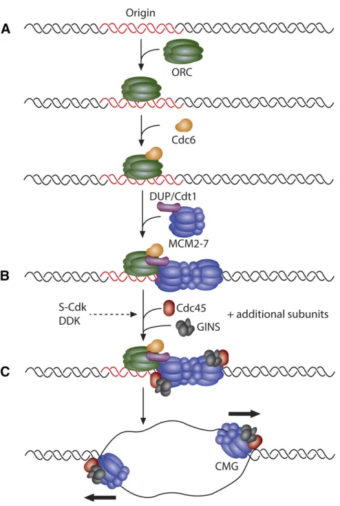
larval and adult development (Painter and Reindorp 1939; King and Burnett 1959; Balls and Billett 1973; Hammond and Laird 1985a,b; Smith and Orr-Weaver 1991; Lilly and Duronio 2005). The neural and imaginal tissues are the only tissues that continue to divide mitotically during embryonic and larval development. The endocycle consists of alternat-ing S and G phases (Figure 2B) without mitosis and cell division that occur during the canonical cell cycle (Figure 2A). During the endocycle, DNA content is increased at the genomic level, thus producing polyploid cells. As organism size is greatly increased throughout larval development, polyploidy is thought to coordinate cell size and tissue growth by generating large, highly metabolically active cells (Edgar et al. 2014; Orr-Weaver 2015). Indeed, blocking polyploidization inhibits cell and larval growth, inhibiting normal tissue function (Edgar and Orr-Weaver 2001).
The endocycle is utilized throughout the plant and animal kingdoms, indicating the importance of this variant cell cycle during development across organisms (Orr-Weaver 2015). Key insights into the regulation of the endocycle and its co-ordination with the replication program have derived from seminal studies in Drosophila. Nearly all larval tissues and many adult tissues inDrosophilahave increased ploidy that is achieved via the endocycle. The replicated DNA duplex
copies are held in register to produce polytene chromosomes with stereotypic banding patterns in mostDrosophila endo-cycling tissues. The most well studied of these polyploid tissues is the larval salivary gland, which undergoes 10 endocycles during larval development to obtain a final ploidy of roughly 1024C (Hammond and Laird 1985b). Dur-ing the endocycle, cells must suppress the mitotic machinery to prevent entry into the mitotic program and subsequent cell division. One strategy that endocycling cells use to achieve this is to downregulate the activity of mitotic Cyclins and mitotic CDKs at the transcriptional level. At the switch from the mitotic cell cycle to the endocycle, cells in the embryo cease expression of the mitotic regulators Cyclin A, Cyclin B, Cyclin B3, String/Cdc25, and CDK1 (Sauer
et al. 1995; Maqbool et al.2010). However, the develop-mental signals that regulate transcription of these regula-tors at this switch are not well understood.
InDrosophila, Cyclin E/CDK2 activity is the major driver of S-phase entry. Mutations in thecycEgene inhibit DNA repli-cation in both mitotic and endocycling cells (Knoblichet al.
1994). Importantly, continuous overexpression of cyclin E in the salivary gland blocks endocycling, suggesting that oscil-lations in Cyclin E/CDK2 activity are required for continued endocycling (Follette et al. 1998; Weiss et al. 1998). The oscillatory expression of cycEis mediated by oscillations in the levels of the transcription factor E2F1, which reaches high levels during G phase and is degraded at the end of S phase (Zielke et al. 2011). E2F1 degradation is mediated by the E3 ubiquitin ligase CRL4-Cdt2 (Shibutani et al. 2007, 2008), whose activity peaks during S phase (Zielke et al.
2011) (Figure 2C). Artificial stabilization of E2F1 prevents endocycling in the salivary gland, indicating that E2F1 deg-radation is required for continued endocycling (Zielkeet al.
2011). At the end of S phase, degradation of E2F1 is fol-lowed by ubiquitin-dependent degradation of Cyclin E via the E3 ubiquitin ligase CRL1-Ago along with its activator Minus (Shcherbata et al. 2004; Szuplewski et al. 2009; Zielkeet al.2011). The degradation of Cyclin E allows for the completion of S phase and the relicensing of replication origins in the subsequent G phase. Additionally, oscillations of theDrosophilaCDK2 inhibitor Dacapo peak similarly to E2F1 during G phase of the endocycle (Hong et al.2003, 2007). Dacapo contributes to the attenuation of Cyclin E/CDK2 activity during G phase and is subsequently degraded dur-ing S phase via its PIP degron (Swanson et al. 2015). Al-though Dacapo is not necessary for the endocycle (Hong
et al. 2003; Zielkeet al.2011), its overexpression inhibits the endocycle, suggesting that Dacapo plays a role in estab-lishing the Cyclin E/CDK2 activity threshold necessary to trigger S phase (Shcherbataet al.2004; Honget al.2007; Zielkeet al.2011; Swansonet al.2015).
Much like during the archetypal cell cycle, endocycling cells must also prevent rereplication during S phase. In the mitotic cell cycle, helicase loading at origins is restricted to late M through G1 phase. At the G1/S transition, the activities of S phase CDK and DDK increase dramatically, allowing for
the assembly and activation of the replicative helicase com-plex to begin DNA replication (Costa et al. 2013). After S-phase onset, high S-phase CDK activity prevents the reload-ing of the helicase complex at origins that have alreadyfired by inhibiting the activity of several replication initiation pro-teins required to load the helicase onto origin DNA (Blow and Dutta 2005). For example, phosphorylation of the DUP/Cdt1 replication initiation factor by Cyclin E/CDK2 during S phase promotes DUP/Cdt1 degradation in mitotic and endocycling cells (Thomeret al.2004). DUP/Cdt1 protein levels oscillate during the endocycle (Hong et al. 2007), and DUP/Cdt1 protein was found to accumulate in the G phase and rapidly decrease once cells enter into S phase (Whittakeret al.2000; Thomeret al.2004). Finally, constitutive overexpression of DUP/Cdt1 is sufficient to induce polyploidy in wing disc cells and results in enlarged nuclei with increased DNA content in endocycling follicle cells, emphasizing the significance of the regulation of DUP/Cdt1 levels by Cyclin E/CDK2 in prevent-ing rereplication (Thomeret al.2004).
InDrosophilaas well as in other metazoans, Geminin is an inhibitor of helicase loading and exhibits high levels during the S phase in the archetypal cell cycle to prevent rereplica-tion (Quinnet al.2001). During M phase, Geminin is targeted for degradation by the anaphase promoting complex (APC)/ cyclosome, allowing for helicase loading in the subsequent G phase (McGarry and Kirschner 1998). In a similar manner, Cyclin E/CDK2 activity peaks during S phase in the endo-cycle (Figure 2C). Additionally, Geminin levels oscillate dur-ing the endocycle, with low levels in G phase to allow for helicase loading and high levels in S phase to prevent reloading of helicases and rereplication. Geminin is targeted for degradation at the end of the endocycle S phase by the APC/cyclosome through the APC activator Fzr/Cdh1, and APC/CFzr/Cdh1activity is inhibited by Cyclin E/CDK2 activity
(Narbonne-Reveau et al. 2008; Zielkeet al. 2008) (Figure 2C). The oscillation of the activity level of Geminin is required for the endocycle, as constitutive expression of Geminin inhibits endocycle progression (Zielkeet al.2008). How-ever, Geminin is not essential for salivary gland develop-ment (Zielkeet al.2011), suggesting that multiple overlapping mechanisms exist to prevent rereplication in endocycling cells.
Tissue specificity of Drosophila origins:To date, the poly-tene larval salivary gland is the only differentiated tissue undergoing genomic replication in which genome-wide ORC localization has been reported (Sheret al.2012). In a survey of ORC binding in Kc, S2, and Bg3 cells, it was found that about a third of the identified ORC binding sites were shared between all three cell types (Eatonet al.2011). Similarly, 31% of the ORC binding sites identified in the larval salivary gland are common with all three cell lines, indicating that a significant level of ORC binding site conservation may exist not only in cell culture lines but in differentiated tissues as well. Notably, 28% of the salivary gland ORC binding sites are unique to this tissue. Consistent with cell culture studies, 73% of the salivary gland ORC binding sites are within a
kilobase of a transcription start site. A total of 57% of the salivary gland-specific ORC binding sites are found near a transcription start site, but the genes controlled by these promoters are not uniquely expressed in the salivary gland. Thus, tissue-specific expression of genes does not correlate with tissue-specific ORC binding (Sheret al.2012).
Insights into Regulation of DNA Replication from Localized Changes in DNA Copy Number
Interestingly, increases in gene copy number in polyploid
Drosophila cells are not uniform throughout the genome. Heterochromatin is repressed for replication in many Drosoph-ilapolyploid cells, and in several larval tissues, defined eukary-otic genomic regions have been shown to be underreplicated (UR) relative to overall ploidy of the cell (Hammond and Laird 1985a,b; Nordmanet al.2011). Additionally in the adult fe-male, follicle cells complete endocycling and begin gene amplification, leading to specific sites within the genome that are increased in copy number (Spradling 1981). The study of underreplication and differential gene amplifi ca-tion in Drosophila has provided important understanding about the developmental regulation of both origin activa-tion and fork progression at the molecular level. In the fol-lowing section, we summarize our current understanding of the molecular parameters of DNA replication from analysis of differential DNA replication.
Underreplication and local copy number reduction
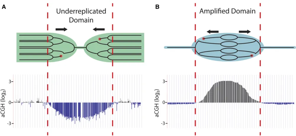
Although polytene cells have increased DNA content per cell, gene copy number is not uniform throughout the genome. For instance, it has long been known that the heterochromatic regions in polyploid salivary gland, follicle cell, and nurse cell chromatin are reduced in copy number relative to overall ploidy, a phenomenon known as underreplication (Zhimulev
et al.1982; Hammond and Laird 1985a,b; Lamb and Laird 1987; Smith and Orr-Weaver 1991) (Figure 3A). In addition to heterochromatin, array-based comparative genome hy-bridization (aCGH) and high-throughput genomic sequenc-ing studies have revealed that larval salivary gland, midgut, and fat body tissues contain precise euchromatic regions that are underreplicated as well (Belyakinet al.2005; Nordman
et al.2011; Sher et al.2012; Yarosh and Spradling 2014). These euchromatic UR regions can be large, ranging up to 450 kb in size. They exhibit features of repressed chromatin and thus also are termed intercalary heterochromatin, al-though as noted below these regions are not necessarily re-pressed for transcription (Belyaeva et al.2008; Filion et al.
2010). Only a third of identified UR regions are common to all the three tissues, highlighting the high degree of tissue specificity of underreplication (Nordmanet al.2011).
In addition to genome-wide profiling approaches in Dro-sophilacell culture, the study of underreplication inDrosophila
genome but is excluded within UR regions (Sheret al.2012). Thisfinding strongly suggests that replication initiation does not occur within these regions, and thus replication of these regions is dependent upon replication forks emanating from outside the region. Interestingly, these UR regions are devoid of RNA polymerase II, strongly inhibited for transcription, and are enriched for the heterochromatic chromatin mark H3K27me3 (Sheret al.2012). These results are consistent with the idea that UR regions in the salivary gland represent repressive chromatin domains that are inhibitory to both transcription and DNA replication initiation. Indeed, nearly all of the UR regions in the salivary gland correspond to domains of re-pressive chromatin as defined in genome-wide chromatin landscape studies (Filion et al. 2010; Kharchenko et al.
2011; Yarosh and Spradling 2014). UR regions in the larval fat body also are devoid of ORC binding, suggesting that ORC repression in these domains may be a common feature of underreplication (B. Hua, H. Kashevsky, G. Bell, J. Von Ste-tina, and T. Orr-Weaver, unpublished data). The analysis of UR regions in fat body shows, however, that underreplication is not causally linked to a chromatin state that is repressive for transcription, because the genes present in URs in the fat body are robustly transcribed (Nordmanet al.2011).
Interestingly,orc1andorc2null mutant salivary glands con-tinue the endocycle, though they reach ploidy levels two- to fourfold lower than wild-type salivary glands (Park and Asano 2008; Sheret al.2012). These results indicate that
the endocycle can occur to a significant extent in the absence of newly synthesized ORC1 and ORC2. However, orc1and orc2mutants exhibit a marked change in the underreplication pattern in the salivary gland where all but the most pro-nounced UR regions become fully replicated (Sher et al.
2012). Thus, ORC plays an important role in the distribution of replication along polyploid chromosomes, and it is possible that replication in the orc1andorc2mutants is allowed by maternal loading of ORC or by residual activity of ORC missing the ORC1 or ORC2 subunits.
Underreplication has been most extensively studied in
Drosophila, but underreplication of defined euchromatic re-gions occurs outside of Diperta as well. A total of 47 rere-gions of the genome in the polyploid mouse trophoblasts giant cells are recurrently and reproducibly underreplicated, although fold underreplication levels are low compared to that ob-served inDrosophila, with most of the identified regions be-ing less than twofold reduced in copy number and thus not called by the cut-off criteria used inDrosophila(Sheret al.
2013; Hannibalet al.2014). Nevertheless, this highlights the importance and relevance of studying underreplicated re-gions infly polyploid tissues as a model for differential rep-lication in polyploid tissues outside ofDrosophila.
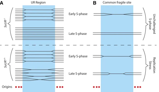
Inhibition of fork progression by SUUR: Underreplication in the salivary gland, fat body, and midgut are dependent upon the Suppressor of Underreplication protein (SUUR), as all
underreplicated regions become fully replicated in theSuUR mutant (Nordmanet al.2011; Sheret al.2012). SUUR is a chromatin protein that as of yet has not been identified out-side ofDrosophila(Nordman and Orr-Weaver 2015). Loss of SUUR function does not restore ORC binding in the under-replicated regions of the salivary gland, indicating that SUUR does not act at the level of replication initiation to inhibit replication (Sheret al.2012). Instead,SuURmutants exhibit enhanced rates of replication fork progression, suggesting that SUUR acts to inhibit replication fork progression (Sheret al.2012). Thesefindings support a model in which underreplicated domains are dependent upon replication forks emanating from origins outside of the region, and underreplication is achieved by the SUUR-mediated inhibi-tion of fork progression through these domains (Figure 4A). Subsequent studies revealed that SUUR coimmunopreci-pitates with the sliding clamp PCNA and the replication fork factor CDC45 in embryonic nuclear extracts and tracks with the replication fork in follicle cells undergoing gene amplifi -cation (detailed in subsequent sections), further supporting the fact that SUUR is recruited to active replication forks (Kolesnikovaet al. 2013; Nordmanet al.2014). Consistent with studies in endocycling tissues, SuUR mutants exhibit significantly enhanced fork progression in amplifying follicle cells, and overexpression of SUUR severely hampers fork pro-gression (Nordman et al.2014). Together, these results in-dicate that SUUR is a general inhibitor of fork progression and acts directly at the replication fork. However, the molec-ular mechanism of fork inhibition by SUUR remains to be elucidated.
Two key questions are whether SUUR inhibits replication in the pericentric heterochromatin and in the dispersed UR regions by the same mechanism, and how SUUR becomes recruited to replication forks in the UR regions. Recentfi nd-ings on the dynamics of histone H1 on salivary gland chro-mosomes during the endocycle provide insights (Andreyeva
et al. 2017). This histone is necessary for underreplication both in the pericentric heterochromatin and the UR regions.
H1 is required for SUUR localization on chromosomes, and the two proteins directly bind each other. Interestingly, early in S phase in the endocycle, H1 is enriched at regions that will replicate late, including those that become underreplicated. Later in the endocycle S phase, H1 becomes more uniformly distributed on the chromosomes. These results provide one mechanism for the regional specificity of SUUR action: that it is directed to specific regions by the presence of H1 histone. This is not sufficient, however, as SUUR localization and underreplication occur at only a subset of H1 localization sites on the euchromatic arms.
Fork instability and DNA damage in UR regions: In the polytene chromosomes of the salivary gland, UR domains are cytologically enriched for a key marker of double-stranded DNA breaks (DSBs), gH2Av (Andreyevaet al.2008). Chro-matin immunoprecipitation studies revealed that gH2Av is enriched throughout the entire region of each UR domain, indicating that UR domains are prone to DNA damage (Nordman et al.2014). Enrichment of gH2Av in these UR regions is dependent upon SUUR function, suggesting that DNA damage in these regions is caused by fork instability mediated by SUUR. Additionally, high-throughput sequenc-ing and analysis of read pairs generated from salivary gland DNA indicate that large deletions ranging 10–500 kb in size may result from DNA damage and local repair in these re-gions (Yarosh and Spradling 2014).
Potential biological functions of underreplication: As SuUR mutants are viable and exhibit normal morphology and fertility (Belyaevaet al.1998), it remains unclear to what extent SUUR is required in normal development. Given that SUUR is a general inhibitor of fork progression, it is possible that SUUR serves to provide an extra level of regulation to ensure proper replication timing in the genome. SUUR may regulate replication timing during S phase by blocking fork progression to ensure that regions of the genome are not replicated until late in S phase. Another function of SUUR
could be to distribute termination events throughout the ge-nome (Hawkinset al.2013). Although these would seem to be critical roles, they may not be essential unless the cells become subject to replication stress.
Because UR regions become fully replicated inSuUR mu-tants, the biological role of underreplication remains to be elucidated. The UR regions in the salivary gland are enriched in genes involved in cell adhesion, segmentation, transcrip-tion factor activity, programmed cell death, mesoderm devel-opment, and cell motility (Sher et al. 2012; Yarosh and Spradling 2014). Additional regions that are consistently underreplicated but to lower extents in the salivary gland are highly enriched in immunoglobulin superfamily genes and genes involved in the nervous system (Yarosh and Spradling 2014). Strikingly, transcription of genes within UR regions is largely repressed in the larval salivary gland and midgut tissues, suggesting that decreased copy number may cause lower gene expression (Nordman et al. 2011; Sher et al.
2012). However, in the SuURmutant in which UR regions are fully replicated, gene expression within the UR regions remains repressed, demonstrating that underreplication is not required for transcriptional repression in these domains (Nordmanet al.2011; Sheret al.2012). Additionally, many genes within the UR regions in the fat body are significantly transcribed, thus underreplication and the repression of transcription can be mechanistically uncoupled (Nordman
et al.2011).
As deletions and rearrangements have been reported throughout UR regions, one proposed role for underreplica-tion is to promote the somatic diversity of genes within these domains (Yarosh and Spradling 2014). This idea is especially interesting in the context of the immunoglobulin superfamily genes found in some UR sites in which gene rearrangements may be advantageous. Nevertheless, the biological role of underreplication has yet to be fully uncovered.
Underreplication as a model for common chromosomal fragile sites: In addition to its utility in understanding the mechanisms underlying differential replication inhibition, underreplication in Drosophilapolyploid tissues serves as a promising model system for human common chromosomal fragile sites. Common fragile sites (CFSs) are chromosomal locations characterized by recurrent breaks, gaps, and con-strictions on metaphase chromosomes upon replication stress (Durkin and Glover 2007). CFSs often are found in euchroma-tin and extend over megabase-long regions of the chromosome (Schwartzet al.2006; Smithet al.2006). It appears that mul-tiple mechanisms can lead to CFSs, but one of these involves replication origins and fork progression (Ozeri-Galai et al.
2014). For example, replication initiation does not occur within a 700-kb region forming the core of the most active human CFS, FRA3B (Letessier et al. 2011). Thus, replication of this large region is dependent entirely upon replication forks ema-nating from origins of replication outside of this domain. A general challenge to replication forks results in incomplete rep-lication of the FRA3B domain, leading to chromosome fragility
and instability (Figure 4B). UR regions in theDrosophila sali-vary gland are also devoid of origins of replication and rely on forks coming fromflanking regions for their replication (Figure 4A). Additionally, UR regions are prone to DNA damage, a property common to CFSs. Combining the genetic and cell bi-ological toolkits of the Drosophilasystem with genome-wide profiling techniques will allow for deeper understanding of the mechanisms that underlie replication initiation repression in these regions, control of fork progression, and the molecular properties of CFSs in human cells.
Developmentally programmed follicle cell gene amplification to increase local copy number
While the underreplication system has allowed study of replication properties and dynamics across large, defined chromatin domains, the molecular dissection of the mecha-nisms that underlie origin activation requires the study of well-defined origins of replication. Additionally, it is neces-sary to know when single origins fire in order to study individual origin activation events. The study ofDrosophila
follicle cell gene amplification has allowed the isolation and detailed molecular characterization of single metazoan ori-gins of replication. In this section, we review the character-istics of follicle cell gene amplification and focus on key studies that have led to critical understanding of the molec-ular parameters that regulate origin activation and fork progression.
To date, aCGH analyses have been performed on seven distinctDrosophilatissues to assay differential DNA repli-cation genome-wide (Kim et al. 2011; Nordman et al.
2011; Sher et al. 2012; B. Hua, H. Kashevsky, G. Bell, J. Von Stetina, and T. Orr-Weaver, unpublished results). Of the examined tissues, only the ovarian somatic follicle cells have been found to exhibit gene amplification or in-creased copy number of distinct genomic regions relative to overall ploidy of the cell.
forks that progress 50 kb to both sides of the origin (Spradling 1981; Claycomb et al. 2002). This results in a gradient of amplified DNA, with the highest copy number at the origin of replication (Figure 3B). Gene amplification continues until stage 13, and follicle cells are ultimately sloughed off the egg chamber at the end of oogenesis.
Most amplicon loci contain genes encoding critical protein components of the egg shell or proteins involved in the in-tegrity of the chorion (Spradling 1981; Claycombet al.2004; Fakhouriet al.2006; Kim and Orr-Weaver 2011; Kimet al.
2011; Tootle et al. 2011) (Table 2). Gene amplification is used as a developmental strategy to increase the template copy number for key chorion components whose protein products must be produced quickly in a relatively short de-velopmental time window (7.5 hr). Female-sterile alleles of essential replication factors demonstrate the requirement of ORC2 (Landis et al. 1997), MCM6 (Schwed et al. 2002), DUP/Cdt1 (Whittaker et al. 2000), Chiffon/Dbf4 (Landis and Tower 1999), and MUS101/TopBP1 (Komitopoulou
et al.1983; Orret al.1984; Yamamotoet al.2000) during gene amplification and egg development, indicating that gene amplification in the follicle cells likely uses the same components as those during normal S phase. Additionally, as egg shell integrity is dependent upon proper execution of the follicle cell gene amplification program, the identifi ca-tion of thin egg shell mutants has been an important and powerful method to uncover key players in gene amplifi ca-tion using forward genetic approaches. These have included the conserved replication proteins noted above as well as new replication proteins such as Humpty Dumpty and the Claspin checkpoint protein (Landis et al.1997; Landis and Tower 1999; Whittaker et al.2000; Schwed et al.2002; Bandura
et al.2005; Choiet al.2017).
The gene amplification system has allowed the molecular characterization of single origins of replication, proving a powerful tool to dissect the mechanisms that underlie origin activation. During gene amplification, originfiring is tightly coordinated with follicle cell differentiation. Amplification is achieved by repeated rounds of origin firing that occur at defined developmental time points during follicle cell differ-entiation, permitting temporal and quantitative resolution of replication initiation events (Table 2). Furthermore, defined sets of replication forks are generated from these single ori-gins of replication, allowing both the cytological and molec-ular characterization of replication fork progression in these
cells (Claycombet al.2002). In the next sections, we sum-marize the keyfindings regarding the molecular mechanisms underlying origin activation and fork progression that have emerged from studying the gene amplification system.
Control of origin activation during gene amplification:
Through aCGH analysis of 16C follicle cells, six distinct sites of amplification have been identified (Kimet al.2011). These sites, termedDrosophilaamplicons in follicle cells (DAFCs), are located at distinct sites within the follicle cell genome and are referred to by their cytological locations. The level of gene amplification varies, ranging from 60- to 80-fold amplifi ca-tion atDAFC-66Dto 4-fold amplification at several amplicons (Spradling 1981; Claycomb et al. 2004; Kim et al. 2011) (Table 2).
Genome-wide ORC mapping from amplification-stage egg chambers revealed that ORC is enriched at all six amplification origins in broad domains ranging from 12 to 32 kb in size (Kimet al.2011). Significant ORC binding was detected at nonamplified regions as well, revealing that ORC binding alone is not sufficient for origin activation during gene am-plification. Further analysis on genome-wide ORC binding from purified amplifying follicle cells will be necessary, how-ever, to rule out the possibility these sites of enrichment are derived from the nurse cells or the oocyte of the egg cham-ber. Interestingly, roughly two-thirds of the identified ORC binding sites overlapped with transcription units, consistent with ORC localization studies in cell culture. However, only a 10th of these ORC binding sites are associated with genes that are expressed at high levels [reads per kilobase per million (RPKM).3], in contrast to cell culture studies in which most ORC binding sites overlap with active promoters (MacAlpineet al.2010).
Many studies have profiled the underlying chromatin sig-nature at amplicon origins. The use of both cytological and molecular techniques have revealed that amplicon origin ac-tivity is correlated with a significant enrichment of histone acetylation marks, namely AcH3, H4K5ac, H4K8ac, H4K12ac, and H4K16ac (Aggarwal and Calvi 2004; Hartlet al.2007; Kim
et al.2011; Liuet al.2012; McConnellet al.2012). Tethering of the histone deacetylase Rpd3 to a transgenic amplicon or-igin significantly reduces its activity (Aggarwal and Calvi 2004; Kimet al.2011), whereas tethering of the histone acetyl transferase HBO1 increases its activity (Aggarwal and Calvi 2004), indicating that histone acetylation plays an important
Table 2 Drosophilaamplicons in follicle cells
Cytological location Max fold amplification Stages of originfiring Genes involved in egg shell function
7F 18–20 10B–11 Cp7Fa,Cp7Fb,Cp7Fc,Cp36,Cp38
22B 4 10B–13 None
30B 4 10B CG11381,CG13113,CG13114a
34B 6 10B, 13 Vm34Ca
62D 4 10B, 13 yellow-g,yellow-g2
66D 60–80 10B–11 Cp18,Cp15,Cp19,Cp16
local role in modulating origin activity. As histone acetylation also is correlated with transcriptional activity, it is thought that these histone modifications serve to establish an open chroma-tin environment that is conducive to the recruitment and load-ing of the large protein complexes involved in transcription as well as DNA replication.
InDrosophilaS2 cells, the histone variants H3.3 and H2Av are enriched at ORC binding sites (MacAlpineet al.2010). In follicle cells, H3.3 is abundant at the amplicon sites before and during amplification, overlapping with ORC binding re-gions (Paranjape and Calvi 2016). H3.3 null mutant flies, however, carry out genomic replication and gene amplifi ca-tion without detectable defects. Thus H3.3 is not essential for origin activation in these cells. These results suggest that although H3.3 is not required for origin activation, it may serve as a marker, possibly along with other histone variants and modifications, for chromatin attributes important for origin function and replication initiation.
Recently, nucleosome density and position have been investigated as regulators of ORC binding and replication initiation at the gene amplification loci. In budding yeast, nucleosomes are strictly and reproducibly positioned around the ARS consensus sequence at origins across the genome (Eaton et al.2011; Belsky et al. 2015). In follicle cells, ORC binding regions at theDAFC-66Dorigin correspond to nucleosome-depleted regions (Liuet al.2015), and ORC binding sites are generally depleted of nucleosomes in S2 cells as well (MacAlpine et al. 2010). ORC binding sites occur preferentially at AT-rich DNA sequences in amplifying follicle cells, suggesting that ORC binding to DNA is not solely a passive effect of the absence of nucleosomes, but rather favors the DNA regions that are disfavored by nucle-osomes (Liu et al.2015). This idea is consistent with the
finding that replication initiation factor binding sites also tend to be AT-rich in cultured cells, and thus this property may be conserved across different replication contexts in
Drosophila development (Comoglio et al. 2015). Nucleo-some positioning in the follicle cells does not correlate with changes in amplicon origin activity, and nucleosome posi-tioning at DAFC-66D is remarkably similar to that in the equivalent region in nonamplifying S2 cells. Therefore nu-cleosome positioning does not fully govern the specificity of ORC binding and origin activity in Drosophila (Liu et al.
2015). Rather, nucleosome positioning may be a passive effect of origin specification to allow for the binding of down-stream replication initiation factors.
Individual characterization of the follicle cell amplicons has revealed that the activation of metazoan origins is regulated by an extremely diverse set of mechanisms. First, it was found that the DAFC-66D origin,orib, requires a 440-bp enhancer ele-ment called amplification control element for the third chro-mosome chorion cluster (ACE3) for activity (Orr-Weaver and Spradling 1986; Carminati et al. 1992). ACE3 directs ORC binding atorib, located 1.5 kb away, to promote originfiring (Austinet al.1999; Chesnokovet al.1999). Additionally, nor-mal DAFC-66Damplification requires the functions of Myb,
Rb, and E2F1. E2F1 and Myb are both localized to ACE3, and an E2F1-Rb-ORC complex can be identified in ovary ex-tracts, suggesting a direct role of these factors in regulating ORC activity duringDAFC-66Dorigin activation (Boscoet al.
2001; Beallet al.2002, 2004). Second, it was found that solely
DAFC-62D exhibits transcription-dependent originfiring. In-terestingly, transcription is required at DAFC-62D in trans, though thistrans-acting mechanism has yet to be elucidated (Xie and Orr-Weaver 2008; Huaet al.2014). Third,DAFC-34B
is unique in that it exhibits originfiring at two separate stages of development, and thefinal round of originfiring occurs in the absence of detectable ORC localization. This raises the possibilities of ORC-independent origin firing or that origin
firing can occur with dramatically reduced ORC enrichment or activity (Kim and Orr-Weaver 2011). Finally, DAFC-22B
exhibits strain-specific amplification. Strikingly, relocation of a 10-kb fragment from the22Blocus from a22B nonamplify-ing strain to an ectopic site restoresDAFC-22Borigin activity, indicating that theDAFC-22Borigin is repressed incisby an inhibitory chromosomal element at the endogenous location (Kimet al.2011). Together, these studies highlight the diver-sity of mechanisms by which the activation of gene amplifi ca-tion origins is regulated.
How is rereplication achieved during gene amplification? One possibility is that the replication initiation factor DUP fails to be inactivated and thus promotes reloading of the helicase and rereplication at the amplicons. During the archetypal S phase, DUP activity is restricted to late M and G1 phase through inhibition by the protein factor Geminin and by CRL4(Cdt2)-mediated degradation during S phase (Lee
et al. 2010). At the most highly amplified locus, DAFC-66D, DUP is detectable cytologically in follicle cells well after the start of amplification and surprisingly tracks with replication forks (Claycombet al.2002) (Figure 5). Addition-ally, excessive DNA amplification is observed in the follicle cells ingemininmutants (Quinnet al.2001), and stabilization of DUP protein leads to excessive DNA amplification and ectopic genomic replication (Thomeret al.2004; Linet al.2009). One possibility for DUP persistence during gene amplification is that CRL4(Cdt2) ubiquitin ligase activity may be attenuated in the follicle cells during these developmental stages (Lee
et al.2010). Consistent with this idea, another target of the CRL4(Cdt2) ubiquitin ligase, E2F1, also persists through the start of amplification (Sunet al. 2008). Low CRL4(Cdt2) activity would allow for the continued presence of DUP even after the first round of origin activation at the amplicons, and this pool of DUP could permit helicase reloading and origin refiring.
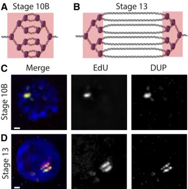
cells with a thymidine analog such as BrdU or 5-ethynyl-29 -deoxyuridine (EdU) (Calviet al.1998; Claycombet al.2002). Sites of amplification can be specifically visualized by BrdU or EdU incorporation because genomic replication is shut off during gene amplification (Calviet al.1998; Claycombet al.
2002). During the initial stages of amplification atDAFC-66D, replication initiation and fork elongation are coupled, which gives rise to a single focus of BrdU/EdU staining. However, during later stages of gene amplification, origin firing at
DAFC-66D ceases, and BrdU/EdU foci solely mark nucleo-tide incorporation at the active replication forks on either side of the origin, resulting in double bars of BrdU/EdU signal (Figure 5). The MCM2–7 helicase complex and the sliding clamp PCNA also can be visualized at sites of BrdU/ EdU incorporation throughout amplification (Claycomb
et al. 2002). Using these cell biological approaches, it has been possible to study fork elongation dynamics as well as the colocalization of other proteins and chromatin factors directly at the replication fork (Claycomb et al.
2002; Park et al. 2007; Nordman et al.2014; Alexander
et al.2015).
Molecular biology tools, both quantitative PCR and aCGH, have permitted replication fork progression to be tracked by changes in copy number at the amplicons in DNA isolated from staged egg chambers. These approaches have allowed for high-resolution analysis of fork progression during amplifi ca-tion, and they have uncovered genomic sites that impede fork
progression, changing the slope of the copy number gradients (Alexander et al.2015). Quantitative analysis of DNA copy number has been used to examine the effects of mutations on replication fork progression.
Mutations that enhance fork elongation:As discussed pre-viously, quantification of amplification domains inSuUR mu-tants demonstrated that loss of function of this protein results in increased fork progression, with replication forks at the amplicons elongating twice as far compared to wild-typeflies over the same developmental time. Thus the normal function of the SUUR protein is to impede fork progression. A cycE allele,cycE1F36, exhibits increased double-bar gap distances
and a wider gradient of amplified DNA copy number without altering originfiring or the developmental timing of the gene amplification program (Parket al.2007). This is a surprising
finding, as it reveals a previously unrecognized role of Cyclin E in fork elongation, a cell cycle factor well known for its role in helicase activation during replication initiation. The repli-cation phenotype of cycE1F36 is semidominant, suggesting
that the allele may be a gain-of-function mutation, promoting the progression of replication forks during amplification. However, how Cyclin E acts at the replication fork remains unclear.
Fork instability and DNA damage during rereplication:
During amplification, repeated originfiring generates multi-ple replication forks in close proximity moving in the same direction. One possible consequence of this close arrangement of trailing forks is collision between forks. Upon collision, replication forks may collapse, resulting in DSBs in the DNA. In support of this idea,gH2Av is enriched at the amplicons spe-cifically at the elongating replication forks, suggesting that rereplication generates a pileup of replication forks that are prone to“rear-end”collisions that may cause the formation of DSBs in the DNA (Alexanderet al.2015) (Figure 6). A paired-end high-throughput sequencing approach in amplifi cation-stage egg chambers highlighted the enrichment of several deletions at theDAFC-66D origin, suggesting that breaks are generated and repaired in this domain (Yarosh and Spradling 2014); however, whether the observed deletions are derived from the follicle cells, nurse cells, or oocyte remains to be determined.
Interestingly, full progression of the replication forks at the amplicons requires DNA damage response signaling, aschk1 andchk2mutants and a separation-of-functionmus101allele that specifically affects DNA damage signaling function (Kondo and Perrimon 2011) exhibit significantly decreased fork progression (Alexanderet al.2015). These results indi-cate that signaling of DNA damage is critical for continued fork progression during rereplication and suggest that repair of DSBs is important for the integrity of forks moving through the region.
As many copies of the amplified region are generated during the endocycle and gene amplification, this would provide many templates from which damaged DNA in the
Figure 5 Visualization of replication forks during follicle cell gene
ampli-fication. (A) At stage 10B, genomic replication is shut off, and originfiring and fork elongation begin at theDAFC-66Damplicon (full copy number not depicted). EdU is incorporated throughout the amplicon (indicated by the pink box), resulting in a single focus of signal. (B) At stage 13, origin
region can be repaired by homologous recombination (HR). Surprisingly, mutants for the key HR factors BRCA2 and SpnA (aDrosophilahomolog of Rad51) do not exhibit ham-pered fork progression (Alexanderet al.2015), and a dou-ble mutant for both homologs of Rad51, SpnA and SpnB, exhibits increased fork progression compared to wild-type controls at all amplicons (Alexanderet al.2016). Thus HR repair is not the main DSB repair mechanism and actually inhibits fork progression during gene amplification. Instead, a mutant for Lig4, a critical component of the nonhomolo-gous end-joining (NHEJ) pathway, shows significantly re-duced fork progression, indicating that NHEJ is a primary repair pathway utilized during gene amplification to repair DSBs and to allow subsequent forks to progress to normal levels (Alexanderet al.2015) (Figure 6). Finally, Mus308, a component of the microhomology-mediated end-joining pathway, allows proper fork progression at a subset of amplicons (Alexander et al. 2016). As amplifying follicle cells are nondividing cells whose functions are required over a short developmental time window (7.5 hr), it is possible that the quick repair of DNA damage offered by end-joining pathways is more advantageous during gene amplification than the homologous recombination pathway. Studies from human and yeast systems indicate that end-joining pathways like NHEJ can be completed in 30–70 min, while HR requires 5–7 hr (Rapp and Greulich 2004; Mao
et al.2008; Hickset al.2011).
Together, follicle cell gene amplification proves to be a powerful developmental replication system to dissect the molecular consequences of rereplication. The generation of two trailing replication forks in close proximity can result in fork collision and collapse, leading to the generation of DNA damage. If this damage is not repaired, this can pose serious consequences for subsequent forks moving through the damaged region, leading to genome instability.
Conclusions, Implications, and Future Directions
Differential regulation of origin activation
Research inDrosophilahas been key in our understanding of what defines a metazoan replication origin and its activation. The ability to identify ORC binding sites in a variety of dif-ferentiated cell types has revealed a high degree of tissue specificity of origin positioning within the genome. Although ORC is enriched at promoter sites, the tissue specificity of ORC binding cannot be explained by promoter activity. A key future direction will be to decipher the chromatin confi g-urations and chromosome conformation that designate ori-gin and ORC positioning. The tools inDrosophilawill permit identification of the state of chromatin modifications and associated proteins at origins and correlation with origin ac-tivity as well as contacts between origins and other chromo-somal sequences. The ability to conditionally eliminate gene function will be a significant advantage in testing causality in regulation of origin activity. The ability to track the activation of specific origins during gene amplification revealed at least three distinct mechanisms of origin activation, including the possibility of ORC-independent initiation. Analyzing whether these mechanisms operate at origins during a canonical S phase and whether the other amplicon origins utilize addi-tional activation mechanisms will be important. The follicle cells provide the opportunity to decipher how controls that normally prevent refiring of a replication origin can be overcome. Given the high frequency of gene amplification in cancer cells and the likelihood that many of these increases in copy number may result from unregulated origin activation (Hook et al. 2007; Beroukhimet al.2010; Greenet al.2010; Matsuiet al.2013), theDrosophilaamplicons will continue to produce relevant in-sights in our understanding of metazoan replication control.
Developmental control of replication timing and fork progression
In S phase in dividing or endocycling cells, replication timing is regulated such that some genomic regions replicate early in S phase while others regulate late, a property shared between
Drosophilaand mammalian cells. Both the mechanism that dictates when origins become active and the biological sig-nificance of replication timing remain to be determined, but it is notable that replication timing profiles are relatively con-served across cell types. Recent advances in analyses of DNA replication inDrosophilamake it an ideal model in which to define the control and role of replication timing. Replication timing profiles have been defined molecularly in cell culture and by cell biological approaches in polytene chromosomes, in which replication protein localization can be correlated with S-phase stages. The function of chromosomal proteins and chromatin modifications also can be linked to time in S phase, exploiting the extensive mutant collection in Dro-sophilaand RNA interference (RNAi) tools. A crucial question to be solved is how genomic regions are established that lack ORC binding. Another is whether genomic rearrangements resulting from underreplication serve biological functions.
Both the differential replication systems in which gene copy number is decreased through underreplication and in which copy number is increased through gene amplification have permitted metazoan replication fork progression and desta-bilization to be visualized and analyzed. This led to the identification of the chromatin protein SUUR as a repressor of replication and inhibitor of fork progression and has un-covered links between this protein and other chromatin pro-teins as well as replication components. Further insights into the tissue specificity of underreplicated domains and the mechanisms of their designation will be critical to our under-standing of how chromatin configuration can affect the elon-gation phase of DNA replication. These principles will be applicable to mammalian cells and thus to our understanding of common chromosomal fragile sites.
Both underreplication and gene amplification lead to ge-nome instability, in the former due to replication fork insta-bility and in the latter due to replication fork collisions. The double-strand breaks that result from these events can lead to genomic rearrangements. These models are powerful in
de-fining repair mechanisms that can restore fork progression to prevent rearrangements, with important implications for ge-nome stability in mammalian cells.
Acknowledgments
This work was supported by National Institutes of Health grants GM057960 and GM118098 to T.L.O.-W. and the Massachusetts Institute of Technology School of Science Fellowship in Cancer Research (to B.L.H.). T.L.O.-W. is an American Cancer Society Research Professor.
Literature Cited
Aggarwal, B. D., and B. R. Calvi, 2004 Chromatin regulates origin activity inDrosophilafollicle cells. Nature 430: 372–376. Alexander, J. L., M. I. Barrasa, and T. L. Orr-Weaver, 2015 Replication
fork progression during re-replication requires the DNA damage checkpoint and double-strand break repair. Curr. Biol. 25: 1654–1660. Alexander, J. L., K. Beagan, T. L. Orr-Weaver, and M. McVey, 2016 Multiple mechanisms contribute to double-strand break repair at rereplication forks in Drosophila follicle cells. Proc. Natl. Acad. Sci. USA 113: 13809–13814.
Almouzni, G., and M. Mechali, 1988 Xenopus egg extracts: a model system for chromatin replication. Biochim. Biophys. Acta 951: 443–450.
Andreyeva, E. N., T. D. Kolesnikova, E. S. Belyaeva, R. L. Glaser, and I. F. Zhimulev, 2008 Local DNA underreplication correlates with accu-mulation of phosphorylated H2Av in the Drosophila melanogaster
polytene chromosomes. Chromosome Res. 16: 851–862.
Andreyeva, E. N., T. J. Bernardo, T. D. Kolesnikova, X. Lu, L. A. Yarinichet al., 2017 Regulatory functions and chromatin load-ing dynamics of linker histone H1 durload-ing endoreplication in
Drosophila. Genes Dev. 31: 603–616.
Aparicio, O. M., 2013 Location, location, location: it’s all in the timing for replication origins. Genes Dev. 27: 117–128. Austin, R. J., T. L. Orr-Weaver, and S. P. Bell, 1999 Drosophila
ORC specifically binds to ACE3, an origin of DNA replication control element. Genes Dev. 13: 2639–2649.
Balls, M., and F. S. Billett, 1973 The Cell Cycle in Development and Differentiation. British Society for Developmental Biology Sym-posium. Cambridge University Press, Cambridge, UK.
Bandura, J. L., E. L. Beall, M. Bell, H. R. Silver, M. R. Botchanet al., 2005 humpty dumptyis required for developmental DNA am-plification and cell proliferation inDrosophila. Curr. Biol. 15: 755–759.
Beall, E. L., J. R. Manak, S. Zhou, M. Bell, J. S. Lipsick et al., 2002 Role for aDrosophilaMyb-containing protein complex in site-specific DNA replication. Nature 420: 833–837. Beall, E. L., M. Bell, D. Georlette, and M. R. Botchan, 2004 Dm-myb
mutant lethality inDrosophilais dependent uponmip130: posi-tive and negaposi-tive regulation of DNA replication. Genes Dev. 18: 1667–1680.
Beck, D. B., A. Burton, H. Oda, C. Ziegler-Birling, M. E. Torres-Padillaet al., 2012 The role of PR-Set7 in replication licensing depends on Suv4–20h. Genes Dev. 26: 2580–2589.
Bell, O., M. Schwaiger, E. J. Oakeley, F. Lienert, C. Beisel et al., 2010 Accessibility of the Drosophila genome discriminates PcG repression, H4K16 acetylation and replication timing. Nat. Struct. Mol. Biol. 17: 894–900.
Bell, S. P., and K. Labib, 2016 Chromosome duplication in Sac-charomyces cerevisiae. Genetics 203: 1027–1067.
Bell, S. P., and B. Stillman, 1992 ATP-dependent recognition of eukaryotic origins of DNA replication by a multiprotein com-plex. Nature 357: 128–134.
Belsky, J. A., H. K. MacAlpine, Y. Lubelsky, A. J. Hartemink, and D. M. MacAlpine, 2015 Genome-wide chromatin footprinting reveals changes in replication origin architecture induced by pre-RC as-sembly. Genes Dev. 29: 212–224.
Belyaeva, E. S., I. F. Zhimulev, E. I. Volkova, A. A. Alekseyenko, Y. M. Moshkin et al., 1998 Su(UR)ES: a gene suppressing DNA underreplication in intercalary and pericentric heterochromatin of Drosophila melanogaster polytene chromosomes. Proc. Natl. Acad. Sci. USA 95: 7532–7537.
Belyaeva, E. S., E. N. Andreyeva, S. N. Belyakin, E. I. Volkova, and I. F. Zhimulev, 2008 Intercalary heterochromatin in polytene chromo-somes ofDrosophila melanogaster. Chromosoma 117: 411–418. Belyakin, S. N., G. K. Christophides, A. A. Alekseyenko, E. V. Kriventseva,
E. S. Belyaevaet al., 2005 Genomic analysis ofDrosophila chro-mosome underreplication reveals a link between replication control and transcriptional territories. Proc. Natl. Acad. Sci. USA 102: 8269–8274.
Beroukhim, R., C. H. Mermel, D. Porter, G. Wei, S. Raychaudhuri
et al., 2010 The landscape of somatic copy-number alteration across human cancers. Nature 463: 899–905.
Bleichert, F., M. R. Botchan, and J. M. Berger, 2015 Crystal struc-ture of the eukaryotic origin recognition complex. Nastruc-ture 519: 321–326.
Bleichert, F., M. R. Botchan, and J. M. Berger, 2017 Mechanisms for initiating cellular DNA replication. Science 355: eaah6317. Blow, J. J., and A. Dutta, 2005 Preventing re-replication of
chro-mosomal DNA. Nat. Rev. Mol. Cell Biol. 6: 476–486.
Blow, J. J., and R. A. Laskey, 1986 Initiation of DNA replication in nuclei and purified DNA by a cell-free extract of Xenopus eggs. Cell 47: 577–587.
Blow, J. J., and J. V. Watson, 1987 Nuclei act as independent and integrated units of replication in a Xenopus cell-free DNA repli-cation system. EMBO J. 6: 1997–2002.
Blumenthal, A. B., H. J. Kriegstein, and D. S. Hogness, 1974 The units of DNA replication in Drosophila melanogaster chromo-somes. Cold Spring Harb. Symp. Quant. Biol. 38: 205–223. Bosco, G., W. Du, and T. L. Orr-Weaver, 2001 DNA replication
control through interaction of E2F-RB and the origin recognition complex. Nat. Cell Biol. 3: 289–295.