CHATTARAJ, PARNA. Gravitropism in Physcomitrella patens : A Microtubule Dependent Process (Under the direction of Dr. Nina Strömgren Allen)
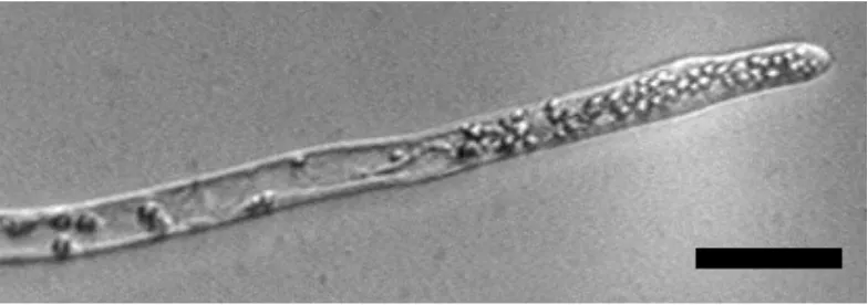
The plant cytoskeleton plays an important role in the early stages of gravisignaling (Kiss, 2000). Although in vascular plants, actin filaments are used predominantly to sense changes in the gravity vector, microtubules have been shown to play an important role in moss gravitropism (Schwuchow et al., 1990). The moss Physcomitrella patens is a model organism and was used here to investigate the role of microtubules with respect to the gravitropic response. Dark grown caulonemal filaments of P. patens are negatively gravitropic and the readily imaged tip growing apical cell is a “single-cell system” which both senses and responds to changes in the gravity vector. MTs were imaged before and after gravistimulation with and without MT depolymerizing agents.Six-day-old filaments were embedded in low melting agarose under dim green light, allowed to recover overnight in darkness and gravistimulated for 15, 30, 60 and 120 min. Using indirect immunofluorecence and high resolution imaging, MTs were seen to accumulate in the lower flank of the gravistimulated tip cell starting 30 min post turning and peaking 60 min after gravistimulation of the cells. The microtubule depolymerizing drug, oryzalin (0.1 µM for 5 min), caused MTs to disintegrate and delayed MT redistribution by 3hrs 30min. Growth of the oryzalin treated filaments was analyzed and a delay in growth was observed for both gravi and non-gravistimulated filaments. Tip cells bulged and sometimes branched after 75 min. This study demonstrates that microtubules are important for growth in P. patens and MT depolymerization leads to a delayed growth and graviresponse.
GRAVITROPISM IN PHYSCOMITRELLA PATENS :
A MICROTUBULE DEPENDENT PROCESS
by
PARNA CHATTARAJ
A thesis submitted to the Graduate Faculty of North Carolina State University
In partial fulfillment of the requirements for the Degree of Master of Science
BOTANY
Raleigh
2003
APPROVED BY:
___________________ __________________
Margaret E Daub Wendy F Boss Committee member Committee member
_________________
DEDICATION
PERSONAL BIOGRAPHY
ACKNOWLEDGEMENTS
I would like to first thank my advisor, Dr. Nina Strömgren Allen, for the tremendous support and excitement for research that she has lend me these past years. Special thanks also to my committee members Dr. Wendy Boss and Dr. Margaret Daub for their invaluable advice and knowledge.
A very special thank you to my laboratory members Dr. Eva Johannes, Ravisha Weerasinghe and Jeff Xu for their immense help and advice.
Special thanks also to Dr. David Collings for his help and advice on antibodies and immunolocalization techniques.
TABLE OF CONTENTS
LIST OF TABLES …………...……….……..vii
LIST OF FIGURES………..viii
LIST OF ABBREVIATIONS ………. ix
1 INTRODUCTION ... 1
1.1 Gravitropism in Plants ... 1
1.1.1 Perception of gravity by plants ... 3
1.1.1.1 Location of statocyte... 3
1.1.1.2 The Susceptor... 8
1.1.1.2.1 The starch-statolith hypothesis ... 9
1.1.1.2.1.1 Reduced sensitivity of starchless and starch-deficient mutants11 1.1.1.2.2 Gravitational Pressure Model ... 12
1.1.1.3 The Receptors ... 14
1.1.1.3.1 Endoplasmic Reticulum ... 14
1.1.1.3.2 The Cytoskeleton ... 16
1.1.1.3.2.1 Role of actin in gravisensing and signaling ... 16
1.1.1.3.2.2 The Role of microtubules in gravisensing and signaling... 19
1.1.1.3.3 The Plasma Membrane ... 21
1.1.2 Signal Transduction ... 22
1.1.2.1 Ions... 22
1.1.2.1.1 Calcium ... 22
1.1.2.1.2 pH... 24
1.1.2.2 The cytoskeleton ... 24
1.1.3 Response ... 25
1.1.3.1 The Cytoskeleton ... 25
1.1.3.1.1 Actin... 25
1.1.3.1.2 Microtubules ... 27
1.2 Tip Growth... 29
1.2.1 Calcium and Tip growth ... 30
1.2.2 Cytoskeleton and Tip growth... 31
1.2.2.1 Actin... 31
1.2.2.2 Microtubules ... 32
2.1 Culture of Physcomitrella patens... 33
2.2 Indirect immunofluorescence... 35
2.3 Imaging ... 35
2.4 Intensity calculations ... 36
2.5 Immunolocalization of oryzalin treated filaments ... 36
2.6 Growth Experiments ... 36
3 RESULTS ... 39
4 DISCUSSION ... 56
4.1 Tubulin distribution changes occur during gravistimulation ... 56
4.2 Effect of Oryzalin ... 58
4.3 Calcium, Gravitropism and Tip growth ... 59
4.4 Proposed model... 60
5 CONCLUSION... 66
LIST OF TABLES
Table 3.1 Total number and percent of cells with or without tubulin distributional changes... 46 Table 3.2 Total number of filaments showing either no change or changes in tubulin
LIST OF FIGURES
Fig 1.1 Different stages of gravitropism. ... 2
Fig 1.2 Life cycle of P. patens... 4
Fig 1.3 Caulonemal filaments grown in darkness... 5
Fig 1.4 Gravitropic response of an apical cell of a caulonemal filament. ... 6
Fig 1.5 Caulonemal tip cell displaying prominent amyloplasts... 7
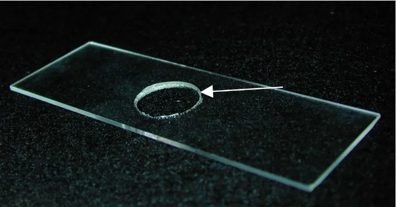
Fig 2.1 Slide with a hole in the center. ... 34
Fig 2.2 A-B Leica Upright Microscope cradled in a sidemount position... 38
Fig 3.1 a-c Microtubule distribution in vertically oriented filaments. ... 40
Fig 3.2 a-g Microtubule distribution in filaments that were gravistimulated for 15, 30, 60 and 120 minutes. ... 42
Fig 3.3 Graphical representation of the ratio of fluorescence intensity of the upper and lower areas of gravistimulated filaments. ... 44
Fig 3.4 Cartoon of MT distribution before and after gravistimulation. ... 45
Fig 3.5 a-f. Microtubule distribution in oryzalin treated filaments... 49
Fig 3.6 a-f. Oryzalin induced growth changes in non-gravistimulated filaments... 53
Fig 3.7 a-l. Oryzalin induced growth changes in gravistimulated filaments. ... 54
Fig 4.1 Calcium influx in apical caulonemal cell of P. patens... 62
Fig4.2 Summary timeline displaying the changes in tubulin intensity and calcium fluxes over time in gravistimulated filaments ... 63
Fig 4.3 Summary timeline of growth and tubulin changes associated with oryzalin treated gravistimulated filaments... 64
LIST OF ABREVIATIONS
Abbreviation
ABP Actin Binding Protein
DIC Differential Interference Contrast
ER Endoplasmic reticulum
F-actin Filamentous actin
G-actin Globular actin
HGMF High Gradient Magnetic Field
IAA Indole-3-acetic acid
IL Intensity of lower flank
IU Intensity of upper flank
IP3 Inositol 1,4,5-triphosphate
IR Infra Red
kDa Kilodalton
MAP Microtubule Associated Protein
MT Microtubule min Minute mM Millimolar
N.A. Numerical aperture
nm Nanometer PBS Phosphate Buffered Saline
pH Negative log of the hydrogen ion concentration S Second
UV Ultraviolet
1
INTRODUCTION
1.1
Gravitropism in Plants
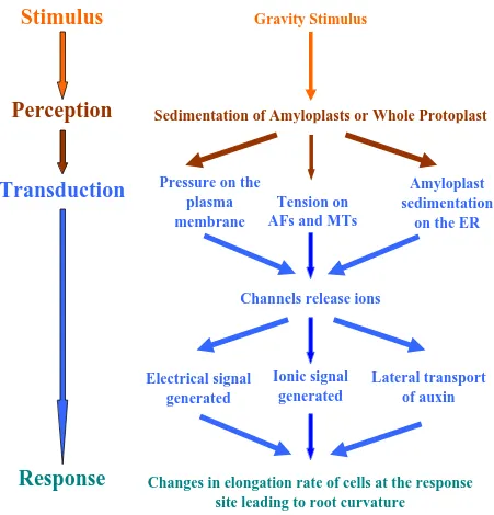
Stimulus
Gravity Stimulus
Fig 1.1 Different stages of gravitropism.
Diagrammatic representation of what is known about different stages of gravitropism and the involvement of various cellular components like the cytoskeleton, the plasma membrane, the ER, ions, hormones etc. in the gravitropic pathway.
Changes in elongation rate of cells at the response
site leading to root curvature
Electrical signal
generated
Ionic signal
generated
Lateral transport
of auxin
Channels release ions
Amyloplast
sedimentation
on the ER
Tension on
AFs and MTs
Pressure on the
plasma
membrane
Sedimentation of Amyloplasts or Whole Protoplast
Perception
Transduction
Response
1.1.1 Perception of gravity by plants
In general, gravity is physically sensed by a cellular component, called the susceptor (Björkman, 1988). The mass of the susceptor falls on the receptor, which turns on the signal transduction phase. The susceptor and receptor are located in the same cell in plants. The cells that harbor the susceptor and receptor are known as statocytes (Björkman, 1988).
1.1.1.1
Location of statocyte
Gravity is sensed in different tissues of roots and shoots in higher plants. Darwin (1881) recognized that when root caps were removed, roots failed to respond to gravity and he concluded that the root cap was the probable sensory site of for a gravistimulus in roots. Other tissues where gravisensing occurs include stems, primary and lateral roots, leaf and stem pulvini, hypocotyls and inflorescence stems. In most cases the perceiving cells in that organ contains amyloplasts.
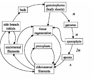
Fig 1.2 Life cycle of P. patens
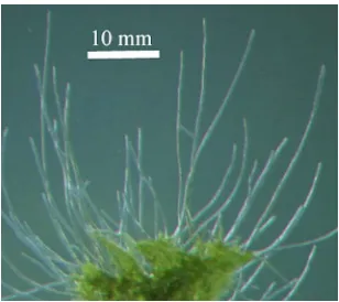
Fig 1.3 Caulonemal filaments grown in darkness.
Vertically growing, negatively gravitropic caulonemal filaments
(gravistimulating filament), Fig. 1.5 (vertically growing filament)} of the protonemata senses changes in the gravity vector.
1.1.1.2
The Susceptor
The susceptor senses gravity at the cellular level. Sack, (1991) proposed 5 different types of susceptors:
1. Statoliths, which are dense, sedimenting e.g. amyloplasts in root columella cells.
2. The entire protoplast e.g. in Chara internodal cells,
3. High density cellular components that stretch and/or compress without sedimentation e.g. cytoskeletal elements
4. Intracellular components that rise and
5. Extracellular components located at or outside the plasmalemma.
According to Björkman (1988) a gravity sensor may be present inside or outside the cell allowing the cell to sense its motion, displacement or position relative to the gravity vector. An effective sensor should be relatively insensitive to random thermal energy but very sensitive to changes in the direction of gravity (Björkman, 1988). The sensor needs to overcome the thermal noise and induce a force that will stimulate a gravi-response.
The presentation time of a normal wild type Arabidopsis root is 0.4 min (Kiss, 2000) and it can be very short (about 7s) at 1g in Lepidium roots (Larsen, 1979). Presentation time in the moss Ceratodon is between 12- 17 min (Walker and Sack, 1990).
There are two major models for gravity perception in plants (Sack, 1997): The starch-statolith hypothesis and the gravitational pressure hypothesis. Both have merit and I will describe each in turn.
1.1.1.2.1 The starch-statolith hypothesis
This classical theory of gravity perception was proposed by Haberlandt and Nemec (1900) and still is one of the most accepted hypothesis for how higher plants as well as mosses sense gravity. According to this theory, specialized dense starch filled organelles called amyloplasts function as statoliths (Sack, 1991). As the amyloplasts sediment, they somehow trigger the transduction of the stimulus. The sedimentation occurs in special cells, at specific areas and in a distinct developmental phase (Sack, 1991). In order to act as statoliths, the amyloplasts must overcome the background thermal noise. According to Björkman (1988), the approximate potential energy produced from a single root cap amyloplast displaced by gravity is about 250 times the thermal noise and about 15 times the activation energy predicted for a gravi-perception.
a statolith based sensing system in mosses.
In Chara rhizoids, statoliths play an important role in gravitropism. Tips of Chara rhizoids contain about 50 small, highly refractive vesicles containing BaSO4 (Schroter et
al., 1975; Sievers et al., 1996). During horizontal stimulation, complete sedimentation of statoliths to the lower flank occurs within 15 min (Sievers and Schröter, 1971) Reduction in numbers of these vesicles by immersing rhizoids in artificial media reduced gravitropism drastically (Kiss, 1994). Displacement of the BaSO4 particles following
manipulation with optical tweezers caused a tip reorientation – corresponding to positive gravitropism (Leitz et al., 1995; Braun, 2002).
1.1.1.2.1.1Reduced sensitivity of starchless and starch-deficient mutants
the reduced gravitropism is probably due to a reduction in sensing of the gravity change (Sack, 1997). Clearly, the amyloplast settling does play a role in sensing gravity and is necessary to get the full gravitropic response even though gravitropic sensing can occur without starch and is not absolutely required for sensing (Sack, 1997) except in Arabidopsis floral stalks. Maybe there are other sensors of gravity in a cell and amyloplasts are but one of these systems. When one sensor does not work, another could take over the sensing mechanism and help respond to the stimulus. This could possibly contribute to the reduced gravitropism in stachless and starch deficient mutants.
The best support for the starch-statolith theory comes from shoot mutants. Shoot gravitropic mutants sgr1 and sgr7 (Fukaki et al., 1998) are insensitive to gravity and lack sedimentable plastids in the shoots. Their growth rates however are normal and their roots are gravitropic with sedimenting amyloplasts.
1.1.1.2.2 Gravitational Pressure Model
streaming reverses when the polar ratio < 1 (Staves et al., 1997a). Calculations show that the energy produced by the settling protoplast in Chara is about 106 times larger than the background noise and is enough to open Ca+2 channels (Wayne et al., 1990).
According to this hypothesis, the entire protoplast senses gravity, and the ability of a cell to sense gravity depends on its static buoyancy (Staves, 1997). So, in a less dense medium a cell’s protoplast will settle on the cell wall or on the extracellular matrix, “producing a differential tension and compression between the plasma membrane and the extracellular matrix at the top and bottom of the cell, respectively” (Staves, 1997). Gravireceptors residing at the lower and upper part of the cell may get activated by these differential pressures (Wayne et al., 1990). According to Staves (1997), when the density of external medium is the same as that of the protoplast, the protoplast will attain neutral buoyancy and will not settle and no gravi-response will occur (polar ratio =1). On the other hand, if the density of the external medium is increased, the protoplast will be buoyant, creating compression between the extracellular matrix and plasma membrane at the cell-top and a tension between these two at the cell-bottom. This condition will ultimately lead to the reversal of the normal graviresponse (polar ratio < 1). This theory explains the presence of gravitropism in cells lacking statoliths e.g. in certain algae, fungi etc. but, the validity of this model in higher plants and gravitropic mosses is highly questionable as few direct data are available.
different ions that might be involved here, most attention has been paid to Ca+2 (calcium will be discussed in detail later in section 1.1.2.1.1). They speculated that tension in the plasma membrane will open the stretch-activated channels leading to Ca+2 release. The calcium ions then can be trapped by putative auxin transport proteins, a kinase (phosphorylating the transport protein) and Calmodulin (Pickard and Ding, 1993). This theory is based on the wall to membrane linkages found in onion epidermal cells (Pontolezica et al., 1993). Mechanoreceptive sensitivity is achieved only if there is a way of transferring shearing stresses from the rigid cell wall to the delicate membrane-filament-channel system, where they can be expressed as strains (Pickard and Ding, 1993). In spite of all these predictions, this theory has not been accepted due to lack of direct evidence for it in gravisensing systems.
1.1.1.3
The Receptors
The receptor is a specialized organ/structure in the cell that interacts with the susceptor in such a way that the physical information of gravity is transduced into a physiological signal (Sack, 1991). The many potential receptors of the signal for higher plants, algae and mosses will be described below, with regard to both plastid based and non-plastid based theories.
1.1.1.3.1 Endoplasmic Reticulum
function in gravi-sensing (Sievers and Volkmann, 1977). But this theory has been challenged as distal enrichment of ER is not universal (Audus, 1979). Recently, a specialized form of ER called nodal ER has been found in freeze substituted columella cells of tobacco and it may play a role in gravisensing (Zheng and Staehelin, 2001).
Two types of ER have been observed in dark grown protonemata of Ceratodon- tubular and cisternal ER (Walker and Sack, 1995). The tubular ER (TER) is distributed evenly throughout all the plastid zones (Ceratodon has 5 zones in the amyloplast region of the apical cell) but the distribution of the cisternal ER (CER) is varied among the zones (Walker and Sack, 1995). Both TER and CER are abundant in and near the apex
(~35 µm from tip). Longitudinal distribution of the TER changes in the apical cell of
gravistimulated protonemata, but the CER remains unchanged (Walker and Sack, 1997). CER is located mostly along the cell periphery in the sedimentation zone and qualitative analysis revealed that plastids sediment on top of the CER in horizontal cells (Walker and Sack, 1997). Although no direct contact between amyloplasts and ER was observed it is possible that ER acts as a gravi-receptor in moss apical cells.
1.1.1.3.2 The Cytoskeleton
All eukaryotic cells have a cytoplasmic network of filamentous proteins and their associated proteins collectively termed the cytoskeleton in their cytoplasm. The cytoskeleton is composed of microtubules, actin filaments (F-actin), intermediate filaments and their associated proteins (Ingber, 1993). In plants, the cytoskeleton has many important roles including cell signaling (Volkmann and Baluška 1999, Nick 1999), tip growth and polarity (Bibikova et al., 1999, Hepler et al., 2001) and perception of environmental stimuli like cold, touch, gravity and light etc (Orvar et al., 2000). Plant gravitropism is a complex process comprised of several well organized, coordinated phases and the cytoskeleton is thought to be an essential component in this pathway.
1.1.1.3.2.1Role of actin in gravisensing and signaling
The involvement of actin in gravisensing is well documented in the green alga Chara. Both positively gravitropic rhizoids and negatively gravitropic protonemata show a complex actin cytoskeleton that plays a role in tip growth as well as in gravisensing by regulating the position and sedimentation of statoliths (Braun and Wasteneys, 1998; Braun et al., 2002). In the rhizoids, the actin cytoskeleton forms thick bundles in the basal region, a meshwork of thin filaments in the subapical zone and a prominent spot of converging filaments in the apex (Braun and Wasteneys, 1998; Braun et al., 1999). In vertical rhizoids treated with cytochalasin D, statoliths fall to the physical bottom of the cell (Hejnowicz and Sievers, 1981). Application of Cytochalasin D leads to loss of the well organized regulation of statolith positioning (Buchen et al., 1993).
gravisensing model, organelles are restricted to their position by the cytoskeleton, which in turn are linked to plasma membrane receptors and ion channels, pumps etc. In response to gravity, these restricted organelles (e.g. statoliths or whole protoplast) can sediment on the cytoskeleton generating a tension that may lead to ion channel activation and triggering of downstream events eventually leading to gravity induced growth (Baluška and Hasenstein 1997; Blancaflor, 2002). According to the unrestrained gravisensing model, the cytoskeleton is not robust enough to control the position and mobility of the organelles and so statoliths sediment to the physical bottom of the cell in response to gravity (Baluška and Hasenstein, 1997). Sedimentation of statoliths onto cellular structures like the ER, the cytoskeleton etc. can then activate downstream events (Blancaflor, 2002). Tensegrity architecture i.e. a cell system built on tensional integrity of the cytoskeleton rather than compressional continuity was first proposed by Ingber et al., (1981). Recently Yoder et al (2001) proposed a new tensegrity based model for gravisensing in roots based on quantitative analysis of statolith sedimentation in corn columella cells. According to this model, sedimenting statoliths disrupt the actin based cytoskeletal network and produce a directional signal that activates/inactivates receptors on the plasma membrane (Yoder et al., 2001).
tensegrity model by Yoder et al, 2001 also shows the absence of actin cables in cryofixed/freeze substituted columella cells. The cytoskeletal matrix in these cells only revealed a meshwork of randomly oriented, single, straight actin-like filaments (Yoder et al, 2001). But recently, prominent actin strands/bundles have been shown to be present in root columella cells by confocal microscopy (Collings et al, 2001; Blancaflor, 2002). These contradictory results arose from lack of proper fixation methods (as the dynamic nature of F-actin is very difficult to image in fixed, dead cells). This problem can be solved by transforming cells with a GFP fusion protein that will accurately label actin filaments.
a greater ease of plastid movement following disruption of the F-actin network (Yamamoto and Kiss, 2002). Although this supports the theory that statoliths are constrained by the F-actin network, another recent result with cytochalasin D treatment contradicts the hypothesis. Application of cytochalasin D in lentil columella cells leads to a decrease in amyloplast displacement velocity (Driss-Ecole et al., 2000). So although the results with actin disrupting drugs are interesting they further complicate the interpretation.
Localization of myosin-related proteins on the surface of sedimenting amyloplasts in cress root statocytes (Wunsch and Volkmann, 1993), maize root caps (Baluška and Hasenstein, 1997) and of profilin (Staiger et al, 1997), the actin binding protein in alfalfa statocytes and maize root apices (Gibbons and Staiger, 2000) suggest that a dynamic actin cytoskeleton maybe responsible for statolith movements.
Recent genetic studies also support the involvement of the cytoskeleton in the early gravisignaling events. The altered response to gravity (arg1) mutant of Arabidopsis encodes a 45kDa DnaJ-like protein whose C-terminal domain contains motifs with sequence homologous to cytoskeleton –interacting proteins (Sedbrook et al., 1999). This means that ARG1 interacts with the cytoskeleton and may help in transmission of gravity signals to the receptors located at the plasma-membrane or ER (Sedbrook et al., 1999)
1.1.1.3.2.2The Role of microtubules in gravisensing and signaling
Hasenstein et al, 1999). So there maybe a different role for MTs in root gravitropism. In Chara rhizoids, the MT inhibitor colcemid, prevents the gravitropic response (Friedrich and Hertel, 1973). Application of colchicine induces MT disassembly and leads to fattening of cylindrical Chara internodal cells to spheres or bulges (Green, 1962). But in contrast to earlier observations oryzalin application does not prevent gravitropism in Chara rhizoids (Braun and Sievers, 1993). Again, in the rhizoids and protonemata of the algae Chara, exclusion of MTs and convergence of actin filaments in the apex suggests an actin-mediated graviresponse system (Braun and Wasteneys, 1998).
The moss life cycle alternates between a haploid gametophyte and a diploid sporophyte. Diploid spores germinate to produce filamentous protonema (Cove et al., 1997). Protonemal stage is comprised of chloronemal and caulonemal filaments. While caulonema exhibit only axially running MTs, chloronema contain axial, transverse and obliquely arranged MTs (Doonan and Cove, 1985).
Moss MTs were first observed by immunofluorescence methods on moss protoplasts (Powell et al., 1980) and later it has been shown that distribution of MTs is very similar in the various moss species (Doonan and Cove, 1985; Schwuchow et al., 1990). The apical cells of moss protonemata contain MT strands/cables that lie axially and also form a focus at the tip (Doonan and Cove, 1985; Wacker et al., 1988; Schwuchow et al., 1990).
amiprophos-methyl (APM) for 30-60 min lead to increased plastid sedimentation compared to that found in untreated cells (Schwuchow and Sack, 1994). Longer application resulted in more dramatic sedimentation and in some cases almost all plastids sedimented to the lower flank of the cell.
The apical cell of negatively gravitropic protonemata of Ceratodon showed a higher density of MTs in the lower flank in comparison to the upper flank when gravistimulated for more than 20 min (Schwuchow et al, 1990). The enrichment of MTs was seen proximal to the sedimenting amyloplasts and near the tip region that elongates to produce curvature (Schwuchow et al, 1990). Application of MT inhibitors like APM and oryzalin lead to disruption of gravitropism and the zonation of the tip cell (Schwuchow et al, 1990).
Application of oryzalin to the protonemata of Funaria leads to disruption of zonation in the tip cell and changes the plastid position (Wacker et al, 1988). I used oryzalin in P. patens to find out the effect of MT disruption on gravitropism. In this thesis, I also demonstrate the involvement of MTs in early phases of gravitropism in the moss P. patens.
1.1.1.3.3 The Plasma Membrane
1.1.2 Signal Transduction
Signal Transduction is the conversion of a signal (ionic/mechanical/thermal) from one form to another. This signal then gets transmitted to the targeted organ. The order of signaling events occurring after reception are unclear, although some of the players have been identified (Blancaflor, 2002). Below are descriptions of some of the components of signal transduction. These components may act alone or in co-ordination with signaling elements from other stimuli such as touch or light that also induce differential growth.
1.1.2.1
Ions
1.1.2.1.1 Calcium
sensing protein) transformed intact Arabidopsis seedlings. Most of the data regarding the involvement of changes in Ca2+ levels with gravitropism are indirect and involved studies using calcium inhibitors. Application of calcium chelators like EGTA, EDTA etc. to maize roots arrested gravitropic curvature and addition of calcium restored the response (Björkman and Cleland, 1991; Lee et al., 1983). Calcium chelators also block gravity induced auxin redistribution (Young and Evans, 1994). On the other hand, auxin transport inhibitors block cap-based polar Ca2+ transport in maize and pea roots (Lee et al., 1984). So there is a relationship between Ca2+ and auxin distribution. Calmodulin, a Ca2+-dependent regulatory protein acts as a plant calcium monitor or modulator and is involved in gravitropic sensing and signal transduction. Calmodulin levels in the root apex are elevated in response to gravity (Sinclair et al., 1996). Application of calmodulin antagonists delayed graviresponse in root tips of maize but the root growth was unhampered (Stinemetz et al., 1992).
More evidence regarding the involvement of Ca2+ in gravisignaling is based on the studies with the Ca2+ related second messenger, inositol-1, 4, 5-triphosphate (IP3). IP3
mobilizes Ca2+ and helps in its release from intercellular reservoirs like ER, vacuole etc. to trigger subsequent calcium dependent signal transduction pathways (Munnik et al., 1998; Kamada and Muto, 1991; Tucker and Boss, 1996). Recently, a rise in IP3 level has
been observed in oat shoot pulvini within 15 sec of gravistimulation (Perera et al., 1999; Perera et al., 2001). Although, the rise in level was equal in both upper and lower flanks initially, it tripled in the lower flank between 10 and 30 min of gravistimulation (Perera et al., 2001).
et al., 2003 observed a Ca2+ influx in the apical dome of growing caulonemal filaments of P. patens. When these filaments were gravistimulated, the Ca2+ influx was not restricted to the apex but extended down the flank for at least 60 µm, where the influx was highest (Allen et al, 2003). So, a differential activation of Ca2+ activating channels in the plasma membrane is established following gravistimulation (Fig 4.1 taken from Allen et al., 2003).
1.1.2.1.2 pH
A number of studies have been conducted to measure extra and intracellular pH in response to gravistimulation and comparing the measurements to normal behavior. Proton efflux has been observed in the upper flank of root caps using the vibrating wire probe (Collings et al., 1992). Scott and Allen, (1999) showed that there is a change in cytoplasmic pH in the columella cells of Arabidopsis roots that have been shown to be most effective in sensing gravity (Blancaflor et al., 1999). Root cap pH changes are necessary for gravitropism in Arabidopsis root (Fasano et al., 2001).
1.1.2.2
The cytoskeleton
work together to transmit a signal as follows. The decrease of pH in a cell can result in a decrease in actin filament / MT tension leading to sedimentation of heavy cellular components like amyloplasts on to the cell bottom. This in turn could activate putative mechanical or stretch induced ion channels. On the other hand an increase in tension can cause strain on the attached membrane and lead to opening of ionic channels. Also, Ca2+ /calmodulin may interact with MTs via MAPs (Microtubule Associated Proteins) (Cyr, 1991) and MT disruption opens Ca2+ channels in Arabidopsis and carrot cells (Thion et al., 1996, 1998). It is likely that a change in calcium concentration can alter the cytoskeletal distribution of either actin filaments or microtubules such that it induces a biochemical signal that gets transmitted to the target organ.
1.1.3 Response
The response phase of gravitropism involves differential growth of the targeted organ that eventually leads to the redirection of the organ in response to gravity. In roots, growth occurs due to elongation of cells in the upper side of the root. In mosses the apical cell responds to gravity by redirecting its growth away from the gravity vector.
1.1.3.1
The Cytoskeleton
1.1.3.1.1 Actin
elongation by mechanically constraining it, by influencing microtubule alignment, or by controlling vesicle transport to active cell wall growth areas (Waller et al., 2000; Baluška et al., 2001b). Presence of severely disrupted actin bundles in the transition zone of the maize lilliputian (lacks cell elongation zone) mutant (Baluška et al, 2001a) and generation of dwarf Arabidopsis and rye plants after application of the actin inhibitor Latrunculin B (Baluška et al., 2001b) implies that actin may be responsible for normal cell elongation. In spite of these correlations between actin structure and elongation of growth, graviresponding maize roots or pulvini do not reveal any organizational differences of their longitudinal actin bundles between the lower and upper flanks (Blancaflor and Hasenstein, 1997; Collings et al, 1998). But actin disruption in maize root cap leads to enhanced gravitropism (Hou et al., 2003). So actin may act as a regulator of the timing and duration of a signal originating from the root cap that allows the root to resume normal vertical growth (Hou et al., 2003). The fine cortical actin arrays can also play a role in plant morphogenesis through an actin-microtubule feedback system (Collings and Allen, 2000). In developing roots, cortical actin undergoes reorientation similar to microtubule reorientation and it may be possible that cortical actin also modifies during gravity induced growth (Blancaflor, 2000, 2002). But it does not do so in pulvini, where the larger actin filaments appeared to unchanged in orientation during gravistimulation. (Collings et al., 1998).
modulating cell expansion during differential growth (Blancaflor, 2002). Both profilin and cofilin interact with phosphoinositide lipids and respond to changes in cytosolic Ca+2 and pH (Gibbon, 2001).
1.1.3.1.2 Microtubules
The involvement of MTs in gravity induced differential growth is based on the parallel alignment of cortical MTs and the newly deposited cellulose microfibrils (Giddings and Staehelin, 1988, 1991). The deposition of microfibrils in the parallel direction restricts the growth (i.e. cell elongation) to a direction perpendicular to its orientation (Green, 1980). Most studies regarding the role of MTs in gravity induced growth have been done in growing roots, pulvini (Collings, 1998) and coleoptiles. Cortical microtubules exhibit a variable but mostly transverse orientation in the meristematic zone that changes to strict transverse MTs in the elongation zone (Barlow and Baluška, 2000). As the cell matures and growth rate decreases, the transverse alignment of MTs changes to a longitudinal pattern (Barlow and Baluška, 2000). Cortical MTs receive directional cues from the environment and transduce this information to different cell developmental processes (Fischer and Schopfer, 1988). In gravitropic roots, the MT orientation changes from transverse to longitudinal in the slower growing lower flank (Blancaflor and Hasenstein, 1993, 1995) while in the lower flank of faster growing graviresponding shoots the orientation changes from longitudinal to transverse (Nick et al., 1990; Himmelspach et al., 1999). So maybe MTs are regulating the gravitropic growth by controlling the directional deposition of cellulose microfibrils (Blancaflor and Hasenstein, 1993).
reorientation of MTs during gravitropism occurs after the growth curvature has started (Blancaflor and Hasenstein, 1995). Again, plant roots do respond to MT depolymerizing drugs eg, oryzalin etc but that does not prevent gravitropic curvature (Baluška and Hasenstein, 1997). Although application of MT disrupting drugs to maize and rice coleoptiles lead to a reduction in gravitropic curvature (Nick et al., 1991), further investigation of maize pulvini revealed no change in MT organization between upper and lower flanks during gravistimulation (Collings et al., 1998). Lastly, Bichet et al., 2001 demonstrated that Arabidopsis BOTERO I mutants with disorganized cortical MTs in the elongation zone can respond to gravity.
In spite of all these studies, some recent findings have again bolstered the fact that MTs play an important role in the plant graviresponse phase. Himmelspach et al., (1999) observed gravity induced reorientation of cortical MTs in tubulin injected live epidermal cells of maize coleoptiles under a confocal laser scanning microscope. A recent study by Himmelspach and Nick, 2001 showed that in maize coleoptiles, gravity induced MT reorientation (30 min in lower flank) preceded gravitropic curvature and growth.
1.2
Tip Growth
The gravitropic response in Physcomitrella patens occurs as stated previously as a differential elongation of the two sides of the apical or tip filament cell. Below is a discussion of what is known about tip growth in plants.
1.2.1 Calcium and Tip growth
Tip growth has been most extensively studied in root hairs and pollen tubes. A calcium influx at the tip is an essential requirement for growth in both. The apical areas of growing cells exhibit a gradient of high concentration of free cytosolic calcium (Bibikova et al., 1997; Felle and Hepler, 1997; Wymer et al., 1997).The steepness of the tip-focused gradient and growth rate of the cells are directly proportional to each other. This local increase in calcium concentration disappears when the growth terminates (Wymer et al., 1997; de Ruijter et al., 1998). Application of calcium ionophores and channel blockers lead to growth inhibition in root hairs and pollen tubes (Herth et al., 1990; Wymer et al., 1997).
The increase in calcium concentration at the apex is due to a calcium influx (Herrmann and Felle, 1995; Jones et al., 1995; Malho et al., 1994). Electrophysiological studies using the ion selective biocurrents probe shows that the calcium influx is higher at the tip than at the sides or base of the growing cell (Schiefelbein et al., 1992; Herrmann and Felle 1995; Jones et al., 1995; Pierson et al., 1994). When growth ceases the calcium channels at the apex are thought to be closed. Vesicle fusion is enhanced in the presence of calcium concentration higher than normal cytosolic level (Zorec and Tester, 1992; Wollheim and Lang, 1994). The concentration of calcium at the cell apex determines the direction in which the secretory vesicles deposit materials (Gilroy and Jones, 2000). In mosses, growing caulonemal filaments of P. patens also have a calcium influx at the apical dome (Allen et al., 2003).
localized photoactivation of a caged calcium ionophore) across root hair tip results in reorientation of root hairs toward the new calcium gradient (Bibikova et al., 1997). UV photoactivation of caged calcium in pollen tubes also results in growth towards the increased calcium level (Malho and Trewavas, 1996). An increase in calcium levels leads to an increase in calcium dependent protein kinase levels (Moutinho et al., 1998). This increase in protein level may then regulate Ca+2 channel activity (Moutinho et al., 1998) that can lead to changes in the actin cytoskeleton (Kohno and Shimmen, 1987).
1.2.2 Cytoskeleton and Tip growth
1.2.2.1
Actin
1.2.2.2
Microtubules
Although MTs are essential component of the cytoskeleton, their involvement in tip growth varies from system to system. MTs are not essential for pollen tube growth and tip extension. When MTs are disrupted, the cytoplasmic organization gets altered but cell elongation and overall cell morphology remains unchanged (Kost et al., 1999; Taylor and Hepler, 1997; Derksen et al., 1995).
In contrast, MTs regulate tip growth in root hairs and moss protonemata. In root hairs, the cortical MTs run parallel to the long axis of the cell and extend to the apex. Bibikova et al., 1999 demonstrated that both stabilization and depolymerization of MTs in Arabidopsis root hairs causes a loss of directionality and induces multiple growth points at the apex. Again, the inhibition of cytoplasmic streaming due to actin depolymerization in Hydrocharis root hairs is reversible in presence of MTs (Tominaga et al., 1997). This indicates that actin organization in root hairs may be regulated by MTs (Kost et al., 1999; Collings and Allen, 2000).
2
MATERIAL AND METHODS
2.1
Culture of Physcomitrella patens
Protonemal fragments of P. patens were grown for 10-12 days under continuous white light at 230C on solid 0.7% Agar with {PPNH4 : CaNO3.4H2O(0.08%),
MgSO4.7H2O(0.025%), FeSO4.7H2O(0.001%), Microelements, NH4tartarate(0.05%),
Glucose(0.5%)} media, but without glucose (Under Annex A: http://www2.unil.ch/lpc/docs/pdf/Schaefer_Thesis.pdf).The fragments were overlaid with
cellophane disks (W.E. Canning, Bristol, UK) After 10-12 days, the protonemal fragments were plated on PPNH4 media (with glucose) and grown in the dark in a vertical
orientation (Fig. 1.3) for 6 days. Filaments were embedded in 1.3% low melting agarose (Sigma, Type VII) under green light, placed on a slide in which a hole had been drilled in the center (Fig. 2.1) and a cover glass (# 0) had been placed at the bottom. This formed a chamber. The agarose was supplemented with glucose medium. Plantlets were allowed to recover overnight in the medium and in darkness.
Fig 2.1 Slide with a hole in the center.
2.2
Indirect immunofluorescence
Filaments were placed for 1.5 hrs. in a fixative containing MTSB (150mM Pipes, 6mM MgSO4 ,15mM EGTA, pH 6.9), 1% DMSO, 0.01% Triton X-100, 8%
Formaldehyde). Filaments were washed 4x in phosphate buffered saline (PBS; 2.68 mM KCl, 1.47 mM KH2PO4, 13.69 mM NaCl, 0.81 mM Na2HPO4, pH 4), placed in acetone
and then kept in the freezer for 10 min. 4 washes of PBS were given after every treatment. Filaments were incubated in blocking buffer (5% BSA, 0.05% Tween-20 in PBS) for 15 min. followed by incubation in monoclonal anti-alpha tubulin (N356; Amersham International plc, Buckinghamshire, England) diluted 1/100 in incubation buffer (1% BSA, 0.05% Tween-20 in PBS). Filaments were then washed and incubated with secondary antibody (Alexa Fluor 488 goat anti-mouse IgG diluted in 1/400 in Incubation buffer, Molecular Probes, Eugene) for 2hrs at 370C. After incubation, the filaments were washed and mounted with PBS, slides were covered with No. 0 cover slips and sealed with VALAP (Vaseline, Lanoline, Paraffin: 1:1:1).
2.3
Imaging
Images were taken with a confocal microscope (Leica TCS Nt or TCS SP or LCS; Wetzlar, Germany) with a 40x, N.A. 1.4 oil immersion objective. Optical stacks were
recorded using approximately 0.2 – 0.3 µm thick sections and each plane was averaged 4
2.4
Intensity calculations
Fluorescence intensities were calculated using the Metamorph Analysis System (Universal Imaging). Approximately the same areas from the upper and lower flanks were selected using the region tool and the intensity of those areas was calculated using the ‘Region measurement’ tool. The intensity data was exported to an excel spreadsheet. The means of upper and lower flank intensities for each time frame were calculated. Next, the ratio of the mean intensities (lower flank/upper flank) was calculated and the data was imported to Microcal Origin software and a graph was constructed.
2.5
Immunolocalization of oryzalin treated filaments
Prior to gravistimulation the filaments were treated with 0.1 µM oryzalin
(microtubule depolymerizing drug) for 5 min and then indirect immunolocalization was performed in exactly the same way as above (under indirect immunolocalization).
2.6
Growth Experiments
P. patens was grown under continuous white light in exactly the same way as described in section 2.1. After 10-12 days, the protonemal fragments were placed on PPNH4 medium (0.5% phytagel was substituted for agar in order to obtain a transparent
medium) with glucose and grown in the dark for 6 days. After 6 days the filaments were
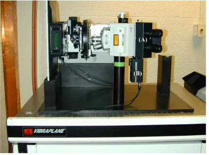
Fig 2.2 A-B Leica Upright Microscope cradled in a sidemount position. (A) Leica upright microscope (DMRX-A) used for growth studies.
3
RESULTS
Microtubule distribution in dark-grown normal filaments
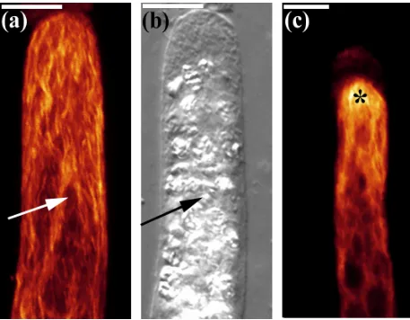
Fig 3.1 a-c Microtubule distribution in vertically oriented filaments.
Full stack projections of fluorescence and transmitted light images of upright filaments as observed through a confocal laser scanning microscope (CLSM). Fluorescently labeled filaments display a net axial distribution of MTs with either no cap (a) or a dense apical cap (c, *). The DIC image (b) is the same filament as seen in (a). Each amyloplast (arrow) is located in a distinct area and is clearly visible in the DIC image (b) Bar = 10
No significant changes are seen in overall MT distribution 15 min after a change in the gravity vector
Caulonemal filaments were gravistimulated by changing their orientation (i.e. 900 to vertical). The distribution of MTs in these filaments (~84%) deviated only slightly from that of the non-gravistimulated ones. MTs were very dense and formed an apical cap in most filaments (Fig.3.2a). The level of fluorescent intensities between the upper and lower flanks revealed no significant difference.
Significant changes in tubulin distribution are observed 30 min after gravistimulation starts
About 76% of the filaments gravistimulated for 30 min were enriched in tubulin
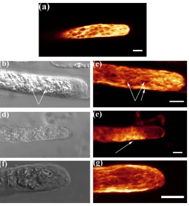
Fig 3.2 a-g Microtubule distribution in filaments that were gravistimulated for 15, 30, 60 and 120 minutes.
60 min after gravistimulation, the greatest accumulation of tubulin occured at the lower flank
When filaments were on their side for 1hr MTs disintegrated, but a very significant enrichment in tubulin fluorescence on the lower flank was observed in 71% of these filaments. The increase occurred in the plastid zone, behind the amyloplast free zone in the tip cell (Fig.3.2e). An arrow indicates the area that had quite intense labeling. Corresponding transmittance light images revealed that all the points of high intensity were in the regions surrounding plastids (fig 3.2d). The preservation of these filaments was difficult, since the MT filaments were not there. The intensity ratio of the fluorescence between the lower and upper flank (IL/IU) was 1.7 with a std. error of 0.22.
Tubulin density returned to low levels 2 hr after gravistimulation
Gravistimulation for 2hrs (Fig.3.2g) appeared to increase the tubulin density in the lower flank, but analysis of the intensity levels revealed that the difference in intensity between the upper and lower flank had decreased relative to that found in cells gravistimulated for 1hr treatment. The tubulin intensity is almost similar to that measured at 30 min and the intensity ratio is also similar to the 30 min level (~1.299) with a std. error of 0.015. Strands of MTs are visible and they lie in the axial direction (3.2g).
Graphical representation of intensity ratio
Fig 3.3 clearly demonstrates the intensity differences occurring between the upper and lower flanks at different time points after gravistimulation.
Fig 3.3 Graphical representation of the ratio of fluorescence intensity of the upper and lower areas of gravistimulated filaments.
The intensity ratio (IL/IU) quantitatively demonstrates that a significant increase in
Fig 3.4 Cartoon of MT distribution before and after gravistimulation.
Time Points
Numbers
% (Approx)
Control (no cap)
4/29
14
Control (with cap)
23/29
79
15 min
21/25
84
30 min
22/29
76
60 min
20/28
71
120 min
24/30
80
In order to determine whether MT redistribution is important for gravistimulation,
I treated the caulonemal filaments with oryzalin (0.1 µM) for 5 min prior to
gravistimulation and recorded their behavior. Gravistimulation was performed for 30 min, 60 min and 4 hrs. This level of oryzalin is non toxic to the filaments as the filaments continue to grow after an initial arrest. Gravistimulation was performed for 30 min and 60 min as the most noticeable tubulin changes in gravistimulated filaments occurred at these time points.
MT depolymerization visible after oryzalin treatment of both upright and gravistimulated filaments
After treatment with oryzalin both upright and horizontal filaments had depolymerized MTs. Long MT strands were not visible and the entire apical cell was filled with short strands of MT (Fig. 3.5a). Diffuse staining indicated the presence of broken MTs and tubulin dimers. Although both vertical controls and horizontal filaments showed an enhanced fluorescence near the tip, the distinct cap like structure that was present in the untreated filaments was absent here (Fig. 3.5a). Many filaments had a bulge a little behind the tip implying that the bulge had formed following MT depolymerization due to oryzalin application (Fig. 3.5a, arrow).
No major changes happened in MT distribution in oryzalin treated 30 and 60 min gravistimulated filaments
and sometimes showed very distinct punctate labeling of MTs (Fig. 3.5 e, f).
Accumulation of MTs in the lower flank of oryzalin treated filaments is delayed, but finally is observed 4 hrs after gravistimulation
About 75 % of oryzalin treated filaments showed a preferential accumulation of MTs in the lower flank in comparison to the upper flank 4hrs after gravistimulation (Fig. 3.5d). The accumulation occurred in approximately the same region as seen in normal gravistimulated filaments earlier. Some filaments also showed reformation of MT strands (Fig. 3.5d).
Fig 3.5 a-f. Microtubule distribution in oryzalin treated filaments.
CLSM fluorescence images showing disruption of MTs following oryzalin application in a vertical control (a). The distribution of MTs between the upper and lower flank of oryzalin treated filaments is the same after 30 min (b) and 60 min (c) of gravistimulation. Oryzalin treated filaments show obvious accumulation of MTs in the lower flank after 4hrs of gravistimulation (d). Bar = 10 µm. CLSM fluorescence images showing punctuate labeling after oryzalin application (e, f, arrows). f, is an example where the apex of the filament bulged due to oryzalin application.Bar = 20 µm.
Time Points
Numbers
% (Approx)
30 min
39/40
98 %
60 min
28/29
97 %
240 min
15/20
75 %
Growth Experiment Results
No visible growth changes occur 15 min after oryzalin application
About 99% of the filaments [both gravistimulated and non-gravistimulated (Fig. 3.6a)] show no visible change in growth 15 min after application of oryzalin. The apical cell has a plastid free zone at the tip that is followed by the dense amyloplast zone (Fig 3.6a). The amyloplasts are arranged through the middle of the cell leaving two thin clear stripes of cytoplasm on either side.
Bulge formation starts 75 min after oryzalin application
About 90 % of both vertical controls and gravistimulated filaments show a bulge either at the tip (Fig 3.6b, 3.7b) or a little behind the tip (Fig 3.7a). In some apical cells the amyloplasts move towards the tip of the cell (Fig 3.7b, arrow). While the bulge is formed there is a temporary arrest in tip growth of the apical cell.
Movement of amyloplasts towards the lower flank in gravistimulated filaments After 105 min approximately 91% of gravistimulated filaments had a bulge with the amyloplasts aggregating and settling to the bottom of the apical cell (Fig 3.7c, 3.7d). After 135 min of oryzalin application 76 % of filaments had a bulge while 24% had branching at the tip. The movement of the amyloplasts towards the lower flank becomes more prominent around this time (Fig 3.7e, 3.7f). The tip growth resumes and in gravistimulated filaments the tip shows prominent negative gravitropism after about 2 hrs 15 min (Fig 3.7e).
Formation of bi and multipolar branches in gravistimulated filaments
filaments had reinitiated tip growth (Fig 3.6c, 3.6d). The amyloplasts form aggregates and move to each branch tip (Fig 3.7h). After about 240 min (4hrs) growth continues at a regular pace and amyloplasts continue to settle to the lower flank of gravistimulated filaments (Fig 3.7i, 3.7j).
24 hrs after oryzalin application growth continues:
Any one of the branches from bi/multipolar tip may continue to grow while others stop growing. Growth continues in both vertical (3.6e, 3.6f) and in horizontal filaments (Fig 3.7k, 3.7l).
Fig 3.6 a-f. Oryzalin induced growth changes in non-gravistimulated filaments. The morphology of oryzalin treated vertical filaments look normal at 15 min after application (a). The filament tips become bulbous (90 %) 75 min after application (b). 27 % tips exhibit branching (c) and 73 % reinitiate tip growth (d) 165 min after oryzalin application. After 24 hrs (e, f) the growing filaments respond to gravity from the new growth point. Bar (a-d & f) = 50 µm, e = 100 µm
Fig 3.7 a-l. Oryzalin induced growth changes in gravistimulated filaments.
Time Points
(gravistimulated)
Numbers %
(Approx)
15 min
30/30
99
75 min (Bulge)
28/31
90
105 min (Bulge)
29/32
91
135 min (Branch)
7/29
24
135 min (Bulge)
22/29
76
165 min (Branch)
18/30
60
165 min (Bulge)
11/30 37
4
DISCUSSION
4.1
Tubulin distribution changes occur during
gravistimulation
This study demonstrates that early stages of gravity sensing and signal transduction cause a dynamic change in tubulin distribution in the caulonemal apical cell of P. patens. The first visible changes appeared in the cells ca. 30 min after the gravity vector was changed. An accumulation of tubulin was seen in the lower flanks of the gravistimulated filaments increasing remarkably around 60 min and then returning at 120 min to levels comparable to those measured at 30 min. The MT changes are illustrated under Fig 4.2. These dynamic microtubule and tubulin movements always occurred in the amyloplast zone about 20 micrometers below the cell apex and leaving the MT distribution in the amyloplast free tip zone unaltered. These data suggest that tubulin changes are associated with gravitropism in Physcomitrella and they confirm previous similar studies on another moss, Ceratodon as described in the introductory section 1.1.1.3.2.2.
In parallel with earlier studies, I show that MTs in P. patens also are on the whole distributed in an axial manner and form a dense cap-like structure at the tip of the cell. Similar to the earlier observations (Doonan and Cove, 1985) I observed that MTs surround the plastids. The ability to make 3D reconstructions from confocal sections allowed visualization of both cortical and endoplasmic MTs and showed that they form an intricate meshwork in the apical cell (physcovertical.avi).
responses of Ceratodon, but it was not known if their role in P. patens relative to gravitropism was similar or not. In Ceratodon the accumulation of MT in the lower flank of the apical cell is proximal to the sedimenting plastid zone (Schwuchow et al., 1990). In this study I report tubulin enrichment in the plastid sedimenting zone of P. patens. The tubulin is observed to be accumulating in the anterior part of the amyloplast zone towards the lower flank, and the tubulin intensity varies at different time points. This is in contrast to events in Ceratodon where the enrichment intensity does not vary with time.
4.2
Effect of Oryzalin
visible (Fig 3.5d) after 4hrs of gravistimulation and a number of MT strands were reformed (Fig 3.5d). This delayed accumulation of MTs may result from delayed gravitropism due to the temporary growth arrest following oryzalin application. Collectively the oryzalin study demonstrates that when MTs are disrupted, changes occur in normal gravitropism and growth patterns of Physcomitrella protonema. The growth and MT changes associated with oryzalin treated filaments have been illustrated in a timeline under Fig 4.3.
4.3
Calcium, Gravitropism and Tip growth
Calcium ion modulation has been linked to both gravitropism and tip growth by many observations. A link between intracellular calcium levels and the cytoskeleton has also been indicated by a number of studies. The stability of MTs depends on Ca2+
(Allen et al., 2003). The timing of this influx coincides with the tubulin accumulation that starts after 30 min. The influx extends about 60 µm along the upper flank and the peak of the influx is about 20 µm from the tip. The calcium ion movements in and out of P. patens filaments have been recorded and this flux data profile is seen in Fig. 4.1 (taken from Allen et al, 2003). The maximum influx of calcium does not occur at the tip, but occurs at a distance from the tip of the cell similar to that seen for tubulin accumulation. It is striking that the tubulin accumulation in the lower flank and Ca+2 influx in the upper flank of P. patens apical filaments correlate in space and time. This correlation has been illustrated in a timeline under Fig 4.2. It can also be said from my growth study data that MTs may control tip growth in P. patens, as MT disruption leads to temporary growth arrest and delayed gravitropism.
4.4
Proposed model
Collectively my results and the calcium data (Allen et al., 2003) would indicate that there is a close relationship between MT reorganization and differential opening of calcium channels in P. patens. But only if one could image the calcium changes relative to dynamic changes in the microtubule pattern would it be possible to say what is cause and what is effect. Unfortunately, ratio imaging techniques cannot be used to image gravitropic events as the phototropic calcium rise overrides any signal from gravitropic events. (Tucker, unpublished data). So based on available data I propose the following model for gravitropism in P. patens.
protein resulting in calmodulin release [higher calmodulin concentrations have been observed in root columella cells, (Allan and Trewavas, 1985)]. Either one of these situations may lead to localized increase in cytosolic calcium concentration. This increased concentration of calcium ion may cause MT disruption in the upper flank. These MTs could relocate to the bottom of the cell and may be visualized as increased fluorescence. Increased MT concentration in the lower flank may lead to an increase in movement of Golgi vesicles containing cell wall materials along these microtubules and to new sites. These vesicles may deposit additional growth materials at the lower side near the tip leading to an increased growth in the lower flank as compared to the upper flank. Fig 4.4 is a schematic representation of the above mentioned interpretations.
Ca
+2influx
in upper flank
Time Points
120 min
15 min
30 min
60 min
0 min
Tubulin
changes
Fig4.2 Summary timeline displaying the changes in tubulin intensity and calcium fluxes over time in gravistimulated filaments
Control No
change
peak
Growth
Growth
resumes
Gravitropism
Fig 4.3 Summary timeline of growth and tubulin changes associated with oryzalin treated gravistimulated filaments
Time
Points
0 min
15 min
30 min
60 min
75 min
90 min
105 min
135 min
165 min
240 min
Tubulin
Oryzalin
application No change
No measure
Accumulation
of tubulin
Growth
Oryzalin
application
No change
Bulge
Plastid
sedimentation
continues
GRAVITY STIMULUS ACTING ON AN APICAL
CELL OF CAULONEMAL P. PATENS FILAMENTS
TENSION ON MTs
CALCIUM CHANNEL OPENING IN THE UPPER FLANK/INCREASE IN CALMODULIN LEVELS
LOCALIZED INCREASE OF CYTOSOLIC CALCIUM IONS
Fig 4.4 Schematic representation of one possible interpretation of gravitropic events in P. patens leading to changes in growth patterns.
DEPOSITION OF MTs IN LOWER FLANK MT DISRUPTION
IN UPPER FLANK
5
CONCLUSION
6
LITERATURE CITED
Alberts B, Bray D, Lewis J, Raff M, Roberts K, Watson JD (1989) Cell Signaling. In Adam R (ed) Molecular biology of the cell. Garland Press, New York, USA p681-726 Allan E, Trewavas AJ (1985) Quantitative changes in calmodulin and NAD kinase during early cell development in the root apex of Pisum sativum. Planta 165: 493-501 Allen NS, Chattaraj P, Collings D, Johannes E (2003) Gravisensing: Ionic responses, cytoskeleton and amyloplast behavior. Adv Space Res (In Publication)
Audus LG (1979) Plant Geosensors. J Exp Bot 30:1051-1073
Ayscough K (1998) Use of latrunculin A, an actin monomer binding drug. Method Enzymol 298:18-25
Baluška F, Hauskrecht M, Barlow PW, Sievers A (1996) Gravitropism of the primary root of maize: a complex pattern of differential cellular growth in the cortex independent of the microtubular cytoskeleton. Planta 198: 310-318
Baluška F, Hasenstein KH (1997) Root cytoskeleton: its role in perception of and response to gravity. Planta 203:S69-S78
Baluška F, Kreibaum A, Vitha S, Parker JS, Barlow PW, Sievers A (1997) Central root cap cells are depleted of endoplasmic microtubules and actin filament bundles: implications for their role as gravity-sensing statocytes. Protoplasma 196:212-223
Baluška F, Busti E, Dolfini S, Gavazzi G, Volkmann D (2001a) Lilliputian mutant of maize lacks cell elongation and shows defects in organization of actin cytoskeleton. Dev Biol 236:478-491
Barlow PW, Baluška F (2000) Cytoskeletal perspectives on root growth and morphogenesis. Annu Rev Plant Physiol Plant Mol Biol 51: 289-322
Bartnik E, Sievers A (1988) In vivo observations of a spherical aggregate of endoplasmic-reticulum and golgi vesicles in the tip of fast growing Chara rhizoids. Planta. 176: 1-9
Bartnik E, Hejnowicz Z, Sievers A (1990) Shuttle like movements of golgi vesicles in the tip of growing Chara rhizoids. Protoplasma. 159: 1-8
Bibikova TN, Zhighilei A, Gilroy S (1997) Root hair growth in Arabidopsis thaliana is directed by calcium and an endogenous polarity. Planta 203:495-505
Bibicova TN, Blancaflor EB, Gilroy S (1999) Microtubules regulate tip growth and orientation in root hairs of Arabidopsis thaliana. Plant J 17: 657-665
Bichet A, Desnos T, Turner S, Grandjean O, Hofte H (2001) BOTERO1 is required for normal orientation of cortical microtubules and anisotropic cell expansion in Arabidopsis. Plant J 25: 137-148
Bjorkman T (1988) Perception of Gravity by Plants. Adv. in Bot Res 15: 1-41
Björkman T, Cleland RE (1991) The role of extracellular free calcium gradients in gravitropic signaling in maize roots. Planta 185:” 379-384
Blancaflor EB, Hasenstein KH (1993) Organization of cortical microtubules in graviresponding maize roots. Planta 191: 231-237
Blancaflor EB, Hasenstein KH (1995) Time course and auxin sensitivity of microtubule reorientation in maize roots. Protoplasma 185:72-82
Blancaflor EB, Fasano JM, Gilroy S (1999) Laser ablation of root cap cells: Implications for models of graviperception. Adv Space Res 24:731-738
Blancaflor EB (2000) Cortical actin filaments potentially interact with cortical microtubules in regulating polarity of cell expansion in primary roots of maize (Zea mays L.). J Plant Growth Reg 19: 406-414
Blancaflor EB (2002) The cytoskeleton and gravitropism in higher plants. J Plant Growth Reg 21: 120-136
Braun M, Sievers A (1993) Centrifugation causes adaptation of microfilaments – studies on the transport of statoliths in gravity sensing Chara-rhizoids. Protoplasma. 174: 50-61 Braun M, Sievers A (1994) Role of microtubule cytoskeleton in gravisensing Chara rhizoids. European J of Cell Biol 63: 289-298
Braun M (1997) Gravitropism in tip-growing cells. Planta 203: S11-S19
Braun M, Wasteneys GO (1998) Distribution and dynamics of the cytoskeleton in graviresponding protonemata and rhizoids of characean algae: exclusion of microtubules and a convergence of actin filaments in the apex suggest an actin mediated gravitropism. Planta 205: 36-50
Braun M, Buchen B and Sievers A (1999) Ultrastructure and cytoskeleton of Chara rhizoids in microgravity. Adv. Space Res. 24: 707-711
Braun M, Buchen B and Sievers A (2002) Actomyosin-mediated statolith positioning in gravisensing plant cells studied in microgravity. J Plant Growth Reg 21: 137-145
Braun M (2002) Gravity perception requires statoliths settled on specific plasma membrane areas in characean rhizoids and protonemata. Protoplasma 219: 150-159
currents associated with cell polarization in Fucoid zygotes - studies of the role of F-actin in embryogenesis. J Cell Biol. 100:1173-1184
Buchen B, Braun, Hejnowicz Z and Sievers A (1993) Statoliths pull on microfilaments. Protoplasma 172: 38-42
Bush DS (1995) Calcium regulation in plant cells and its role in signaling. Annu Rev Plant Physiol Plant Mol Biol 46: 95-122
Cai G, Moscatelli A, Cresti M (1997) Cytoskeleton organization and pollen tube growth. Trends Plant Sci 2: 86-91
Caspar T, Pickard B (1989) Gravitropism in a starchless mutant of Arabidopsis. Planta 177:185-197
Casper T (1994) Genetic dissection of the biosynthesis, degradation, and biological functions of starch. In: Meyerowitz EM, Somerville CR (Eds) Arabidopsis. Cold Spring Harbor Laboratory Press, Plainview, New York, USA, pp 913-936
Chen R, Rosen E, Masson PH (1999) Gravitropism in higher plants. Plant Physiol 120:343-350
Collings DA, White RG, Overall RL (1992) Ionic current changes associated with the gravity-induced bending response in roots of Zea mays L. Plant Physiol 100: 1417-1426 Collings DA, Winter H, Wyatt SE, Allen NS (1998) Growth dynamics and cytoskeleton organization during stem maturation and gravity-induced stem bending in Zea mays L. Planta 207: 246-258
The Netherlands, pp 145-164
Collings DA, Zsuppan G, Allen NS, Blancaflor EB (2001) Demonstration of prominent actin filaments in the root columella. Planta 212: 392-403
Corti B (1774) Osservazioni microscopiche sulls tremella e sulls circolacione del fluido in una planta acquajuola. Apresso Guiseppe Rocchi, Luca
Coue M, Brenner SL, Spector I, Korn ED (1987) Inhibition of actin polymerization by latrunculin A. FEBS Letters 213:316-318
Cove DJ, Knight CD, Lamparter T (1997) Mosses as model systems. Trends Plant Sci 2: 99-105
Cyr RJ (1991) Calcium calmodulin affects microtubule stability in lysed protoplasts, J. Cell Sci. 100: 311-317
Cyr DJ (1994) Microtubules in plant morphogenesis: role of the cortical array. Annu Rev Cell Biol 10: 153-180
Derksen J, Rutten T, Vanamstel T, Dewin A, Doris F, Steer M (1995) Regulation of pollen tube growth. Acta Bot. Neer. 44: 93-119
de Ruijter NCA, Rook MB, Bisseling T, Emons AMC (1998) Lipochito-oligosaccharides re-initiate root hair tip growth in Vicia sativa with high calcium and spectrin-like antigen at the tip. Plant J 13: 341-350
Doonan JH, Cove DJ (1985) Immunofluorescence microscopy of microtubules in intact cell lineages of the moss, Physcomitrella patens. J. Cell Sci 75: 131-147