| GENETICS OF SEX
Primary Sex Determination in
Drosophila
melanogaster
Does Not Rely on the
Male-Speci
fi
c Lethal Complex
James W. Erickson Department of Biology, Texas A&M University, College Station, Texas 77843 ORCID ID: 0000-0002-4378-9449 (J.W.E.)
ABSTRACTIt has been proposed that the Male Specific Lethal (MSL) complex is active inDrosophila melanogasterembryos of both sexes prior to the maternal-to-zygotic transition. Elevated gene expression from the two X chromosomes of female embryos is pro-posed to facilitate the stable establishment ofSex-lethal(Sxl) expression, which determines sex and represses further activity of the MSL complex, leaving it active only in males. Important supporting data included female-lethal genetic interactions between the sevenmsl genes and either SxlorscuteandsisterlessA, two of the X-signal elements (XSE) that regulate early Sxlexpression. Here I report contraryfindings that there are no female-lethal genetic interactions between themslgenes andSxlor its XSE regulators. Fly stocks containing themsl31allele were found to exhibit a maternal-effect interaction withSxl,scute, andsisterlessAmutations, but genetic
complementation experiments showed thatmsl3is neither necessary nor sufficient for the female-lethal interactions, which appear to be due to an unidentified maternal regulator ofSxl. Published data cited as evidence for an early function of the MSL complex in females, including a maternal effect ofmsl2, have been reevaluated and found not to support a maternal, or other effect, of the MSL complex in sex determination. Thesefindings suggest that the MSL complex is not involved in primary sex determination or in X chromosome dosage compensation prior to the maternal-to-zygotic transition.
KEYWORDSsex chromosomes;Sxl; X-signal element; maternal-to-zygotic transition; dosage compensation; Genetics of Sex
O
NE of thefirst developmental decisions confronting a Drosophilaembryo is to determine its chromosomal sex. The urgency of the decision likely reflects the need to ensure that the genic imbalances that result from having large het-eromorphic X and Y sex chromosomes are compensated be-fore they can exact deleterious effects (Vicoso and Bachtrog 2009; Conrad and Akhtar 2011; Stenberg and Larsson 2011). Genic imbalances in several X-encoded genes, known as the X-signal elements (XSEs), actually serve as the signals of chromosomal sex (Cline 1988; Erickson and Quintero 2007). The XSE products function prior to the large-scale onset of zygotic gene expression to initiate the sex determination pro-gram. Quickly engaged, the sex determination program thenlocks in place a mechanism that helps ensure balanced gene expression before development can be adversely affected (reviewed in Cline and Meyer 1996; Salz and Erickson 2010; Salz 2011).
The connection between sex and balanced genomic ex-pression is the master regulatory geneSex-lethal(Sxl) (Cline 1984). Male embryos, with one X chromosome and one set of XSEs per cell, never expressSxlprotein and produce all the protein and RNA components of the Male Specific Lethal (MSL) complex (Cline and Meyer 1996; Salz and Erickson 2010; Salz 2011). The MSL complex then assembles and binds to the male X chromosome. There, acting in conjunc-tion with more general chromosome buffering effects, the MSL complex helps adjust the relative expression of the male X so that it is in balance with expression from the autosomes (Conrad and Akhtar 2011; Stenberg and Larsson 2011; Sunet al.2013a; Sunet al.2013b; Figueiredoet al.2014; Lucchesi and Kuroda 2015). In contrast, female embryos with two Xs and two sets of XSEs per cell transiently activate the
Sxlestablishment promoter,SxlPe, producing a brief pulse of
Copyright © 2016 by the Genetics Society of America doi: 10.1534/genetics.115.182931
Manuscript received September 21, 2015; accepted for publication November 21, 2015; published Early Online November 24, 2015.
Supporting information is available online at www.genetics.org/lookup/suppl/ doi:10.1534/genetics.115.182931/-/DC1.
Sxlprotein (Keyeset al.1992; Erickson and Quintero 2007). ThePe-derivedSxlprotein directs a rapid transition to a sta-ble maintenance mode of Sxl expression that depends on positive autoregulation of the splicing of the premessenger RNA (pre-mRNA) products of the sex nonspecific promoter SxlPm (Bell et al. 1991; Gonzalezet al. 2008). Once fully engaged,Sxlblocks the translation of key dosage compensa-tion regulator msl2(Bashaw and Baker 1995; Kelley et al. 1995; Lim and Kelley 2012). The absence of msl2 protein prevents the formation of the MSL complex and ensures that females also express their X chromosomes in concordance with the autosomes (Bashaw and Baker 1995; Kelleyet al. 1995; Lymanet al.1997).
TheSxlregulatory mechanism requires that the XSE genes be expressed differentially in early XX and XY embryos and indeed the strong XSE genes,scute(sc) andsisterlessA(sisA), encoding transcriptional activators ofSxlPe, appear to be ex-pressed in proportion to their gene dose (Cline 1988; Deshpande et al. 1995; Erickson and Quintero 2007; Lott et al. 2011). Curiously, this is not the case for every early expressed X-linked gene. RNA-sequencing (RNA-seq) analy-sis of staged single embryos has revealed that most X-linked genes undergo a partial dosage compensation prior to the time that the canonical MSL complex is thought to be active (Lott et al. 2011). The nature of this early zygotic dosage compensation (EZDC) is unknown, but could reflect gene-specific or more general MSL-independent“buffering” mech-anisms to adjust genic balance (Stenberg et al.2009; Lott et al.2011; Stenberg and Larsson 2011; Philip and Stenberg 2013; Chen and Oliver 2015). Curiously, twoDrosophila spe-cies with larger X chromosomes appear to lack this early dosage compensation (Lottet al.2014).
The conventional view of sex and dosage compensation posits a temporally linear regulatory pathway in females: the XSE proteins initiateSxlexpression, autoregulatory splicing maintains Sxl activity, and Sxl protein thereafter prevents formation of the MSL complex ensuring an appropriate bal-ance of X and autosomal expression. There is, however, an alternative model that upends this regulatory hierarchy. Based on reported genetic interactions between mutations affecting the msl complex and mutations in Sxl, and the XSE genes sisAandscute, as well as on correlated changes in gene expression, Gladstein et al. (2010) and Horabin (2012) have proposed that the MSL complex acts in early embryos of both sexes to generally elevate X-linked gene expression. With respect to sex determination, the alternative model posits that the MSL complex is part of an X-signal amplification mechanism that increases the fidelity of Sxl
activation by elevating the concentrations of XSE proteins, both in absolute terms, and relative to maternal-supplied and autosomally expressed inhibitors. Likewise, the MSL complex facilitates the transition to stable splicing ofSxlPm transcripts by doubling the amount of early Sxl protein produced in females and by doubling the levels of its SxlPm-derived pre-mRNA substrates (Gladsteinet al.2010; Horabin 2012).
The early embryonic MSL complex was also proposed to explain much of the EZDC activity observed by Lott et al. (2011). Initially, X-linked gene expression is elevated in both sexes but as Sxlprotein is produced, it downregulates the production ofmsl2 protein reducing the relative activity of the MSL complex in females. The net result is partial dosage compensation of X-linked genes in the period before msl2 is fully repressed bySxlprotein (Horabin 2012).
Despite the appeal of a model that interconnects the beginning and ends of a regulatory pathway, and that offers a unified explanation for sex signal amplification and early zygotic dosage compensation, there are reasons to be skeptical of an early embryonic MSL function. One reason for concern is that the early embryonic MSL model has paradoxical and counterintuitive implications for the regulation of sex deter-mination. The model postulates that the MSL complex nor-mally elevates expression of the XSEs twofold, effectively providing females with a 4X and males a 2X concentration of XSE proteins (Gladsteinet al.2010). If this is so, how can it be thatmslmutant females activateSxlPe, and wild-type ma-les do not, when both should have equivalent 2X levels of XSE proteins? The proposal thatSxldownregulatesmsl2and the MSL complex in females before autoregulatorySxlsplicing is fully established (Horabin 2012) implies that theSxl regula-tory scheme is programmed to decrease its ability to discrim-inate between the male and female signals before that task is actually accomplished. This seems odd for a high-fidelity reg-ulatory system that otherwise seems constructed to continu-ally amplify the differences between males and females (Gonzalez et al.2008). A second reason for caution is that the proposed early embryonic function of the MSL complex depends on a reported maternal effect of msl2 (Gladstein et al.2010; Horabin 2012). There is, however, no evidence suggesting that MSL2 could be deposited in eggs, nor do precisely staged single embryos contain maternally deposited msl2 mRNA (Bashaw and Baker 1995; Kelley et al. 1995; Rastelli et al. 1995; Franke et al. 1996; McDowell et al. 1996; Lottet al.2011, 2014). A third reason for doubt is that the genetic evidence used to support an early embryonic role of the MSL complex is unpersuasive. Most importantly, the critical genetic experiments presented by Gladstein et al. (2010) relied on nonstandard controls and on comparisons with a reference strain that may have been particularly in-sensitive to the biological effects measured (Cline 1988).
For these reasons, I reexamined the proposed functions of themslgenes inSxlregulation. My data and conclusions contradict those of Gladstein et al.(2010) and Horabin (2012). I found that none of themslgenes tested exhibited the genetic interactions previously reported to occur with components of the sex determination system. I discovered that the genetic interactions previously ascribed to msl31
complex is unlikely to underlie the little understood early zygotic X dosage compensation process (Lottet al.2011).
Materials and Methods
Experiments were done at 25°, except for those with sc3-1,
which were done at 29°. Flies were raised in uncrowded conditions on a standard yeast, sucrose, cornmeal, and mo-lasses medium. Eclosion was the criterion for viability. Muta-tions and chromosomes not referenced in the text are described at FlyBase. MSL complex alleles were obtained from the Bloomington Drosophila Stock Center: mle9,
msl1KmB,msl1g216,msl2227,msl2KmA,msl31,Df(3L)Exel6110,
and Df(3L)BSC224; from James Birchler (University of Missouri):msl2P17,msl2P22,msl31, andmsl3MAK-1; from Mitzi
Kuroda (Harvard University):msl1L60,msl31, andP(msl3+-tap);
and from Victoria Meller (Wayne State University): mle1,
msl11,msl1g269,msl1L60,mslundefined,msl21,msl31, androx-1Ex6.
All mslalleles employed are thought to be null alleles or strong hypomorphs:mle9(internal deletion);mle1(nonsense
codon after 125 aa); msl1g269 (large internal deletion);
msl1L60 (deletion of most coding sequences); msl1KmB
(amorph or strong hypomorph); msl1g216 (deletion of 39
end of gene); msl21 (amino acid substitution, amorph, or
hypomorph); msl2227 (early frameshift); msl2KmA (amorph
or hypomorph); msl2P17 (internal deletion/inversion);
msl2P22(internal deletion/inversion);msl2undefined(unknown,
fully male lethal); msl31 (amorph or strong hypomorph);
msl3MAK-1 (amorph or strong hypomorph); see FlyBase for
details of sequence changes or genetic evidence of function. Thesc71andsc96mutations are loss-of-function alleles with
bristle phenotypes and reduced male viability that were re-covered in a screen used to identify sisA null mutations (Walkeret al.2000). I used the null alleleSxlf1in the
exper-iments detailed here instead of the SxlfP7BOdeletion allele
employed by Gladstein et al.(2010). Control experiments showed that both nullSxlalleles produced equivalent results in experiments withmsl1,msl2,and msl3mutations (data not shown). Strains not carried by the BloomingtonDrosophila Stock Center are available upon request.
Results and Discussion
The experiments described here rely on well-defined genetic interactions among components of the primary sex determi-nation signal and their molecular target, Sxl (Cline 1984, 1988, 1993). The key principle is that the dose-sensitive na-ture of the X-chromosome counting process means that there is an expectation that any two mutations affecting X counting will exhibit sex-specific lethal interactions between them. Females individually heterozygous for the X-signal elements sisterlessA(sisA), orscute(sc), or forSxl, are fully viable, but are sensitized to the loss of another X-signal component. Fe-males carrying a second mutation,sc sisA /++orsisA Sxl/++, for example, suffer reduced viability because of reduced expression ofSxlPeand a decreased ability to transition to
stable autoregulatorySxlpre-mRNA splicing.Sxl,scute, and
sisAmutations also show female-lethal interactions with ma-ternal effect loci, such asdaughterless, that are defective for maternally supplied factors needed to controlSxlexpression. Most experiments in this paper address female-lethal inter-actions that have been reported to occur between maternal components of the MSL complex and scute, sisA, or Sxl
(Gladstein et al. 2010). My general strategy was to cross mslmutant or control mothers with males carrying an X chro-mosome withSxlor XSE mutations and to compare the via-bility of the experimental daughters with their appropriate male or female siblings. While the genetic interactions be-tweenSxl,scute, andsisAare well defined, the magnitudes of the lethal effects observed can vary dramatically depending on genetic background (Cline 1988). For this reason, whenever possible, viability comparisons were made between siblings or between the offspring of sibling mothers.
Neither mle, nor msl1, nor msl2 interact with Sxl or XSE mutations
The data most supportive of a role of the MSL complex in primary sex determination are the apparent female-lethal synergisms between the null SxlfP7BO allele and maternal
msl mutations reported in figure 1A of Gladstein et al. (2010). The strong female-lethal effects reported for SxlfP7BO/+ progeny of msl31 (1.4% survival), msl21 (31%
survival),msl1L60(23% survival), and even the weak effect
ofmaleless1(mle1) mothers (76% survival), under conditions
where all female progeny would be expected to survive, hint that components of the MSL complex may be needed to en-sure thatSxlis activated in females (Gladsteinet al.2010).
Because of the central importance of these genetic inter-actions, I reexamined the female-lethal synergisms reported between maternalmslmutations andSxlnull alleles. I also added tests for interactions with the X-signal elements,sisA
and scute, which should exhibit female-lethal interactions if the MSL complex is involved in the establishment phase of sex determination (Cline 1988; Gladstein et al.2010). I observed a strong female-lethal interaction between mater-nalmsl31and all three zygotic sex determination mutations
(Table 1, line 4), as reported previously (figures 1A and 2A Table 1 Test for lethal interactions between maternalmslmutations and individual sex determination mutations
Viability (%) of females of indicated X chromosome genotype compared
to sibling males (no. reference)
Maternal 2nd or 3rd chromosome
genotype Sxlf1/ + sisA1/ + scM6/ +
mle9 99 (99) 97 (87) 123 (100)
msl1KmB 107 (122) 99 (80) 91 (67)
msl2227 101 (147) 95 (94) 115 (80)
msl31 15 (88) 26 (95) 12.5 (183)
of Gladstein et al. 2010). In contrast, I found that neither
mle9, normsl1KmB, normsl2227mutants showed any
indica-tion of lethal genetic interacindica-tions withSxlor the sex signal components (Table 1, lines 1–3). Because themle,msl1, and msl2 alleles in Table 1 are different from those used in the earlier study, I tested for lethal interactions using addi-tionalmle,msl1, andmsl2alleles, including the three used by Gladstein et al. (2010). In every case, I found that female viability was unaffected by the maternalmslgenotype (Sup-porting Information,Table S1).
mle, msl1, and msl2 do not enhance the female lethality of sc sisA heterozygotes
A possible explanation for the discrepancies between my
findings and those of Gladsteinet al.(2010) would be if some undefined aspect of the genetic background or experimental conditions rendered thefly lines I used less sensitive to the lethal synergism between maternal mslmutations and fe-males heterozygous for a single sex determination mutation. In other words, because females heterozygous forSxlf1,sisA1,
orscM6mutations are fully viable, perhaps the demand for a
lethal synergism with maternalmslalleles was too stringent a criterion. To test this possibility, I carried out sensitized crosses in which female viability was partially compromised by virtue of being heterozygous for both thesc3-1andsisA1
alleles. Two control crosses (A and B of Table 2) illustrate an important point about the lethal interactions between the
two XSE mutations—viability is sensitive to undefined as-pects of genetic background (see Cline 1988). Here that dif-ference is manifest as a threefold difdif-ference in viability between thesc3-1sisA1/++progeny of two lab stocks,yellow
white(9.5% survival) andyellow white singed(33% survival), that would naively be expected to produce equivalent out-comes. In table 2 of his seminal paper defining the sex de-termination signal, Cline (1988) documented the magnitude of this variation on the related female-lethal synergism be-tweenSxlf1andsisA1. Depending on the wild-type stock used,
viability ofSxlf1sisA1/++daughters ranged from,1% up to
79%. Because of the potential for undefined aspects of genetic background to influence results, each sensitized test performed here compared the viability of the progeny of sibling mothers either homozygous or heterozygous for themslalleles.
I usedmsl31, the onlymslmutation that appeared to
in-teract with individual XSE components in the experiments of Table 1 andTable S1, as a proof of principle for the maternal-effect enhancement ofsc sisA/++lethality (Table 2, crosses M and N). The sc sisAcombination exerted a considerable lethal effect on its own, as only 15% (20/133) ofsc sisA/++ daughters ofmsl31/+heterozygotes survived (cross N). As
expected, the lethal impact of the XSE mutations was strongly enhanced (.50-fold) when mothers were homozy-gous formsl31(0 daughters/401 sons) (Table 2, cross M). In
contrast, and in agreement with the results in Table 1 and
Table S1, I found that none of the other msl mutations Table 2 mle, msl1, and msl2show no female-lethal interactions with XSE mutations in sensitized
crosses
Progeny recovered from crosses withsc3-1sisA1/Y males
Experimental daughterssc3-1sisA1/+ +
Control sons sc+sisA+/Y
Cross
Maternal
MSL genotype Zygotic MSL genotype
Viability (%) relative
to sibling males No. No.
A + (y wcontrol) +/+ 9.5 12 126
B + (y w sncontrol) +/+ 33 28 84
C mle9 mle9/+ 49 38 77
D mle9/CyO mle9/+ 17 16 93
CyO/+ 11 10 89
E mle1 mle1/+ 22* 32 147
F mle1/In(2L)Gla mle1/+ 25 33 132
In(2LR)Gla/+ 34 41 122
G msl1L60 msl1L60/+ 12 21 171
H msl1L60/ In(2L)Gla msl1L60/+ 12 7 57
In(2LR)Gla/+ 12 6 51
I msl1KmB msl1KmB/+ 11 7 63
J msl1KmB/ In(2L)Gla msl1KmB/+ 10 13 125
In(2LR)Gla/+ 2.4 3 122
K msl2227 msl2227/+ 50 92 183
L msl2227/CyO msl2227/+ 27 23 85
CyO/+ 25 21 83
M msl31 msl31/+ ,0.25 0 401
N msl31/TM3,Sb1 msl31/+ 18 12 68
TM3/+ 12 8 65
Mothers homozygous and heterozygous for MSL mutations were siblings from crosses of the form:msl/msl♀♀3♂♂msl/ Balancer. Full genotypes of stocks used as in Table 1 andTable S1, excepty w;y w sn, andmsl1KmB/In(2L2R)BcGla1. Males were: sc3-1w cm1ct6sisA1/Y. Crosses at 29°.*Female viability in cross E was not significantly different from either the total, or theGla/+,
enhanced the lethality of sc3-1 sisA1 /++heterozygotes
(Table 2, crosses C–L).
There are no zygotic lethal interactions between msl and XSE mutations
The results of the crosses shown in Table 1, Table 2, andTable S1provide no support for the claim (Gladsteinet al.2010; Horabin 2012) that maternalmle,msl1, ormsl2 mutations affect the assessment of chromosomal sex or influence the activity of Sxl. Might there still be a role for zygotically expressed products of these, or other, MSL complex genes in Sxl activation? The question is important because data presented infigure 2B of Gladsteinet al.(2010) were inter-preted as showing that zygotically contributedmle1,msl1L60,
msl21, msl31, males absent on thefirst2(mof2), andRNA on the
Xex6(roxex6) alleles all enhanced the lethality ofsc3-1sisA1/++
females. Any such zygotic lethal interactions should have been apparent in the results shown here in Table 2 as a re-duction in the number ofmsl/+female offspring compared to their balancer-bearing sisters. In no case was such a deficit observed (Table 2, crosses D, F, H, J, L, and N). Reasoning that reducing the dose of two zygotic components of the MSL complex should have a stronger impact on viability than the loss of one, I asked whether amsl1L60msl2double mutant
chromosome enhanced the lethality of females carrying the
scandsisAmutations. Once again, I found no evidence of a lethal interaction as both the doublemsl1 msl2mutant and balancer chromosome-bearingsc71sisA1/++females were
equally viable (Table 3A).
Therox-1RNA component of the MSL complex is not pro-vided maternally and the transcript is expressed zygotically in early embryos (Lim and Kelley 2012). Gladstein et al. (2010) reported that the rox-1ex6 allele exhibited a strong
zygotic female-lethal synergism withscandsisAmutations. Cross C of Table 3 shows the results of a cross in which
females carrying markedrox-1ex6androx-1+X chromosomes
were crossed tosc3-1sisA1/Y males. I scored male and female
progeny nonrecombinant for thew ctinterval and found no evidence for a lethal interaction between therox-1ex6
muta-tion and the XSEs. Indeed, the viability of sc3-1 sisA1/++
females inheriting the wild-type rox-1+ allele (57%) was
more compromised than those bearing therox-1ex6mutation
(80% survival).
msl31, alone among themsllines I tested, exhibited lethal
interactions with XSE mutants (Table 1, Table 2, andTable S1). Gladsteinet al.(2010) reported that this lethal interac-tion also had a zygotic component. The results of cross N of Table 2 indicate, however, that the lethal interaction with msl31 flies is exclusively maternal as both msl31/+ and
TM3/+ female progeny were equally viable. Because bal-ancer chromosomes are not necessarily neutral in their ef-fects on primary sex determination (Cline 1988), I wished to determine if there was any evidence of a zygotic interac-tion betweenmsl31andscandsisAunder conditions where
the TM3balancer normally carried in the msl31 stock was
absent. Accordingly, I crossed males transheterozygous for msl31and the dominant eye markerRap1with females
car-rying the sc96andsisA1alleles balanced withFM7.
Hetero-zygous sc96 sisA1/++ progeny were equivalently viable
whether they carriedmsl31orRap1, confirming there is no
zygotic component of the lethal interaction between the msl31chromosome and the XSEs (Table 3, cross B).
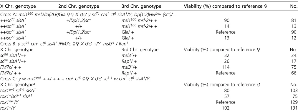
The data presented here do not support the hypothesis that the MSL complex functions in the early regulation of Sxl. Despite extensive tests, I was unable to identify any of the genetic interactions that would be predicted to occur be-tweenmle,msl1, ormsl2and eitherSxlor the two key zygotic regulators ofSxl,scute, andsisA. Nor was I able tofind sup-port for the claimed zygotic role ofrox-1in earlySxl activa-tion. In this context, the strong female-lethal maternal effect Table 3 There are no zygotic female-lethal interactions betweenmsl1andmsl2,msl3orrox1mutations and XSE mutations
X Chr. genotype 2nd Chr. genotype 3rd Chr. genotype Viability (%) compared to reference♀ No.
Cross A:msl1L60msl2/In(2LR)Gla♀♀X♂♂y sc71cm1ct6sisA1/Y;Dp(1;2)Hwbap(sc+)/+
++/sc71sisA1 +/Dp(1;2)sc+ msl1L60msl-2/+ + 90 81
++/sc71sisA1 +/+ msl1L60msl-2/+ + 14 13
++/sc71sisA1 +/Dp(1;2)sc+ Gla/ + Reference 90
++/sc71sisA1 +/+ Gla/ + 13 12
Cross B:y sc96cm1ct6sisA1/FM7c♀♀X♂♂+/Y;msl31/ Rap1
X Chr. genotype 3rd Chr. genotype Viability (%) compared to reference♀ No.
sc96sisA1/++ msl31/+ 32 24
sc96sisA1/++ Rap1/ + 26 17
FM7c/ + + msl31/+ 114 75
FM7c/ + + Rap1/ + Reference 66
Cross C:y w rox1ex6+ +/ + + + cm1ct6♀♀X♂♂sc3-1w cm1ct6sisA1/Y
X Chr. genotypea Viability (%) compared to reference♂ No.
rox1ex6sc3-1sisA1 80 103
rox1+/sc3-1sisA1 57 75
rox1ex6/Y Reference 129
rox1+/Y 102 131
observed with msl31females suggests one of two
explana-tions: Eithermsl3regulatesSxlindependent of the MSL com-plex or themsl31stock contains an unrecognized mutation(s)
responsible for the maternal effect interaction.
An unidentified mutation is responsible for the interactions seen with an msl3 mutant
To determine which of these alternatives is true, I examined othermsl31isolates and additionalmsl3alleles to see if they
retained the interaction with the X-signal components. I obtained four additionalmsl31isolates from J. Birchler
(Uni-versity of Missouri), V. Meller (Wayne State Uni(Uni-versity), and M. Kuroda (Harvard University), some labeled by their his-torical synonyms, mle(3)132 and msl3P. Each of the msl31
stocks, regardless of origin, exhibited maternal-effect lethal interactions withSxl,scute, andsisA(data not shown). I also received themsl3MAK-1allele from J. Birchler but found that
the stock had acquired a mutation, rendering the second chromosome homozygous lethal, so I was unable to test di-rectly ifmsl3MAK-1mutants exhibited the maternal-effect
le-thal interaction. Instead, I carried out complementation tests with msl31, msl3MAK-1, and two chromosomal deficiencies, Df(3L)Exel6110 and Df(3L)BSC224, which are deleted for allmsl3coding sequences (Table 4). I found thatmsl3MAK-1
and the two deficiencies fully complemented the female-lethal maternal effect of themsl31chromosome as evidenced
by the absence of any lethal interaction withsisA1orscM6
(Table 4). The complementation tests thus demonstrate the female-specific maternal effects previously attributed tomsl3 are likely due, at least in part, to one or more additional mutations inmsl31fly stocks.
An important question is whether the maternal effect lethal interaction requires the loss ofmsl3function, as might be the case if both a deficit inmsl3and another gene were required, or if the lethal effect is entirely independent ofmsl3. To ad-dress this point, I asked ifmsl31females that carried anmsl3+
transgene that fully complements msl3 male lethality also exhibited the maternal effect. Initially I tested ay w;P[w+;
msl3+-TAP];msl31stock (Alekseyenkoet al.2006) and found
that the stock did not exhibit the female-lethal maternal ef-fect (data not shown). This result could have occurred either
if an msl3 mutation is necessary, but not sufficient for the maternal defect, or if the responsible mutation(s) had been lost from the strain. I therefore generated two independent
w1118; msl31/TM3 stocks that retained the maternal-effect
female-lethal interaction and introduced theP[w+;msl3+-TAP]
transgene into each, taking care to ensure that the suspect msl31 chromosome from the y w transgenic stock was
excluded. Sibling w1118;msl31mothers, differing only in
whether or not they carried a copy of P[w+; msl3+-TAP],
were tested to determine if there was a female-lethal inter-action with thescM6allele (Table 5). I found that the
pres-ence of the fully functionalmsl3+transgene had no effect on
the female-lethal interaction asscM6/+female viability was
equally compromised regardless of whether the maternal ge-notype was functionallymsl3+ormsl32(Table 5). Note that crosses A and B in Table 5 incorporated a second chromo-some duplication of sc+ as a control to ensure that the
female-lethal interactions observed remained dependent on thescM6allele present in the progeny females. Taken
to-gether, the complementation experiments withmsl3mutants (Table 4) andmsl3+transgenes (Table 5) establish thatmsl3
mutations are neither necessary nor sufficient for the observed maternal-effect interactions with the XSE genes.
Collectively, the data presented here argue that neither the MSL complex nor its component parts participate in the early embryonic activation ofSxl. This undercuts the notion that females use the MSL complex to amplify the X chromosome signal (Gladstein et al.2010) and eliminates an important challenge to standard models for sex determination and dos-age compensation. My experiments also suggest the exis-tence of at least one novel maternal regulator of primary sex determination. The molecular identification of this locus, which has escaped detection in a variety of screens, is a high priority, as it is likely to offer new insights into the mechanism of X-chromosome counting.
Comparisons of differing results
The conclusions I reach in this paper are in direct opposition to those made earlier by Gladsteinet al. (2010) and Horabin (2012), and the data also appear to be in conflict. How then can one explain the differences between the results in this report and those published previously? With respect tomsl3 the answer is clear. Because Gladsteinet al.(2010) neither mapped the responsible lesion nor performed a complemen-tation test, they reached the incorrect conclusion about the involvement of msl3in sex determination. Remarkably, Uenoyamaet al.(1982) offers evidence that the original iso-late ofmsl31contained a maternal-effect mutation exhibiting
a female-lethal interaction withSxlf1. For convenience, data
from tables 1 and 2 of Uenoymaet al.(1982) are reproduced here with explanatory notes and current nomenclature as
Table S2. Crosses 1a and 1b of Uenoymaet al.(1982), com-paring siblingmsl31redandmsl31red/TM3mothers, showed
a modest female-lethal maternal-effect interaction withSxlf1.
Crosses 2a and 2b, however, reveal that when the experimen-tal mothers carried a recombinant msl31 red chromosome
Table 4 msl3is not sufficient for the maternal-effect female-lethal interaction with XSE mutations
Viability (%) of females of indicated X Chr. genotype compared to sibling males (no. reference)
Maternal 3rd
Chr. genotype sisA1/ + scM6/ +
msl31 42 (117) 9.2 (174)
msl3MAK-1/msl31 110 (152) 103 (241)
Df(3L)Exel6110/msl31 102 (101) 102 (477)
Df(3L)BSC224/msl31 92 (104) 99 (135)
Experimental mothers derived from crosses:msl31red♀♀X♂♂msl31red/TM3, Sb1Ser1or♂♂msl3MAK-1/TM3, Sb1Ser1or♂♂w1118;Df(3L)Exel6110/TM6, Tb1, or
that also included the distal markerebony(e), the maternal-effect lethal interaction was lost (seeTable S2). In retrospect, it is not surprising that themsl31stock would carry additional
mutations. The msl31 chromosome was recovered from a
wild population in Japan in the 1970s (Uchidaet al.1981) and may well have experienced mobilization ofP-elements while introducing markers from laboratory strains. In fact, Uchidaet al.(1981) reported the original chromosome also bore a nonsex-specific maternal-effect lethal that mapped be-tweenredandebony. The relationship between that mutation and the one responsible for the interactions withSxland the XSEs is not known.
It is less obvious what might explain the differences be-tween what is reported here and what was published earlier formle,msl1, andmsl2. I suggest the resolution is that the differences are more illusory than real because Gladstein et al.(2010) drew their conclusions from equivocal genetic experiments. Considerfigures 1B and 2B of Gladsteinet al. (2010), which addressed whether maternal or zygotic msl mutations enhanced the female-lethal effects of thesc3-1sisA1
X chromosome. Instead of comparing the viability of the off-spring of heterozygous and homozygous msl mothers, or between sibling sc sis/++; msl/+ and sc sisA/++; +/+ females, Gladsteinet al.(2010) measured viability with ref-erence to the unrelated wild-type Oregon R strain. They con-cluded that each msl had maternal and zygotic effects on female viability because the lethal effects observed with themslmutants were greater than seen with the Oregon R controls. The problem with this approach is that it relies on quantitative comparisons betweenflies of unrelated genetic backgrounds. This was compounded by the choice of Oregon R, a strain particularly ill-suited to serve as a reference be-cause it is among those least sensitive to the female-lethal effects of reduced XSE gene dose (Cline 1988). In effect, the choice of control strain may have predetermined that stron-ger female-lethal effects would be observed with themsl mu-tant strains than with the control, which would have given
the appearance of female-lethal interactions where none existed.
The only cases where there appear to be actual conflicts between my findings and those published earlier are with respect to the abilities of maternalmsl1L60,msl21, and
per-hapsmle1, to create synthetic female-lethal interactions with
aSxlnull allele (figure 1B in Gladsteinet al.2010vs.Table 1 andTable S1). I cannot explain the discordant data because Sxl/+heterozygotes should be fully viable in the absence of interacting mutations, but note that Gladsteinet al.(2010) examined only a single mutant line for each locus and did not determine if the effects were dominant or recessive or if they were maternal. The latter points are important because if the maternal effects were equal between mothers homozygous and heterozygous for these null, or near null,mslalleles, that result would suggest the lethally interacting loci were un-likely to be themslgenes.
Maternal effects of the msl2 gene?
The question of whether there are maternally contributed msl gene products is of crucial importance for assessing a possible role of the MSL complex in the early embryo, whether related to sex determination or early dosage com-pensation (Gladsteinet al. 2010; Lottet al.2011; Horabin 2012). There is strong evidence that maternally providedmle
andmsl1products function in the embryo as assembly of the MSL complex is delayed, and the lethal period shortened, in the male progeny of mothers mutant for the two loci (Belote and Lucchesi 1980; Frankeet al.1996). The most crucial questions concern msl2 as the protein is essential for the formation and function of the MSL complex. Numerous ex-perimental measures suggest that there is no maternal con-tribution (Bashaw and Baker 1995; Kelleyet al.1995; Rastelli et al.1995; Frankeet al.1996; McDowellet al.1996; Lott et al.2011, 2014). RNA-seq data are potentially helpful, but
findings from precisely staged embryos that lackmsl2mRNA (Lott et al.2011) have been criticized for lack of statistical Table 5 Themsl31mutation is not necessary for the female-lethal maternal-effect interactions with the XSEscute
♀♀X♂♂scM6w/Y;Dp(1;2)Hwbap(sc+)/+
Crosses A and B Maternal genotype 2nd Chr. genotype
Female viability (%) compared to brothers
No. female progeny
No. male progeny (reference)
Cross A w;msl31 +/Dp(sc+) 98 65 66
“ +/+ 4 3 79
Cross B w: P{msl3+-TAP}/+;msl31 P{msl3+-TAP}/Dp(sc+) 98 63 64
“ P{msl3+-TAP}/+ 7 4 60
“ +/Dp(sc+) 127 71 56
“ +/+ 8 6 72
Crosses C and D ♀♀X♂♂scM6w/ Y
Maternal genotype 2nd Chr. genotype Female viability (%) compared to brothers
No. female progeny
No. male progeny (reference)
Cross C w;msl31 +/+ 2 6 284
Cross D w: P{msl3+-TAP}/+;msl31 +/+ 0.9 1 113
“ P{msl3+-TAP}/+ ,1.1 0 94
Females in crosses A–D were derived from independently isolatedw1118;msl31lines. It is not known if themsl31chromosomes retain theredallele. Experimental mothers
derived from crosses betweenw1118;msl31females andw1118;P{w+,msl3+-TAP]/+;msl31males and differ only in the presence or absence of themsl3+transgene. Male
power (Horabin 2012), whereas bulk samples from 0- to 2-hr-old embryos that contain msl2 mRNA (FlyBase, cited by Gladstein et al. 2010 and Horabin 2012) could be explained by even moderate contamination with older em-bryos. It is thus worth reexamining the genetic evidence cited (Gladsteinet al.2010; Horabin 2012) in support of a mater-nal effect ofmsl2. Apart from the incomplete experiment in
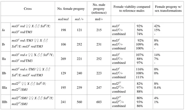
figure 1A of Gladsteinet al.(2010), which does not formally address a maternal effect, that evidence comes from crosses performed by Uenoyamaet al.(1982). As reprinted inTable S2, two effects were observed in crosses between msl227
mothers and Sxlf1/Y; msl227/SM1 fathers: a small
female-lethal effect and a low frequency of apparent sex transforma-tions in homozygousmsl227female progeny (Uenoyamaet al.
1982). With respect to viability, there was no evidence for a maternal effect as there was no difference between the female progeny of homozygous and heterozygous msl227
mothers (88vs.86% viability). With respect to sex transfor-mation, 8% of femalemsl227progeny of homozygous mothers
exhibited at least one sex-transformed structure compared to 4% of themsl227progeny of heterozygous mothers (Table S2,
crosses IIIa, IIIb; Uenoyamaet al.1982). Given that the basis of the sex transformations remains mysterious, that they occur only in 4–8% of progeny, and are affected by the zygotic ge-notype, the 1982 sex transformation data do not make a con-vincing case for anmsl2maternal effect. In light of the strong evidence that there is no detectable maternalmsl2protein, or mRNA, and the absence of genetic evidence for anmsl2 func-tion in embryos, the simplest interpretafunc-tion is thatmsl2mRNA and proteinfirst appear, and the MSL complexfirst assembles, afterSxlactivity has been set in cycle 14 (Frankeet al.1996; Lottet al.2011, 2014).
Conclusions
The data presented here indicate that the MSL complex does not participate in the early regulation ofSxl in female em-bryos. Mostmslmutants did not exhibit the genetic interac-tions predicted to occur if the complex directly, or indirectly, affects the early steps in sex determination. In the sole case where anmslmutant did interact as predicted, themsl3 mu-tation was shown to be unnecessary for the effects, which appear instead, to be the result of at least one unidentified maternal-effect locus. My experimental findings nullify the rationale behind an important alternative model of primary sex determination. By extension, they also undercut the pro-posal that the MSL complex regulates the process by which some X-encoded genes are dosage compensation prior to the large-scale activation of the zygotic genome (Lottet al.2011).
Acknowledgments
Keith Maggert, L. Rene Garcia, Sharvani Mahadevaraju, Jayashre Rajendren, and Yi Sun offered useful comments and criticisms throughout this work. Keith Maggert and Teresa Lamb provided useful comments on the manuscript.
Stocks obtained from the BloomingtonDrosophilaStock Cen-ter (National Institutes of Health P40OD018537) were used in this study. I thank James Birchler, Victoria Meller, and Mitzi Kuroda for additional fly strains. This work was supported in part by a grant from the National Science Foundation.
Literature Cited
Alekseyenko, A. A., E. Larschan, W. R. Lai, P. J. Park, and M. I. Kuroda, 2006 High-resolution ChIP-chip analysis reveals that the Drosophila MSL complex selectively identifies active genes on the male X chromosome. Genes Dev. 20: 848–857.
Bashaw, G. J., and B. S. Baker, 1995 Themsl-2dosage compen-sation gene of Drosophila encodes a putative DNA-binding pro-tein whose expression is sex specifically regulated bySex-lethal. Development 121: 3245–3258.
Bell, L. R., J. I. Horabin, P. Schedl, and T. W. Cline, 1991 Positive autoregulation ofSex-lethalby alternative splicing maintains the female determined state in Drosophila. Cell 65: 229–239. Belote, J. M., and J. C. Lucchesi, 1980 Male-specific lethal
muta-tions ofDrosophila melanogaster. Genetics 96: 165–186. Chen, Z. X., and B. Oliver, 2015 X chromosome and autosome
dosage responses inDrosophila melanogasterheads. G3 (Bethesda) 5: 1057–1063.
Cline, T. W., 1984 Autoregulatory functioning of a Drosophila gene product that establishes and maintains the sexually deter-mined state. Genetics 107: 231–277.
Cline, T. W., 1988 Evidence that sisterless-aand sisterless-b are two of several discrete “numerator elements” of the X:A sex determination signal in Drosophila that switch Sex-lethal be-tween two alternative stable expression states. Genetics 119: 829–862.
Cline, T. W., 1993 The Drosophila sex determination signal: How doflies count to two? Trends Genet. 9: 385–390.
Cline, T. W., and B. J. Meyer, 1996 Vive la difference: malesvs. females infliesvs.worms. Annu. Rev. Genet. 30: 637–702. Conrad, T., and A. Akhtar, 2011 Dosage compensation inDrosophila
melanogaster:epigeneticfine-tuning of chromosome-wide tran-scription. Nat. Rev. Genet. 13: 123–134.
Deshpande, G., J. Stukey, and P. Schedl, 1995 scute (sis-b) function in Drosophila sex determination. Mol. Cell. Biol. 15: 4430–4440. Erickson, J. W., and J. J. Quintero, 2007 Indirect effects of ploidy suggest X chromosome dose, not the X:A ratio, signals sex in Drosophila. PLoS Biol. 5: e332.
Figueiredo, M. L., M. Kim, P. Philip, A. Allgardsson, P. Stenberg et al., 2014 Non-codingroXRNAs prevent the binding of the MSL-complex to heterochromatic regions. PLoS Genet. 10: e1004865.
Franke, A., A. Dernburg, G. J. Bashaw, and B. S. Baker, 1996 Evidence that MSL-mediated dosage compensation in Drosophila begins at blastoderm. Development 122: 2751–2760.
Gladstein, N., M. N. McKeon, and J. I. Horabin, 2010 Requirement of male-specific dosage compensation in Drosophila females: implications of early X chromosome gene expression. PLoS Genet. 6: e1001041.
Gonzalez, A. N., H. Lu, and J. W. Erickson, 2008 A shared en-hancer controls a temporal switch between promoters during Drosophila primary sex determination. Proc. Natl. Acad. Sci. USA 105: 18436–18441.
Horabin, J. I., 2012 Balancing sex chromosome expression and satisfying the sexes. Fly (Austin) 6: 26–29.
Keyes, L. N., T. W. Cline, and P. Schedl, 1992 The primary sex determination signal of Drosophila acts at the level of transcrip-tion. Cell 68: 933–943.
Lim, C. K., and R. L. Kelley, 2012 Autoregulation of the Drosophila noncodingroX1RNA gene. PLoS Genet. 8: e1002564. Lott, S. E., J. E. Villalta, G. P. Schroth, S. Luo, L. A. Tonkinet al.,
2011 Noncanonical compensation of zygotic X transcription in early Drosophila melanogaster development revealed through single-embryo RNA-seq. PLoS Biol. 9: e1000590.
Lott, S. E., J. E. Villalta, Q. Zhou, D. Bachtrog, and M. B. Eisen, 2014 Sex-specific embryonic gene expression in species with newly evolved sex chromosomes. PLoS Genet. 10: e1004159. Lucchesi, J. C., and M. I. Kuroda, 2015 Dosage compensation in
Drosophila. Cold Spring Harb. Perspect. Biol. 7: a019398. Lyman, L. M., K. Copps, L. Rastelli, R. L. Kelley, and M. I. Kuroda,
1997 Drosophilamale-specific lethal-2protein: structure/func-tion analysis and dependence on MSL-1 for chromosome asso-ciation. Genetics 147: 1743–1753.
McDowell, K. A., A. Hilfiker, and J. C. Lucchesi, 1996 Dosage compensation in Drosophila: the X chromosome binding of MSL-1 and MSL-2 in female embryos is prevented by the early expression of the Sxl gene. Mech. Dev. 57: 113–119.
Philip, P., and P. Stenberg, 2013 Male X-linked genes in Drosoph-ila melanogaster are compensated independently of the Male-Specific Lethal complex. Epigenetics Chromatin 6: 35. Rastelli, L., R. Richman, and M. I. Kuroda, 1995 The dosage
com-pensation regulators MLE, MSL-1 and MSL-2 are interdepen-dent since early embryogenesis in Drosophila. Mech. Dev. 53: 223–233.
Salz, H. K., 2011 Sex determination in insects: a binary decision based on alternative splicing. Curr. Opin. Genet. Dev. 21: 395–400.
Salz, H. K., and J. W. Erickson, 2010 Sex determination in Dro-sophila: the view from the top. Fly (Austin) 4: 1–11.
Stenberg, P., and J. Larsson, 2011 Buffering and the evolution of chromosome-wide gene regulation. Chromosoma 120: 213–225. Stenberg, P., L. E. Lundberg, A. M. Johansson, P. Ryden, M. J. Svenssonet al., 2009 Buffering of segmental and chromosomal aneuploidies inDrosophila melanogaster. PLoS Genet. 5: e1000465. Sun, L., H. R. Fernandez, R. C. Donohue, J. Li, J. Cheng et al., 2013a Male-specific lethal complex in Drosophila counteracts histone acetylation and does not mediate dosage compensation. Proc. Natl. Acad. Sci. USA 110: E808–E817.
Sun, L., A. F. Johnson, R. C. Donohue, J. Li, J. Cheng et al., 2013b Dosage compensation and inverse effects in triple X metafemales of Drosophila. Proc. Natl. Acad. Sci. USA 110: 7383–7388.
Uchida, S., T. Uenoyama, and K. Oishi, 1981 Studies on the sex-specific lethals ofDrosophila melanogaster. Jpn. J. Genet. 56: 523–527.
Uenoyama, T., A. Fukunaga, and K. Ioshi, 1982 Studies on the sex-specific lethals ofDrosophila melanogaster. V. Sex transfor-mation caused by interactions between a female-specific lethal, Sxlf1, and the male-specific lethalsmle(3)132, msl-2(27), and
mle. Genetics 102: 233–243.
Vicoso, B., and D. Bachtrog, 2009 Progress and prospects toward our understanding of the evolution of dosage compensation. Chromosome Res. 17: 585–602.
Walker, J. J., K. K. Lee, R. N. Desai, and J. W. Erickson, 2000 The Drosophila melanogastersex determination genesisAis required in yolk nuclei for midgut formation. Genetics 155: 191–202.
GENETICS
Supporting Information
www.genetics.org/lookup/suppl/doi:10.1534/genetics.115.182931/-/DC1
Primary Sex Determination in
Drosophila
melanogaster
Does Not Rely on the
Male-Speci
fi
c Lethal Complex
James W. Erickson
TABLE S1. There are no female-lethal interactions between maternal
mle, msl1
, or
msl2
mutations
and zygotic sex determination mutations.
Viability (%) of females of indicated X chromosome genotype relative to sibling males (# reference).
maternal MSL genotype Sxlf1/+ sisA1/+ scM6/+
mle1 110 (212) 97 (173)
mle1/mle9 99 (205) 107 (183) 96 (119)
msl11 103 (119) 101 (131)
msl11/ msl1KmB 95 (118) 102 (194)
msl11/msl1γ216 104 (113)
msl1L60 104 (168) 123 (109)
msl1L60 msl2/msl1KmB 97 (150)
msl1L60 msl2/msl11 110 (182)
msl21 111 (98)
msl21/msl2227 104 (98)
+ msl21/msl1L60 msl2 110 (70)
msl2P17/ msl2227 97 (115) 100 (95)
msl2P22/ msl2KmA 97 (175)