Medical Image Compression by using Threshold
Predicting Wavelet-Based Algorithm
Nik Shahidah Afifi Bt Md Taujuddin1, Rosziati Ibrahim1
1 Faculty of Computer Science and Information Technology, Universiti Tun Hussein Onn
Malaysia, 86400 Parit Raja, Johor, Malaysia.
{shahidah, rosziati}@uthm.edu.my
Abstract. In recent decades with the rapid development in biomedical engineering, digital medical images have been becoming increasingly important in hospitals and clinical environment. Apparently, traversing medical images between hospitals need a complicated process. Many techniques have been developed to resolve these problems. Compressing an image will reduces the amount of redundant data with the good quality of the reproduced image sufficiently high, depending on the application. In the case of medical images, it is important to reproduce the image close to the original image so that even the smallest details readable. The aim of this paper is to reveal our new proposed compression algorithm. It started by segmenting the image area into Region of Interest (ROI) and Region of Background (ROB) and use the special features provide by wavelet algorithm to produce efficient coefficients. These coefficients is then will be thresholded by using our new proposed thresholding predicting algorithm. This method will provide a low computational cost algorithm besides decreasing the image size without tolerating with the precision of image quality.
1 Introduction
Advances over the past decade in many aspects of digital technology especially devices for image acquisition, data storage, and bitmapped printing and display have brought about many applications of digital imaging. However, problems involving storage space and network bandwidth requirements arise when large volumes of images are to be stored or transmitted, as is the case with medical images. From the diagnostic imaging point of view, the challenge is how to deliver clinically critical information in the shortest time possible. A solution to this problem is through image compression. The main objective of this compression is to reduce redundancy of the data image in order to be able to stored or transmit data in an efficient form[1][3][4][5][6].
capabilities of available technologies. The recent growth of data intensive multimedia-based web applications have not only sustained the need for more efficient ways to encode signals and images but have made compression of such signals central to storage and communication technology.
2 Related Work on Image Compression
Even though nearly all image processing applications can accept some information loss, but in numerous critical areas such as medical, satellite, and legal imaging, lossless compression algorithms are preferred. Context Based, Adaptive, Lossless Image Codec (CALIC), JPEG-LS, and JPEG2000 are among outstanding lossless image compression algorithms that give high compression ratio in a practical time [6][7]. Compression ratio for typical image is best offered by CALIC, while JPEG-LS provide low complexitity alternative. As, JPEG provides a unified approach to lossy-to-lossless compression
Meanwhile, lossy method offer high compression ratio but with some tolerate corrupted data. It is always producing a sufficient enough data size for transmission and storage purpose. Some of the prominant lossy image-coding techniques are DCT, DFT, fractal-based coding, wavelet-based coding and vector quantization method.
CALIC[8] is a compression technique based on the setting of the pixels of some predetermined pattern of neighbor pixels. The method is capable of learning from the errors made in the previous predictions and in this way it can improve its prediction adaptively when the compression proceeds. Pixel values are calculated by a nonlinear predictor, which choose the prediction pixels and the particular prediction function amongst numerous potential prediction functions on the basis of the local context. The context is built up of the local gradient magnitudes in horizontal and vertical directions. The coefficients are chosen based on the training set drawn from the type of images to be compressed. The final set of prediction errors is coded by entropy coder. CALIC is proved provides best compression ratios in a reasonable time over typical images.
JPEG-LS[9] is the lossless/near lossless compression standard for continuous-tone image. It is based on context modeling and predictive coding combined with Huffman coding. The values of pixel are predicted adaptively based on an edge detector’s output. The predicted value is chosen based on three neighboring pixel.
The Joint Photographic Expert Group (JPEG) standard is already used for almost 2 decades. This standard is used for the digital compression and coding of continuous tone. JPEG 2000 in other hand, provide international standard for image compression that give extra features where it can be operated in network and mobile environment.
3 Related Work on Wavelet-Based Image Compression
Wavelet-based image compression techniques have raise interest amongst the medical community. Beside compression, wavelet also widely been used in noise reduction, detection of microcalcifications, image analysis and image enhancement. Wavelets has offer great compression ratio without harming the image quality and it became a solemn challenger to DCT [11].
Wavelet is well known because of its energy compactness in the frequency domain. Besides, the multiresolution analysis (MRA) offered by wavelet is providing much higher compression ratio at the same time preserving good image quality. In addition, wavelet-based compression scheme give the most excellent rate-distortion performance.
There are many Wavelets generic being introduced nowadays. Sophisticated wavelets produce smoother and more satisfactory compressed image. It makes no assumption concerning periodicity of the data. As a result, Wavelets are suitable for demonstrating sharp changes or even discontinuities. Some of the common Wavelet methods used in image processing are Haar, Embedded Zerotree Wavelet (EZW) and Set Partitioning in Hierarchical Trees (SPIHT). While the extended versions that already being well accepted in committee are Wavelet Difference Reduction (WDR) and Adaptively Scanned Wavelet Difference Reduction (ASWDR). Wavelets has offer great compression ratio without harming the image quality and it became a solemn challenger to DCT[11].
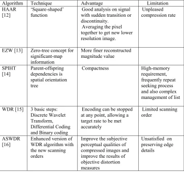
Table 1: Summary of various wavelet-based compression algorithm
Algorithm Technique Advantage Limitation
HAAR
[12] ‘Square-shaped’ function with sudden transition or Good analysis on signal discontinuity.
Averaging the pixel together to get new lower resolution image.
Unpleased compression rate
EZW [13] Zero-tree concept for significant-map information
More finer reconstructed magnitude value
SPIHT
[14] Parent-offspring dependencies is spatial orientation tree
Compactness High-memory
requirement, frequently repeat seeking process and also complex management of list
WDR [15] 3 basic steps: Discrete Wavelet Transform, Differential Coding and Binary coding
Encoding can be stopped at any point, allowing a target rate to be met accurately
Limited scanning order
ASWDR [16]
Enhanced version of WDR algorithm with the new scanning orders
Improve the subjective perceptual qualities of compressed images and improve the results of objective distortion measures
Unsatisfied on preserving edge details
4 Experimental Results on Wavelet-Based Compression Methods
To analyze the performance of SPIHT, EZW, WDR and ASWDR, three sample standard images used; Lena.bmp, Cameraman.tif and Barbara.bmp. The image dimension of Lena.bmp is 256 x 256, while Cameraman.tif and Barbara.bmp is 512 x 512. These standard images are used with the assumption that the image compression performance of the selected wavelet-based algorithm (SPIHT, EZW, WDR and ASWDR) will be imitate if the medical images are used.
image size, while the BPP is the number of bits required to store one pixel of the image [14].
[image:5.595.119.477.262.355.2]For perceptual quality, Mean Square Error (MSE) and Peak Signal to Noise Ratio (PSNR) are used [15]. MSE represent the mean squared error between compresses and the original image, while PSNR stand for a measure of the peak error and is expressed in decibels [16].
Table 2: Quantitative and Perceptual Quality Measures of Compression performance on Lena.bmp
Methods
Compression
ratio
PSNR
MSE
BPP
SPIHT
2.3239
32.1101
40.0013
0.1859
STW
3.3707
32.9988
32.5986
0.2697
EZW
3.4946
32.8449
33.7743
0.2796
WDR
3.7579
32.8449
33.7743
0.3006
ASWDR
3.6617
32.8449
33.7743
0.2929
[image:5.595.122.480.493.584.2]Table 2 shows the objective compressions performance of Lena image respectively. It is difficult to observe the differences between these images. This illustrates that all compression methods produce equally good compressions at moderately high bit rates. WDR shows the greatest performance in compression ratio, PSNR, MSE and BPP compared to another 4 algorithm. In almost every case the PSNR and MSE value for EZW, WDR and ASDWR is the same. These numerical results are consistent with the increased preservation of details within EZW, WDR and ASDWR.
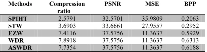
Table 3: Quantitative and Perceptual Quality Measures of Compression performance on Cameraman.tif
Methods
Compression
ratio
PSNR
MSE
BPP
SPIHT
2.5791
32.5701
35.9809
0.2063
STW
3.6903
33.6661
27.9557
0.2952
EZW
7.4116
37.5756
11.3637
0.5929
WDR
7.8918
37.5756
11.3637
0.6313
ASWDR
7.7354
37.5756
11.3637
0.6188
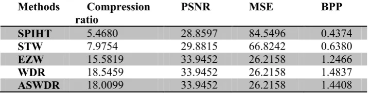
Table 4: Quantitative and Perceptual Quality Measures of Compression performance on Barbara.bmp
Methods
Compression
ratio
PSNR
MSE
BPP
SPIHT
5.4680
28.8597
84.5496
0.4374
STW
7.9754
29.8815
66.8242
0.6380
EZW
15.5819
33.9452
26.2158
1.2466
WDR
18.5459
33.9452
26.2158
1.4837
ASWDR
18.0099
33.9452
26.2158
1.4408
By referring to Table 4, it can be noticed that the compression ratio and BPP of WDR for Barbara image is clearly superior to both ASWDR and SPHIT. While PSNR and MSE value for EZW, WDR and ASWDR retain the same.
5 Methodology
Although there are numerous number of research are done on medical image compression in the wavelet domain, there still have rooms for improvement. Especially in predicting the accurate threshold value for wavelet coefficient because by using the typical hard threshold in quantizing the coefficients it will lead to blocky artifact on medical image [17]. Medical community also raise a high intention to produce a low computational cost algorithm with high speed compression and decompression to assist the existence network bandwidth capability while reducing the image size to upkeep the limited storage size.
So, to archive this aim, we propose a new algorithm that can encode these coefficients effectively. Below are the proposed algorithm steps:
1. The original image is segmented to Region of Interest (ROI) and Region of Background (ROB).
2. DWT is used to produce sequence of wavelet coefficient and separate it to low frequency and high frequency subband.
3. Analyze the correlation between adjacent wavelet coefficients to get the best suit coefficient relationship.
4. Resulting wavelet coefficient are thresholded by using proposed efficient prediction scheme to get the best truncated threshold. Then the prediction equation is applied for thresholding process to get the significant predicted wavelet coefficient.
6 Conclusion
Based on the preliminary analysis on existence wavelet-based algorithm, there is a need to develop an effective algorithm that can provide the improvement preservation details at low bit rates. Protecting details at low bit rates is crucial for sensitive data such as remote medical diagnosis via rapid transmission of compressed image and rapid retrieval of image in databases.
Therefore, modification on prediction procedure as proposed in our algorithm should be performed to eliminate the blocking and edge effect while increasing the effectiveness and reliability of the compressed image.
Acknowledgments.
The authors would like to thank the Universiti Tun Hussein Onn Malaysia (UTHM) and Malaysian Ministry of Education for providing the research grant for facilitating this research activity.
References
[1] M. G. Strintzis, “A review of compression methods for medical images in PACS.,” Int. J. Med. Inform., vol. 52, no. 1–3, pp. 159–65, 1998.
[2] S. Wong, L. Zaremba, D. Gooden, and H. K. Huang, “Radiologic image compression-a review,” Proc. IEEE, vol. 83, no. 2, pp. 194–219, 1995.
[3] S. Burak, G. Carlo, T. Bernd, and G. Chris, “Medical Image Compression Based on Region of Interest, With Application to Colon CT Images,” in 23rd
Annual EMBS International Conference, 2001, pp. 2453–2456.
[4] E. Kofidis, N. Kolokotronis, A. Vassilarakou, S. Theodoridis, and D. Cavouras, “Wavelet-based medical image compression,” Futur. Gener. Comput. Syst., vol. 15, no. 2, pp. 223–243, 1999.
[5] R. Janaki, “Enhanced ROI ( Region of Interest Algorithms ) for Medical Image Compression,” Int. J. Comput. Appl., vol. 38, no. 2, pp. 38–44, 2012.
[6] M. U. Celik, G. Sharma, and A. M. Tekalp, “Gray-level-embedded lossless image compression,” Signal Process. Image Commun., vol. 18, no. 6, pp. 443–454, Jul. 2003.
[7] J. Kivijärvi, T. Ojala, T. Kaukoranta, a Kuba, L. Nyúl, and O. Nevalainen, “A comparison of lossless compression methods for medical images.,” Comput. Med. Imaging Graph., vol. 22, no. 4, pp. 323–39, 1998.
[9] M. J. Weinberger, G. Seroussi, and G. Sapiro, “The LOCO-I lossless image compression algorithm: principles and standardization into JPEG-LS.,” IEEE Trans. Image Process., vol. 9, no. 8, pp. 1309–24, Jan. 2000.
[10] A. Skodras, C. Christopoulos, and T. Ebrahimi, “The JPEG 2000 Still Image,” IEEE Signal Process. Mag., no. September, pp. 36–58, 2001.
[11] A. Bruckmann and A. Uhl, “Selective medical image compression techniques for telemedical and archiving applications.,” Comput. Biol. Med., vol. 30, no. 3, pp. 153–69, May 2000.
[12] D. I. Belc, “A Hybrid Wavelet Filter for Medical Image Compression,” Florida State University, Florida, 2006.
[13] J. M. Shapiro, “A fast technique for identifying zerotrees in the EZW algorithm,” 1996 IEEE Int. Conf. Acoust. Speech, Signal Process. Conf. Proc., vol. 3, pp. 1455–1458.
[14] A. Said, W. A. Pearlman, and S. Member, “A New , Fast , and Efficient Image Codec Based on Set Partitioning in Hierarchical Trees,” vol. 6, no. 3, 1996.
[15] J. Tian and R. Wells, “A Lossy Image Codec Based on Index Coding,” IEEE Signal Process. Mag., p. 456, 1996.
[16] J. S. Walker, “Lossy image codec based on adaptively scanned wavelet difference reduction,” Opt. Eng., vol. 39, no. 7, p. 1891, Jul. 2000.