Defects of T-cell effector function and
post-thymic maturation in X-linked hyper-IgM
syndrome
Ashish Jain, … , David L. Nelson, Warren Strober
J Clin Invest. 1999;
103(8)
:1151-1158.
https://doi.org/10.1172/JCI5891
.
X-linked hyper-IgM syndrome (XHIM) results from mutations in the gene encoding for CD40
ligand (CD154). Patients with the syndrome suffer from infections with opportunistic
pathogens such as Cryptosporidium and Pneumocystis carinii. In this study, we
demonstrate that activated T cells from patients with XHIM produce markedly reduced levels
of IFN-
g
, fail to induce antigen-presenting cells to synthesize IL-12, and induce greatly
reduced levels of TNF-
a
. In addition, we show that the patients’ circulating T lymphocytes of
both the CD4
+and CD8
+subsets contain a markedly reduced antigen-primed population,
as determined by CD45RO expression. Finally, we demonstrate that the defects in antigen
priming are likely due to the lack of CD154 expression and insufficient costimulation of T
cells by CD80/CD86 interactions. Taken together, this study offers a basis for the increased
susceptibility of patients with XHIM to certain opportunistic infections.
Article
Introduction
X-linked hyper-IgM syndrome (XHIM) is an immunod-eficiency disorder characterized by low levels of IgG, IgE, and IgA, but normal-to-elevated levels of IgM (1, 2). The syndrome is caused by mutations in the gene encoding CD40 ligand (CD154), a molecule expressed on the sur-face of activated CD4+T cells that interacts with CD40
on the surface of B cells to provide an essential signal for B-cell proliferation and differentiation (3–7). As a con-sequence, patients with XHIM, as well as CD154 knock-out mice, exhibit profoundly depressed B-cell activation, fail to form germinal centers, do not generate IgG-, IgA-, and IgE-producing B cells, and mount poor primary and secondary antibody responses (8–10).
Patients with XHIM are unlike other patients with defects in immunoglobulin production (e.g., Bruton’s agammaglobulinemia) in that they frequently suffer from infection with opportunistic pathogens such as
Cryptosporidiumand Pneumocystis carinii, despite treat-ment with γ-globulin (2, 11). A possible basis for this clinical predisposition comes from CD154 knockout mice, which also manifest increased susceptibility to cer-tain infectious agents. In such mice, it has been shown that deficient CD154 expression on activated T cells leads to impaired IL-12 production and reduced Th1 T-cell responses (12, 13). Furthermore, deficient CD154 expression in mice is associated with susceptibility to
Leishmania infection, which is reversed by pretreatment with IL-12 (13). Thus, the CD154 knockout model sug-gests that opportunistic infections in patients with
XHIM can be attributed to defective IL-12 responses and Th1 T-cell differentiation. However, this possibility has not been explored in patients with XHIM.
In this report, we demonstrate that activated T cells from patients with XHIM produce markedly reduced levels of IFN-γ,fail to induce antigen-presenting cells (APCs) to synthesize IL-12, and induce greatly reduced levels of TNF-α. We further demonstrate that patients with XHIM have a defect in post-thymic maturation of CD4+and CD8+T cells characterized by a diminished
capacity to generate mature CD45RO+T cells. As shown
here, this is likely due to the lack of CD154 expression and insufficient costimulation of T cells by CD80/CD86 interactions. These results imply that the increased sus-ceptibility of patients with XHIM is in fact caused by defective induction of IL-12, leading to reduced elabora-tion of mature Th1 T cells.
Methods
Patients and protocols. Six patients with XHIM were studied at the Clinical Center, National Institute of Allergy and Infectious Diseases/National Institutes of Health (NIAID/NIH) (protocol no. 89-1-0158). The diagnosis was established in each patient by medical and family history, immunoglobulin profile, and sequencing of the CD154 gene. Unaffected family members of patients, other normal volunteers, and immunologically nor-mal individuals on other NIH protocols served as controls. The Institutional Review Board of NIAID approved the open pro-tocol, and informed consent was obtained from all patients or their parents before enrollment in the study.
CD40 ligand gene analysis. DNA and total RNA were extracted
Defects of T-cell effector function and
post-thymic maturation in X-linked hyper-IgM syndrome
Ashish Jain,
1T. Prescott Atkinson,
2Peter E. Lipsky,
3Jay E. Slater,
4David L. Nelson,
5and Warren Strober
11Mucosal Immunity Section, Laboratory of Clinical Investigation, National Institute of Allergy and Infectious Diseases/National
Institutes of Health, Bethesda, Maryland 20892, USA
2Department of Pediatrics, University of Alabama–Birmingham, Birmingham, Alabama 35294, USA
3Department of Rheumatology and Immunology, University of Texas–Southwestern, Dallas, Texas 75235, USA 4Department of Pediatrics, Children’s National Medical Center, Washington, DC 20010, USA
5Immunophysiology Section, Metabolism Branch, National Cancer Institute/National Institutes of Health, Bethesda,
Maryland 20892, USA
Address correspondence to: Warren Strober, Room 11N238, Building 10, National Institutes of Health, Bethesda, Maryland 20892, USA. Phone: (301) 496-6810; Fax: (301) 402-2240; E-mail: [email protected]
Received for publication November 24, 1998, and accepted in revised form March 15, 1999.
X-linked hyper-IgM syndrome (XHIM) results from mutations in the gene encoding for CD40 ligand (CD154). Patients with the syndrome suffer from infections with opportunistic pathogens such as Cryp-tosporidiumand Pneumocystis carinii. In this study, we demonstrate that activated T cells from patients with XHIM produce markedly reduced levels of IFN-γ, fail to induce antigen-presenting cells to syn-thesize IL-12, and induce greatly reduced levels of TNF-α. In addition, we show that the patients’ circu-lating T lymphocytes of both the CD4+and CD8+subsets contain a markedly reduced antigen-primed population, as determined by CD45RO expression. Finally, we demonstrate that the defects in antigen priming are likely due to the lack of CD154 expression and insufficient costimulation of T cells by CD80/CD86 interactions. Taken together, this study offers a basis for the increased susceptibility of patients with XHIM to certain opportunistic infections.
from activated lymphocytes using standard methods. cDNA was obtained from RNA by RT-PCR using CD154-specific primers. PCR of genomic DNA was performed with primers flanking each exon using primers and PCR cycling conditions described previously (14–16). The PCR products were purified and cycle sequenced at both the 5′and 3′ends using dye termi-nator dideoxynucleotides.
Flow cytometry.Specimens of peripheral blood were obtained during routine patient clinic visits and were handled according to established clinical guidelines. The specimens were stained by flow cytometry with the whole-blood lysis technique and analyzed with a FACScan (Becton Dickinson Immunocytome-try Systems, San Jose, California, USA) with Lysis II software by methods described previously (17). The monoclonal antibod-ies used included anti-CD3ε(Leu-4) and anti-CD4 (Leu-3) (Bec-ton Dickinson Immunocytometry Systems), anti-CD45RO (UCHL-1) (DAKO Corp., Carpinteria, California, USA), and anti-CD8 (Leu-2a) and anti-CD45RA (Alb11) (Gentrack, Ply-mouth Meeting, Pennsylvania, USA). Irrelevant antibodies of the IgG1, IgG2a, IgG2b subclasses were used to ascertain back-ground staining. To calculate absolute numbers of each lym-phocyte subgroup, the percentage of cells staining positive was multiplied by the absolute count of peripheral blood lympho-cytes, as determined by Coulter Counter (Coulter Electronics Ltd., Hialeah, Florida, USA), followed by a differential leuko-cyte count in a blood sample obtained simultaneously.
Cell preparation and culture conditions. PBMCs were obtained from patients and healthy adult volunteers by centrifugation of heparinized blood over Ficoll-Hypaque density gradient lym-phocyte separation medium (Pharmacia Biotech, Inc., Piscat-away, New Jersey, USA), using standard techniques (18). All patients had a complete blood count with differential formed on the same day that the cytokine studies were per-formed. Their monocyte counts were comparable to those seen in the normal volunteers, and no adjustments were made in PBMCs. To measure IL-12 production, 2 ×10⁶PBMCs were cul-tured in 1 ml of RPMI-1640 complete medium for 36 h. For anti-CD3εstimulation, 64.1 antibody (gift of Bristol-Myers Squibb, Princeton, New Jersey, USA) was first dissolved in car-bonate buffer (pH 9.6) at a concentration of 2.5 µg/ml and aliquoted into 24-well tissue culture plates at 250 µl/well. After a two-hour incubation at 37°C, the plates were washed twice in sterile PBS. For anti-CD3ε, OKT3 antibody (gift of Ortho Biotech, Somerset, New Jersey, USA) was first dissolved in car-bonate buffer at a concentration of 10 µg/ml and aliquoted into 24-well plates at 250 µl/well. After overnight incubation at 4°C, the plates were washed twice in sterile PBS. Cell suspen-sions were then added. Human IFN-γwas used at 1,000 U/ml,
[image:3.612.66.528.54.198.2]Staphylococcus aureus,Cowan’s strain I (SAC) (Pansorbin cells; Calbiochem Corp., La Jolla, California, USA) at 1:10,000 (wt/vol), LPS from Escherichia coli01127:B8 (Sigma Chemical Co., St. Louis, Missouri, USA) at 1 µg/ml, phytohemagglutinin
Figure 1
(PHA) at 1:100 (wt/vol), and CD154 trimer (gift of Immunex Corp., Seattle, Washington, USA) at 3.5 µg/ml. After 36 h, supernatants were removed. IFN-γ, IL-12, and TNF-α concen-trations were determined by specific ELISA (R&D Systems Inc., Minneapolis, Minnesota, USA) according to the manufactur-er’s instructions. CD45RA-enriched PBMCs were prepared by negative selection using anti-CD45RO antibody (UCHL-1) and immunomagnetic beads Dynal Inc. (Lake Success, New York, USA). The procedures followed were in accordance to the man-ufacturer’s instructions and with methods described previous-ly (19). In the resulting population, CD3+T cells were >95% CD45RA+. In in vitro culture, 106cells were stimulated with OKT3 or with SAC and human IgG at 1 µg/ml (Jackson ImmunoResearch Laboratories Inc., West Grove, Pennsylvania, USA) or SAC with CTLA4-Fc at 1 µg/ml (R&D Systems Inc.). The cells were then harvested 72 h later, and two-color flow cytometric analysis was performed by methods described pre-viously (20).
Statistical analysis. Wilcoxon rank-sum test was used to com-pare measurements between patients and normal volunteers. All Pvalues are two-sided.
Results
Clinical features of the patients with XHIM. As shown in Table 1, five of the six patients with XHIM who were studied had histories of P. cariniiinfection, an oppor-tunistic infection not commonly associated with other humoral immunodeficiencies. In addition, most of the patients have histories suggesting other opportunistic infections or immune surveillance defects. Patient 1 has cerebellar ataxia of unknown etiology despite intensive investigation. Patient 2 has malignant carcinoid tumor and carcinoid syndrome; 12 months before the study, he had a partial pancreas resection and has since complet-ed five cycles of chemotherapy with cisplatin/etoposide. He was studied six weeks after the completion of chemotherapy, when he was in remission. Patient 5 has hepatitis C infection presumably acquired from prior infusions of fresh frozen plasma. Patient 6 had recurrent herpes simplex stomatitis requiring maintenance on suppressive therapy with acyclovir. Laboratory evalua-tions revealed that three of six patients had a lymphocy-tosis primarily of CD4+ T cells. All six patients were
maintained on monthly intravenous immunoglobulin (IVIG) therapy, and four of the six patients were main-tained on G-CSF for neutropenia.
[image:4.612.320.508.49.660.2]CD154 mutations in affected patients. Five of the six patients with XHIM studied have mutations on exon 5 of the extracellular domain (21). Patient 1 has a donor splice site mutation at exon 1 GT→TT (Slater, J.E., unpublished observations); expression of the mutant protein was not detectable by flow cytometry using anti-CD154 antibody (Jain, A., and Strober, W., unpublished observations). Patient 2 has an A insertion between nt424G and nt430G, resulting in a frameshift of the CD154 coding frame; thus, his CD40 ligand gene would be expected to yield a trun-cated protein consisting of 163 amino acids (14). Patient 3 has an A→G substitution at nt509, resulting in a change in the amino acid Y→C substitution at position 170 (22). Patient 4 has a 6-bp in-frame deletion at nt424–429 that causes the deletion of amino acids G→L at positions 142 and 143 (23). Patient 5 has C→A substitution at nt623, resulting in an amino acid substitution A→D at position
Figure 2
208 (Nelson, D.L., unpublished observations). Patient 6 has a G→A substitution at nt655, resulting in an amino acid substitution of G→R at position 219, and a C→T substitution at 695, resulting in a stop codon and a trun-cated protein of 232 amino acids (15).
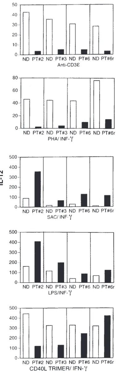
APCs from patients with XHIM do not produce IL-12 and pro-duce markedly repro-duced levels of TNF-αupon stimulation with antigen-activated T cells.Previous studies have shown that activated T cells stimulate APCs to produce cytokines by CD154/CD40 interaction (24, 25). Thus, in our initial studies of immune function in patients with XHIM, we determined the capacity of their PBMCs to produce IL-12 when stimulated with T-cell activators. To this end, we cultured PBMCs from three patients with XHIM (patient
6 was studied twice) and normal volunteers with either PHA/IFN-γ or anti-CD3ε. Activation of T cells with either anti-CD3εor PHA stimulation in patients with XHIM was noted by enlargement of cell size (determined by FACS on forward-/side-scatter dot plot analysis) and increased [3H]thymidine incorporation (one study). After
36 h, we measured IL-12 secretion into culture fluid by specific ELISA. Monocytes in PBMCs from the patients with XHIM failed to produce measurable levels of IL-12 in response to either stimulant compared with controls (see Figure 2). This reduced production of IL-12 was not due to an intrinsic inability to synthesize this cytokine, because in patients with XHIM, monocytes in PBMCs cultured with either trimeric CD154/IFN-γ, LPS/IFN-γ, or SAC/IFN-γproduced, if anything, increased amounts of IL-12. Such increased IL-12 production may in part be due to prior conditioning of the patients’ monocytes by exogenous G-CSF, used for treatment of neutropenia.
In further studies, we determined the capacity of monocytes in PBMCs of patients with XHIM to produce TNF-αafter stimulation with T-cell activators. Accord-ingly, supernatants from the culture conditions already described were also tested for TNF-αby specific ELISA. As shown in Figure 3, monocytes in PBMCs from patients with XHIM produced significantly reduced lev-els of TNF-αcompared with those of normal volunteers. Again, the reduced TNF-αproduction was not due to an inability to produce TNF-α,as patients with XHIM pro-duce normal levels of TNF-αwhen their monocytes in PBMCs are stimulated with LPS/IFN-γor SAC/IFN-γ. Taken together, these studies demonstrate that mono-cytes of patients with XHIM are markedly deficient in their capacity to produce cytokines when stimulated by activated T cells, because of the absence of CD154/CD40 interaction.
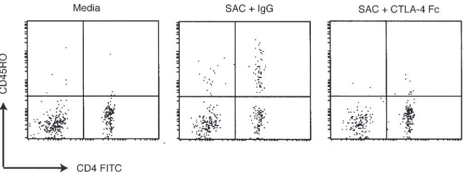
T cells of patients with XHIM manifest deficient IFN-γ pro-duction that can be restored with CD154 trimer stimulation.
The deficiency in monocyte IL-12 production upon stimulation with activated T cells in patients with XHIM suggests that cognate interactions between APCs and T cells would result in reduced IFN-γproduction (25, 26). To explore this possibility, we cultured PBMCs with anti-CD3 or PHA for 36 h and then measured
IFN-γsecretion into the culture fluid by specific ELISA. As shown in Figure 4, patients with XHIM produce markedly reduced amounts of IFN-γcompared with controls. To demonstrate that the reduced IFN-γ pro-duction was due to the lack of CD154, we stimulated PBMCs from controls and patients with XHIM with anti-CD3εas before, but this time in the presence of CD154 trimer. CD154 trimer restored IFN-γproduction in the three patients with XHIM who were studied.
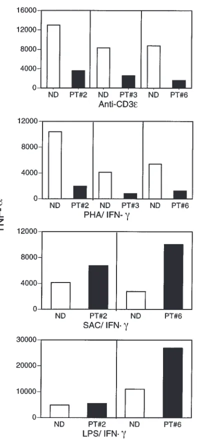
[image:5.612.73.270.231.670.2]Patients with XHIM have a markedly reduced pool of mature (CD45RO+) T cells. In humans, antigen priming of T cells is associated with a change in T-cell CD45 isoform expression (27). Naive T cells express the CD45RA iso-form, whereas antigen-primed (memory) T cells express the CD45RO isoform (28, 29). Given the occurrence of T-cell priming defects in CD154 knockout mice (30), as well as the evidence of cytokine production deficiency in the patients discussed here, we evaluated the expression of the CD45 isoforms by the T cells of patients with Figure 3
XHIM by flow cytometry. As indicated in Figure 5a, all six patients studied had CD4+T-cell populations
con-taining reduced percentage of cells expressing CD45RO and increased percentage of cells expressing CD45RA. Thus, in five of six patients, the percentage value of CD4+
T cells expressing CD45RO fell below the range deter-mined for normal volunteers, whereas the fifth patient’s percentage value fell in the lower limits of the normal range (this patient’s CD45RO percentage value may have been transiently elevated because he had recently com-pleted chemotherapy, a procedure known to reduce the naive CD45RA T-cell population; ref. 17). In keeping with these findings, the population of CD4+T cells
expressing CD45RO in patients with XHIM was signifi-cantly reduced both in absolute number (P > 0.007) and in percentage (P > 0.0002) when compared with controls. In further studies, we also showed that CD8+T-cell
pop-ulation in patients with XHIM contained a reduced per-centage of cells expressing CD45RO (P > 0.002) and an increased percentage of cells expressing CD45RA (P >
0.0001). Finally, given the fact that CD45RO memory cells increase with age (31), and four of six patients were 16 years of age or younger (mean age of 9.3 years), the patient values were also compared with a large age-matched control group (n = 15) with a mean age of 12.7 years (P > 0.17). Patients with XHIM still manifested sig-nificantly low CD45RO T-cell values (CD4+: %CD45RO,
P > 0.01, %CD45RA, P > 0.004; CD8+: %CD45RO, P >
0.04, %CD45RA, P > 0.003). Of interest, analysis of two obligate carriers of the CD154 gene mutation indicated that carriers also had reduced numbers of T cells with exclusive expression of CD45RO. In studies of two mothers of patients with XHIM, the percentages of CD4+
T cells expressing CD45RO were 11% and 20%, respec-tively, both values well below the 95% confidence inter-val for normal subjects (Jain, A., and Strober, W., unpub-lished observations). These studies demonstrate that patients with XHIM have a diminished capacity to prime T cells and induce a mature T-cell phenotype.
Inhibition of the CD28/B7 interaction blocks T-cell conver-sion from CD45RA+to CD45RO+. Previous studies have shown that in vitro primary stimulation of T cells by PHA or anti-CD3ε (32) alone induces transition of naive CD4+/CD45RA+T cells into the mature CD45RO+
phe-notype; indeed, we confirmed this finding in two nor-mal volunteers and one patient with XHIM (Jain, A., and Strober, W., unpublished observations). However, the marked reduction in the CD45RO+T-cell population in
patients with XHIM suggests that in vivo CD154 pro-vides an advantage in T-cell maturation and that engagement of the antigen receptor alone by pep-tide/MHC is usually not adequate for the generation of mature T cells. One possible explanation for this dis-crepancy is that the generation of CD45RO+T cells in
vivo normally requires costimulation of T cells by B7 (CD80/86)/CD28 interaction and that the reduction in CD45RO+ T cells in patients with XHIM is due the
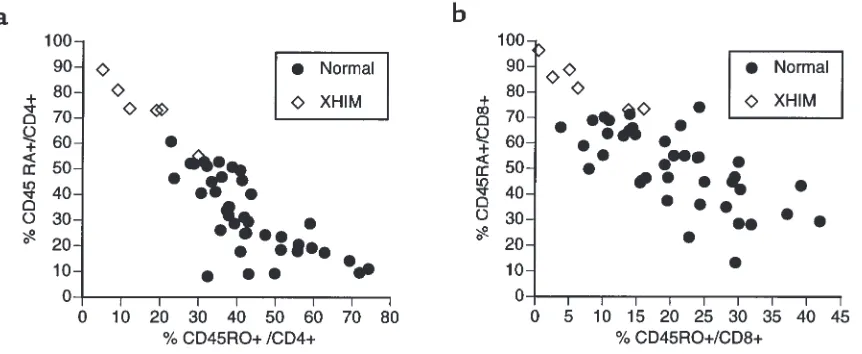
absence of CD154/CD40 interaction and the subse-quent failure to upregulate CD80/86 on APCs (33–35). To investigate this possibility, we first determined whether CD80/86 regulates the conversion of normal naive CD45RA+T cells to a mature CD45RO+cells. In
these studies, we stimulated normal donor PBMCs (depleted of CD45RO+cells) with SAC, a potent APC
activator. As shown in Figure 6, when macrophages in PBMCs are stimulated with SAC, the CD4+T cells
tran-sition from CD45RA+-expressing cells to CD45RO+
[image:6.612.319.526.325.670.2]-expressing cells during the three days of culture. In con-trast, when PBMCs are stimulated in the same way in the presence of CTLA4-Fc, the cells fail to undergo such a transition. We next determined whether patients with XHIM manifest a defect in the upregulation of B7 mol-ecules. In these studies, we found that, unlike those from normal volunteers, B cells in PBMCs from patients with XHIM fail to induce CD86 surface expression when stimulated with PHA. Furthermore, CD86 expres-sion on B cells can be restored when PBMCs are stimu-lated with PHA and CD154 trimer (Jain, A., and Strober, W., unpublished observations). Together, these findings confirm the importance of costimulatory molecules normally induced by CD154 in the maturation of T cells into cells expressing CD45RO. As such, they provide a compelling explanation for the reduced induction of mature memory cells in patients with XHIM.
Figure 4
Discussion
In the present study, we demonstrate that activated T cells in patients with XHIM fail to produce meaningful levels of IFN-γ, fail to induce APC secretion of IL-12, and induce markedly reduced secretion of TNF-α. In addition, we report that patients with XHIM have a defect in post-thymic maturation of T cells, manifested by a greatly dimin-ished capacity to generate mature (memory) CD45RO+T
cells of both the CD4 and CD8 subsets. These abnormali-ties are directly attributable to deficient CD154 signaling and provide a basis for the susceptibility of patients with XHIM to opportunistic infection.
Cell-surface interaction between CD154 on CD4+T
cells and CD40 on B cells and other APCs is now well rec-ognized as a central requirement for most types of the host immune response. Initially, the importance of CD154 was noted in relation to B-cell isotype switching (36–38). B cells interact with antigen by means of their immunoglobulin receptor, internalize antigen, and then present processed antigen to CD4+T cells; the CD4+T
cells stimulated in this way express surface CD154 and then backstimulate the B cells via CD40 to undergo iso-type switching and terminal B-cell differentiation. Because IgM secretion can occur in the absence of CD154 signaling, these T-cell/B-cell interactions explain why ineffective CD154/CD40 interaction leads to low levels of IgG, IgA, and IgE, and to normal-to-elevated lev-els of IgM, both in patients with XHIM and in CD154 knockout mice. Recently, normal levels of CD27+/IgD+
B cells and an absence of CD27+/IgD–B cells, the latter a
more mature B-cell form, have been shown in patients with XHIM (39). This finding is analogous to our find-ing in T cells and presumably relates to the same defect of CD154/CD40 interaction.
To expand this latter point, it has recently become apparent that CD154 signaling also plays a key role in T-cell differentiation. The locus of interaction, in this case, is between dendritic cells or monocytes and T cells.
Acti-vated T cells expressing CD154 interact with CD40 on APCs and induce upregulation of CD80/86 and IL-12 secretion. IL-12 secretion by APCs leads to the differenti-ation of naive T cells into Th1 cells. The importance of this interaction in vivo has been shown in a Th1 T cell–dependent murine colitis model (TNBS-colitis), in which it was observed that its blockade by anti-CD154 antibody prevented TNBS-induced inflammation of the colon (40). Similarly, as already noted, CD154 knockout mice infected with Leishmaniahave T cells that fail to pro-duce INF-γand monocytes that fail to produce IL-12 (13). Finally, it has recently been shown that the interaction between CD154 on antigen-specific CD4+T cells and
CD40 on dendritic cells is also critical to the development of CD8+cytolytic T cells (CTLs) (41–43). This was
demon-strated by in vitro studies in which it was shown that interference with this interaction inhibits the maturation of antigen-specific CD8+T cells into effector CTLs.
The role of CD154/CD40 interaction in various aspects of T-cell function provides a basis for the finding that patients with XHIM manifest diverse T-cell defects. Perhaps the most profound of these defects is the inabil-ity of activated T cells to induce cytokine production by APCs. In the present study, we demonstrate that although patients with XHIM have the capacity to pro-duce IL-12 and TNF-αwhen their PBMCs are stimulat-ed with SAC/IFN-γor LPS/IFN-γ, they fail to produce IL-12 and produce greatly reduced levels of TNF-αwhen their PBMCs are stimulated by anti-CD3εor
PHA/IFN-γ. These results imply that patients with XHIM produce IL-12 and TNF-αby direct stimulation of monocytes by certain microbes but manifest defects when such cytokine production is elicited by antigen-activated CD4+T cells expressing CD154. In turn, APC
stimula-tion defects have a major effect on T-cell cytokine pro-duction. Thus, PBMCs stimulated by the T-cell activa-tors PHA and anti-CD3ε normally produce large amounts of IFN-γin normal individuals; similar stimu-Figure 5
[image:7.612.66.497.53.230.2]lation of PBMCs in patients with XHIM is associated with greatly reduced IFN-γproduction.
A second and interlocking abnormality of T-cell func-tion in patients with XHIM relates to their defective T-cell priming. Thus, patients with XHIM have a marked-ly reduced percentage of antigen-primed T cells, as defined by the surface CD45RO expression by both CD4+and CD8+T cells. The relationship of this
abnor-mality to defective CD154 expression and signaling was indicated by two additional findings. First, we showed that two obligate carriers of the CD154 gene mutation also manifest reduced numbers of mature CD4+T cells.
Given the random nature of X-chromosome inactiva-tion, these female carriers may have a large portion of the circulating CD4+T cells with nonfunctional CD154
and, as a result, manifest defects in post-thymic matu-ration of T cells analogous to their male offspring. Sec-ond, we demonstrated that isoform switching from CD45RA to CD45RO on T cells is blocked in vitro by CTLA4-Fc (an inhibitor of CD28 interaction with CD80/86) and that patients with XHIM manifest defective upregulation of CD86 due to the lack of CD154 signaling. These finding suggest that the reduced CD45RO expression in patients with XHIM is a direct manifestation of the core defect in CD154 expression.
Studies of immune responses in patients with XHIM, as well as their clinical course, correlate well with their immunological findings. Thus, the limited number of studies of in vitro T cell–proliferative responses to spe-cific antigens have revealed either normal responses to tetanus toxoid or lectins or reduced responses to various antigens, which generally are corrected with booster immunization (11, 44, 45). In this regard, two of the three patients tested in our series demonstrate normal delayed-type skin test responses without booster immu-nization (one to tetanus, one to candida and mumps). These observations demonstrate that
CD154-independ-ent mechanisms do exist for T-cell activation and differ-entiation in humans. Clinically, this partial preservation of T-cell immunity accounts for the fact that the infec-tious complications in patients with XHIM are not as grave as those seen in severe combined immunodefi-ciency. Nevertheless, despite alternative routes to T-cell effector function, patients with XHIM routinely suffer from fulminant viral hepatitis, Cryptosporidium infection, and P. cariniipneumonia (11). In addition, reports of hepatobiliary cancer, carcinoid tumor, and other neo-plasms in patients with XHIM suggest defective immune surveillance (11, 14, 46).
[image:8.612.69.531.500.676.2]The immune defects in patients with XHIM described here appear to have consequences early in life. In the only large retrospective study of 56 patients, the Kaplan-Meier survival rate was 20% by the age of 25 years despite IVIG therapy, with the vast majority of deaths occurring in the second decade of life (11). Such a high mortality rate has not been reported in other immunodeficiencies, such as Bruton’s agammaglobulinemia, in which the humoral immune abnormality is controlled by IVIG treatment. Because defective CD154 signaling and the subsequent defects of T-cell effector function are a con-stant feature of XHIM, we speculate that the increased mortality in patients with XHIM in the second decade of life is due to their defect in the post-thymic maturation of antigen-specific T cells. This view is based on the fact that after the first decade of life, the host cannot expand his or her repertoire of antigen-specific T cells because of thymic involution and the failure of post-thymic T cells to undergo somatic mutation. As a result, the host becomes more dependent on the preexisting antigen-primed population of T cells for defense against infec-tious disease and immune surveillance. Thus, the con-tracted population of antigen-primed T cells, caused by a failure of post-thymic maturation, renders these patients particularly susceptible to opportunistic infec-tion and cancer early in life.
Figure 6
Kruisebeek, D.H. Margulies, E.M. Shevach, and W. Strober, editors. Greene Publishing and Wiley Interscience. New York, NY. 5.3.1–5.3.5. 21. Villa, A., et al. 1994. Organization of the human CD40L gene: implica-tions of molecular defects in X chromosome-linked hyper-IgM syn-drome and prenatal diagnosis. Proc. Natl. Acad. Sci. USA. 91:2110–2114. 22. Atkinson, T.P., et al. 1998. Leukocyte transfusion–associated granulo-cyte responses in a patient with X-linked hyper-IgM syndrome. J. Clin. Immunol. 18:430–439.
23. Chu, Y.W., et al. 1995. Somatic mutation of human immunoglobulin V genes in the X-linked hyper IgM syndrome. J. Clin. Invest. 95:1389–1393. 24. Grewal, I.S., and Flavell, R.A. 1988. CD40 and CD154 in cell mediated
immunity. Annu. Rev. Immunol. 16:111–135.
25. Shu, U., et al. 1995. Activated T cells induce interleukin-12 production by monocytes via CD40-CD40 ligand interaction. Eur. J. Immunol.
25:1125–1128.
26. Trinchieri, G. 1988. Proinflammatory and immunoregulatory functions of interleukin-12. Int. Rev. Immunol. 16:365–396.
27. Mackay, C.R. 1993. Homing of naive, memory and effector lymphocytes.
Curr. Opin. Immunol. 5:423–427.
28. Pilarski, L.M., Gillitzer, R., Zola, H., Shortman, K., and Scollay, R. 1989. Definition of the thymic generation lineage by selective expression of high molecular weight isoforms of CD45 (T200). Eur. J. Immunol. 19:589–597.
29. Fujii, Y., Okumura, M., Inada, K., Nakahara, K., Matusda, H. 1992. CD45 isoform expression during T cell development in the thymus. Eur. J. Immunol. 22:1843–1850.
30. Grewal, I.S., Xu, J., and Flavell, R.A. 1995. Impairment of antigen-specif-ic T-cell priming in mantigen-specif-ice lacking CD40 ligand. Nature. 378:617–620. 31. Erkeller-Yuksel, F.M., et al. 1992. Age related changes in human blood
lymphocytes subpopulations. J. Pediatr. 120:216–222.
32. Serra, H.M., Krowka, J.F., Ledbetter, J.A., and Pilarski, L.M. 1988. Loss of CD45R(Lp220) represents a post-thymic T cell differentiation event. J. Immunol. 140:1435–1441.
33. Cella, M., et al. 1996. Ligation of CD40 on dendritic cells triggers pro-duction of high levels of interleukin-12 and enhances T cell stimulato-ry capacity: T-T help via APC activation. J. Exp. Med. 184:747–752. 34. Caux, C., et al. 1994. Activation of human dendritic cells through CD40
cross-linking. J. Exp. Med. 180:1263–1272.
35. Kennedy, M.K., et al. 1994. Induction of B cell costimulatory function by recombinant murine CD40 ligand. Eur. J. Immunol. 24:116–123. 36. Kawabe, T., et al. 1994. The immune responses in CD40-deficient mice:
impaired immunoglobulin class switching and germinal center forma-tion. Immunity. 1:167–178.
37. Defrance, T.B., et al. 1992. Interleukin 10 and transforming growth fac-tor beta cooperate to induce anti-CD40-activated naive human B cells to secrete immunoglobulin A. J. Exp. Med. 175:671–682.
38. Gascan, H., Gauchat, J.F., Aversa, G., van Vlasselaer, P., and de Vries, J.E. 1991. Anti-CD40 monoclonal antibodies or CD4+ T cell clones and IL-4 induce IgGIL-4 and IgE switching in purified human B cells via different signaling pathways. J. Immunol. 147:8–13.
39. Agematsu, K., et al. 1998. Absence of IgD-CD27(+) memory B cell pop-ulation in X-linked hyper-IgM syndrome. J. Clin. Invest. 102:853–860. 40. Stuber, E., Strober, W., and Neurath, M. 1996. Blocking the CD40L-CD40 interaction in vivo specifically prevents the priming of T helper 1 cells through the inhibition of interleukin 12 secretion. J. Exp. Med. 183:693–698.
41. Schoenberger, S.P., Toes, R.E., van der Voort, E.I., Offringa, R., and Melief, C.J. 1998. T-cell help for cytotoxic T lymphocytes is mediated by CD40-CD40L interactions. Nature. 393:480–483.
42. Bennett, S.R., et al. 1998. Help for cytotoxic-T-cell responses is mediat-ed by CD40 signalling. Nature. 393:478–480.
43. Ridge, J.P., Di Rosa, F., and Matzinger, P. 1988. A conditioned dendritic cell can be a temporal bridge between a CD4+ T- helper and a T-killer cell. Nature. 393:474–478.
44. Ameratunga, R., et al. 1997. Defective antigen-induced lymphocyte pro-liferation in the X-linked hyper-IgM syndrome. J. Pediatr. 131:147–150. 45. Geha, R.H., et al. 1979. Hyperimmunoglobulin M immunodeficiency. J.
Clin. Invest. 64:385–391.
46. Hayward, A.R., et al. 1997. Cholangiopathy and tumors of the pancreas, liver, and biliary tree in boys with X-linked immunodeficiency with hyper-IgM. J. Immunol. 158:977–983.
47. The International Chronic Granulomatous Disease Cooperative Study Group. 1991. A controlled trial of interferon gamma to prevent infection in chronic granulomatous disease. N. Engl. J. Med. 324:509–516.
An important therapeutic possibility that arises from these studies is that the poor Th1 T-cell responses in patients with XHIM may be ameliorated by supplemen-tal IFN-γtherapy (47). Studies in patients with chronic granulomatous disease have demonstrated IFN-γhas limited side effects and is associated with improved host defense. Another rational approach to therapy would be CD154 administration; this form of replacement thera-py is becoming feasible with the availability of recombi-nant CD154.
Acknowledgments
We thank Thomas Fleisher and Margaret Brown for immuno-logical evaluations, and Hans Ochs, Luigi Notarangelo, and Lynda Schneider for assistance with sequencing information on individual patients.
1. Callard, R.E., Armitage, R.J., Fanslow, W.C., and Spriggs, M.K. 1993. CD40 ligand and its role in X-linked hyper-IgM syndrome. Immunol. Today. 14:559–564.
2. Notarangelo, L.D., Duse, M., and Ugazio, A.G. 1992. Immunodeficien-cy with hyper-IgM (HIM). Immunodefic. Rev. 3:101–122.
3. Fuleihan, R., et al. 1993. Defective expression of CD40 ligand in X chro-mosome-linked immunoglobulin deficiency with normal or elevated IgM. Proc. Natl. Acad. Sci. USA. 90:2170–2173.
4. DiSanto, J.P., Bonnefoy, J.Y., Gauchat, J.F., Fischer, A., and deSaint Basile, G. 1993. CD40 ligand mutations in X-linked immunodeficiency with hyper-IgM. Nature. 361:541–543.
5. Allen, R.C., et al. 1993. CD40 ligand gene defects responsible for X-linked hyper-IgM syndrome. Science. 259:990–993.
6. Aruffo, A., et al. 1993. The CD40 ligand, gp39, is defective in activated T cells from patients with X-linked hyper-IgM syndrome. Cell. 72:291–300. 7. Korthauer, U., et al. 1993. Defective expression of T cell CD40 ligand causes X-linked immunodeficiency with Hyper-IgM. Nature. 361:539–541.
8. Xu, J., et al. 1994. Mice deficient for the CD40 ligand. Immunity. 1:423–431.
9. Han, S., et al. 1995. Cellular interaction in germinal centers. Roles of CD40 ligand and B7-2 in established germinal centers. J. Immunol. 155:556–567.
10. Renshaw, B.R., et al. 1994. Humoral immune responses in CD40 ligand-deficient mice. J. Exp. Med. 180:1189–1900.
11. Levy, J., et al. 1997. Clinical spectrum of X-linked hyper-IgM syndrome.
J. Pediatr. 131:47–54.
12. Soong, L., et al. 1996. Disruption of CD40-CD40 ligand interaction results in an enhanced susceptibility to Leishmania amazonensisinfection.
Immunity. 4:263–273.
13. Campbell, K.A., et al. 1996. CD40 ligand is required for protective cell-mediated immunity to Leishmania major. Immunity. 3:283–289. 14. Seyama, K., et al. 1998. Mutations of the CD40 ligand gene and its effect
on CD40 ligand expression in patients with X-linked hyper IgM syn-drome. Blood. 7:2421–2434.
15. Grammer, A.C., et al. 1995. The CD40 ligand expressed by human B cells costimulates B cell responses. J. Immunol. 154:4996–5010.
16. Ramesh, N., et al. 1993. Deletions in the ligand for CD40 in X-linked immunoglobulin deficiency with normal to elevated IgM (HIGMX-1).
Int. Immunol. 5:769–773.