The basic helix-loop-helix transcription factor,
dHAND, is required for vascular development
Hiroyuki Yamagishi, … , Eric N. Olson, Deepak Srivastava
J Clin Invest.
2000;
105(3)
:261-270.
https://doi.org/10.1172/JCI8856
.
Reciprocal interactions between vascular endothelial cells and vascular mesenchymal cells
are essential for angiogenesis. Here we show that the basic helix-loop-helix transcription
factor, dHAND/Hand2, is expressed in the developing vascular mesenchyme and its
derivative, vascular smooth muscle cells (VSMCs). Targeted deletion of the
dHAND
gene in
mice revealed severe defects of embryonic and yolk sac vascular development by
embryonic day 9.5. Vascular endothelial cells expressed most markers of differentiation.
Vascular mesenchymal cells migrated appropriately but failed to make contact with vascular
endothelial cells and did not differentiate into VSMCs. In a screen for genes whose
expression was dependent upon dHAND (using subtractive hybridization comparing
wild-type and
dHAND
-null hearts), the VEGF
165receptor, neuropilin-1, was found to be
downregulated in
dHAND
mutants. These results suggest that dHAND is required for
vascular development and regulates angiogenesis, possibly through a VEGF signaling
pathway.
Article
Introduction
Establishment of the mammalian vasculature is con-trolled by a complex process involving differentiation and assembly of multiple cell lineages. Rapid expansion of blood vessels is necessary for the tissue growth observed during embryonic development and tumor growth. Dysregulation of vascular development can result in excessive proliferation of vessels, as seen in hemangiomas, retinopathy of prematurity, and venous malformations (1). Because of the fundamental signif-icance of vascular development to human biology and disease, there has been much interest in understanding the molecular mechanisms underlying the develop-ment and proliferation of vessels.
Anatomically, formation of blood vessels can be divid-ed into 2 distinct steps. Vasculogenesis, the initial step, occurs when mesodermally derived angioblasts differ-entiate into vascular endothelial cells and assemble into vascular tubes (2). Subsequently, in a process known as angiogenesis, preexisting vessels sprout new branches that become remodeled into mature vessels (3). This step requires recruitment of vascular mesenchymal cells that encase endothelial tubes and differentiate into vascular smooth muscle cells (VSMCs), pericytes, and fibroblasts to promote the assembly and stabilization of the mature blood vessel wall (4). Reciprocal endothelial-mesenchy-mal interactions are essential for angiogenesis.
Recent studies have revealed several signaling pathways important during vasculogenesis and angiogenesis (1, 4). The secreted ligand angiopoietin-1 (Ang-1) and its recep-tor tyrosine kinase Tie2 (5–8), and ephrinB2 and its receptor EphB4 (9, 10), are critical for
endothelial-mes-enchymal interactions during angiogenesis. Within the heart, Ang-1 secretion from the endocardium is also nec-essary for trabeculation of the neighboring myocardium (8). TGF-βand its receptors play a role in vascular stabi-lization and remodeling and function through the Smad family of transcription factors (11–14). In contrast to these molecules that function mainly during angiogen-esis, VEGF, a heparin-binding growth factor with high specificity for endothelial cells, is a central mediator of angiogenesis and vasculogenesis (4, 15). There are 5 iso-forms produced from a singleVEGFgene by alternative splicing. VEGF induces endothelial proliferation, pro-motes cell migration, and inhibits apoptosis (3). Inhibi-tion of VEGF activity results in tumor regression, indi-cating that VEGF is necessary for the neovascularization associated with tumors (16). VEGF signals through its receptor tyrosine kinases, 1/Flt-1 and VEGFR-2/KDR/Flk-1, which are expressed specifically on the surface of vascular endothelial cells and are required for both vasculogenesis and angiogenesis (17–20). Endothe-lial cells of VEGFR-1 mutant mouse embryos differenti-ate, but do not assemble properly into vessels (18), whereas VEGFR-2 mutants display more severe defects of vasculogenesis (17). Recently, another VEGF receptor, VEGFR-3, has been shown to be important for the remodeling and maturation of primary vascular net-works into larger blood vessels (21). These data suggest that VEGF controls unique steps of vascular develop-ment through distinct receptors.
Neuropilin-1 is a specific receptor for the 165-amino acid form of VEGF (VEGF165) and is a potential
regula-tor of VEGF-induced angiogenesis (22). Neuropilin-1
The basic helix-loop-helix transcription factor, dHAND,
is required for vascular development
Hiroyuki Yamagishi,
1,2Eric N. Olson,
2and Deepak Srivastava
1,21Department of Pediatrics, Division of Cardiology, and
2Department of Molecular Biology, University of Texas Southwestern Medical Center, Dallas, Texas 75235, USA
Address correspondence to: Deepak Srivastava, Department of Pediatrics and Molecular Biology, 6000 Harry Hines Boulevard, Dallas, Texas 75235-9148, USA.
Phone: (214) 648-1246; Fax: (214) 648-1196; E-mail: [email protected].
Received for publication November 4, 1999, and accepted in revised form December 17, 1999.
Reciprocal interactions between vascular endothelial cells and vascular mesenchymal cells are essen-tial for angiogenesis. Here we show that the basic helix-loop-helix transcription factor, dHAND/Hand2, is expressed in the developing vascular mesenchyme and its derivative, vascular smooth muscle cells (VSMCs). Targeted deletion of the dHANDgene in mice revealed severe defects of embryonic and yolk sac vascular development by embryonic day 9.5. Vascular endothelial cells expressed most markers of differentiation. Vascular mesenchymal cells migrated appropriately but failed to make contact with vascular endothelial cells and did not differentiate into VSMCs. In a screen for genes whose expression was dependent upon dHAND (using subtractive hybridization comparing wild-type and dHAND-null hearts), the VEGF165receptor, neuropilin-1, was found to be
downregulated in dHANDmutants. These results suggest that dHAND is required for vascular devel-opment and regulates angiogenesis, possibly through a VEGF signaling pathway.
was first identified in the Xenopustadpole nervous sys-tem (23) and found to play a role in semaphorin-medi-ated axonal chemorepellence (24). Neuropilin-1 is expressed in the endothelial and mesenchymal cells of the murine vasculature (25) and may mediate interac-tions between endothelial and mesenchymal cells. Mouse embryos overexpressing neuropilin-1 exhibit car-diovascular, peripheral nervous system (PNS) and limb defects (25). Mice deficient in neuropilin-1 have defects in PNS axonal guidance and die between embryonic day 10.5 and 12.5 from cardiovascular defects (26).
Despite recent progress in identification of signaling molecules and cell-surface receptors that regulate blood vessel formation, little is known currently about the nuclear transcriptional pathways that regulate or are regulated by the vascular signaling cascades. Tissue-specific members of the basic helix-loop-helix (bHLH) family of transcription factors are essential for devel-opment of many cell lineages (27–29), but have not been demonstrated to play a role in vascular develop-ment. The bHLH transcription factor dHAND (decidu-um, heart, autonomic nervous system, neural crest-derived)/Hand2 is required for cardiac morphogenesis (30–32). Mice lacking dHAND have a single ventricle and aortic arch defects and die by embryonic day 11.0 (E11.0) from heart failure (31). Although the cardiac defects of dHANDmutants are well characterized as hypoplasia of the right ventricle segment and poor tra-beculation of the remaining ventricle, the role of dHAND in development of the vasculature has not been examined previously. Here we demonstrate that dHAND is expressed in the developing vasculature and VSMCs and is required for normal vascular develop-ment. The dHAND-null endothelial cells differentiate but mesenchymal cells fail to differentiate into VSMCs. The vascular defects in dHAND-mutant embryos are similar to the angiogenic defects observed in VEGFand
Flt1mutants. In a screen to identify dHAND-depend-ent genes in the heart by suppressive subtractive hybridization, we found that expression of the VEGF165
receptor, neuropilin-1, was dHAND dependent in the heart and portion of the vasculature. These data indi-cate that dHAND plays a role in angiogenesis and may function through a VEGF signaling pathway.
Methods
Section and whole-mount in situ hybridization. In situ hybridization to mouse embryo sections was per-formed with riboprobe labeled with 35S as described
previously (30). Antisense riboprobe was synthesized with SP6 RNA polymerase (MAXIScript; Ambion Inc., Austin, Texas, USA) from dHAND full-length cDNA. Hybridization was performed on tissue sec-tions of E9.5 and E10.5 mouse embryos. After hybridization, sections were washed and dipped in Kodak NTB-2 emulsion. Slides were exposed for 14 days and then developed in D19 (Eastman Kodak Co. Scientific Imaging Systems, New Haven, Connecti-cut, USA) for 3 minutes.
Whole-mount RNA in situ hybridizations for dHAND, Ang-1, COUP-TFII, SM22α,and neuropilin-1were performed using digoxigenin-labeled antisense riboprobes. In situ hybridizations were performed as described previously (31). Briefly, embryos were hybridized with digoxigenin-labeled riboprobes at 60°C for 18 hours. After a series of washes, embryos were incubated with alkaline phosphatase–conjugat-ed anti-digoxigenin antibodies at room temperature for 1 hour. After another series of washes, embryos were incubated in a substrate color-reaction mixture (Roche Molecular Biochemicals, Indianapolis, Indiana, USA) for 10–12 hours in darkness. Color reaction was terminated by fixing embryos in 4% paraformaldehyde and 0.1% glutaraldehyde.
Breeding and genotyping of mice. Mice heterozygous for the dHANDmutation were generated as described pre-viously (31). To generate transgenic mice in a dHAND -null background, male mice heterozygous for the Tie2-lacZ transgene (33) and the SM22lacZtransgene (34) were mated to dHANDheterozygous females. Male off-spring that were heterozygous fordHANDand the lacZ
transgene were mated with dHAND heterozygous females to obtain embryos that expressed the lacZ trans-gene and were heterozygous or homozygous for the tar-geted dHANDallele. Mothers were sacrificed and their uteri dissected to harvest E8.0–10.5 embryos. Genomic DNA was prepared from tail biopsies or from embry-onic yolk sacs and subjected to PCR and/or Southern blot for genotyping as described previously (31–34).
β- galactosidase staining. Embryos were dissected in 4°C PBS and fixed in 2% paraformaldehyde/PBS with phe-nol red for 30 minutes on ice. After rinsing with PBS, embryos were incubated at room temperature over-night in 0.1% X-gal, 5 mM potassium ferricyanide, 5 mM potassium ferrocyanide, 1 mM magnesium chlo-ride, 0.002% NP-40, 0.01% sodium deoxycholate, PBS, pH 7.0. After staining, the embryos were rinsed in PBS and postfixed at 4°C overnight in 4% paraformalde-hyde and 0.1% glutaraldeparaformalde-hyde in PBS. Embryos were dehydrated and cleared in a solution of benzyl ben-zoate/benzyl alcohol (2:1).
Histology. For histologic analysis, embryos were embed-ded in paraffin after fixation. Transverse and sagittal sec-tions were made at 7-µm intervals throughout the embryos. Paraffin was cleared in xylene, and photographs of sections were taken without counterstaining to illus-trate color reaction after either immunochemistry, β-gal reaction, or whole-mount in situ hybridization.
Electron microscopy. Embryos were fixed in 2.5% glu-taraldehyde in PBS at 4°C overnight and post-fixed with 1% osmium tetroxide for 1 hour. After washings, embryos were dehydrated in increasing concentrations of ethanol and infiltrated and embedded in LK-112 medium. Ultrathin transverse sections were cut with a diamond knife, mounted on 200-mesh grids, and stained with uranyl acetate and lead citrate. Sections were viewed with and photomicrographs obtained through an electron microscope.
RT-PCR. Total RNA was purified from whole body, hearts, or yolk sacs of wild-type and dHAND-null embryos at E9.5, and adult mouse heart, aorta, and liver (Trizol; GIBCO BRL, Rockville, Maryland, USA) and A10 cell line (ATCC #CRL-1476; American Type Culture Collection, Rockville, Maryland, USA). After
RT reaction, PCR analysis of several endothelial mark-ers was performed using oligonucleotides specific for Flk-1, Tie-1, and Tie-2, described previously (17, 36), and numerous other vascular markers (primer sequences available upon request). PCR primers specif-ic for dHAND (upper: 5′-TACCAGCTACATCGCCTACCT-3′; lower: 5′-TCACTGCTTGAGCTCCAGGG-3′) and neu-ropilin-1 (upper: 5′- TGAAAAATACCCCAACTG-3′; lower: 5′- CCTGAATGATGACACCTCTT-3′) were used under the following conditions: 94°C, 5 minutes; cycles of 94°C, 1 minute; 60°C, 1 minute; 72°C, 1 minute; and 72°C, 7 minutes. PCR products were analyzed after serial cycles in the linear range of amplification. RNA load-ing was controlled by amplification of the housekeep-ing gene, GAPDH. Negative controls were performed for each sample using non–reverse transcribed RNA.
Suppressive subtractive hybridization. One microgram of total RNA, after DNase treatment, from pooled wild-type or dHAND-null E9.5 hearts was used for generation of cDNA by the SMART cDNA synthesis kit (CLON-TECH Laboratories Inc., Palo Alto, California, USA). PCR-Select (CLONTECH) was used for subtractive hybridization as described by CLONTECH. Briefly, 2 pools of RsaI-digested wild-type cDNA were used as tester and ligated to unique adapters. RsaI-digested
[image:4.612.57.390.49.301.2]dHAND-null cDNA was used as a driver and was not lig-ated to any adapter sequences. Hybridization of the tester population with excess driver and subsequent amplification of subtracted species with the 2 unique adapters present on the tester cDNA resulted in a pool of PCR fragments theoretically present in wild-type, but not dHAND-null, cDNA. TA cloning (Invitrogen Corp., San Diego, California, USA) of the PCR product and subsequent DNA isolation and sequencing of selected clones was performed. Forty clones were initially
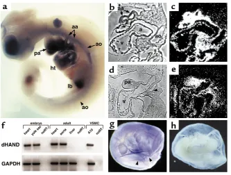
Figure 1
sequenced. Basic local alignment search tool (BLAST) was used for searching sequence homology to DNA and expressed sequence tag (EST) databases.
Results
Vascular expression of dHAND. The expression of dHAND in the vasculature has not been examined in detail pre-viously. Whole-mount in situ hybridization at E10.0 revealed dHAND expression in the aorta and aortic arch arteries in addition to the pharyngeal arches and heart (Figure 1a). Expression of dHAND was enhanced in the rostral, compared with the caudal, vasculature. Histo-logically, dHAND was expressed in the walls of the dor-sal aorta and aortic arch arteries at E9.5 (Figure 1, b and c) and in the heart (endocardium and myocardium) and pharyngeal arches. The hybridization signal in the blood vessels was wider than the thickness of the endothelium, suggesting that the mesenchymal cells surrounding the vessels expressed dHAND. At E10.5,
dHANDexpression was localized to the vascular mes-enchyme between the third and fourth aortic arch arter-ies (Figure 1, d and e), which later gives rise to VSMCs and to the mesenchyme of the pharyngeal arch. The vas-cular expression of dHAND was subsequently down-regulated and became undetectable by in situ hybridiza-tion around E12.5. However, dHAND expression was detectable by RT-PCR in the adult mouse aorta (Figure 1f). Consistent with aortic expression of dHAND, we
found that dHAND mRNA was present in a vascular cell line (A-10; ATCC #CRL-1476) that is derived from rat aortic smooth muscle cells (Figure 1f). In addition to expression in the embryo proper, dHAND was also detectable in the developing yolk sac. In situ hybridiza-tion detected dHAND expression along the yolk sac ves-sels during the process of remodeling at E9.5–10.0 (Fig-ure 1, g and h). RT-PCR confirmed dHAND expression in the yolk sac at E9.5 (Figure 1f).
Abnormal vascular development in dHAND mutants. Mouse embryos homozygous-null for dHANDlack a right ventricle and have a poorly trabeculated left ven-tricle, resulting in death by E11.0 from heart failure (31). Although dHAND is also thought to play a role in the neural crest component of aortic arch develop-ment, we sought to determine its role during vasculo-genesis and/or angiovasculo-genesis. Because heart failure might cause secondary vascular defects, all analyses of
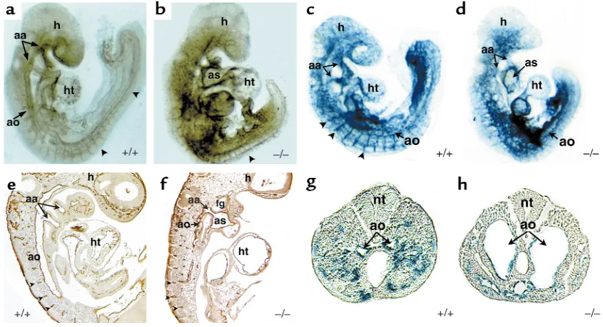
dHANDmutants were performed at E9.5, before any evidence of cardiac or growth failure. Unlike wild-type E9.5 embryos that had a grossly visible vascular pat-tern, dHANDmutants did not have an apparent vas-culature, but rather had pooling of blood in extravas-cular regions of the embryo. Whole-mount immuno-chemical staining of E9.5 dHAND-null embryos using a mAb against the endothelial-specific marker PECAM-1 (Figure 2, a and b) revealed the presence of differentiated endothelial cells. However, the vascula-Figure 2
Endothelial development in dHANDmutants. Whole-mount immunochemistry revealed that endothelial cells expressed PECAM-1 protein appropriately in wild-type (a) and mutant (b) E9.5 embryos, but displayed a disorganized pattern in dHANDmutants (b). The rostral por-tion ofdHAND-null embryos was more severely affected than the caudal region, where the aorta (ao) and somitic arteries (arrowheads) were visible. Sagittal section of PECAM-1 antibody–stained wild-type (e) and mutant (f) embryos revealed disorganization of the dorsal aorta of
[image:5.612.80.518.53.291.2]ture was grossly disorganized throughout the embryo with the more rostral vessels being more severely affected. Histological analysis of E9.5 mutant embryos showed that the aortic arch arteries were patent but the rostral aorta was poorly patterned, without an apparent lumen (Figure 2, e and f).
To further examine endothelial development in
dHANDmutants, transgenic mice containing a lacZ marker under control of the endothelial-specific Tie2 promoter (33) were crossed into the dHAND-null background. As observed with PECAM expression, Tie2 expression was intact, but vascular development was disrupted in the dHAND-null embryos at E9.5 (Figure 2, c and d). Interestingly, histologic analysis revealed that the vasculature was formed but dilated in the caudal portion of the embryo (Figure 2, g and h). The disorganization of the rostral vasculature and dilation of caudal vessels were similar to that seen in VEGF-deficient or MEF2Chomozygous-null embryos, although the etiology of this observation in either model is unclear (19, 20, 37).
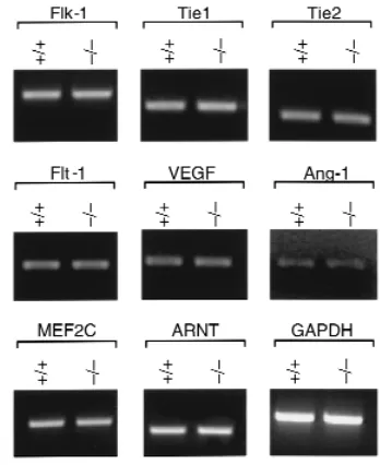
Because endothelial differentiation appeared rela-tively normal in dHANDmutants, we examined the expression of several endothelial markers, as well as other factors implicated in vasculogenesis and angio-genesis. By semiquantitative RT-PCR, transcripts of VEGF and its receptors Flk-1 and Flt-1, and Ang-1 and its receptors, Tie1 and Tie2, were detected at similar lev-els in wild-type and dHANDmutant embryos at E9.5 (Figure 3). Similarly, expression of the bHLH tran-scription factor, ARNT (38), and the trantran-scription fac-tor, MEF2C (37), both implicated in vascular develop-ment, were unaffected in dHANDmutants (Figure 3). These results suggest that the vascular defects of
dHAND-null embryos do not result from altered expression of the factors examined, although we can-not eliminate the possibility of subtle changes in gene expression in specific regions of mutant embryos.
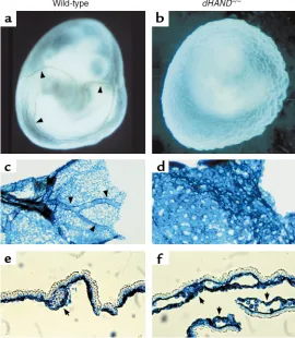
Abnormal vascular remodeling in dHAND-null yolk sacs. The vitelline circulation in the embryonic yolk sac rep-resents the earliest circulatory system and is the first site of vasculogenesis and angiogenesis in the embryo. Normally, a honeycomb-like vascular plexus is evident by E8.5; subsequent remodeling of the vasculature (angiogenesis) results in defined vessels by E9.5 (Figure 4a). However, dHAND-null yolk sacs failed to form a normal vasculature at E9.5 (Figure 4b). Expression of lacZ under control of the Tie2 promoter revealed that endothelial cells were present in both wild-type and
dHAND-null yolk sacs at E9.5 (Figure 4, c and d). In wild-type yolk sacs, lacZ expression demarcated the for-mation of large vitelline vessels and a fine network of smaller vessels (Figure 4c). In contrast, lacZ expression in yolk sacs lacking dHAND displayed a honeycomb-like plexus pattern of endothelial cells with no remod-eling into a vascular network (Figure 4d). Histologic analyses revealed that although capillary-like vessels containing blood cells with an endothelial cell lining (Figure 4e) were seen in the wild-type yolk sac, no
dis-tinct blood vessels were evident in the dHAND-mutant yolk sac (Figure 4f). Large cavities containing blood cells and lined by endothelial cells (Figure 4f) were observed in dHANDmutants suggesting that vasculo-genesis proceeds, but angiovasculo-genesis is blocked in the mutant yolk sac.
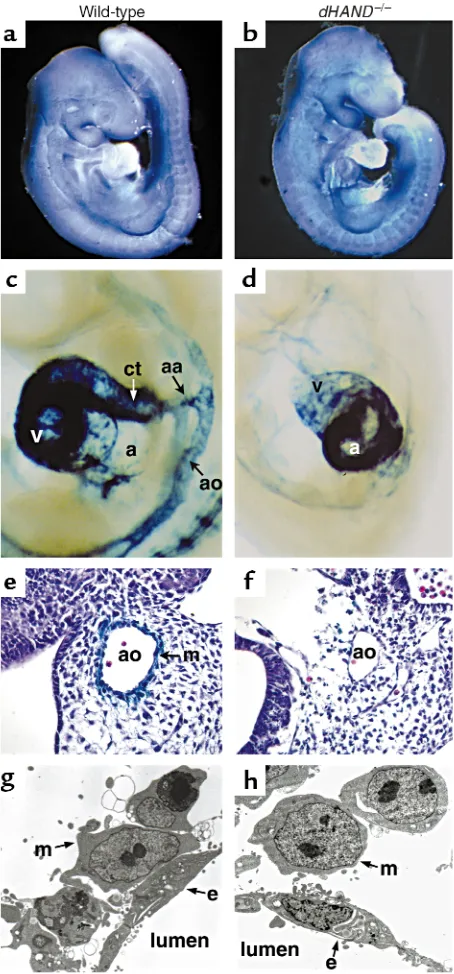
Vascular mesenchyme cells fail to differentiate into VSMCs in dHAND mutants. Mesenchymal cells begin to invade the vasculature around E9.0 and later differentiate into VSMCs. Because dHAND was expressed in vas-cular mesenchymal cells and VSMCs, we examined the development of vascular mesenchymal cells in
dHANDmutants. Ang-1 and COUP-TFII are expres-sed in mesenchymal cells and play a role in angiogen-esis, possibly in a common molecular pathway (9, 39). Transcripts of these 2 genes were examined in
dHAND-null embryos by RT-PCR and whole-mount in situ hybridization, and no difference was found between wild-type and dHAND-null embryos (Figure 3 and Figure 5, a and b; data not shown).
[image:6.612.324.498.425.638.2]SM22αis an early marker for differentiated VSMCs and marks the developing vasculature only after vas-cular mesenchymal cells differentiate into VSMCs around E9.0 (40, 41). Unlike Ang-1 and COUP-TFII, SM22αwas not detected in the dHAND-null vascula-ture by in situ hybridization (data not shown). To genetically confirm the absence of SM22αexpression, transgenic mice containing a lacZ marker under con-trol of a SM22αarterial VSMC-specific promoter (34) were crossed into the dHAND-null background. No
Figure 3
Normal endothelial cell differentiation in dHAND-null embryos. Semiquantitative RT-PCR analysis of wild-type (+/+) and dHAND-null (–/–) embryos using primers specific to the endothelial cell markers Flk-1, Tie1, Tie2, Flt-1, and VEGF revealed equal levels of expression suggesting normal differentiation and quantity of endothelial cells in
expression of lacZ was detected in the vasculature of mice lacking dHAND at E9.5 or E10.5 (Figure 5, c and d). Ventricular expression of SM22αwas not detected in dHANDmutants, although atrial expression was present (Figure 5, c and d). Histologic analysis con-firmed absence of SM22α-expressing cells around the vasculature of mutants (Figure 5, e and f). These results suggest that mesenchymal cells were present in the absence of dHAND, but fail to differentiate into SM22α-expressing VSMCs.
To more accurately determine if vascular mesenchy-mal cells had migrated to the developing vasculature and to assess the relationship of vascular endothelial cells with mesenchymal cells, we performed electron microscopy of wild-type and dHAND-null E9.5 embryos. Transverse sections revealed that mesenchymal cells had migrated around the aorta of dHANDmutants, similar to wild-type (Figure 5, g and h). However, dHAND-null mesenchymal cells remained rounded without cyto-plasmic processes that normally make contact with endothelial cells. Endothelial cells also began to appear abnormal at this stage, possibly secondary to insuffi-cient interactions with the mesenchyme. Together, these observations suggest a role for dHAND in promoting vascular mesenchyme development and possibly endothelial-mesenchymal interactions.
Neuropilin-1 functions downstream of dHAND. To gain insight into potential mechanisms through which dHAND might function, we sought to identify dHAND-dependent genes by subtraction cloning. Subtraction
cloning was performed by suppressive-subtrac-tive hybridization in which E9.5dHAND-null heart cDNA was subtracted from wild-type E9.5 heart cDNA, resulting in isolation of genes that were expressed in wild-type, but not dHAND -mutant, hearts (Figure 6a). Whereas numerous genes were identified and will be described else-where, one of the dHAND-dependent factors represented neuropilin-1, the VEGF165receptor
(22). Downregulation of neuropilin-1 in
dHAND-mutant hearts was confirmed by semi-quantitative RT-PCR (Figure 6b).
The expression pattern of neuropilin-1 in early embryonic development has not been described previously. In addition to its expres-sion in the heart, neuropilin-1 mRNA was found to be most prominent in the aorta, aor-tic arch arteries, pharyngeal arches, and limb bud of wild-type embryos by whole-mount in situ hybridiza-tion (Figure 6, c and d). The expression of neuropilin-1 described previously in the PNS and placenta and that described here is very similar to that of dHAND expression. Because neuropilin-1 was downregulated in the hearts of dHANDmutants, we examined the spatial regulation of neuropilin-1 expression in
dHAND-null embryos. Neuropilin-1 was downregu-lated in the ventricle of the heart, as expected (Figure 6e). Interestingly, neuropilin-1 mRNA was severely downregulated in the rostral aorta and pharyngeal arches but was still detectable in the caudal aorta, cor-relating with the expression pattern of dHAND. Expression in the limb buds and septum transversum was normal in dHANDmutants (Figure 6e), providing internal controls for RNA integrity within the embryo. Histologic analysis also demonstrated downregulation of neuropilin-1 expression in the rostral dHAND-null vasculature (Figure 6, f and g). Neuropilin-1 expres-sion was also seen in the yolk sac of E9.5 wild-type embryos (Figure 6b). Transcripts of neuropilin-1 were downregulated in the dHAND-null yolk sac by semi-quantitative RT-PCR (Figure 6b).
Discussion
[image:7.612.57.327.54.364.2]The results in this study demonstrate that dHAND is expressed in the developing vasculature and VSMCs and plays a role in vascular development. Whereas endothelial cell differentiation and vasculogenesis were relatively unaffected in dHAND mutants,
Figure 4
endothelial cell patterning was disrupted. Defects in angiogenesis were observed with failure of VSMC dif-ferentiation. Finally, by subtraction cloning, the VEGF165receptor, neuropilin-1, was found to be
down-regulated in dHANDmutants, suggesting one poten-tial mechanism through which dHAND may function in cardiac and vascular development.
Reciprocal interactions between vascular endothelial cells and mesenchymal cells are essential for angio-genesis and maintenance of the vasculature. Based on a model of endothelial-mesenchymal interaction pro-posed by Folkman and D’Amore, vascular mesenchy-mal cells are recruited to endothelial cells through Ang-1-Tie2 signaling, then differentiate into VSMCs or pericytes, possibly being induced by TGF-β
signal-ing (1). IndHANDmutants, numerous markers of endothelial cell development were normally expressed and the mesenchymal cells surrounded endothelial tubes, suggesting that the step of mesenchymal re-cruitment is intact. This observation is consistent with the results of marker analysis showing that Ang-1, Tie2, and COUP-TFII, which is a putative upstream regulator of the Ang-1-Tie2 signaling pathway (39), are expressed normally in dHAND-null embryos. Howev-er, in the absence of dHAND, VSMC differentiation was arrested based on the lack of expression of SM22α. The failure of vascular mesenchymal cells to express the VSMC marker SM-22αcould be a result of inade-quate interactions with endothelial cells or could be a direct effect of dHAND transcriptional regulation. A proximal CArG box in the SM-22αpromoter is essen-tial for smooth muscle–specific gene expression, but no binding sites for bHLH proteins (E-boxes) are required for SM-22αtranscription (42). Therefore, SM-22αis unlikely to be a direct target of dHAND, but more likely serves as a marker for abnormal vascular smooth muscle differentiation.
TGF-β signaling pathways are also important for angiogenesis. Mouse embryos lacking endoglin, a TGF-β–binding protein expressed on the surface of endothe-lial cells, have vascular defects similar to those described here. Specifically, endoglin-null embryos have poor VSMC development and arrested endothelial remodel-ing, whereas vasculogenesis was normal (43). Targeted deletion of a number of other genes involved in the TGF-βsignaling pathways have defects in remodeling of the yolk sac vasculature (11–14). It will be interesting to determine if dHAND-related pathways and TGF-β signaling converge during vascular development.
[image:8.612.57.284.51.538.2]Similar to the endothelial-mesenchymal interaction in angiogenesis, reciprocal interactions between the endocardium and myocardium are essential for car-diogenesis. Development of myocardial trabeculations is dependent upon secreted signals from the endo-cardium, many of which are common to the vascula-ture, including VEGF, Ang-1, and their respective recep-tors. The phenotype of dHAND-null embryos was Figure 5
similar to VEGF-heterozygous embryos (19), not only in the vasculature, but also in the myocardium where trabeculations failed to form. An unbiased search for genes downstream of dHANDin the heart resulted in cloning of neuropilin-1 and demonstration of neu-ropilin-1 downregulation in the hearts of dHAND
mutants. Further evidence of neuropilin-1 downregu-lation in the yolk sac and other portions of the dHAND -null vasculature suggests a possible link between VEGF signaling and dHAND function in the heart and ves-sels. Neuropilin-1 potentiates binding of VEGF165with
Flk-1, and mice lacking neuropilin-1 die from cardio-vascular defects between E10.5 and E12.5, similar to dHAND. Thus, it is tempting to speculate that regula-tion of neuropilin-1 is important for dHAND’s role in not only cardiac but also vascular development.
Although our data indicate that vascular defects in
dHAND-null embryos may be partly mediated by dys-regulation of neuropilin-1, deficiencies in many yet
unknown genes downstream of dHAND likely con-tribute to the phenotype. In addition, we cannot for-mally eliminate the possibility that a subtle hemody-namic change from the cardiac defect might contribute to some part of the dHAND-null phenotype, although all our analyses were performed before any signs of car-diac failure was apparent. Hemodynamic forces (shear stress of blood flow) are important for vascular main-tenance (44), but its role in early vascular development remains unclear. Classic experiments in chick embryos suggest that hemodynamic forces may not be essential for initial development of the vasculature in vivo (45). Further studies are required to elucidate how gene defects and hemodynamic changes, respectively, could contribute to defects of early vascular development.
[image:9.612.95.509.51.460.2]Although little is known about transcriptional regu-lation of vascular development, several recent reports have described mouse mutants harboring vascular defects overlapping those described here (37–39, 46, 47). Figure 6
Neuropilin-1 is downregulated in dHAND mutants. Neuropilin-1 was isolated as a dHAND-dependent gene by subtractive hybridization between dHAND -mutant and wild-type hearts (a). Quantitative RT-PCR of neuropilin-1 in wild-type (+/+) and dHAND-null (–/–) hearts confirmed downregulation of neu-ropilin-1 in dHAND-mutant heart and yolk sac RNA (b). Cycles of PCR amplification are shown. RNA loading was controlled by assaying GAPDH expression. Whole-mount in situ hybridization demonstrated neuropilin-1 expression in the heart (ht) and aorta (arrowheads) of an E9.0 embryo in right lateral view (c). At E9.5 (d), neuropilin-1 expression, viewed from the left lat-eral side, was seen in the aorta and aortic arch arteries (arrowheads), heart, pharyngeal arch (pa), limb bud (lb), and septum transversum (st). In dHAND
A member of the myocyte enhancer factor-2 family of transcription factors, MEF2C, is required for VSMC dif-ferentiation and vascular morphogenesis (37). Embryos lacking MEF2C or dHAND have overlapping pheno-types in the heart (48) and vasculature (37). The MyoD family of bHLH proteins and the MEF2 family of tran-scription factors interact with one another and function synergistically during skeletal myogenesis (49). Because dHAND, rather than MyoD family members, is expressed in smooth and cardiac muscle, it is possible that dHAND functions together with MEF2 factors during cardiovascular development. The phenotypes after genetic alteration of dHAND and MEF2C in mice is consistent with such a model warranting further studies of direct interaction among these proteins.
The studies here showed that an unbiased screen for potential mediators of dHAND function in the cardio-vascular system may have provided potential insight into the mechanism through which dHAND might support cardiovascular development. Subtraction of dHAND-null heart from wild-type heart cDNA provided a tractable approach to screening for many genes in molecular path-ways regulated by dHAND (50). This type of screen selects genes that may be one or several molecular steps downstream. Taking advantage of this approach, we found neuropilin-1 as one of many genes downstream of dHAND. This could be a powerful method for function-al genomics and can be used for any genes that are tar-geted in mice. Whether neuropilin-1 is a direct or indirect target for dHAND transcriptional activation remains to be determined and awaits identification and analysis of the neuropilin-1 promoter. Given the important role of VEGF signaling pathways in development and disease, it will be interesting to determine how neuropilin-1 and other dHAND-dependent genes are involved in vascular maintenance during embryogenesis, tumorigenesis, and other pathologic conditions.
After submission of this manuscript, a detailed analy-sis of the vascular defects in neuropilin-1mutant embryos was reported (Kawasaki, T., et al. 1999. A requirement of neuropilin-1 in embryonic vessel formation. Development.
126:4895–4902). The vascular abnormalities overlapped those described here and those observed in endothelin-1
mutants where dHAND is downregulated.
Acknowledgements
The authors thank T. Sato for use of Tie2-lacZ trans-genic mice and helpful discussions, X. Chao for tech-nical assistance, S. Tsai for the COUPTFII probe, Michael Bennett for electron microscopy, and S. John-son for manuscript preparation and graphics. This work was supported by grants from the NIH/NHLBI (R01 HL5781-01 and R01 HLDE62591-01) and March of Dimes to D. Srivastava.
1. Folkman, J., and D’Amore, P.A. 1996. Blood vessel formation: what is its molecular basis? Cell.87:1153–1155.
2. Risau, W., and Flamme, I. 1995. Vasculogenesis. Annu. Rev. Cell Dev. Biol.
11:73–91.
3. Risau, W. 1997. Mechanisms of angiogenesis. Nature.386:671–674. 4. Hanahan, D. 1997. Signaling vascular morphogenesis and maintenance.
Science.277:48–50.
5. Dumont, D.J., et al. 1994. Dominant-negative and targeted null muta-tions in the endothelial receptor tyrosine kinase, tek, reveal a critical in vasculogenesis of the embryo. Genes Dev.8:1897–1909.
6. Sato, T.N., et al. 1995. Distinct roles of the receptor tyrosine kinases Tie-1 and Tie-2 in blood vessel formation. Nature.376:70–74.
7. Davis, S., et al. 1996. Isolation of angiopoietien-1, a ligand for the TIE2 receptor, by secretion-trap expression cloning. Cell.87:1161–1169. 8. Suri, C., et al. 1996. Requisite role of angiopoietin-1, a ligand for the Tie2
receptor, during embryonic angiogenesis. Cell.87:1171–1180. 9. Wang, H.U., Chen, Z., and Anderson, D.J. 1998. Molecular distinction
and angiogenic interaction between embryonic arteries and veins revealed by ephrin-B2 and its receptor Eph-B4. Cell.93:741–753. 10. Adams, R.H., et al. 1999. Roles of ephrinB ligands and EphB receptors in
cardiovascular development: demarcation of arterial/venous domains, vascular morphogenesis, and sprouting angiogenesis. Genes Dev.
13:295–306.
11. Dickson, M.C., et al. 1995. Defective haematopoiesis and vasculogenesis in transforming growth factor-beta 1 knock out mice. Development.
121:1845–1854.
12. Oshima, M., Oshima, H., and Taketo, M.M. 1996. TGF-beta receptor type II deficiency results in defects of yolk sac hematopoiesis and vascu-logenesis. Dev. Biol.179:297–302.
13. Chang, H., et al. 1999. Smad5 knockout mice die at mid-gestation due to multiple embryonic and extraembryonic defects. Development.
126:1631–1642.
14. Yang, X., et al. 1999. Angiogenesis defects and mesenchymal apoptosis in mice lacking SMAD5. Development.126:1571–1580.
15. Neufeld, G., Cohen, T., Gengrinovitch, S., Poltorak, Z. 1999. Vascular endothelial growth factor (VEGF) and its receptors. FASEB J.13:9–22. 16. Kim, K.J., et al. 1993. Inhibition of vascular endothelial growth factor-induced angiogenesis suppresses tumour growth in vivo. Nature.
362:841–844.
17. Shalaby, F., et al. 1995. Failure of blood-island formation and vasculo-genesis in Flk-1-deficient mice. Nature.376:62–66.
18. Fong, G.H., Rossant, J., Gertsenstein, M., and Breitman, M.L. 1995. Role of the Flt-1 receptor tyrosine kinase in regulating the assembly of vas-cular endothelium. Nature.376:66–70.
19. Carmeliet, P., et al. 1996. Abnormal blood vessel development and lethal-ity in embryos lacking a single VEGF allele.Nature.380:435–439. 20. Ferrara, N., et al. 1996. Heterozygous embryonic lethality induced by
tar-geted inactivation of the VEGF gene. Nature.380:439–442.
21. Dumont, D.J., et al. 1998. Cardiovascular failure in mouse embryos defi-cient in VEGF receptor-3. Science.282:946–949.
22. Soker, S., Takashima, S., Miao, H.Q., Neufeld, G., and Klagsburn, M. 1998. Neuropilin-1 is expressed by endothelial and tumor cells as an iso-form-specific receptor for vascular endothelial growth factor. Cell.
92:735–745.
23. Takagi, S., Tsuj, T., Amagai, T., Takamatsu, T., and Fujisawa, H. 1987. Specific cell surface labels in the visual centers of xenopus laevis tadpole identified using monoclonal antibodies. Dev. Biol.122:90–100. 24. He, Z., and Tessier-Lavigne, M. 1997. Neuropilin is a receptor for the
axonal chemorepellent semaphorin III. Cell.90:739–751.
25. Kitsukawa, T., Shimono, A., Kawakami, A., Kondoh, H., and Fujisawa, H. 1995. Overexpression of a membrane protein, neuropilin, in chimeric mice causes anomalies in the cardiovascular system, nervous system and limbs.Development.121:4309–4318.
26. Kitsukawa, T., et al. 1997. Neuropilin-semaphorin III/D-mediated chemorepulsive signals play a crucial role in peripheral nerve projection in mice. Neuron. 19:995–1005.
27. Jan, Y.N., and Jan, L.Y. 1993. HLH proteins, fly neurogenesis, and verte-brate myogenesis. Cell.75:827–830.
28. Olson, E.N., and Klein, W.H. 1994. bHLH factors in muscle develop-ment: dead lines and commitments, what to leave in and what to leave out. Genes Dev.8:1–8.
29. Shivdasani, R.A., Mayer, E.L., Orkin, S.H. 1995. Absence of blood for-mation in mice lacking the T-cell leukaemia oncoprotein tal-1/SCL. Nature. 373:432–434.
30. Srivastava, D., Cserjesi, P., and Olson, E.N. 1995. A subclass of bHLH proteins required for cardiogenesis. Science. 70:1995–1999.
31. Srivastava, D., et al. 1997. Regulation of cardiac mesoderm and neural crest by the bHLH protein, dHAND. Nat. Genet.16:154–160. 32. Thomas, T., Yamagishi, H., Overbeek, P.A., Olson, E.N., and Srivastava,
D. 1998. The bHLH factors, dHAND and eHAND, specify pulmonary and systemic cardiac ventricles independent of left-right sidedness. Dev. Biol.196:228–236.
33. Schlaeger, T.M., Qin, Y., Fujiwara, Y., Magram, J., and Sato, T.N. 1995. Vascular endothelial cell lineage-specific promoter in transgenic mice. Development. 121:1089–1098.
cells. J. Cell Biol.132:1–11.
35. Baldwin, H.S., et al. 1994. Platelet endothelial cell adhesion molecule-1 (PECAM-1/CD31): alternatively spliced, functionally distinct isoforms expressed during mammalian cardiovascular development. Development.
120:2539–2553.
36. Iwama, A., et al. 1993. Molecular cloning and characterization of mouse TIE and TEK receptor tyrosine kinase genes and their expression in hematopoietic stem cells. Biochem. Biophys. Res. Commun.195:301–309. 37. Lin, Q., et al. 1998. Requirement of the MADS-box transcription factor
MEF2C for vascular development. Development.125:4565–4574. 38. Maltepe, E., Schmidt, J.V., Baunoch, D., Bradfield, C.A., and Simon, C.M.
1997. Abnormal angiogenesis and response to glucose and oxygen dep-rivation in mice lacking the protein ARNT. Nature.386:403–407. 39. Pereira, F.A., Qiu, Y., Zhou, G., Tsai, M.J., and Tsai, S.Y. 1999. The orphan
nuclear receptor COUP-TFII is required for angiogenesis and heart development. Genes Dev.13:1037–1049.
40. Solway, J., et al. 1995. Structure and expression of a smooth muscle cell-specific gene, SM22 alpha.J. Biol. Chem.270:13460–13469.
41. Li, L., Miano, J.M., Cserjesi, P., and Olson, E.N. 1996. SM22 alpha, a marker of adult smooth muscle, is expressed in multiple myogenic line-ages during embryogenesis. Circ. Res. 78:188–195.
42. Li, L., Liu, Z.-C., Mercer, M., Overbeek, P., and Olson, E.N. 1997. Distinct serum response factor-mediated regulatory networks governing SM22
transcription in smooth, skeletal and cardiac muscle cells. Dev. Biol.
187:311–321.
43. Li, D.Y., et al. 1999. Defective angiogenesis in mice lacking endoglin. Sci-ence.284:1534–1537.
44. Papadaki, M., Eskin, S.G. 1997. Effects of fluid shear stress on gene reg-ulation of vascular cells. Biotechnol. Prog.13:209–221.
45. Chapman, W.B. 1918. The effect of the heart-beat upon the development of the vascular system in the chick. Am. J. Anat.23:175–203.
46. Kuo, C.T., et al. 1997. The LKLF transcription factor is required for nor-mal tunica media formation and blood vessel stabilization during murine embryogenesis. Genes Dev.11:2996–3006.
47. Tanaka, M., Chen, Z., Bartunkova, S., Yamasaki, N., and Izumo, S. 1999. The cardiac homeobox gene Csx/Nkx2.5 lies genetically upstream of mul-tiple genes essential for heart development. Development.126:1269–1280. 48. Lin, Q., Schwarz, J., Bucana, C., and Olson, E.N. 1997. Control of mouse cardiac morphogenesis and myogenesis by transcription factor MEF2C. Science.276:1404–1407.
49. Molkentin, J.D., Black, B.L., Martin, J.F., and Olson, E.N. 1995. Cooper-ative activation of muscle gene expression by MEF2 and myogenic bHLH proteins.Cell. 83:1125–1136.