0095-1137/95/$04.0010
Copyrightq1995, American Society for Microbiology
Vancomycin-Resistant Enterococci Colonizing the
Intestinal Tracts of Hospitalized Patients
BART GORDTS,1* HERMAN VAN LANDUYT,1MARGARETHA IEVEN,2PETER VANDAMME,2,3
ANDHERMAN GOOSSENS2
Departments of Microbiology and Hospital Hygiene, Sint Jan General Hospital, B-8000 Bruges,1
Department of Clinical Microbiology, Antwerp University Hospital, B-2650 Edegem,2and
Microbiology Laboratory, University of Gent, B-9000 Gent,3Belgium
Received 27 April 1995/Returned for modification 30 June 1995/Accepted 3 August 1995
A point prevalence culture survey was carried out to investigate the prevalence of fecal carriage of vanco-mycin-resistant enterococci (VRE) among patients admitted to an 800-bed general hospital where no VRE had been isolated previously. Twenty-two of 636 patients (3.5%) were found to be VRE carriers. Eighteen strains were identified asEnterococcus faecium, three were identified asEnterococcus gallinarum, and one was identified asEnterococcus faecalis. The susceptibilities of the enterococci to ampicillin, vancomycin, and teicoplanin were determined by the disk diffusion and the agar dilution methods. High-level resistance (HLR) to gentamicin and streptomycin was determined by the agar screening method. Eighteen strains (82%) were highly resistant to vancomycin, and four strains (18%) were moderately resistant to vancomycin. Five strains were susceptible to teicoplanin (23%; MICs,<8mg/ml). Only one strain (4.5%,E. faecium) showed HLR to gentamicin, and six strains (27%) showed HLR to streptomycin (oneE. faecalisand fiveE. faeciumstrains). All 18E. faeciumand 1E. faecalisstrain carried thevanAgene, and 3E. gallinarumstrains carried thevanCgene. An epidemiological investigation revealed several risk factors for VRE colonization: hospitalization and duration of stay in the hematology department and prior vancomycin treatment. The study demonstrates that the patient’s gastro-intestinal tract is a possible reservoir for VRE, even in hospitals where VRE infections have not yet been observed. Therefore, we conclude that infection control precautions and restriction of glycopeptide usage may be key issues in limiting the emergence and spread of nosocomial VRE infections.
Although enterococci as such are not particularly virulent, they are becoming more important as nosocomial pathogens (14). This is related to their resistance to several antimicrobial agents, and this resistance can be intrinsic (low-level resistance to penicillin, cephalosporins, and aminoglycosides), as well as acquired (glycopeptides, high concentrations of aminoglyco-sides). In the United States, the Centers for Disease Control and Prevention recorded a 20-fold increase in the incidence of vancomycin-resistant enterococci (VRE) associated with nos-ocomial infections between 1989 and 1993 (4). Although VRE have been reported in Europe since 1990 (18), infections caused by these organisms are still extremely rare in Belgium (6, 23). A multicenter study carried out in 1993 (23) indicated that 17% of the Enterococcus faecium strains but none of the
Enterococcus faecalis strains causing infections in hospitalized
patients showed resistance to glycopeptides. To our knowl-edge, no reports on VRE colonization of patients admitted to a hospital where VRE infections have not yet been observed have been published. Therefore, a point prevalence culture survey was carried out to investigate the prevalence of intesti-nal colonization with VRE among patients admitted to our hospital.
MATERIALS AND METHODS
Prevalence study.In 1993, 25,000 patients were admitted to the St. Jan Gen-eral Hospital, an 800-bed acute- and chronic-care facility with a university affil-iation, for an average of 11 days per admission. The study protocol was approved by the hospital’s Ethical Committee. On 22 or 23 November 1993, a rectal swab or fecal sample (as preferred by the patient) was taken from all patients
con-senting verbally to the study protocol (636 of the 775 patients admitted to the hospital) and was transported to the microbiology laboratory for selective culture of glycopeptide-resistant enterococci.
Selective culture and biochemical identification of VRE.All samples were plated onto two different selective D-Enterococcosel agar plates (bioMe´rieux, Marcy-l’Etoile, France). The first one was supplemented with 8mg of vancomy-cin per ml, and the second one was supplemented with 8mg of teicoplanin per ml. All plates were incubated aerobically at 358C for 48 h. From each plate, one or more colonies morphologically resembling enterococci (i.e., dark brown halo) were initially identified by Gram staining, growth in 6.5% NaCl broth, and bile esculin hydrolysis. All presumed enterococci were further identified as described by Facklam and Collins (8) by using the following physiological tests: motility; pyrrolidonyl arylamidase (Rosco Diagnostica, Taestrup, Denmark); leucine aminopeptidase (Rosco Diagnostica); acid formation from mannitol, sorbitol, sorbose, arabinose, raffinose, and sucrose; arginine hydrolysis; and tolerance to tellurite. In parallel, all isolates were also identified by the API-20 S Streptococ-cus system (bioMe´rieux), concomitantly with the vancomycin-susceptible refer-ence strain E. faecalis ATCC 29212 and two vancomycin-resistant strains E. faecium Iowa 1 and E. faecium Iowa 2 (11).
PAGE of whole-cell proteins.All strains were grown for 24 h at 378C on brain heart infusion agar (Difco Laboratories, Detroit, Mich.) and were incubated in a microaerobic atmosphere containing 5% O2, 10% CO2, and 85% N2.
Polyacryl-amide gel electrophoresis (PAGE) of whole-cell proteins, densitometric analysis, normalization and interpolation of the protein profiles, and numerical analysis were performed as described by Pot et al. (17). All strains were identified by using a database comprising protein patterns for more than 700 strains repre-senting all enterococcal species.
Susceptibility testing.Disk diffusion tests for vancomycin and teicoplanin (Becton Dickinson Microbiology Systems, Cockeysville, Md.) were performed on Mueller-Hinton agar (Becton Dickinson Microbiology Systems) according to the standards of the National Committee for Clinical Laboratory Standards (15). The MICs of vancomycin (Eli Lilly & Co., Indianapolis, Ind.), teicoplanin (Gruppo Lepetit, Milan, Italy), and ampicillin (SmithKline Beecham, Genval, Belgium) were determined by agar dilution on Mueller-Hinton agar with serial twofold dilutions of between 256 and 0.125mg/ml, and the results were inter-preted according to the standards of the National Committee for Clinical Lab-oratory Standards (16) (for vancomycin, resistance was an MIC of$32mg/ml and susceptibility was an MIC of#4mg/ml; for teicoplanin, resistance was an MIC of$32mg/ml and susceptibility was an MIC of#8mg/ml). The susceptible reference strain E. faecalis ATCC 29212 and the two vancomycin-resistant strains E. faecium Iowa 1 and Iowa 2 (11) were included in each run.
* Corresponding author. Mailing address: Sint Jan General Hospi-tal, Ruddershove 10, B-8000 Brugge, Belgium. Phone (32) 50-45 26 03. Fax: (32) 50-45 26 09.
2842
on May 15, 2020 by guest
http://jcm.asm.org/
High-level resistance to aminoglycosides was determined by growth of the iso-lates on four Mueller-Hinton agars containing 500 and 2,000mg of streptomycin per ml and 500 and 1,000mg of gentamicin per ml, respectively.
Epidemiological investigation.A case-control study was performed. For each patient colonized with VRE, two patients present in the same ward at the time of study were chosen as controls. The following factors were registered: age, sex, hospital ward at time of sampling, principal diagnosis, length of stay in the hospital during the previous 324 days (i.e., from 1 January 1993 onward), number of days in the hematology or intensive care ward, and antibiotic treatment in the last 324 days (days with any antibiotic and for each group of antibiotics sepa-rately). Odds ratio’s (ORs) were calculated by using the Epi Info software (7) with 95% confidence limits (Cornfield’s approximation as described by Fleiss [9]). Statistical means for cases and controls were compared by the Kruskal-Wallis one-way analysis of variance (19).
Amplification ofvanA,vanB, andvanCgenes by PCR.For amplification of the vanA, vanB, and vanC genes, the oligonucleotide primers published by Clark et al. (5) were chosen. The PCR mixture was slightly modified. All reactions were performed in a 50-ml volume. Briefly, between 5 and 10 bacterial colonies from a blood agar plate incubated overnight were suspended in the reaction mixture, which contained 10 mM Tris HCl (pH 8.3), 50 mM KCl, 1.5 mM MgCl2, 200mM
(each) dATP, dCTP, dGTP, and dTTP, 0.1mM (each) primer, and 1 U of Taq polymerase (Goldstar; Eurogentec, Seraing, Belgium). A Perkin-Elmer Cetus model 9600 DNA Thermocycler was used and was programmed as follows: lysis and denaturation for 30 s at 948C, hybridization for 30 s at 588C, and elongation for 30 s at 728C for 30 cycles; the first lysis and denaturation step was for 10 min at 958C, and the last elongation step was for 10 min at 728C. All glycopeptide-resistant enterococci and 10 glycopeptide-susceptible control strains were ana-lyzed for the vanA, vanB, and vanC resistance genes. The reference strains included were E. faecium Iowa 1 (carrying the vanA gene) and E. faecium Iowa 2 (carrying the vanB gene) (11). The amplicon was revealed by agarose gel electrophoresis on a 2.0% agarose (Hispanagar, Burgos, Spain) gel prepared in 0.53TBE (Tris-borate-EDTA) buffer and was stained with ethidium bromide. Amplicons were visualized by using UV transillumination.
RESULTS
Prevalence of VRE colonization.Six hundred thirty-six of the 775 hospitalized patients (82.1%) agreed to participate in the study. Enterococcus-like organisms were obtained from 135 of the patients (21.2%), with a dark brown coloration of the medium. The isolates grew massively on at least one selective medium. However, 113 (83.7%) of these isolates were identi-fied as Leuconostoc spp. or vancomycin- and teicoplanin-sus-ceptible enterococci. Twenty-two patients (3.5%) carried
en-terococci with decreased susceptibility to vancomycin (MICs, .4mg/ml). Isolates from 17 patients (2.7%) also showed de-creased susceptibility to teicoplanin (MICs,.8mg/ml), and 5 isolates were teicoplanin susceptible (MICs,#8mg/ml).
Identification.All 22 strains grew at 458C and in 6.5% NaCl and were pyrrolidonyl arylamidase and leucine aminopepti-dase positive. None of the strains was pigmented. Eighteen of the 22 strains were identified as E. faecium, 1 was identified as
E. faecalis, and 2 were identified as Enterococcus gallinarum; 1
of the last two strains was presumed to be E. gallinarum, al-though it did not ferment raffinose. The API-20 S Streptococ-cus system misidentified the three E. gallinarum strains as E.
faecium but correctly identified all other isolates. The
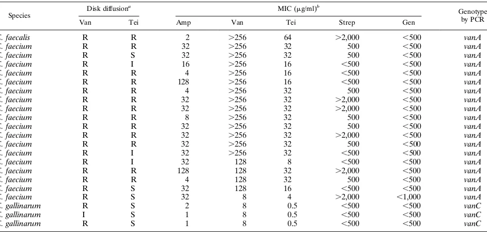
identi-fication of the 22 strains was confirmed by comparative analysis of their whole-cell protein profiles by PAGE (data not shown). Susceptibility to antimicrobial agents.Table 1 provides the results of the in vitro susceptibility tests. By the disk diffusion method, 21 of the 22 strains were resistant to vancomycin (zone diameter,#14 mm) and one strain showed an interme-diate resistant result (zone diameter,,17 mm but.14 mm). Thirteen strains were also resistant to teicoplanin (12 E.
fae-cium strains and 1 E. faecalis strain), 3 strains showed
inter-mediate resistance, and 6 strains were susceptible. Determina-tion of the MIC by the agar diluDetermina-tion test confirmed decreased susceptibility to vancomycin for all 22 strains. Twelve E.
fae-cium and one E. faecalis strains were resistant to vancomycin
and teicoplanin (MICs,$32mg/ml). Four E. faecium isolates were resistant to vancomycin and had intermediate resistance to teicoplanin (MICs, 16mg/ml). All three E. gallinarum strains and one E. faecium strain had intermediate resistance to van-comycin (MICs, 8mg/ml) and were susceptible to teicoplanin (MICs, 0.5 and 4mg/ml for the E. gallinarum strains and the E.
faecium strain, respectively). Finally, one E. faecium strain was
[image:2.612.62.554.82.317.2]resistant to vancomycin (MIC, 128mg/ml) and susceptible to teicoplanin (MIC, 8mg/ml). Eight enterococci remained sus-ceptible to ampicillin (MICs, ,16 mg/ml). Only 1 of the 22 enterococci showed high-level resistance to gentamicin (MIC, TABLE 1. Antimicrobial susceptibilities of 22 VRE
Species Disk diffusion a
MIC (mg/ml)b
Genotype by PCR
Van Tei Amp Van Tei Strep Gen
E. faecalis R R 2 .256 64 .2,000 ,500 vanA
E. faecium R R 32 .256 32 500 ,500 vanA
E. faecium R S 32 .256 32 500 ,500 vanA
E. faecium R I 16 .256 16 ,500 ,500 vanA
E. faecium R R 4 .256 16 ,500 ,500 vanA
E. faecium R R 128 .256 16 ,500 ,500 vanA
E. faecium R R 4 .256 32 500 ,500 vanA
E. faecium R R 32 .256 32 .2,000 ,500 vanA
E. faecium R R 32 .256 32 .2,000 ,500 vanA
E. faecium R R 8 .256 32 500 ,500 vanA
E. faecium R R 32 .256 32 500 ,500 vanA
E. faecium R R 32 .256 32 .2,000 ,500 vanA
E. faecium R R 32 .256 32 500 ,500 vanA
E. faecium R I 32 .256 32 ,500 ,500 vanA
E. faecium R I 32 128 8 ,500 ,500 vanA
E. faecium R R 128 128 32 .2,000 ,500 vanA
E. faecium R R 4 128 32 500 ,500 vanA
E. faecium R S 32 128 16 ,500 ,500 vanA
E. faecium R S 32 8 4 .2,000 ,1,000 vanA
E. gallinarum R S 2 8 0.5 ,500 ,500 vanC
E. gallinarum I S 1 8 0.5 ,500 ,500 vanC
E. gallinarum R S 1 8 0.5 ,500 ,500 vanC
a
Van, vancomycin; Tei, teicoplanin; R, resistant; S, susceptible; I, intermediate resistance. b
Amp, ampicillin; Van, vancomycin; Tei, teicoplanin; Strep, streptomycin; Gen, gentamicin.
on May 15, 2020 by guest
http://jcm.asm.org/
.1,000mg/ml), and 6 of them showed high-level resistance to streptomycin (MICs,.2,000mg/ml).
Mechanisms of resistance.PCR demonstrated that all 18 E.
faecium strains and 1 E. faecalis strain carried the vanA gene.
However, two E. faecium isolates were phenotypically not clas-sified as vanA according to the criteria published by Arthur and Courvalin (1). Isolate 175 had low-level resistance to vanco-mycin and was susceptible to teicoplanin, and isolate 11 was borderline susceptible to teicoplanin (MIC, 8mg/ml). All three
E. gallinarum strains were phenotypically classified as vanC,
and the vanC gene was detected by PCR.
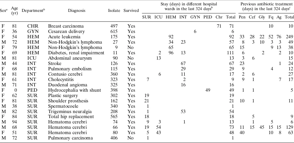
Distribution of VRE carriers in the hospital.Table 2 pro-vides information on the 22 VRE carriers encountered in the study. Their distribution throughout the hospital was not ho-mogeneous. Nine of the 22 carriers were situated in the surgi-cal wards, but the prevalence in this department was only 4.9%. Four patients were admitted to the hematology ward, where the prevalence was 10.8%, more than double in comparison with that on other wards (P,0.05). Carriers were also iden-tified, although in small numbers, in several departments con-sidered less at risk for nosocomial transmission (maternity ward, 1 carrier among 34 patients; chronic care, 1 carrier among 125 patients; pediatrics, 1 carrier among 62 patients). Carriers were not found more frequently in the intensive care ward than in the rest of the hospital (5.4%; P.0.1).
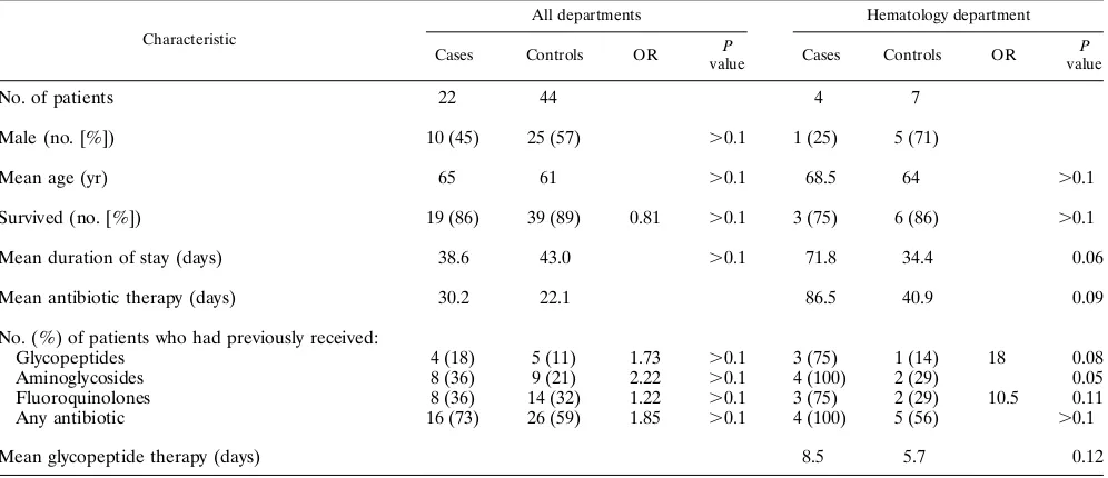
Risk factors for VRE colonization.An overview of the epi-demiologic data for the VRE carriers and for the subjects in the case-control study is provided in Tables 2 and 3. Sex, mean age, and survival rates were comparable in both groups. The mean duration of hospital stay for VRE carriers was more than 38 days. This is significantly longer (P,0.05) than the average of 13 days for all patients admitted in 1993. The mean numbers of antibiotic treatment days in the 324 days prior to the study were 22.1 and 30.2 days in the control patients and the VRE carriers, respectively (P.0.1). Carriers had received the
fol-lowing drug classes more often than controls: glycopeptides (OR, 1.73), aminoglycosides (OR, 2.22), and antibiotics all together (OR, 1.85). The differences were not statistically sig-nificant (P .0.1), however. Because VRE carriers were sig-nificantly more frequently found in the hematology ward than in other departments (10.8 versus 3.1%, respectively; P , 0.05), we investigated the relation of risk factors between cases and controls from this department separately (Table 3). No difference was found regarding sex, mean age, or survival rate. However, the mean duration of stay was considerably longer for cases than for controls (72 versus 34 days, respectively; P5 0.06). Each of the four cases had received antibiotics in the 324 days prior to the study, but so had 56% of the controls. How-ever, ORs of 18 and 10, respectively, suggested that VRE carriers had received glycopeptides (P50.08) or quinolones (P50.11) more often, although the differences were statisti-cally not significant. Carriers tended to have received antibiotic treatment longer than controls (mean of 87 versus 41 days, respectively, P50.09), and the difference in the duration of glycopeptide treatment was important, but it also was not sta-tistically significant (mean, 8.5 versus 5.7 days, respectively;
P50.12).
DISCUSSION
Glycopeptide-resistant enterococci have become a major threat to hospitalized patients. Like methicillin-resistant
Staph-ylococcus aureus, VRE can cause important nosocomial
[image:3.612.59.554.84.325.2]epi-demics and can, increase morbidity, mortality, and costs re-lated to admission to the hospital. The presence of VRE in clinical samples in Belgium is still very low, and no VRE epidemics have been described. In the St. Jan General Hospi-tal of Bruges, no infection caused by VRE was ever diagnosed. Little is known about the epidemiology of VRE colonization outside of the hospital environment. The investigations of TABLE 2. Epidemiological data for patients colonized with VRE
Sexa Age
(yr) Department
b Diagnosis Isolate Survived
Stay (days) in different hospital
wards in the last 324 daysb Previous antibiotic treatment (days) in the last 324 daysc
SUR ICU HEM INT GYN PED Chr Total Pen Cef Gly Fq Ag Total
F 81 CHR Breast carcinoma 497 Yes 71 71 10 10
F 36 GYN Cesarean delivery 615 Yes 6 6
F 54 HEM Acute leukemia 175 Yes 92 92 33 28 22 52 76 249
M 72 HEM Non-Hodgkin’s lymphoma 27 Yes 34 23 57 8 3 10 3 3 49
F 79 HEM Non-Hodgkin’s lymphoma 9 No 65 65 15 9 13 38
F 69 HEM Diabetes, renal impairment 11 Yes 2 96 13 111 6 2 2 10
M 81 ICU Abdominal aneurysm 90 No 13 13 3 6 15
M 44 INT Stroke 126 Yes 67 67 23 1 24
F 68 INT Pulmonary embolism 115 Yes 29 29 9 4 12
M 81 INT Contusio cerebri 360 Yes 6 11 17 2 6 27
F 61 INT Cholecystitis 323 Yes 7 2 9 9 1 7 17
M 71 INT Duodenal angioma 325 Yes 16 16
F 0 PED Hydrocephalia with shunt 398 Yes 49 49 1 1 5
F 62 SUR Plastic surgery 302 Yes 19 19
F 81 SUR Shoulder prosthesis 162 Yes 21 21 10 1 11
M 38 SUR Spermatocoele 340 Yes 1 1
M 82 SUR Trigeminus neuralgia 208 Yes 1 53 54
F 84 SUR Total hip replacement 565 Yes 18 18 5 9
M 94 SUR Hematoma cerebri 74 Yes 9 3 1 13 1 5 6
M 68 SUR Hematoma cerebri 66 Yes 19 54 73 11 15 45 15 15 129
F 51 SUR Hematoma cerebri 80 Yes 5 43 48 40 10 8 63
M 72 SUR Pulmonary carcinoma 406 No 1 1
a
F, female; M, male. b
SUR, surgery; ICU, intensive care; HEM, hematology; INT, internal medicine; GYN, gynecology; PED, pediatrics; CHR, chronic care. c
Pen, all penicillins; Cef, all cephalosporins; Gly, glycopeptides; Fq, fluoroquinolones; Ag, aminoglycosides.
on May 15, 2020 by guest
http://jcm.asm.org/
Torres et al. (22) and Jordens et al. (12) have suggested that VRE can be part of the intestinal microflora of patients inside and outside of the hospital. The latter investigators also dem-onstrated vancomycin-resistant enterococci from animal reser-voirs (2). However, those studies investigated colonization in areas where nosocomial VRE infections and epidemics were ongoing. Therefore, contamination of the environment from the hospital could not be excluded. The present study demon-strates that about 1 of every 29 patients in St. Jan General Hospital may be colonized with VRE. Detection of VRE in patients without signs of infection or without evidence of a hospital epidemic may suggest that the nosocomial spread is secondary to the emergence of VRE, possibly prior to hospital admission, and selection from the intestinal flora of the pa-tients after admission to the hospital. Moreover, since two patients in the present study had been in the hospital for only 1 day before VRE were detected, we suspect that VRE colo-nization can be acquired in the community outside the hospital environment, as suggested by Torres et al. (22) and Jordens et al. (12).
The present study detected not only vancomycin-resistant E.
faecium but also E. faecalis and E. gallinarum. The question
may be asked why vancomycin-resistant E. faecalis isolates are only rarely encountered in patients with nosocomial infections even though they are present in the human enteric tract.
Our study also confirms the difficulty in identifying glyco-peptide resistance in Enterococcus spp. by the disk diffusion method, as previously mentioned by Tenover et al. (21) and Swenson et al. (20), since it failed to detect 1 of 13 teicoplanin-resistant enterococci. The results of PCR indicate that the phenotypic appearance represented by the susceptibility pat-terns can also be unreliable in identifying the presence of vanA,
vanB, or vanC genes. Six of 19 E. faecium isolates (31.6%) with vanA genotypes would have been missed on the basis of the
results of the disk diffusion method, and MICs for 2 isolates (10.5%) were beyond the accepted range for isolates of the
vanA phenotype (1). Future prevalence culture surveys must
also take into account the fact that both Leuconostoc spp. and glycopeptide-susceptible enterococci can be mistaken for VRE on the basis of growth and colony morphology on culture
medium containing 8mg of vancomycin per ml. Identification of enterococci with the API-20 S Streptococcus system appears to be unreliable since this system misidentified all three E.
gallinarum isolates. It is therefore imperative to perform
stan-dardized reference methods for identification, susceptibility testing, and PCR in all epidemiological studies on VRE.
[image:4.612.59.557.84.299.2]As to the epidemiology of VRE colonization, certain risk factors reviewed by Korten and Murray (13) also seem to be involved in the present study. Colonized patients had been admitted to the hospital longer than the average patient, but such times were not significantly different from those for VRE-free control patients hospitalized in the same wards. There-fore, we conclude that VRE colonization is more frequently found in wards where patients tend to stay longer. The relation between VRE colonization and previous antimicrobial therapy and its duration, investigated for all 22 cases and controls, is not obvious. The mean number of antibiotic treatment days was high, and determination of the OR suggested a relation with the kinds of antibiotics administered, especially for glyco-peptides and aminoglycosides. However, the differences be-tween controls and cases were not statistically significant and do not confirm the relation with risk factors reported during nosocomial VRE epidemics in other hospitals (3, 10) except for patients in the hematology ward. This may be due to the limited number of cases and controls in our study, but it may also indicate that patients colonized with VRE represent pop-ulation epidemiologically different from patients who acquire VRE during a nosocomial outbreak. However, a relation be-tween the discovery of VRE and admission on the hematology ward was demonstrated; VRE carriers were significantly more frequently found on this ward (P,0.05) and tended to have stayed there longer than control patients, although this differ-ence was not statistically significant. In this ward, there was a trend for VRE-colonized patients to have received glycopep-tides and aminoglycosides, and possibly fluoroquinolones, more often than controls (but the differences not statistically significant). A difference in the duration of glycopeptide ther-apy was detected, although this difference was not statistically significant. On the basis of the conclusions presented above, we believe that the use of antibiotics, in particular, glycopeptides, TABLE 3. Comparison of risk factors between 22 patients colonized with VRE and 44 controls
Characteristic
All departments Hematology department
Cases Controls OR P
value Cases Controls OR P value
No. of patients 22 44 4 7
Male (no. [%]) 10 (45) 25 (57) .0.1 1 (25) 5 (71)
Mean age (yr) 65 61 .0.1 68.5 64 .0.1
Survived (no. [%]) 19 (86) 39 (89) 0.81 .0.1 3 (75) 6 (86) .0.1
Mean duration of stay (days) 38.6 43.0 .0.1 71.8 34.4 0.06
Mean antibiotic therapy (days) 30.2 22.1 86.5 40.9 0.09
No. (%) of patients who had previously received:
Glycopeptides 4 (18) 5 (11) 1.73 .0.1 3 (75) 1 (14) 18 0.08
Aminoglycosides 8 (36) 9 (21) 2.22 .0.1 4 (100) 2 (29) 0.05
Fluoroquinolones 8 (36) 14 (32) 1.22 .0.1 3 (75) 2 (29) 10.5 0.11
Any antibiotic 16 (73) 26 (59) 1.85 .0.1 4 (100) 5 (56) .0.1
Mean glycopeptide therapy (days) 8.5 5.7 0.12
on May 15, 2020 by guest
http://jcm.asm.org/
should probably be dramatically restricted in order to avoid the selection of VRE, which are already part of the human micro-flora.
Our institution maintains a policy of nursing patients colo-nized with multiresistant bacteria (e.g., methicillin-resistant S.
aureus and cefotaxime-resistant Klebsiella pneumoniae) in
iso-lation. However, since the present study indicates that at least 3.5% of hospitalized patients are healthy VRE carriers, we conclude that the spread of nosocomial VRE epidemics prob-ably cannot be prevented only by nursing VRE-infected pa-tients in isolation. Preferentially, compliance with basic hy-gienic measures for all patient care, like systematic hand disinfection, wearing gloves, and taking universal precautions when handling blood and body fluid, should be increased.
ACKNOWLEDGMENTS
P.V. is indebted to the National Fund for Scientific Research (Bel-gium) for a position as a postdoctoral research fellow. This work was partially supported by a grant from Merrell Dow Belgium.
We thank Kathy Claeys and Hilde Jannes for logistic support during the study, Annemie Lambert and Jean-Marie Fossepre´ for technical assistance in the laboratory, and B. Pot (University of Ghent) for permission to use the Enterococcus reference database for comparison of whole-cell protein electrophoresis. The authors are grateful to R. N. Jones (Iowa City) for the reference strains E. faecium Iowa 1 and Iowa 2.
REFERENCES
1. Arthur, M., and P. Courvalin. 1993. Genetics and mechanisms of glycopep-tide resistance in enterococci. Antimicrob. Agents Chemother. 37:1563– 1571.
2. Bates, J., J. Z. Jordens, and D. T. Griffiths. 1994. Farm animals as a putative reservoir for vancomycin-resistant enterococcal infection in man. J. Antimi-crob. Chemother. 34:507–516.
3. Boyle, J. F., S. A. Soumakis, A. Rendo, J. A. Herrington, D. G. Gianarkis,
B. E. Thurberg, and B. G. Painter.1993. Epidemiologic analysis and geno-typic characterization of a nosocomial outbreak of vancomycin-resistant en-terococci. J. Clin. Microbiol. 31:1280–1285.
4. Centers for Disease Control and Prevention. 1993. Nosocomial enterococci resistant to vancomycin—United States 1989–1993. Morbid. Mortal. Weekly Rep. 42:597–600.
5. Clark, H. C., R. C. Cooksey, B. C. Hill, J. M. Swenson, and F. C. Tenover. 1993. Characterization of glycopeptide-resistant enterococci from U.S. hos-pitals. Antimicrob. Agents Chemother. 37:2311–2317.
6. Coene, J., E. De Brauwer, B. Gordts, and H. W. Van Landuyt. 1995. Van-comycin resistant Enterococcus faecalis: now also in Belgium. Acta Clin. Belg. 50:46–47.
7. Dean, A. D., J. A. Dean, A. H. Burton, and R. C. Dicker. 1990. Epi Info version 5: a word processing, database, and statistics program for epidemi-ology on micro-computers. USD, Inc., Stone Mountain, Ga.
8. Facklam, R. R., and M. D. Collins. 1989. Identification of Enterococcus species isolated from human infections by a conventional test scheme. J. Clin. Microbiol. 27:731–734.
9. Fleiss, J. L. 1981. Statistical methods for rates and proportions, p. 71–75. John Wiley & Sons, Inc., New York.
10. Handwerger, S., B. Raucher, D. Altarac, J. Monka, S. Marchione, K. V.
Singh, B. E. Murray, J. Wolff, and B. Walters.1993. Nosocomial outbreak due to Enterococcus faecium highly resistant to vancomycin, penicillin and gentamicin. Clin. Infect. Dis. 16:750–755.
11. Jones, R. N., H. S. Sader, M. E. Erwin, and S. C. Anderson. 1995. Emerging multiply resistant enterococci among clinical isolates. Diagn. Microbiol. In-fect. Dis. 21:85–94.
12. Jordens, J. Z., J. Bates, and D. T. Griffiths. 1994. Faecal carriage and nosocomial spread of vancomycin resistant Enterococcus faecium. J. Anti-microb. Chemother. 34:515–528.
13. Korten, V., and B. E. Murray. 1993. The nosocomial transmission of entero-cocci. Curr. Opinion Infect. Dis. 6:498–505.
14. Moellering, R. C. 1992. Emergence of Enterococcus as a significant patho-gen. Clin. Infect. Dis. 14:1173–1178.
15. National Committee for Clinical Laboratory Standards. 1990. Performance standards for antimicrobial susceptibility tests, 4th ed. Approved standards. NCCLS document M2-A4. National Committee for Clinical Laboratory Standards, Villanova, Pa.
16. National Committee for Clinical Laboratory Standards. 1990. Performance standards for antimicrobial susceptibility tests, 4th ed. Approved standards. NCCLS document M7-A2. National Committee for Clinical Laboratory Standards, Villanova, Pa.
17. Pot, B., P. Vandamme, and K. Kersters. 1993. Analysis of electrophoretic whole organism protein fingerprinting, p. 493–521. In M. Goodfellow and A. G. O’Donnel (ed.), Modern microbiological methods. Chemical methods in bacterial systematics. John Wiley & Sons, Chichester, United Kingdom. 18. Shlaes, D. M., and B. Binczewski. 1990. Enterococcal resistance to vanco-mycin and related cyclic glycopeptide antibiotics. Eur. J. Clin. Microbiol. Infect. Dis. 9:106–110.
19. Siegel, S. 1956. Nonparametric statistics for the behavioral sciences, p. 184. McGraw-Hill Book Co., New York.
20. Swenson, J. M., B. C. Hill, and C. Thornsberry. 1989. Problems with disk diffusion test for detection of vancomycin resistance in enterococci. J. Clin. Microbiol. 27:2140–2142.
21. Tenover, F. C., J. Tokars, J. Swenson, S. Paul, K. Spitalny, and W. Jarvis. 1993. Ability of clinical laboratories to detect antimicrobial agent-resistant enterococci. J. Clin. Microbiol. 31:1695–1699.
22. Torres, C., J. A. Reguera, M. J. Sanmartin, J. C. Perez-Diaz, and F. Baquero. 1994. Van-A mediated vancomycin resistant Enterococcus spp. in sewage. J. Antimicrob. Chemother. 33:553–561.
23. Vercauteren, E., R. Leclercq, and H. Goossens. 1994. Belgian surveillance study of Enterococcal susceptibility patterns, abstr. E10, p. 48. In Program and abstracts of the 34th Interscience Conference on Antimicrobial Agents and Chemotherapy. American Society for Microbiology, Washington, D.C.