IL-1 produced and released endogenously
within human islets inhibits beta cell function.
M Arnush, … , P T Manning, J A Corbett
J Clin Invest.
1998;
102(3)
:516-526.
https://doi.org/10.1172/JCI844
.
Resident macrophages have been suggested to participate in the initiation of beta cell
damage during the development of autoimmune diabetes. The purpose of this study was to
determine if the endogenous production and release of interleukin 1 (IL-1) in human islets of
Langerhans by resident macrophages results in the inhibition of beta cell function.
Treatment of human islets with a combination of tumor necrosis factor (TNF) +
lipopolysaccharide (LPS) + interferon-gamma (IFN-gamma) stimulates inducible nitric oxide
synthase (iNOS) expression, nitric oxide production, and inhibits glucose-stimulated insulin
secretion. The IL-1 receptor antagonist protein (IRAP) prevents TNF + LPS +
IFN-gamma-induced iNOS expression and nitrite production, and attenuates the inhibitory effects on
glucose-stimulated insulin secretion by human islets. Inhibition of iNOS activity by
aminoguanidine also attenuates TNF + LPS + IFN-gamma-induced inhibition of insulin
secretion by human islets. These results indicate that the inhibitory effects of TNF + LPS +
IFN-gamma are mediated by nitric oxide, produced by the actions of IL-1 released
endogenously within human islets. Reverse transcriptase polymerase chain reaction was
used to confirm that TNF + LPS + IFN-gamma stimulates the expression of both IL-1alpha
and IL-1beta in human islets. Two forms of evidence indicate that resident macrophages are
the human islet cellular source of IL-1: culture conditions that deplete islet lymphoid cells
prevent TNF + LPS + IFN-gamma-induced […]
Research Article
J. Clin. Invest.
© The American Society for Clinical Investigation, Inc. 0021-9738/98/08/0516/11 $2.00
Volume 102, Number 3, August 1998, 516–526 http://www.jci.org
IL-1 Produced and Released Endogenously within Human Islets Inhibits
b
Cell Function
Marc Arnush,* Monique R. Heitmeier,* Anna L. Scarim,* Margaret H. Marino,‡ Pamela T. Manning,§ and John A. Corbett*
*Edward A. Doisy Department of Biochemistry and Molecular Biology, St. Louis University School of Medicine, St. Louis, Missouri 63104; and ‡Department of Protein Biochemistry and §Department of Molecular Pharmacology, Inflammation Disease Research, G.D.
Searle, St. Louis, Missouri 63167
Abstract
Resident macrophages have been suggested to participate in the initiation of b cell damage during the development of autoimmune diabetes. The purpose of this study was to de-termine if the endogenous production and release of inter-leukin 1 (IL-1) in human islets of Langerhans by resident macrophages results in the inhibition of b cell function. Treatment of human islets with a combination of tumor ne-crosis factor (TNF) 1 lipopolysaccharide (LPS) 1 inter-feron-g (IFN-g) stimulates inducible nitric oxide synthase (iNOS) expression, nitric oxide production, and inhibits glu-cose-stimulated insulin secretion. The IL-1 receptor antago-nist protein (IRAP) prevents TNF 1 LPS 1 IFN-g–induced iNOS expression and nitrite production, and attenuates the inhibitory effects on glucose-stimulated insulin secretion by human islets. Inhibition of iNOS activity by aminoguani-dine also attenuates TNF 1 LPS 1 IFN-g–induced inhibi-tion of insulin secreinhibi-tion by human islets. These results indi-cate that the inhibitory effects of TNF 1 LPS 1 IFN-g are mediated by nitric oxide, produced by the actions of IL-1 re-leased endogenously within human islets. Reverse tran-scriptase polymerase chain reaction was used to confirm that TNF 1 LPS 1 IFN-g stimulates the expression of both IL-1a and IL-1b in human islets. Two forms of evidence in-dicate that resident macrophages are the human islet cellu-lar source of IL-1: culture conditions that deplete islet lym-phoid cells prevent TNF 1 LPS 1 IFN-g–induced iNOS expression, nitric oxide production, and IL-1 mRNA ex-pression by human islets; and IL-1 and the macrophage sur-face marker CD69 colocalize in human islets treated with TNF 1 LPS 1 IFN-g as determined by immunohistochemi-cal analysis. Lastly, nitric oxide production is not required for TNF 1 LPS 1 IFN-g–induced IL-1 release in human lets. However, cellular damage stimulates IL-1 release by is-let macrophages. These findings support the hypothesis that activated islet macrophages may mediate b cell damage during the development of insulin-dependent diabetes by
releasing IL-1 in human islets followed by cytokine-induced iNOS expression by b cells. (J. Clin. Invest. 1998. 102:516– 526.) Key words: human islets • macrophages • inducible
nitric oxide synthase • interleukin-1 • insulin-dependent di-abetes mellitus
Introduction
Insulin-dependent diabetes mellitus (IDDM)1 is characterized
by a local inflammatory reaction in and around islets of Langerhans, followed by selective destruction of pancreatic is-let b cells. Resident macrophages have been proposed to be one islet cell type that may initiate b cell damage during the development of autoimmune diabetes (1). Evidence to support this proposal includes studies showing that macrophage deple-tion by feeding a diet deficient in essential fatty acids (2) pre-vents the development of diabetes induced by multiple injec-tions of streptozotocin in CD-1 mice, and attenuates the natural occurrence of diabetes in the Bio Breeding (BB) rat (3, 4). In addition, macrophage depletion by silica treatment at-tenuates the natural development of diabetes in BB rats (5). While these studies support a role for macrophage involve-ment in autoimmune diabetes, the mechanisms by which in-traislet macrophages participate in the development of au-toimmune diabetes are unknown.
In 1991, Lacy and Finke (6) showed that treatment of iso-lated islets with high concentrations of rat IFN-g resulted in is-let destruction after a 4–7-d culture. Culturing isis-lets for 7 d at 248C, a treatment that results in the depletion of . 98% of islet class II–positive lymphoid cells (presumed to be macrophages), prevents IFN-g–induced islet destruction (6). Recently, we have shown that treatment of rat islets with TNF 1 LPS results in a potent inhibition of insulin secretion that is mediated by the intraislet release of IL-1, followed by IL-1–induced induc-ible nitric oxide synthase (iNOS) expression by b cells (7). Al-though these studies indicate that islet lymphoid cells and the intraislet production of IL-1 can inhibit b cell function, the ex-tent to which resident human macrophages participate in the development of IDDM, and the mechanisms by which these cells mediate b cell damage are unknown. In this study we have evaluated the effects of resident macrophage activation on b cell function in human islets of Langerhans. We show that treatment of human islets with TNF 1 LPS 1 IFN-g, condi-tions known to activate macrophages, results in the expression of iNOS, the increased production of nitric oxide, and a potent inhibition of insulin secretion. In addition, evidence is pre-sented that supports TNF 1 LPS 1 IFN-g–induced resident
Address correspondence to Dr. Corbett, St. Louis University School of Medicine, Department of Biochemistry and Molecular Biology, 1402 South Grand Boulevard, St. Louis, MO 63104. FAX: 314-577-8156; E-mail: [email protected]
Received for publication 6 June 1997 and accepted in revised form 28 May 1998.
macrophage expression and release of IL-1 in islets, followed by cytokine-induced inhibition of b cell function as a mecha-nism by which resident macrophages mediate b cell damage in human islets.
Methods
Materials.Human islets were provided by the Islet Isolation Core Fa-cility at Washington University School of Medicine (St. Louis, MO) and the Diabetes Research Institute at the University of Miami (Mi-ami, FL). Human recombinant IL-1b was from Cistron Biotechnol-ogy (Pine Brook, NJ). Human recombinant IFN-g and human recom-binant TNF-a were from Boehringer Mannheim (Indianapolis, IN). Human recombinant IL-1 receptor antagonist protein (IRAP) was a gift from Dr. Charles Hall (Upjohn, Kalamazoo, MI). CMRL-1066 tissue culture medium, penicillin, streptomycin, L-glutamine, and all PCR oligonucleotide primers were from GIBCO BRL (Gaithers-burg, MD). FCS was obtained from Hyclone Labs (Logan, UT). Ami-noguanidine hemisulfate (AG) was from Sigma Chemical Co. (St. Louis, MO). ECL reagents were obtained from Amersham (Arling-ton Heights, IL). Horseradish peroxidase–conjugated donkey anti– rabbit IgG, FITC-conjugated donkey anti–guinea pig and donkey anti–mouse IgG, and CY3-conjugated donkey anti–rabbit and don-key anti–goat IgG were from Jackson ImmunoResearch Laborato-ries, Inc. (West Grove, PA). Affinity-purified goat anti–human IL-1b and mouse anti–human macrophage CD68 were from R&D Systems (Minneapolis, MN) and Biomeda Corp. (Foster City, CA), respec-tively. Rabbit anti–human iNOS antiserum used in Fig. 1 was pre-pared as described previously (8). All other Western blots were performed with rabbit anti–human iNOS prepared against the COOH-terminal 19 amino acids of human iNOS (Santa Cruz Bio-technology, Santa Cruz, Ca). All other reagents were from commer-cially available sources.
Islet culture, lymphoid cell depletion, and insulin secretion. Freshly isolated human islets were cultured for 3 d at 378C in complete CMRL-1066 (CMRL-1066 containing 2 mM L-glutamine, 10% heat-inactivated FCS, 100 U/ml penicillin, and 100 mg/ml streptomycin) before experimentation. Resident lymphoid cells were depleted from human islets as previously described for rat islets (6). In brief, iso-lated human islets were cultured at 248C in an atmosphere of 95% air and 5% CO2 in complete CMRL-1066 tissue culture medium for 7 d. Islets were removed from the 248C culture, washed three times with fresh complete CMRL-1066, and were then cultured for 3 d at 378C in complete CMRL-1066. Resident macrophage depletion was con-firmed by immunohistochemical analysis of CD68-positive macro-phages using methods outlined below. Glucose-stimulated insulin secretion by human islets, incubated for 40 h with the indicated concentrations of IRAP, IFN-g, TNF, LPS, and AG, was per-formed as described previously (9). Where indicated, human islets were pretreated for 15 min with IRAP before incubation with cyto-kines and LPS.
Human islet dispersion. Isolated human islets were dispersed into individual cells by treatment with trypsin (1.0 mg/ml) in Ca21- and
Mg21-free Hanks’ solution at 378C (7, 10). The dispersed islet cells
were counted and immediately aliquoted into 24-well microtiter plates (120,000 cells/400 ml of complete CMRL-1066) and cultured for 1 h. Experiments were initiated by the addition of cytokines as de-scribed above.
Western blot analysis and nitrite determination. iNOS protein ex-pression was determined by Western blot analysis as described previ-ously for human islets (8). Nitrite production was determined as de-scribed previously (11) by mixing 50-ml portions of culture media with 50 ml of Griess reagent and the absorbance at 540 nm was deter-mined.
Immunohistochemistry. Immunohistochemistry was performed as previously described for sequential double staining of intact rat is-lets (7). In brief, after cytokine stimulation, isis-lets (2,000/3 ml of
com-plete CMRL-1066) were isolated by centrifugation, washed three times in 0.1 M PBS, pH 7.4, and then fixed overnight in 2% paraform-aldehyde in 0.1 M PBS at 48C. The islets were cryopreserved by incu-bation at 48C in 0.1 M PBS containing 10% sucrose. Sections were prepared (z 8 mm) and sequentially stained for human iNOS using rabbit anti–human iNOS (1:500 dilution) and indocarbocyanine (CY-3)-conjugated donkey anti–rabbit secondary antiserum (1:200 dilution; red fluorescence), and insulin using guinea pig anti–human insulin (1:500 dilution) and FITC-conjugated donkey anti–guinea pig secondary antiserum (1:200 dilution; green fluorescence).
For immunohistochemical identification of IL-1–expressing cells, human islets (150/400 ml of complete CMRL-1066), cultured for 4 h with or without 50 ng/ml TNF 1 10 mM LPS 1 750 U/ml IFN-g, were isolated and dispersed into individual cells (see above). Islet cells were washed three times with 0.1 M PBS (pH 7.4) and diluted to a concentration of 40,000 cells/100 ml. The cells were transferred to Su-perfrost/Plus microscope slides by cytospin. The slides were fixed in 4% paraformaldehyde for 1 h at 48C and then blocked for 1 h with 5% BSA (in 0.1 M PBS). The slides were incubated overnight at room temperature with a 1:25 dilution (in 0.1 M PBS containing 1% BSA) of goat anti–human IL-1b antiserum, followed by a 1-h incuba-tion with a 1:100 diluincuba-tion of mouse anti–human macrophage antise-rum (CD68). The slides were washed three times with 150 ml of PBS and then incubated for 1 h with a 1:200 dilution of FITC-conjugated donkey anti–mouse and CY3-conjugated donkey anti–goat secondary antibody for IL-1b and CD68, respectively. Immunofluorescence mi-croscopy was used for the detection of IL-1b–expressing cells and CD68-positive macrophages.
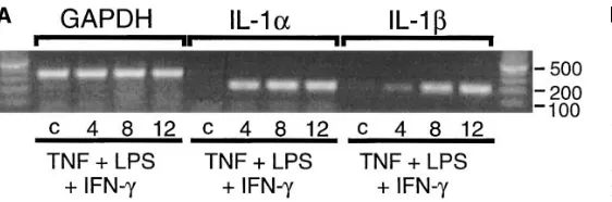
PCR.Total RNA was isolated from islets using the Qiagen RNeasy RNA isolation kit. Total RNA (2.5 mg) from each sample was then used to prepare first-strand cDNA with the GIBCO BRL Preamplifi-cation Superscript kit. A standard 25-ml PCR reaction containing 2.5 ml from the RT reaction, 200 mM each of dATP, dCTP, dGTP, and dUTP, 50 pmol each of forward and reverse primers, 2 U of Taq DNA polymerase (Promega, Madison, WI), 1.5 mM MgCl2, and 2.5 ml of the supplied 103 reaction buffer was performed. IL-1a, IL-1b, iNOS, and GAPDH primers used for PCR were: (a) IL-1a forward primer 59-CCACTCCATGAAGGCTGCATG-39, reverse primer 59-GGTGCTGACCTAGGCTTGATG-39 (PCR product size 5 204 bp); (b) IL-1b forward primer 59 -CCTGTGGCCTTGGGCCTCAA-39, reverse primer 59-GGTGCTGATGTACCAGTTGGG-39 (PCR product size 5 204 bp); (c) iNOS forward primer 59 -ACATTGAT-CAGAAGCTGTCCCAC-39, reverse primer 59 -CAAAGGCTGT-GAGTCCTGCAC-39 (PCR product size 5 236 bp); (d) GAPDH forward primer 59-GCTGGGGCTCACCTGAAGGG-39, reverse primer 59-GGATGACCTTGCCCACAGCC-39 (PCR product size 5 343 bp).
Each PCR reaction mixture was overlaid with one drop of min-eral oil, and then incubated in a thermal cycler using the following profile: an initial denaturation step at 948C for 5 min, followed by 30 cycles of 948C for 45 s, 608C for 45 s, and 728C for 75 s. The samples were finally incubated at 308C for 2 min. To each reaction, 5 ml of 63 loading dye (Promega) was added, and then 12 ml from each PCR re-action was run alongside 5 ml of 100 BP marker (Promega) on a 1.5% agarose gel containing 0.5 mg/ml ethidium bromide. PCR products were visualized with ultraviolet light and photographed.
of reducing the cycle number to 22. From each PCR reaction 12 ml was run on a 1.5% agarose gel, the gels were dried, and phosphorim-age analysis was conducted using a PhosphorImphosphorim-ager and Imphosphorim-ageQuant Software version 3.3 (Molecular Dynamics, Inc., Sunnyvale, CA). The values obtained for each IL-1 PCR reaction were normalized as a percentage of the values obtained for each GAPDH control. Under the conditions used, the PCR product signal is proportional to the amount of cDNA subjected to PCR amplification.
Figure preparation. Ethidium-stained agarose gels and Western blot autoradiograms were scanned into NIH Image version 1.59 using a high performance CCD camera (COHU, Brookfield, WI). The im-ages were then imported into Canvas 3.5 (Deneba Software, Miami, FL) for the preparation of figures.
Results and Discussion
Human islet cellular source of iNOS. Our previous studies have identified IL-1b1 IFN-g as the minimal combination of cy-tokines required to stimulate iNOS expression and nitric oxide production by human islets (9, 14). Alone, IL-1, TNF, or IFN-g does not stimulate iNOS expression by human islets, nor do the combinations of TNF 1 IFN-g or IL-1 1 TNF (9, 14). As shown in Fig. 1 A, treatment of human islets with 75 U/ml IL-1b1 750 U/ml IFN-g results in the time-dependent expression
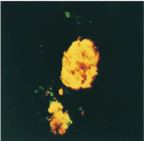
of iNOS that is first apparent after a 12-h exposure and maxi-mal after a 24- and 48-h incubation. The cellular source of iNOS in human islets treated for 40 h with IL-1b1 IFN-g was examined by immunohistochemical colocalization of iNOS and insulin (Fig. 1 B). The combination of IL-1b 1 IFN-g stimulates high levels of iNOS expression in human islets (red fluorescence) and iNOS expression nearly completely colocal-izes with insulin-containing cells (green) as evidenced by the intense yellow fluorescence upon double exposure. IL-1 1 IFN-g also appear to stimulate iNOS expression in a number of islet cells that do not contain insulin. However, human b cells release insulin in culture, thus it is not clear if these cells are b cells with depleted insulin stores or if a second popula-tion of islet cells also expresses iNOS in response to this cyto-kine combination. In controls, iNOS expression was not de-tected in untreated human islets and staining was not observed using secondary antisera alone (data not shown). These find-ings indicate that b cells are a cellular source of iNOS in hu-man islets after treatment with IL-1b1 IFN-g.
TNF 1 LPS 1 IFN-g–induced iNOS expression and nitrite production by human islets requires the intraislet release of IL-1. Treatment of rat islets with TNF 1 LPS results in a po-tent inhibition of insulin secretion that is mediated by the
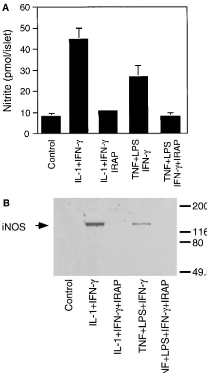
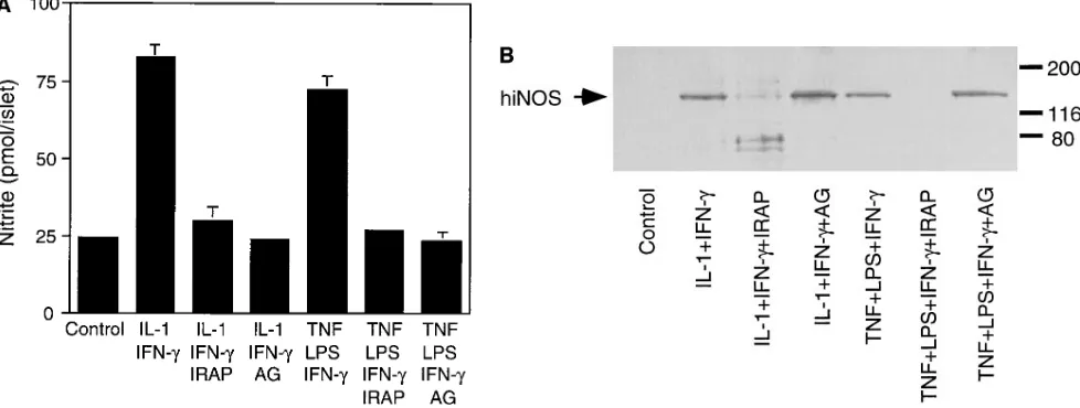
[image:5.612.286.535.347.506.2]traislet release of IL-1 followed by IL-1–induced iNOS expres-sion by b cells (7). Therefore, we examined the effects of TNF 1 LPS on iNOS expression and nitrite production by hu-man islets. Either alone or in combination, TNF (10 ng/ml) 1 LPS (10 mg/ml) fail to stimulate iNOS expression or nitrite production by human islets after a 40-h exposure (data not shown). Since human b cells require a combination of IL-1 1 IFN-g to stimulate iNOS expression (Fig. 1), the lack of iNOS expression in response to TNF 1 LPS may be due to the ab-sence of IFN-g. Therefore, the effects of TNF 1 LPS 1 IFN-g on iNOS expression and nitrite production by human islets were examined. Incubation of human islets for 40 h with TNF 1 LPS 1 IFN-g results in iNOS expression and a three-fold increase in nitrite production (Fig. 2). IRAP (5 mg/ml) prevents TNF 1 LPS 1 IFN-g–induced nitrite production and iNOS protein expression by human islets (Fig. 2, A and B, respectively), indicating that iNOS expression and nitrite production require the intraislet release of IL-1. In addition, antisera specific for IL-1a and IL-1b attenuate TNF 1 LPS 1 IFN-g–induced nitrite formation and iNOS expression by hu-man islets (data not shown).
TNF 1 LPS 1 IFN-g also stimulate the accumulation of iNOS mRNA by human islets after an 8-h exposure (Fig. 2 C). Although IRAP prevents TNF 1 LPS 1 IFN-g–induced iNOS protein expression and nitrite formation by human islets, this receptor antagonist does not inhibit iNOS mRNA accumula-tion under these condiaccumula-tions. We believe that resident mac-rophages may be the islet cellular source of iNOS mRNA that is stimulated by TNF 1 LPS 1 IFN-g, and that it is insensitive to IRAP. Evidence to support this conclusion includes the finding that macrophage depletion by culturing islets for 7 d at 248C prevents TNF 1 LPS 1 IFN-g–induced iNOS mRNA ex-pression (see Fig. 4 C). In addition, IRAP has been shown to prevent TNF 1 LPS–induced iNOS protein expression by b cells, but without inhibiting iNOS expression by a subpopula-tion of rat islet cells (z 5–10/islet) believed to be resident mac-rophages (7). Thus, TNF 1 LPS 1 IFN-g–induced iNOS ex-pression and nitric oxide production by human islets appear to require the intraislet release of IL-1 followed by IL-1 1
IFN-g–induced iNOS expression by b cells. TNF 1 LPS 1 IFN-g also stimulate iNOS mRNA expression by a second popula-tion of cells believed to be resident macrophages.
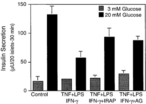
TNF 1 LPS 1 IFN-g–induced inhibition of insulin secre-tion is attenuated by IRAP and AG. To determine if b cell function is inhibited by the endogenous production of IL-1 in human islets, the effects of TNF 1 LPS 1 IFN-g on glucose-stimulated insulin secretion were examined. Treatment of hu-man islets for 40 h with a combination of TNF 1 LPS 1 IFN-g results in z 70% inhibition of glucose-stimulated insulin secre-tion (Fig. 3). The inhibitory effects of TNF 1 LPS 1 IFN-g on insulin secretion are attenuated by IRAP. This result supports the hypothesis that activation of resident islet macrophages re-sults in an impairment of b cell function by a mechanism that requires the intraislet release of IL-1. Also, the iNOS-selective inhibitor AG (15, 16) attenuates TNF 1 LPS 1 IFN-g–induced inhibition of insulin secretion to levels that are comparable to those seen with IRAP. This result is consistent with our previ-ous studies showing that IL-1 1 TNF 1 IFN-g–induced inhibi-tion of insulin secreinhibi-tion by human islets is attenuated by NMMA (9), and indicates that nitric oxide mediates, in part, the inhibitory effects of cytokines on human islet function. The lack of complete prevention of TNF 1 LPS 1 IFN-g–induced
inhibition of insulin secretion by either IRAP or AG suggests that nitric oxide may not be the only effector molecule that participates in the inhibition of b cell secretory function under these conditions.
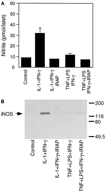
Lymphoid cell depletion attenuates TNF 1 LPS 1 IFN-g– induced iNOS expression by human islets. Results from Figs. 2 and 3 indicate that TNF 1 LPS 1 IFN-g stimulate iNOS ex-pression and inhibit insulin secretion by a mechanism that re-quires the endogenous production and release of IL-1 in hu-man islets. To determine if resident macrophages are an islet source of IL-1, the effects of TNF 1 LPS 1 IFN-g on nitrite production and iNOS expression by human islets depleted of resident macrophages have been evaluated. The combination of TNF 1 LPS 1 IFN-g fails to stimulate nitrite production or iNOS protein expression by human islets cultured for 7 d at 248C (Fig. 4, A and B). Low levels of iNOS mRNA are de-tected under these conditions (Fig. 4 C). However, the level of expression is considerably reduced as compared with iNOS ex-pression by freshly isolated human islets (Fig. 2 C). In addi-tion, a 7-d culture at 248C does not inhibit b cell function as IL-1b1 IFN-g stimulate the expression of iNOS and produc-tion of nitrite to levels nearly equivalent to those produced by freshly isolated human islets (Figs. 2 and 4). These results show that a 7-d culture at 248C does not inhibit b cell expression of iNOS (in response to IL-1 1 IFN-g) but prevents iNOS ex-pression stimulated by the intraislet release of IL-1 (TNF 1 LPS 1 IFN-g), and provides evidence to suggest that resident macrophages are a cellular source of IL-1 in human islets.
[image:6.612.314.555.57.229.2]To confirm that a 7-d culture at 248C depletes human islets of resident macrophages, the effects of this culture condition on the presence of resident macrophages was examined by immunohistochemistry. For these experiments, islets either freshly isolated or cultured for 7 d at 248C were dispersed into individual cells and plated onto slides by cytospin. The number
of macrophages in human islets was determined in a double-blind fashion by immunofluorescent microscopy using antisera specific for human macrophages (mouse anti–human CD68). Freshly isolated human islets contain 5.561.1 CD68-positive macrophages/100 islet cells, while islets cultured for 7 d at 248C contain 0.7760.42 CD68-positive macrophages/100 islet cells. These results, which are the average6SEM of two experi-ments containing two replicates per condition, provide evi-dence that a 7-d culture at 248C results in the depletion of
. 80% of resident islet macrophages, although it does not re-sult in the complete loss of this islet cell population.
Isoform(s) of IL-1 expressed in human islets in response to TNF 1 LPS 1 IFN-g. Recently, IL-1a mRNA expression has been detected in mononuclear leukocytes purified from islets isolated from nonobese diabetic (NOD) mice recently diag-nosed with autoimmune diabetes (13, 17). Using RT-PCR and semiquantitative PCR, we have examined the effects of TNF 1 LPS 1 IFN-g on the time-dependent expression of IL-1a and IL-1b mRNA, and quantitated the levels of each mRNA spe-cies in human islets. TNF 1 LPS 1 IFN-g stimulate the time-dependent expression of both IL-1a and IL-1b mRNA that is first apparent at 4 h and appears to be maximal after a 12-h in-cubation (Fig. 5 A). Neither IL-1a nor IL-1b expression is
de-tected in untreated control human islets. The levels of IL-1a and IL-1b mRNA which accumulate in response to TNF 1 LPS 1 IFN-g were directly compared by semiquantitative PCR. As shown in Fig. 5 B, TNF 1 LPS 1 IFN-g stimulate a nearly twofold increase in the levels of IL-1b as compared with IL-1a mRNA after a 12-h incubation. Although both isoforms of IL-1 are expressed, these results suggest that the predomi-nate isoform expressed in human islets in response to a 12-h incubation with TNF 1 LPS 1 IFN-g is IL-1b. In further sup-port for an imsup-portant role of IL-1b in the initiation of autoim-mune diabetes, Cailleau et al. (18) have shown recently that neutralizing antibodies specific for IL-1b prevent cyclophos-phamide-induced diabetes in the NOD mouse.
The effects of a 7-d culture at 248C on IL-1a and IL-1b mRNA accumulation in response to a 4, 8, or 12 h of incuba-tion with TNF 1 LPS 1 IFN-g have also been examined. Mac-rophage-depleted human islets do not express IL-1a or IL-1b mRNA in response to TNF 1 LPS 1 IFN-g (data not shown). The lack of IL-1 expression by human islets that have been de-pleted of resident macrophages provides additional evidence that resident macrophages may be one islet cellular source of IL-1.
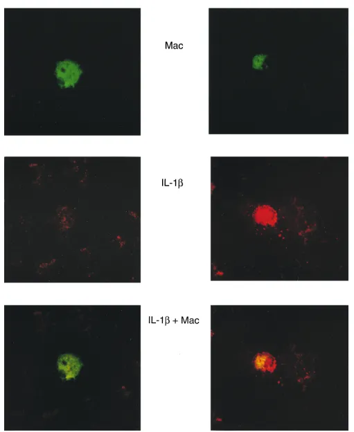
[image:7.612.55.273.57.452.2]Identification of resident macrophages as the human islet
cellular source of IL-1. To determine the cellular source of IL-1, human islets were cultured for 4 h with TNF 1 LPS 1 IFN-g. The islets were isolated, dispersed into individual cells, fixed onto slides, and then stained for IL-1 and the macro-phage surface marker CD68. As shown in Fig. 6, TNF 1 LPS 1 IFN-g stimulate IL-1b expression in a limited number of islet cells as evidenced by the punctate red fluorescence (Fig. 6, middle right), and IL-1b expression localizes with CD68 posi-tive macrophages (Fig. 6, green fluorescence), as evidenced by the yellow fluorescence after double exposure (Fig. 6, bottom right). Importantly, not every CD68-positive macrophage stains positive for IL-1b (5.561.07 CD68-positive macro-phages/100 islet cells as compared with 3.660.7 IL-1b-positive cells/100 islet cells). However, IL-1b was not detected in any islet cell type other than CD68-positive macrophages. In addi-tion, IL-1b expression is not detected in untreated islet endo-crine or nonendoendo-crine cells although a similar number of CD68-positive macrophages were detected, as compared with TNF 1 LPS 1 IFN-g–treated human islets (Fig. 6, left). In ad-dition, cross-reactivity of secondary antisera and nonspecific primary antisera of the same species was not observed. These findings provide direct evidence that CD68-positive macro-phages are the islet cellular source of IL-1b in response to TNF 1 LPS 1 IFN-g.
Mechanisms of IL-1 release in human islets. Recent stud-ies suggest that nitric oxide is required for the release of bio-logically active IL-1 from mouse peritoneal exudate cells and RAW 264.7 macrophages (19). iNOS inhibitors NMMA and AG prevent LPS 1 IFN-g– and LPS-induced IL-1 release and ni-tric oxide production by mouse peritoneal exudate cells and RAW 264.7 cells, respectively (19). If nitric oxide participates in IL-1 release by resident islet macrophages, then the inhibi-tion of nitric oxide producinhibi-tion should prevent TNF 1 LPS 1 IFN-g–induced iNOS expression by human islets. As shown in Fig. 7, AG (and NMMA, data not shown) completely prevents TNF 1 LPS 1 IFN-g–induced nitrite formation, but does not inhibit iNOS expression by human islets. However, IRAP pre-vents TNF 1 LPS 1 IFN-g–induced iNOS expression and ni-trite formation. These results indicate that nitric oxide is not required for TNF 1 LPS 1 IFN-g–induced IL-1 release in hu-man islets. As a control, the ability of AG to attenuate IL-1b1
IFN-g–induced nitrite production without inhibiting iNOS ex-pression by human islets is also shown (Fig. 7). These findings indicate that the intraislet release of IL-1 in human islets is by a mechanism that is independent of nitric oxide production.
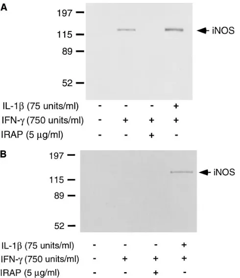
Several studies have shown that macrophage damage or death (either by apoptosis or necrosis) results in the release of IL-1 (20, 21). To determine if cellular damage participates in intraislet IL-1 release, the individual effects of IFN-g on iNOS expression by human islets physically dispersed into individual cells were examined. Islet dispersion involves the treatment of intact islets with trypsin, an experimental manipulation that re-sults in the destruction of z 5% of islet cells (based on trypan blue exclusion, data not shown). Recently, we have shown that macrophage damage after the physical dispersion of rat islets into individual cells results in the release of IL-1 to levels that are sufficient to stimulate iNOS expression in the presence of exogenously added IFN-g (22). Treatment of dispersed human islet cells with IFN-g results in the expression of iNOS as de-termined by Western blot analysis (Fig. 8 A). IRAP com-pletely prevents IFN-g–induced iNOS expression, indicating that physical dispersion of human islets into individual cells re-sults in the endogenous release of IL-1. In addition, antisera specific for IL-1a and IL-1b also significantly attenuate iNOS expression by dispersed islet cells stimulated by IFN-g (data not shown). To determine if resident macrophages are a cellu-lar source of IL-1, the effects of macrophage depletion on
IFN-g–induced iNOS expression by dispersed human islet cells were examined (Fig. 8 B). Although IFN-g stimulates the ex-pression of iNOS by freshly isolated dispersed human islet cells, IFN-g does not induce the iNOS expression by dispersed islet cells prepared from macrophage-depleted human islets. In addition, the 7-d culture at 248C does not inhibit b cell ex-pression of iNOS in response to IL-1 1 IFN-g. These results suggest that cellular damage may be one mechanism that leads to the release of IL-1 in human islets by resident macrophages. In this study evidence is provided in support of the hypoth-esis that activation of resident macrophages in human islets re-sults in the impairment of b cell function by a mechanism that is dependent on the intraislet release of IL-1. The inhibitory effects of resident macrophage activation on b cell function ap-pear to be mediated by the expression of iNOS and the
[image:8.612.59.340.57.150.2] [image:8.612.322.529.58.227.2]creased production of nitric oxide. Aminoguanidine attenuates the inhibitory effects of resident macrophage activation on in-sulin secretion by human islets, and IRAP prevents both the inhibitory effects of TNF 1 LPS 1 IFN-g on insulin secretion and the stimulatory effects on iNOS expression and nitric ox-ide production. These results suggest that nitric oxox-ide, pro-duced by human islet macrophages, does not mediate b cell damage. However, the release of IL-1 in islets followed by IL-1 1 IFN-g–induced iNOS expression by b cells results in
b cell damage. Islets contain several nonendocrine cells that are potential sources of IL-1. These include endothelial cells, fibroblasts, macrophages, and dendritic cells. In rat islets, endo-thelial cells appear to “ball up” and are extruded after a 48-h culture and fibroblasts are normally found in only a thin cap-sule around islets and this capcap-sule is lost during isolation (Bon-ner-Weir, S., personal communication). In this study, human islets were cultured for 72 h before initiation of experiments, or conditions under which endothelial cells would be extruded from islets. We believe that macrophages are the source of IL-1 in human islets, as culture conditions known to deplete islets of lymphoid cells prevent TNF 1 LPS 1 IFN-g–induced iNOS and IL-1 expression and nitrite production by human islets. In addition, immunohistochemical colocalization of IL-1 with the macrophage surface marker CD68 provides direct evidence that resident macrophages are the islet cellular source of IL-1.
It is clear that human macrophages express iNOS (23–25). However, it has been difficult to demonstrate increased pro-duction of nitric oxide by activated human macrophages to levels comparable to the levels produced by rodent macro-phages (26). Our studies suggest that resident human
mac-rophage production of nitric oxide does not inhibit b cell func-tion. However, macrophage activation and cytokine release, followed by cytokine-induced iNOS expression by target b cells, may be one mechanism by which resident human mac-rophages mediate tissue damage during the development of IDDM.
These studies also raise the possibility that resident islet macrophages may participate in the initial destruction of b cells leading to the activation of an immune process resulting in the continued destruction of remaining b cells. The precipi-tating events that lead to initial b cell destruction during the development of IDDM are unknown (27). However, environ-mental factors appear to play an important role. Evidence for environmental factors comes from the relatively low concor-dance of IDDM between identical twins (, 40%) (28). Envi-ronmental factors that have been implicated in the develop-ment of IDDM include chemical toxins or viral infections (29, 30). Several hypotheses have suggested that environmental factors either “trigger” b cell death by directly targeting b cells, or contribute to b cell autoimmunity by unknown mecha-nisms (31). Our findings provide biochemical evidence to sup-port a role for resident macrophages as a potential target for an environmental insult that may trigger the initiation of IDDM in genetically predisposed individuals. Viral infections are known stimuli for macrophage activation and cytokine re-lease (32, 33), and cellular damage is one mechanism associ-ated with IL-1 release by macrophages (20, 21). In addition, we have shown recently that dsRNA, the active component of a viral infection that stimulates antiviral activities in infected cells, stimulates IL-1 release by mouse macrophages (34). In
[image:10.612.59.548.62.247.2]fluorescence), and macrophages were identified using mouse anti–human macrophage (CD68) and FITC-conjugated donkey anti–mouse sec-ondary antisera (red fluorescence). Colocalization of IL-1b– and CD68-positive macrophages is shown by yellow fluorescence after double ex-posure. Results are representative of two individual experiments.
Figure 7. Effects of AG on TNF 1 LPS 1 IFN-g–induced IL-1 release in human islets. Human islets (150/400 ml of complete CMRL-1066) were cultured for 40 h at 378C with 75 U/ml IL-1b, 750 U/ml IFN-g, 50 ng/ml TNF-a, 10 mg/ml LPS, 5 mg/ml IRAP, and 1 mM AG as indicated. (A) The culture media were removed for nitrite determinations, and (B) the islets were isolated for Western blot detection of iNOS expression. The results are representative of three independent experiments from three separate human islet isolations.
this study, we show that a combination of TNF 1 LPS 1 IFN-g inhibits b cell function by a mechanism that is dependent on the intraislet release of IL-1 and the production of nitric oxide. The islet cellular source of IL-1 is believed to be resident mac-rophages, and cellular damage appears to be one mechanism by which resident macrophages release IL-1 in human islets. iNOS expression by human b cells requires IFN-g in addition to IL-1. T cells are the most likely cellular source of IFN-g. Vi-ral infection would be expected to stimulate macrophage re-lease of IL-1, and to stimulate T cell migration (to islets) and activation resulting in the release of IFN-g. The release of these cytokines in the microenvironment of the islet would be sufficient to stimulate iNOS expression by b cells, resulting in
b cell damage and the release of potential autoantigens or b cell neoantigens leading to autoimmunity against the b cell.
Acknowledgments
We would like to thank Colleen Kelly for expert technical assistance, Denise Dorsey for assistance with immunohistochemistry of human islets, and Dr. Paul Lacy for many helpful discussions and advice dur-ing the course of this work. We would also like to thank the Islet Iso-lation Core Facility and the Diabetes Research and Training Center
at Washington University School of Medicine for supplying human is-lets and performing insulin RIAs, and the Diabetes Research Insti-tute at the University of Miami for supplying human islets.
This work was supported by research grants from Alteon Inc., The Tobacco Research Council, The National Institutes of Health (DK-52194), and a Career Development Award from the Juvenile Diabetes Foundation International (J.A. Corbett).
References
1. Lacy, P.E. 1994. The intraislet macrophage and type I diabetes. Mount
Si-nai J. Med. 61:170–174.
2. Schreiner, G.F., W. Fly, E. Brunt, K. Korber, and J.B. Lefkowith. 1988. Essential fatty acid depletion of renal allografts and prevention of rejection.
Science. 240:1032–1033.
3. Wright, J.R., J.B. Lefkowith, G. Schreiner, and P.E. Lacy. 1988. Essential fatty acid deficiency prevents multiple low-dose streptozotocin-induced diabe-tes in CD-1 mice. Proc. Natl. Acad. Sci. USA. 85:6137–6141.
4. Lefkowith, J.B., G. Schreiner, J. Cormier, E.S. Handler, H.K. Driscoll, D. Greiner, J.P. Mordes, and A.A. Rossini. 1990. Prevention of diabetes in the BB rat by essential fatty acid-deficiency. Relationship between physiological and biochemical changes. J. Exp. Med. 171:729–743.
5. Oschilewski, U., U. Kiesel, and H. Kolb. 1985. Administration of silica prevents diabetes in BB rats. Diabetes. 34:197–199.
6. Lacy, P.E., and E.H. Finke. 1991. Activation of intraislet lymphoid cells causes destruction of islet cells. Am. J. Pathol. 138:1183–1190.
7. Corbett, J.A., and M.L. McDaniel. 1995. Intraislet release of IL-1 inhibits
b cell function by inducing b cell expression of iNOS. J. Exp. Med. 181:559–568. 8. Corbett, J.A., G. Kwon, M. Marino, C.P. Rodi, P.M. Sullivan, and M.L. McDaniel. 1996. Tyrosine kinase inhibitors prevent cytokine-induced expres-sion of iNOS and COX-2 by human islets of Langerhans. Am. J. Physiol. 39: C1581–C1587.
9. Corbett, J.A., M.A. Sweetland, J.L. Wang, J.R. Lancaster, Jr., and M.L. McDaniel. 1993. Nitric oxide mediates cytokine-induced inhibition of insulin secretion by human islets of Langerhans. Proc. Natl. Acad. Sci. USA. 90:1731– 1735.
10. Ono, J., R. Takaki, and M. Fukuma. 1977. Preparation of single cells from pancreatic islets of adult rat by the use of dispase. Endocrinol. Jpn. 24: 265–270.
11. Green, L.C., D.A. Wagner, J. Glogowski, P.L. Skipper, J.S. Wishnok, and S.R. Tannenbaum. 1982. Analysis of nitrate, nitrite, and [15N] nitrate in
bio-logical fluids. Anal. Biochem. 126:131–138.
12. Kennedy, M.K., D.S. Torrance, K.S. Picha, and K.M. Mohler. 1992. Analysis of cytokine mRNA expression in the central nervous system of mice with experimental autoimmune encephalomyelitis reveals that IL-10 mRNA correlates with recovery. J. Immunol. 149:2496–2505.
13. Rabinovitch, A., W.L. Saurez-Pinzon, O. Sorensen, R.C. Bleackley, and R.F. Power. 1995. IFN-g gene expression in pancreatic islet-infiltrating mono-nuclear cells correlates with autoimmune diabetes in nonobese diabetic mice. J.
Immunol. 154:4874–4882.
14. Eizirik, D.L., S. Sandler, N. Welsh, M. Cetkovic-Cvrlje, A. Nieman, D.A. Geller, D.G. Pipeleers, K. Bendtzen, and C. Hellerstrom. 1994. Cytokines suppress human islet function irrespective of their effects on nitric oxide gener-ation. J. Clin. Invest. 93:1968–1974.
15. Corbett, J.A., R.G. Tilton, K. Chang, K.S. Hasan, Y. Ido, J.L. Wang, M.A. Sweetland, J.R. Lancaster, Jr., J.R. Williamson, and M.L. McDaniel. 1991. Aminoguanidine, a novel inhibitor of nitric oxide formation, prevents dia-betic vascular dysfunction. Diabetes. 41:552–556.
16. Misko, T.P., W.M. Moore, T.P. Kasten, G.A. Nickols, J.A. Corbett, R.G. Tilton, M.L. McDaniel, J.R. Williamson, and M.G. Currie. 1993. Selective inhibition of the inducible isoform of nitric oxide synthase by aminoguanidine.
Eur. J. Pharmacol. 233:119–125.
17. Rabinovitch, A., W.L. Saurez-Pinzon, O. Sorensen, and R.C. Bleackley. 1996. Inducible nitric oxide synthase (iNOS) in pancreatic islets of nonobese di-abetic mice. Identification of iNOS expression cells and relationship to cyto-kines expressed in the islets. Endocrinology. 137:2093–2099.
18. Cailleau, C., A. Diu-Hercend, E. Ruuth, R. Westwood, and C. Carnaud. 1997. Treatment with neutralizing antibodies specific for IL-1b prevents cyclo-phosphamide-induced diabetes in nonobese diabetic mice. Diabetes. 46:937– 940.
19. Hill, J.R., J.A. Corbett, G. Kwon, C.A. Marshall, and M.L. McDaniel. 1996. Nitric oxide regulates interleukin-1 bioactivity released from murine mac-rophages. J. Biol. Chem. 271:22672–22678.
20. Hogquist, K.A., M.A. Nett, E.R. Unanue, and D.D. Chaplin. 1991. In-terleukin-1 is processed and released during apoptosis. Proc. Natl. Acad. Sci.
USA. 88:8485–8489.
21. Zychlinsky, A., C. Fitting, J.M. Cavaillon, and P.J. Sansonetti. 1994. In-terleukin-1 is released by murine macrophages during apoptosis induced by
Shigella flexneri. J. Clin. Invest. 94:1328–1332.
[image:11.612.57.302.57.341.2]22. Heitmeier, M.R., A.L. Scarim, and J.A. Corbett. 1997. IFN-g increases
the sensitivity of islets of Langerhans for iNOS expression induced by IL-1. J.
Biol. Chem. 272:13653–13661.
23. Nicholson, S., M.G. Bonecini-Almeida, L.E. Silva, Jr., C. Nathan, Q.-W. Xie, R. Mumford, J.R. Weidner, J. Calaycay, J. Geng, N. Boechat, et al. 1996. Inducible nitric oxide synthase in pulmonary alveolar macrophages from pa-tients with tuberculosis. J. Exp. Med. 183:2293–2302.
24. Guo, F.H., H.R. DeRaeve, T.W. Rice, D.J. Stuehr, F.B. Thunnissen, and S.C. Erzurum. 1995. Continuous nitric oxide synthesis by inducible nitric oxide synthase in normal human airway epithelium in vivo. Proc. Natl. Acad.
Sci. USA. 92:7809–7813.
25. Watkins, S.C., W. Macaulay, D. Turner, R. Kang, H.E. Rubash, and C.H. Evans. 1997. Identification of inducible nitric oxide synthase in human macrophages surrounding loosened hip prostheses. Am. J. Pathol. 150:1199– 1206.
26. Albina, J.E. 1995. On the expression of nitric oxide synthase by human macrophages. Why no NO? J. Leukocyte Biol. 58:643–649.
27. Benoist, C., and D. Mathis. 1997. Cell death mediators in autoimmune diabetes. No shortage of suspects. Cell. 89:1–3.
28. Barnett, A.H., C. Eff, R.D. Leslie, and D.A. Pyke. 1981. Diabetes in identical twins: a study of 200 pairs. Diabetologia. 20:87–93.
29. Bach, J.-F. 1994. Insulin-dependent diabetes mellitus as an autoimmune disease. Endocrinol. Rev. 15:516–541.
30. Yoon, J.W. 1991. Role of viruses in the pathogenesis of IDDM. Ann.
Med. 23:437–445.
31. Nerup, J., and A. Lernmark. 1981. Autoimmunity in insulin-dependent diabetes mellitus. Am. J. Med. 70:135–141.
32. Peschke, T., A. Bender, M. Nain, and D. Gemsa. 1993. Role of mac-rophage cytokines in influenza A virus infections. Immunobiology. 189:340– 355.
33. Henke, A., C. Mohr, C. Graebner, A. Stelzner, M. Nain, and D. Gernsa. 1992. Coxsackie virus B3-induced production of tumor necrosis factor-alpha, IL-1 beta and IL-6 in human monocytes. J. Immunol. 148:2270–2277.