Syntaxin-4 is localized to the apical plasma
membrane of rat renal collecting duct cells:
possible role in aquaporin-2 trafficking.
B Mandon, … , S Nielsen, M A Knepper
J Clin Invest.
1996;
98(4)
:906-913.
https://doi.org/10.1172/JCI118873
.
To evaluate the possible role of a putative vesicle-targeting protein, syntaxin-4, in
vasopressin-regulated trafficking of aquaporin-2 water channel vesicles to the apical
plasma membrane of renal collecting duct cells, we have carried out immunoblotting,
immunocytochemistry, and reverse transcription (RT)-PCR experiments in rat kidney.
Immunochemical studies used an affinity-purified, peptide-directed polyclonal antibody to
rat syntaxin-4. Immunoblots using membrane fractions from inner medullary collecting duct
(IMCD) cell suspensions revealed a solitary protein of 36 kD, the expected molecular mass
of syntaxin-4. This protein was enriched in a plasma membrane-enriched membrane
fraction from IMCD cells. Immunoperoxidase immunocytochemistry in 0.85-microm
cryosections from rat inner medulla revealed discrete labeling of the apical plasma
membrane of IMCD cells. RT-PCR demonstrated the presence of syntaxin-4 mRNA in
microdissected IMCD segments, confirmed by direct sequencing of the PCR product. In
addition, RT-PCR experiments demonstrated syntaxin-4 mRNA in glomeruli, vasa recta,
connecting tubules, and thin descending limbs of Henle's loops. The demonstrated
localization of syntaxin-4 in the apical plasma membrane of collecting duct principal cells,
coupled with previous demonstration of syntaxin-4's putative cognate receptor VAMP2 in
aquaporin-2-containing vesicles, supports the view that these proteins could play a role of
aquaporin-2 vesicle targeting to the apical plasma membrane.
Research Article
Find the latest version:
The Journal of Clinical Investigation Volume 98, Number 4, August, 1996, 906–913
Syntaxin-4 Is Localized to the Apical Plasma Membrane of Rat
Renal Collecting Duct Cells: Possible Role in Aquaporin-2 Trafficking
Béatrice Mandon,* Chung-Lin Chou,* Søren Nielsen,‡ and Mark A. Knepper*
*Laboratory of Kidney and Electrolyte Metabolism, National Heart, Lung, and Blood Institute, National Institutes of Health, Bethesda, Maryland 20892-1598; and ‡Department of Cell Biology, Institute of Anatomy, University of Aarhus, Aarhus, Denmark
Abstract
To evaluate the possible role of a putative vesicle-targeting
protein, syntaxin-4, in vasopressin-regulated trafficking of
aquaporin-2 water channel vesicles to the apical plasma
membrane of renal collecting duct cells, we have carried out
immunoblotting, immunocytochemistry, and reverse
tran-scription (RT)-PCR experiments in rat kidney.
Immuno-chemical studies used an affinity-purified, peptide-directed
polyclonal antibody to rat syntaxin-4. Immunoblots using
membrane fractions from inner medullary collecting duct
(IMCD) cell suspensions revealed a solitary protein of 36
kD, the expected molecular mass of syntaxin-4. This protein
was enriched in a plasma membrane–enriched membrane
fraction from IMCD cells. Immunoperoxidase
immunocy-tochemistry in 0.85-
m
m cryosections from rat inner medulla
revealed discrete labeling of the apical plasma membrane of
IMCD cells. RT-PCR demonstrated the presence of
syn-taxin-4 mRNA in microdissected IMCD segments,
con-firmed by direct sequencing of the PCR product. In
addi-tion, RT-PCR experiments demonstrated syntaxin-4 mRNA
in glomeruli, vasa recta, connecting tubules, and thin
de-scending limbs of Henle’s loops. The demonstrated
localiza-tion of syntaxin-4 in the apical plasma membrane of
collect-ing duct principal cells, coupled with previous demonstration
of syntaxin-4’s putative cognate receptor VAMP2 in
aqua-porin-2–containing vesicles, supports the view that these
proteins could play a role of aquaporin-2 vesicle targeting to
the apical plasma membrane. (
J. Clin. Invest.
1996. 98:906–
913.) Key words: syntaxin
•collecting duct
•vesicle-targeting
receptors
•water channel
•aquaporin
Introduction
The peptide hormone, vasopressin, regulates water excretion in part through its action to increase the osmotic water permeabil-ity of the principal cells of the renal collecting duct (CD)1 (1,
2). This action is mediated by cAMP, whose intracellular levels increase as a result of binding of vasopressin to V2 receptors. We
have recently demonstrated in isolated perfused collecting ducts that vasopressin increases osmotic water permeability of the principal cells by triggering translocation of aquaporin-2–con-taining vesicles to the apical plasma membrane (3). Similar obser-vations have also been made recently in in vivo studies (4–6).
The molecular mechanisms responsible for targeting of aquaporin-2–containing vesicles to the apical plasma mem-brane are as yet unknown. One plausible paradigm has arisen largely on the basis of studies of the molecular mechanisms by which synaptic vesicles dock and fuse with plasma membrane (7–9), namely the “SNARE hypothesis.” In this model, it has been proposed that specificity of docking and fusion in vesicu-lar trafficking is mediated by specialized integral membrane proteins present in vesicles and in the target membrane which act as vesicle-targeting receptors, or so-called “SNAp REcep-tors” or “SNAREs” (7). Three such proteins have been identi-fied in translocating vesicles, namely VAMP1 (vesicle-associated membrane protein-1 or synaptobrevin-1), VAMP2 (synapto-brevin-2), and cellubrevin. These have been termed “v-SNAREs.” We have demonstrated recently that VAMP2 or a closely related homologue is present in aquaporin-2–contain-ing vesicles of collectaquaporin-2–contain-ing duct cells, suggestaquaporin-2–contain-ing that VAMP2 may function as a vesicle-targeting receptor for vasopressin-regulated exocytosis of these vesicles (10). The presence of VAMP2 in water channel vesicles of collecting duct cells has also been demonstrated by Jo et al. (11) and Liebenhoff and Rosenthal (12). In addition, another VAMP isoform, cellubre-vin, has been detected recently in rat inner medullary collect-ing duct (IMCD) cells and intracellular vesicles in these cells are colabeled with antibodies to cellubrevin and aquaporin-2 (13). VAMPs or synaptobrevins are proposed to bind to cognate vesicle-targeting receptors in target membranes, which include several members of the syntaxin family and SNAP-25 (9, 14). These have been termed “t-SNAREs” (7). Among the known syntaxin isoforms, only syntaxin-1 and syntaxin-4 bind VAMP2 with high affinity (15, 16). Thus, syntaxin-1 and syntaxin-4 could be considered candidates for a role in targeting of the aquaporin-2/VAMP2–containing vesicles to the apical plasma membrane in collecting duct cells. In contrast to syntaxin-1, which is expressed predominantly in the central nervous sys-tem, syntaxin-4 is expressed in a variety of tissues including the kidney (14). Thus, we hypothesize that syntaxin-4 may be ex-pressed in collecting duct cells where it could play a role in the targeting of aquaporin-2/VAMP2–containing vesicles to the apical plasma membrane of renal collecting duct cells. In this study, we address this hypothesis using immunoblotting, im-munocytochemistry, and reverse transcription (RT)-PCR to localize syntaxin-4 and its mRNA within the kidney.
Methods
Polyclonal antibodies
To obtain a polyclonal antibody which specifically recognizes syn-taxin-4, a 23–amino acid peptide corresponding to the amino termi-Address correspondence to Mark A. Knepper, M.D., Ph.D., National
Institutes of Health, Building 10, Room 6N307, 10 Center Drive MSC 1598, Bethesda, MD 20892-1598. Phone: 496-3064; FAX: 301-402-1443; E-mail: knep@helix.nih.gov
Received for publication 13 March 1996 and accepted in revised form 3 June 1996.
nus of the rat syntaxin-4 (with an added carboxy-terminal cysteine) was synthesized by standard solid peptide synthesis techniques yield-ing a sequence from the amino to carboxy termini of RDRTHEL-RQGDNISDDEDEVRVC. This sequence is distinct from the amino-terminal sequence of the other known syntaxins. A search of the available protein sequence data bases using the BLAST computer program revealed that this sequence is distinct from that of all other known eukaryotic proteins. The peptide was purified by HPLC and was conjugated to maleimide-activated keyhole limpet hemocyanin (KLH) via covalent linkage to the amino-terminal cysteine. Two rab-bits were immunized with this conjugate using a combination of Freund’s complete and incomplete adjuvants. One rabbit developed an ELISA titer . 1:32,000 before exsanguination and all subsequent studies were done with this antiserum (LL279). The antiserum was af-finity-purified using a column on which 2 mg of the same synthetic peptide was immobilized via covalent linkage to activated agarose beads (immunobilization kit No. 2; Pierce, Rockford, IL). An IgG fraction of the preimmune serum was purified on a protein A affinity column (Pierce) for use as a negative control in immunolabeling ex-periments.
To obtain a polyclonal antibody that specifically recognizes VAMP2 we used a synthetic-peptide approach similar to that de-scribed above for the syntaxin-4 antibody. The sequence of the immu-nizing peptide from amino to carboxy termini was SATAATVP-PAAPAGEGGC. This corresponds to the initial 17 amino acids of rat VAMP2 (17) with an added cysteine at the carboxy end to allow conjugation of the peptide to KLH or to agarose beads as described above. Two rabbits were immunized with the peptide–KLH conju-gate. One developed an ELISA titer . 1:8,000 and all studies were done with this antiserum (LL220). The antiserum was affinity-puri-fied before use as described above (IgG concentration, 0.10 mg/ml).
An affinity-purified polyclonal antibody to the vasopressin-regu-lated water channel, aquaporin-2, was characterized previously by DiGiovanni et al. (18). Similarly, an affinity-purified polyclonal anti-body to aquaporin-3, a water channel found in the plasma mem-branes of collecting duct cells, was characterized previously by Ecel-barger et al. (19).
Immunoblotting studies
Preparation of membrane fractions from rat tissues. Sprague-Dawley rats (National Cancer Institute-Frederick Cancer Research Center, Frederick, MD) were killed by decapitation and tissues (lung, liver, total kidney, heart ventricle, brain cortex) were quickly removed. Ap-proximately 1 gram of each tissue was homogenized using a tissue ho-mogenizer (Omni 1000 fitted with a micro-sawtooth generator) in ice-cold isolation solution (250 mM sucrose, 10 mM triethanolamine) with added protease inhibitors leupeptin (1 mg/ml; Bachem Califor-nia, Torrance, CA) and phenylmethyl sulfonyl fluoride (0.1 mg/ml; United States Biochemical Corporation, Toledo, OH). The homoge-nates were initially spun at low speed (800 g) for 10 min to pellet in-completely homogenized fragments and nuclei. The supernatants were then spun at 17,000 g for 20 min in a Sorvall RC2-B centrifuge with an SS34 rotor to obtain a membrane fraction enriched in plasma membranes (5). The pellets were resuspended in isolation solution with protease inhibitors and the total protein concentration was mea-sured using the Pierce BCA Protein Assay Reagent kit.
Preparation of IMCD suspensions. A cell suspension enriched in IMCD cells was prepared using a technique described by Stokes et al. (20) as modified by Chou et al. (21). Inner medullas were removed from kidneys of six Sprague-Dawley rats and were minced finely with a razor blade. The minced tissue was transferred into 8 ml of bicar-bonate buffer solution (125 mM NaCl; 25 mM NaHCO3; 2 mM
K2HPO4; 1.2 mM MgSO4; 2 mM CaCl2; 5.5 mM glucose; 5 mM Na
ac-etate; 6 mM l-alanine) with 2 mg/ml collagenase B and 0.7 mg/ml
hy-aluronidase. The tissue was incubated at 378C with CO2/air
superfu-sion for 60 min. Every 15 min the cells were aspirated through a broad-tip Pasteur pipette 10–12 times to break up clumps of cells. Af-ter addition of 0.001% DNase, samples were incubated at 378C for
another 20 min. After removing an aliquot (“whole inner medulla”), the resulting cell suspension was centrifuged at 50 g and the superna-tant, containing most of the thin limb segments and small blood ves-sels, was discarded. This procedure was repeated twice, resulting in a suspension that consisted almost entirely of small collecting duct frag-ments with very few thin limbs. After the final spin, the cells were re-suspended in isolation solution with protease inhibitors (see above) and homogenized as above. The homogenate was subjected to a lim-ited subcellular fractionation procedure as follows. An initial spin was carried out at 800 g in a Tomy centrifuge (TMA-3E rotor) to remove nuclei and incompletely homogenized cellular debris. The superna-tant was then spun at 17,000 g for 20 min as described above to yield a membrane fraction enriched in plasma membranes (5, 19). The 17,000 g supernatant was either retained or subjected to a further high speed centrifugation (200,000 g for 60 min in a Beckman L8-M ultracentrifuge with an 80TI rotor) to obtain a low density microsome fraction enriched in intracellular vesicles. The total protein concen-trations of all fractions were measured using the Pierce BCA Protein Assay Reagent kit.
Electrophoresis and immunoblotting of membranes.The membrane fractions were solubilized at 608C for 15 min in Laemmli buffer and SDS/PAGE was carried out on minigels of 12% polyacrylamide (Novex, San Diego, CA). The proteins were transferred from the gels electrophoretically to nitrocellulose membranes using a Bio Rad transfer apparatus (Bio Rad Laboratories, Hercules, CA). After blocking with 50 grams/liter nonfat dry milk in wash buffer (150 mM NaCl, 50 mM NaH2PO4, 50 mg/dl Tween-20, pH 7.25), the
nitrocellu-lose membranes were probed with the affinity-purified polyclonal an-tibody to the syntaxin-4 at IgG concentration of 0.5 mg/ml in antibody dilution buffer solution (150 mM NaCl, 50 mM sodium NaH2PO4, 100
mg/liter sodium azide, 50 mg/liter Tween-20, 10 grams/liter bovine se-rum albumin; pH 7.5). The secondary antibody was donkey anti–rab-bit IgG conjugated to horseradish peroxidase (Pierce No. 31458) used at a concentration of 0.16 mg/ml in the antibody dilution buffer solu-tion. Sites of antibody–antigen reaction were visualized using Lumi-nol-based enhanced chemiluminescence (LumiGLO; Kirkegaard and Perry Laboratories, Gaithersburg, MD) before exposure to light-sen-sitive imaging film (Kodak No. 165-1579 Scientific Imaging Film). Controls were carried out, as described in Results, using the IgG frac-tion of the preimmune serum and affinity-purified antiserum pread-sorbed with the immunizing peptide.
Immunocytochemistry
Male Wistar rats (Mollegaard Breeding Centre, Denmark), weighing 250–300 grams, were anesthetized with intraperitoneal sodium pento-barbital, and the kidneys were perfused retrograde via the abdominal aorta with ice-cold fixative (4% paraformaldehyde in 0.1 M Na ca-codylate buffer, pH 7.4) for 3 min. The kidneys were then removed and prepared for immunochemistry. Tissue blocks were cut from in-ner medulla with a razor blade. They were postfixed for 2 h in 4% paraformaldehyde, and then infiltrated with 2.3 M sucrose/2% paraformaldehyde for 30 min. The blocks were then mounted on holders and rapidly frozen in liquid nitrogen.
Thin (0.85 mm) cryosections, cut on a Reichert Ultracut FCS cryo-ultramicrotome, were incubated with the affinity-purified anti-bodies against syntaxin-4 (0.7–2.8 mg IgG/ml) as described previously (10). The labeling was visualized using horseradish peroxidase–conju-gated secondary antibodies (DAKO P448, 1:100, DAKO A/S, Glos-trup, Denmark). Controls using preimmune serum, nonimmune IgG, or omission of primary or secondary antibody revealed no labeling.
RT-PCR amplification of syntaxin mRNAs
RT-PCR experiments were carried out to localize syntaxin mRNAs in (a) total RNA samples extracted from various tissues or (b) from microdissected renal tubule segments and vascular elements.
or from the major regions of the kidney (IM, outer medulla [OM], and cortex [C]) using a method adapted from that of Chomczynski and Sacchi (22). Sprague-Dawley rats were killed by decapitation and both kidneys were removed. 100–200 mg of each tissue was homoge-nized in 4 ml of RNAzol (4 M guanidium thiocyanate, 25 mM diso-dium-citrate, pH 7.0) containing 3.6 ml/ml b-mercaptoethanol. RNAs were extracted using chloroform, purified by isopropanol precipita-tion, and washed with 70% ethanol. Then, the RNA pellets were re-suspended in 200 ml of RNA dilution buffer and stored at 2808C until use for RT-PCR. The RNA dilution buffer contained 10 mM Tris (pH 7.5), 0.1 mM EDTA, 2 mM DTT, and 40 U/ml RNase inhibitor (Boehringer-Mannheim, Indianapolis, IN).
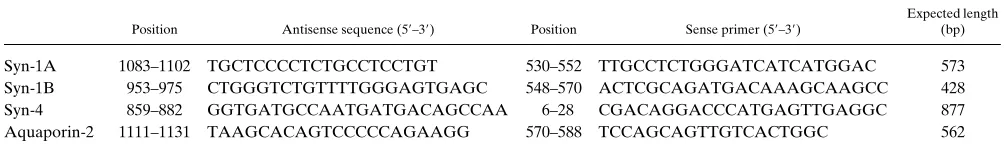
Primers corresponding to syntaxin-1A, syntaxin-1B, and syntaxin-4 were chosen (Table I) with the aid of the computer program OLIGO 5.0 (National Biosciences, Plymouth, MN). The chief criteria were: specificity, Tm close to 608C, lack of predicted internal structure. In
general the sense primers were located in the coding region whereas the antisense primers were in the 39-untranslated region. RT-PCR re-actions were performed as described previously (23). Briefly, RT was carried out with the antisense primer (6.25 pmol) in a 50-ml reaction volume containing 20 mM Tris-HCl (pH 8.3), 50 mM KCl, 3.5 mM MgCl2, 200 mM of each deoxynucleotide triphosphate, 6.4 mM DTT,
0.1 mg/ml gelatin, and 200 U of MMLV reverse transcriptase (Life Technologies, Gaithersburg, MD). Each reaction used 2 mg of total
RNA. After completion of RT (45 min, 428C), the temperature was raised to 968C for 30 s to inactivate the enzyme and denature the RNA-cDNA hybrids, then lowered to 808C. PCR was initiated by adding 50 ml of a mix containing 20 mM Tris-HCl (pH 8.3), 50 mM KCl, 0.1 mg/ml gelatin, 1.25 U of Taq polymerase (Perkin-Elmer Corp., Norwalk, CT) and the appropriate sense primer (6.25 pmol). In addition, either 2.5 or 3.5 mM MgCl2 was used as designated in the
figure captions. The samples were overlaid with mineral oil and pro-cessed for 31 cycles (968C for 30 s; 59–618C, depending on the prim-ers, for 30 s; 748C for 1 min). At the end of the last cycle the elonga-tion time at 748C was extended to 10 min. 10 ml of each PCR product was electrophoresed using 1.5% agarose gels, stained with ethidium bromide, destained, and photographed.
RT-PCR in isolated renal tubule segments. Renal tubule segments were microdissected as described (24) and RT-PCR was performed as described previously (21, 25) and as summarized briefly in the fol-lowing. The rats were killed by decapitation. Quickly, the left kidney was perfused with ice-cold dissection solution (135 mM NaCl, 0.5 mM KCl, 0.1 mM Na2HPO4, 0.3 mM NaC3COONa, 0.12 mM Na2SO4, 2.5
mM CaCl2, 1.2 mM MgSO4, 5 mM Hepes, and 5.5 mM glucose, at pH
7.4) and followed by 5 ml of collagenase-hyaluronidase solution (10 mg collagenase, and 5,000 U hyaluronidase, 3 mg BSA in 5 ml of dis-section solution). Then, the kidney was removed, sliced, and incu-bated in the collagenase-hyaluronidase solution at 378C with con-tinuous oxygenation for 10–40 min. The different segments were microdissected freehand using Dumont No. 5 forceps in dissection so-lution containing 1:40 vanadyl ribonucleoside complex (VRC), a ri-bonuclease inhibitor. 1–2-mm lengths of these microdissected seg-ments were washed in dissection solution with added 0.1% BSA but without VRC, and then transferred with 2 ml of the dissection solu-tion with 0.1% BSA into PCR tubes containing 6.7 ml of Triton X-100 solution (mix of 96 ml 2% Triton X-100, 3.5 ml RNase placental inhib-itor [Boehringer-Mannheim], and 0.5 ml 1 M DTT). Finally, the sam-ples were frozen on dry ice and then thawed before RT-PCR was ini-tiated. To monitor for RNA or cDNA contamination of reagents, blanks were included in each assay with 2 ml of the wash solution in lieu of tubules.
We used the same specific primers described above (Table I). Primers specific for the aquaporin-2 water channel were also used in some reactions with collecting ducts to provide a positive control (Table I). 11.3 ml of the following mix was added to each PCR tube: 4 ml of 53
Boehringer-Mannheim incubation buffer, 50 U RNase inhibitor, 20 nmol of each deoxynucleotide, 1.6 mg of poly(dT)15, and 50 U of AMV
reverse transcriptase (Boehringer-Mannheim). The RT was carried out for 60 min at 428C, followed by 5 min at 958C. The tubes were then placed on ice and 80 ml of the following mix was added to each tube: 10 ml of 103 Perkin-Elmer reaction buffer, 200 mmol of each deoxy-nucleotide, 50 pmol of sense and antisense specific primers, and 2.5 U DNA polymerase (AmpliTaq; Perkin-Elmer). The samples were over-laid with mineral oil and processed for 31 cycles (948C, 1 min [3 min for the initial cycle]; 59–618C according to the primers, 1 min; 728C, 1 min). The elongation period in the last cycle was extended to 7 min. 90 ml of the PCR product was precipitated by adding 1/10 vol of 3 M Na
ace-Table I. Oligonucleotide Primers Used in This Study
Position Antisense sequence (59–39) Position Sense primer (59–39)
Expected length (bp)
Syn-1A 1083–1102 TGCTCCCCTCTGCCTCCTGT 530–552 TTGCCTCTGGGATCATCATGGAC 573 Syn-1B 953–975 CTGGGTCTGTTTTGGGAGTGAGC 548–570 ACTCGCAGATGACAAAGCAAGCC 428 Syn-4 859–882 GGTGATGCCAATGATGACAGCCAA 6–28 CGACAGGACCCATGAGTTGAGGC 877 Aquaporin-2 1111–1131 TAAGCACAGTCCCCCAGAAGG 570–588 TCCAGCAGTTGTCACTGGC 562
[image:4.612.55.557.76.155.2]Positions of the primers are numbered from the ATG initiation codon of the syntaxin cDNA sequences reported by Bennett et al. (14) and of the AQP-2 cDNA sequence reported by Fushimi et al. (33).
[image:4.612.57.304.485.742.2]tate (pH 5.2) and 2 vol of 100% ethanol. The pellet were washed with 70% ethanol. The samples were resuspended in 10 ml of TE (10 mM Tris, 1 mM EDTA, pH 7.5), electrophoresed on a 1% agarose gel, stained with ethidium bromide, destained, and photographed as de-scribed above. Selected products were sequenced commercially by a combination of direct cycle sequencing and standard sequencing of the cloned PCR product (Lofstrand Laboratories, Gaithersburg, MD) to confirm the identity of the amplified cDNA.
Results
Syntaxin-4 protein is expressed in renal collecting duct. We
pre-pared a rabbit polyclonal antibody to a 23–amino acid thetic peptide corresponding to the amino terminus of rat
syn-taxin-4. This peptide was chosen to produce an antibody with a high degree of specificity for syntaxin-4 (see Discussion). Fig. 1 shows immunoblots run with crude membrane fractions pre-pared from rat IMCD cell suspensions and probed with the af-finity-purified anti–syntaxin-4 (left), with the same antibody preadsorbed with an excess of the immunizing peptide ( mid-dle), and with the IgG fraction from the preimmune serum
(right). The antibody labeled a distinct 36-kD band, consistent
[image:5.612.57.297.58.288.2]with the expected molecular mass of syntaxin-4 (14). This band was not seen with the peptide-adsorbed antibody or with the preimmune IgG.
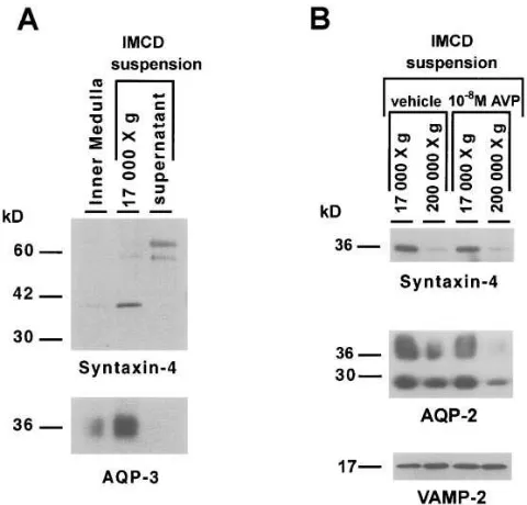
Fig. 2 A shows immunoblots prepared from SDS-poly-acrylamide gels loaded with a homogenate from the whole in-ner medulla and with membrane fractions from an IMCD cell suspension. These were probed with the affinity-purified anti-body to syntaxin-4 (upper blot) and an affinity-purified poly-clonal antibody to aquaporin-3, a plasma membrane marker for collecting duct principal cells (19) (lower blot). As shown, there was a parallel enrichment of syntaxin-4 and aquaporin-3 in the 17,000 g fraction from IMCD cells relative to whole in-ner medulla, suggesting that syntaxin-4 is present in the plasma membranes of collecting duct cells.
Further differential centrifugation experiments were car-ried out using IMCD cell suspensions to assess the distribution of syntaxin-4 immunoreactivity between a plasma membrane– enriched low-speed (LS) membrane fraction and a high-speed (HS) membrane fraction, enriched in intracellular vesicles (5) (Fig. 2 B). Syntaxin-4 (upper blot) appears to be abundant in the plasma membrane–enriched LS fraction, while little syn-taxin-4 is present in the HS membrane fraction, which contains most of the aquaporin-2–bearing intracellular vesicles (5). In contrast, aquaporin-2 was abundant in both fractions (middle blot). As previously reported (5), vasopressin shifts aquaporin-2 out of the HS fraction, presumably as a result of stimulation of exocytosis of aquaporin-2–bearing vesicles. However, vaso-pressin did not affect the distribution of syntaxin-4 immunore-activity between the HS and LS fractions. As shown, in the lower blot in Fig. 2 B, VAMP-2 was also present in membrane fractions from IMCD cell suspensions. In contrast to syntaxin-4, VAMP-2 was predominantly found in the HS fraction. How-ever, a significant amount of VAMP-2 was also present in the LS fraction, suggesting either that VAMP-2 may be present in plasma membrane or, more likely, that the LS fraction con-tains a substantial amount of non–plasma membrane mem-brane protein including VAMP-2–bearing intracellular vesi-cles. Unlike AQP-2, VAMP-2 was not decreased in the HS fraction in response to vasopressin.
To determine which plasma membrane domain contains syntaxin-4, we carried out immunoperoxidase labeling in thin cryosections of rat inner medulla (Fig. 3). The anti–syntaxin-4 antibody gave discrete labeling of the apical plasma membrane domain of the IMCD cells as seen both in the longitudinal sec-tion and the cross-secsec-tion (Fig. 3, inset). There was no percep-tible labeling of the basolateral plasma membrane. A control using preimmune IgG gave no labeling. This demonstrates that the apical plasma membrane labeling is specific for syntaxin-4 and is not associated with the weak higher molecular mass band seen in the 17,000 g supernatant (Fig. 2) which was present when blots were probed with preimmune serum. Therefore, these results point to the presence of syntaxin-4 in the apical plasma membrane, the target membrane for aqua-porin-2 water channel vesicles.
Syntaxin-4 mRNA is expressed in renal collecting duct. To further establish the presence of syntaxin-4 expression in the renal collecting duct, we examined whether syntaxin-4 mRNA is present in collecting duct cells. RT-PCR experiments were carried out in microdissected renal tubule segments (Fig. 4). Collecting ducts from all three renal regions (CCD, OMCD, and IMCD) appear to express syntaxin-4 mRNA. Restriction analysis of the PCR product using Pst1 gave products of sizes consistent with those predicted for syntaxin-4 (not shown). Di-rect sequencing of the 877-bp product yielded a sequence which was . 99% identical to that reported for rat syntaxin-4 (14), demonstrating that the amplified cDNA in collecting ducts indeed corresponds to syntaxin-4 rather than to some other syntaxin isoform. Therefore, this result confirms the con-clusion from immunoblotting and immunolocalization studies that syntaxin-4 is expressed in collecting duct principal cells. Parallel amplifications using primers for rat aquaporin-2 pro-vided a positive control for collecting ducts, confirming that the collecting duct segments were correctly identified in the microdissection. Controls run without added tissue were uni-formly negative, ruling out the possibility that the signal de-rives from cDNA contamination of reagents or pipettes.
Con-trols run on collecting ducts without the RT step were negative, indicating that the syntaxin-4 signal derives from mRNA present in the collecting duct cells rather than from ge-nomic DNA. In addition to substantial levels in collecting ducts, relatively high levels of syntaxin-4 mRNA expression were also seen in glomeruli, vasa recta, thin descending limbs, cortical thick ascending limbs, and connecting tubules. There-fore, these results indicate that syntaxin-4 expression in the kidney is not limited to collecting ducts.
Distribution of syntaxin-4 protein and mRNA among
non-renal tissues. Immunoblots were also run to assess the
[image:6.612.57.403.58.472.2]distri-bution of syntaxin-4 immunoreactivity among nonrenal tis-sues. As shown in Fig. 5, syntaxin-4 was present in membrane fractions (17,000 g pellets) from lung, liver, brain, and heart. These results indicate that syntaxin-4 protein expression is broadly distributed among epithelial and nonepithelial tissues. Fig. 6 shows the results of RT-PCR experiments using total RNA isolated from several tissues. Syntaxin-4 mRNA was de-tectable in lung, liver, brain, heart, and kidney, confirming the conclusion that syntaxin-4 mRNA is broadly expressed (14). Substantial syntaxin-4 signals were obtained in all three re-gions of the kidney, namely cortex, outer medulla, and inner
medulla. In parallel experiments using primers for syntaxin-1A and syntaxin-1B (not shown), a detectable band was found only in total RNA samples from the brain.
Discussion
Vasopressin (the “antidiuretic hormone”), acting via cAMP, increases the osmotic water permeability of the collecting duct principal cells by stimulating the translocation of aquaporin-2– containing intracellular vesicles to the apical plasma mem-brane (3–6). The resulting vesicular fusion adds new aqua-porin-2 water channels to the apical plasma membrane and
[image:7.612.57.291.58.371.2]thereby increases its water permeability. As is true for regu-lated exocytosis in other tissues, important questions remain with regard to the mechanisms by which aquaporin-2–contain-ing vesicles are targeted to a specific membrane domain and with regard to the mechanisms by which vesicle docking and fusion are regulated. Investigation of regulated exocytosis of synaptic vesicles has recently led to a new paradigm, the so-called “SNARE hypothesis” (7), which may be useful in the analysis of regulated exocytosis in the renal collecting duct. A component of this hypothesis is the concept that specificity of vesicular targeting may be mediated by a group of membrane-associated proteins which behave as vesicle-targeting receptors
[image:7.612.316.554.79.236.2]Figure 4. RT-PCR determination of relative distribution of syntax-in-4 mRNA (877 bp) in microdissected collecting ducts and other re-nal structures. After RT-PCR was run for 31 cycles, products were electrophoresed on 1.5% agarose gels which were stained with ethid-ium bromide as described in Methods. Most reactions were run in du-plicate or tridu-plicate as shown. Tissue substrate amounts for syntaxin-4 reactions: glomerulus, four tufts per sample; proximal convoluted tu-bule (PCT), 2.2 and 2.0 mm; cortical thick ascending limb (cTAL), 1.9 and 1.7 mm; distal convoluted tubule (DCT), 2.0 and 0.6 mm; con-necting tubule (CNT), 1.8 mm; CCD, 2.1 and 2.0 mm; vasa recta, one outer medullary vascular bundle per sample; thin descending limb type 1 (TDL-I), 2.0 and 2.5 mm; TDL type 2 (TDL-II), 2.4 and 2.5 mm; medullary thick ascending limb (mTAL), 2.2 and 2.8 mm; OMCD, 2.2 and 2.2 mm; inner medullary TDL type 3 (TDL-III), 3.2 and 2.8 mm; thin ascending limb (tAL), 2.8 and 3.2 mm; IMCD, 1.3, 1.3, and 1.4 mm. In addition, parallel reactions were run using primers for aquaporin-2 (AQP-2) in microdissected collecting ducts (562-bp product) to confirm identification of segments and success of reverse transcription step.
Figure 5.Distribution of syntaxin-4 immunoreactivity among various tissues. Membrane fractions (17,000 g pellets) from lung, liver, brain, and heart were loaded on 12% polyacrylamide gels at 10 mg protein per lane. Blot was probed with affinity-purified anti–syntaxin-4 poly-clonal antibody (IgG concentration, 0.56 mg/ml).
Figure 6.Distribution of syntaxin-4 mRNAs among various tissues as determined by RT-PCR. For each organ, 2 mg of total RNA was loaded into each tube. Amplifications were carried out using either 2.5 mM of Mg21 (left lane of each pair) or 3.5 mM Mg21 (right lane of
[image:7.612.317.553.481.653.2]or SNAREs. These proteins are proposed to be associated with the vesicular membrane (vesicle-SNAREs or v-SNAREs) or with the target membrane (t-SNAREs). In this paper, we have demonstrated that a particular t-SNARE protein, syntaxin-4, is expressed in collecting duct principal cells and that its distri-bution within the cell is compatible with a role in targeting of aquaporin-2–containing vesicles to the apical plasma membrane. The syntaxins are a family of integral membrane proteins of z 300 amino acids with a single membrane-spanning region near the carboxy terminus (14). In contrast to the archetypal member of the family, syntaxin-1, which is expressed chiefly in the central nervous system, syntaxin-4 is believed to be ex-pressed in a variety of tissues including the kidney (14). Based on indirect immunofluorescence microscopy of transfected COS cells, syntaxin-4 is thought to be localized predominantly in the plasma membrane (14) where it has been proposed to be involved in targeting of secretory vesicles to the plasma mem-brane. In the current study, both subcellular fractionation ex-periments using differential centrifugation (Fig. 2) and immu-noperoxidase localization (Fig. 3) support the view that syntaxin-4 is expressed chiefly in the plasma membrane of col-lecting duct cells. Furthermore, the immunoperoxidase label-ing was restricted to the apical plasma membrane with no evi-dence of basolateral localization. Thus, the localization is fully compatible with a possible role in the targeting of aquaporin-2 vesicles to the apical plasma membrane in response to vasopres-sin. In previous studies we have demonstrated that a v-SNARE, VAMP2, is present in large amounts in aquaporin-2 vesicles (10), a finding also supported by Jo et al. (11) and Liebenhoff et al. (12). In vitro binding studies (15, 16) have demonstrated that, although VAMP2 does not bind with high affinity to syn-taxin-2 and syntaxin-3, two other broadly expressed homo-logues, it does bind syntaxin-4 with relatively high affinity. Hence, in analogy to VAMP–syntaxin interactions that are proposed to participate in targeting of synaptic vesicles to the active zone of the presynaptic plasma membrane, we propose that VAMP2 and syntaxin-4 may fulfill a similar function in collecting duct cells to direct aquaporin-2 to the appropriate membrane domain.
Recent studies of syntaxin localization in neurons have raised doubts about the role of syntaxin as a determinant of specificity of targeting of synaptic vesicles to the active zone. Specifically, several investigators have demonstrated that im-munoreactive syntaxin is present, not only in the active zone membrane, but throughout the axon (26–28). Thus, it appears that syntaxin may lack the unique localization in the neuron to allow a role in determining specificity of synaptic vesicle tar-geting. It appeared to us that one possible explanation for the broad distribution of immunoreactive syntaxin in the neuron could be that more than one syntaxin isoform could be recog-nized by the antibodies used for immunolocalization. These antibodies were raised to bacterial fusion proteins which may exhibit sequence overlap with other syntaxins. To avoid this problem, we chose a site-directed approach designed to maxi-mize antibody specificity by immunizing with a synthetic pep-tide which lacks significant sequence overlap with other known syntaxins. The resulting antibody, after affinity purifi-cation, recognized a solitary band on immunoblots of renal in-ner medullary membranes of apparent molecular mass of 36,000 D (Fig. 1), consistent with the expected size of syntaxin-4 (14). This band was not present on blots probed with the pre-immune IgG fraction or with the anti–syntaxin-4 antibody
after preadsorption with the immunizing peptide. Both immu-noblotting of membrane proteins from IMCD cell suspensions and immunocytochemistry demonstrated that this protein is present in collecting duct cells. Furthermore, RT-PCR in mi-crodissected collecting ducts, followed by sequencing of the PCR product, confirmed the conclusion that syntaxin-4 is ex-pressed in collecting duct cells.
Aside from the collecting duct, we found evidence from RT-PCR experiments that syntaxin-4 mRNA is expressed in several other renal structures (Fig. 4). Constitutive membrane trafficking is a vital process in all cells and, consequently, broad distribution of syntaxins and other SNARE proteins is not an unexpected finding, assuming that the SNARE mecha-nism may be involved in both constitutive and regulated traf-ficking. Furthermore, in addition to vasopressin-regulated wa-ter channel trafficking in collecting duct cells, there are a number of other examples of regulated exocytosis in the kid-ney, e.g., adrenergic regulation of renin secretion and oxygen-regulated erythropoietin secretion, that may use similar mech-anisms of vesicular targeting.
Bennett et al. (14) observed by Northern blot analysis of various tissues that several of the syntaxins, including syntaxin-4, are widely distributed among tissues. Our results are in accord with that view. Both immunoblotting and RT-PCR experi-ments provided evidence for expression of syntaxin-4 in a vari-ety of tissues including lung, liver, heart, and brain (Figs. 5 and 6). In addition, several recent reports have documented the presence of either syntax4 or its mRNA in several tissues in-cluding liver (29), pancreatic islets (30), gastric mucosa (31), and skeletal muscle (32).
In summary, we have demonstrated that at least one mem-ber of the syntaxin family, namely syntaxin-4, is expressed in the principal cells of the renal collecting duct, the site of vaso-pressin-regulated water transport in the kidney. Furthermore, immunoperoxidase labeling of thin cryosections showed that, within the principal cells, syntaxin-4 is localized to the apical plasma membrane. Thus, our findings have established that both VAMP2 (10) and syntaxin-4 (this paper) are in appropri-ate membrane domains of collecting duct cells to be involved in targeting of aquaporin-2–containing vesicles to the apical plasma membrane. These results, therefore, provide further support for the hypothesis that the SNARE mechanism could be responsible for the specificity of water channel trafficking to the apical plasma membrane in collecting duct cells.
Acknowledgments
We thank Hanne Weiling for excellent technical assistance.
Funding for this paper was derived from the intramural budget of the Heart, Lung, and Blood Institute of the National Institutes of Health, project number ZO1-HL-01282-KE (M.A. Knepper), Novo Nordic Foundation (S. Nielsen), and the Danish Medical Research Council (S. Nielsen).
References
1. Knepper, M.A., and F.C. Rector, Jr. 1995. Urinary concentration and di-lution. In The Kidney. B.M. Brenner and F.C. Rector, Jr., editors. W.B. Saun-ders Co., Philadelphia. 532–570.
2. Knepper, M.A., S. Nielsen, C.-L. Chou, and S.R. DiGiovanni. 1994. Mechanism of vasopressin action in the renal collecting duct. Semin. Nephrol.
14:302–321.
col-lecting duct by inducing translocation of aquaporin-CD water channels to plasma membrane. Proc. Natl. Acad. Sci. USA. 92:1013–1017.
4. Sabolic, I., T. Katsura, J.M. Verbabatz, and D. Brown. 1995. The AQP2 water channel: effect of vasopressin treatment, microtubule disruption, and dis-tribution in neonatal rats. J. Membr. Biol. 143:165–177.
5. Marples, D., M.A. Knepper, E.I. Christensen, and S. Nielsen. 1995. Re-distribution of aquaporin-2 water channels induced by vasopressin in rat kidney inner medullary collecting duct. Am. J. Physiol. 269:C655–C664.
6. Yamamoto, N., S. Sasaki, K. Fushimi, K. Ishibashi, E. Yaiota, K. Ka-wasaki, F. Marumo, and I. Kihara. 1995. Vasopressin increases AQP-CD water channel in the apical membrane of collecting duct cells without affecting AQP3 distribution in Brattleboro rat. Am. J. Physiol. 268:C1546–C1551.
7. Söllner, T., S.W. Whiteheart, M. Brunner, H. Erdjument-Bromage, S. Germanos, P. Tempst, and J.E. Rothman. 1993. SNAP receptors implicated in vesicle targeting and fusion. Nature (Lond.). 362:318–324.
8. Südhoff, T.C., P. De Camilli, H. Niemann, and R. Jahn. 1993. Membrane fusion machinery: insights from synaptic proteins. Cell. 75:1–4.
9. Bajjalieh, S.M., and R.H. Scheller. 1995. The biochemistry of neurotrans-mitter secretion. J. Biol. Chem. 270:1971–1974.
10. Nielsen, S., D. Marples, M. Mohtashami, N.O. Dalby, W. Trimble, and M. Knepper. 1995. Expression of VAMP2-like protein in kidney collecting duct intracellular vesicles: colocalization with aquaporin-2 water channels. J. Clin. Invest. 96:1834–1844.
11. Jo, I., H.W. Harris, A.M. Amendt-Raduege, R.R. Majewski, and T.G. Hammond. 1995. Rat kidney papilla contains abundant synaptobrevin protein that participates in the fusion of antidiuretic hormone-regulated water channel-containing endosomes in vitro. Proc. Natl. Acad. Sci. USA. 92:1876–1880.
12. Liebenhoff, U., and W. Rosenthal. 1995. Identification of Rab3-, Rab5a and synaptobrevin II-like proteins in a preparation of rat kidney vesicles con-taining the vasopressin-regulated water channel. FEBS Lett. 365:209–213.
13. Franki, N., F. Macaluso, W. Schubert, L. Gunther, and R.M. Hays. 1995. Water channel-carrying vesicles in the rat IMCD contain cellubrevin. Am. J. Physiol. 269:C797–C801.
14. Bennett, M.K., J.E. Garcia-Arraras, L.A. Elferink, K. Peterson, A.M. Fleming, C.D. Hazuka, and R.H. Scheller. 1993. The syntaxin family of vesicu-lar transport receptors. Cell. 74:863–873.
15. Calakos, N., M.K. Bennett, K.E. Peterson, and R.H. Scheller. 1994. Pro-tein-protein interactions contributing to the specificity of intracellular vesicular trafficking. Science (Wash. DC). 263:1146–1149.
16. Pevsner, J., S.-C. Hsu, J.E.A. Braun, N. Calakos, A.E. Ting, M.K. Ben-nett, and R.H. Scheller. 1994. Specificity and regulation of a synaptic vesicle docking complex. Neuron. 13:353–361.
17. Elferink, L.A., W.S. Trimble, and R.H. Scheller. 1989. Two vesicle-asso-ciated membrane protein genes are differentially expressed in the rat central nervous system. J. Biol. Chem. 264:11061–11064.
18. DiGiovanni, S.R., S. Nielsen, E.I. Christensen, and M.A. Knepper. 1994. Regulation of collecting duct water channel expression by vasopressin in Brat-tleboro rat. Proc. Natl. Acad. Sci. USA. 91:8984–8988.
19. Ecelbarger, C.A., J. Terris, G. Frindt, M. Echevarria, D. Marples, S. Nielsen, and M.A. Knepper. 1995. Aquaporin-3 water channel localization and regulation in rat kidney. Am. J. Physiol. 269:F663–F672.
20. Stokes, J.B., C. Grupp, and R.K.H. Kinne. 1987. Purification of rat pap-illary collecting duct cells: functional and metabolic assessment. Am. J. Physiol.
253:F251–F262.
21. Chou, C.-L., S.R. DiGiovanni, A. Luther, S.J. Lolait, and M.A. Knepper. 1995. Oxytocin as an antidiuretic hormone. II. Role of V2 vasopressin receptor. Am. J. Physiol. 268:F78–F85.
22. Chomczynski, P., and N. Sacchi. 1987. Single-step method of RNA isola-tion by acid guanidinium thiocyanate-phenol-chloroform extracisola-tion. Anal. Bio-chem. 162:156–159.
23. Elalouf, J.M., J.M. Buhler, C. Tessiot, A.C. Bellanger, I. Dublineau, and C. de Rouffignac. 1993. Predominant expression of b1-adrenergic receptor in thick ascending limb of rat kidney. Absolute mRNA quantitation by reverse transcription and polymerase chain reaction. J. Clin. Invest. 91:264–272.
24. Wright, P.A., M.B. Burg, and M.A. Knepper. 1990. Microdissection of kidney tubule segments. In Methods in Enzymology. S. Fleischer and B. Fleis-cher, editors. Academic Press, San Diego. 191–231.
25. Terada, Y., T. Moriyama, B.M. Martin, M.A. Knepper, and A. Garcia-Perez. 1991. RT-PCR localization of mRNA for guanylyl cyclase-coupled ANF receptor in rat kidney. Am. J. Physiol. 261:F1080–F1087.
26. Koh, S., A. Yamamoto, A. Inoue, Y. Inoue, K. Akagawa, Y. Kawamura, K. Kawamoto, and Y. Tashiro. 1993. Immunoelectron microscopic localization of the HPC-1 antigen in rat cerebellum. J. Neurocytol. 22:995–1005.
27. Sesack, S.R., and C.L. Snyder. 1995. Cellular and subcellular localiza-tion of syntaxin-like immunoreactivity in the rat striatum and cortex. Neuro-science. 67:993–1007.
28. Garcia, E.P., P.S. McPherson, T.J. Chilcote, K. Takei, and P. De Cam-illi. 1995. rbSec1A and B colocalize with syntaxin 1 and SNAP-25 throughout the axon, but are not in a stable complex with syntaxin. J. Cell Biol. 129:105– 120.
29. Fujita, H., L. Locco, and A.L. Hubbard. 1995. Localization and quanti-tation of syntaxin 2 and 4 in rat liver and WIF-B cells. Mol. Biol. Cell. 6:183a. (Abstr.)
30. Jacobsson, G., A.J. Bean, R.H. Scheller, L. Juntti-Berggren, J.T. Deeney, P.O. Berggren, and B. Meister. 1995. Identification of synaptic pro-teins and their isoform mRNAs in compartments of pancreatic endocrine cells.
Proc. Natl. Acad. Sci. USA. 91:12487–12491.
31. Scott, D.R., S.J. Hersey, H.G. Helander, and G. Sachs. 1995. Localiza-tion of syntaxin in the chief cell of the rabbit gastric mucosa. Mol. Biol. Cell. 6: 183a. (Abstr.)
32. Sumitani, S., T. Ramlal, Z. Liu, and A. Klip. 1995. Expression of syn-taxin 4 in rat skeletal muscle and rat skeletal muscle cells in culture. Biochem. Biophys. Res. Commun. 213:462–468.