Copyright © 1998, American Society for Microbiology. All Rights Reserved.
Rapid Identification of Candida Species with
Species-Specific DNA Probes
CHERYL M. ELIE, TIMOTHY J. LOTT, ERROL REISS, ANDCHRISTINE J. MORRISON*
Division of Bacterial and Mycotic Diseases, National Center for Infectious Diseases, Centers for Disease Control and Prevention, Atlanta, Georgia
Received 30 March 1998/Returned for modification 26 May 1998/Accepted 7 August 1998
Rapid identification of Candida species has become more important because of an increase in infections caused by species other than Candida albicans, including species innately resistant to azole antifungal drugs. We previously developed a PCR assay with an enzyme immunoassay (EIA) format to detect amplicons from the five most common Candida species by using universal fungal primers and species-specific probes directed to the ITS2 region of the gene for rRNA. We designed probes to detect seven additional Candida species (C. guilli-ermondii, C. kefyr, C. lambica, C. lusitaniae, C. pelliculosa, C. rugosa, and C. zeylanoides) included in the API 20C sugar assimilation panel, five probes for species not identified by API 20C (C. haemulonii, C. norvegica, C. nor-vegensis, C. utilis, and C. viswanathii), and a probe for the newly described species C. dubliniensis, creating a panel of 18 Candida species probes. The PCR-EIA correctly identified multiple strains of each species tested, including five identified as C. albicans by the currently available API 20C database but determined to be C. dubliniensis by genotypic and nonroutine phenotypic characteristics. Species identification time was reduced from a mean of 3.5 days by conventional identification methods to 7 h by the PCR-EIA. This method is simple, rapid, and feasible for identifying Candida species in clinical laboratories that utilize molecular identification techniques and provides a novel method to differentiate the new species, C. dubliniensis, from C. albicans.
Rapid identification of Candida isolates to the species level in the clinical laboratory has become more important because the incidence of candidiasis continues to rise in proportion to a growing number of patients at risk for infection with Candida albicans and, recently, with innately azole-resistant non-albi-cans Candida species (5, 7, 29). This patient population has in-creased as a result of more intensive regimens of cancer ther-apy, complications of abdominal or cardiothoracic surgery, organ transplantations, burns, and trauma. Affected patients may be immunocompromised or not, and common risk factors include prolonged broad-spectrum antibiotic therapy, invasive devices such as indwelling Hickman catheters, and/or pro-longed hospital stays (5, 7, 26). Under these conditions, an antibiotic-resistant replacement flora, including Candida spe-cies, can proliferate in the gut and invade deep tissues from mucosal foci. This is especially the case when mucosal integrity has been disrupted as a result of chemotherapy or surgery. In addition, as the number of risk factors increases, the odds of developing candidiasis multiply (26). Some Candida species, including C. glabrata and C. krusei, are emerging, possibly be-cause they are innately less susceptible to azole drugs (16, 18, 29). In the case of C. parapsilosis, its ability to survive in the hospital environment, i.e., on the hands of healthcare workers, on intravenous devices, and in solutions, increases the possi-bility of its nosocomial transmission (5, 16). Consequently, rapid identification to the species level is necessary for more timely, targeted, and effective antifungal therapy and to facil-itate hospital infection control measures.
Identification of Candida species by conventional morphol-ogy and assimilation tests can require 3 to 5 days or even longer for more difficult or unusual species (25). We
previ-ously employed universal fungal primers, multicopy gene targets, and species-specific probes directed to the ITS2 region of the rRNA-encoding gene (rDNA) to develop a rapid (1-day) PCR assay to detect candidemia (6, 20). Amplicons were detected in an enzyme immunoassay (EIA) format, and the method was referred to as PCR-EIA. Since the API 20C carbohydrate assimilation panel is limited to the identification of only certain species, DNA probes were designed to detect a total of 18 Candida species. Of these, the following 12 species can be identified by the current API 20C panel: C. albicans, C. glabrata, C. guilliermondii, C. kefyr, C. krusei, C. lambica, C. lusitaniae, C. parapsilosis, C. pelliculosa, C. rugosa, C. tropi-calis, and C. zeylanoides. Five other, newly emerging Candida species, not identified by API 20C but readily identified by molecular probes, are C. haemulonii, C. norvegensis, C. nor-vegica, C. utilis, and C. viswanathii. A species-specific probe for the newly described Candida species, C. dubliniensis, was also designed. The resulting PCR-EIA identification matrix is simple, rapid, sensitive, and feasible for identifying Candida species in clinical laboratories.
MATERIALS AND METHODS
Microorganisms.Clinical isolates or cultures obtained from the American Type Culture Collection (ATCC) were used in this study (see Tables 2 and 5). Isolates of Candida spp., Cryptococcus humicolus, Stephanoascus ciferrii, and
Trichosporon cutaneum were grown in 50-ml Erlenmeyer flasks by seeding one
10-ml loopful of growth from an agar slant into 10 ml of YPD broth (1% yeast extract, 2% Bacto Peptone, 2% dextrose; Difco Laboratories, Detroit, Mich.).
Cryptococcus neoformans serotypes A, B, C, and D were grown similarly;
how-ever, YPD broth was supplemented with 2.9% NaCl to reduce capsule formation. All broth cultures were grown at 35°C for 18 h in a rotary shaker set at 150 rpm prior to DNA extraction for prototype testing.
DNA isolation.DNA was extracted from all yeast species by using the Pure-gene DNA Isolation Kit (Gentra Systems Inc., Minneapolis, Minn.). This kit facilitates the rapid recovery of sufficient DNA for PCR amplification and allows multiple samples to be extracted in parallel. For example, multiple yeast isolates could be extracted at the same time so that a large number of samples could be processed quickly and efficiently on a given day. DNAs from filamentous and dimorphic fungi were obtained as previously described (6) or were a gift from * Corresponding author. Mailing address: Mailstop G-11, Centers
for Disease Control and Prevention, Atlanta, GA 30333. Phone: (404) 639-3128. Fax: (404) 639-3546. E-mail: cjm3@cdc.gov.
3260
on May 15, 2020 by guest
http://jcm.asm.org/
Liliana de Aguirre, Instituto Investigaciones Veterinarias, Maracay, Venezuela. Quantification of DNA was performed by using a fluorometer and Hoechst 33258 Dye (Dyna Quant 200; Pharmacia Biotech, Piscataway, N.J.). DNA was diluted in TE buffer (10 mM Tris, 1 mM EDTA [pH 8.0]) so that a total of 1 ng of template DNA was added to each PCR vial.
Oligonucleotide synthesis of primers and probes.Oligodeoxyribonucleotide primers and probes were synthesized as described previously (6). Universal fun-gal primers ITS3 and ITS4 (28) were used to amplify the ITS2 region. Oligonu-cleotide probes were designed from sequence data for the ITS2 region of the
Candida sp. rDNA (13, 14).
PCR amplification.The reaction mixture (100ml) contained 10ml of 103PCR buffer (100 mM Tris-HCl, 500 mM KCl [pH 8.3]; Boehringer Mannheim, Indi-anapolis, Ind.), 6ml of 25 mM MgCl2, 8ml of a deoxynucleotide triphosphate mixture (1.25 mM each dATP, dCTP, dGTP, and dTTP), 1ml of each primer (20mM), 2.5 U of Taq DNA polymerase (TaKaRa Shuzo Co., Ltd., Shiga, Japan), 2ml of template DNA (0.5 ng/ml), and sterile distilled water to bring the total volume to 100ml. Vials were placed in the heating block of a model 9600 thermal cycler (Perkin-Elmer, Emeryville, Calif.) equilibrated at 95°C. PCR amplification conditions were 5 min of denaturation at 95°C, followed by 30 cycles of 95°C for 30 s, 58°C for 30 s, and 72°C for 1 min. A final extension step of 72°C for 5 min was then conducted. Appropriate positive and negative controls were included, and PCR contamination precautions were followed (6, 9).
Agarose gel electrophoresis.Electrophoresis was conducted in TBE (0.1 M Tris, 0.09 M boric acid, 0.001 M EDTA [pH 8.4]) buffer at 76 V for approxi-mately 1 h in gels composed of 1% (wt/vol) agarose (Boehringer Mannheim) and 1% (wt/vol) NuSieve (FMC Bioproducts, Rockland, Maine). Gels were stained with 0.5mg of ethidium bromide per ml of deionized water for 30 min, followed by a 30-min wash in deionized water. DNA bands confirming a positive PCR were visualized with a UV transilluminator and photographed.
PCR-EIA.PCR-amplified DNA was hybridized to species-specific digoxigenin-labeled probes and to a generic biotinylated probe, and then the complex was added to streptavidin-coated microtitration plates and captured as previously described (6, 20). A colorimetric EIA was then conducted to detect captured DNA by using horseradish peroxidase-conjugated anti-digoxigenin antibodies (6, 20) in a manner very similar to that of other EIAs performed routinely in many clinical microbiology laboratories. All probes were tested in a matrix format against DNA from other Candida species, as well as against DNAs from other fungi (see Table 3). In this manner, all probes were tested against all of the target DNAs so that fungi could be identified by a discrete pattern of reactivity. When a probe cross-reacted with heterologous DNA, probes specific to the heterolo-gous DNA were designed. Therefore, use of both probes as part of the matrix allows species-specific identification by a process of elimination and does not require additional steps or retesting of samples because all probes and all targets are included in the complete matrix from the beginning.
Statistical analyses.Student’s t test was used to determine significant differ-ences between mean absorbance values of homologous and nonhomologous
probe reactions. Differences were considered significant when the value of P was less than or equal to 0.05.
Nucleotide sequence accession numbers.The GenBank accession numbers for
C. dubliniensis and C. pelliculosa sequences are U96719 and U96720,
respec-tively. The accession numbers for the DNA sequences of the other Candida species used in this study are published in references 13 and 14.
RESULTS
Specificity of digoxigenin-labeled probes.Eighteen Candida species probes (Table 1) were designed and tested in the PCR-EIA against the DNAs from the fungi listed in Table 2. The absorbance value obtained for each probe tested against its homologous DNA was significantly greater than that obtained when probes were tested against nonhomologous DNA (P# 0.05), with two exceptions (Table 3). In the first instance, the probe for C. guilliermondii (GU) cross-reacted with DNA from C. zeylanoides (CZ). However, the probe for C. zeylanoides (CZ) did not cross-react with DNA from C. guilliermondii (GU). Therefore, by using both probes as part of the complete matrix, species-specific identification could be achieved by a process of elimination (Table 3). In the second instance, as previously described (6), the probe for C. glabrata, CG, cross-reacted with Saccharomyces cerevisiae DNA (mean absorbance, 0.882). In the present study, the CG probe also cross-reacted with C. pel-liculosa and C. utilis DNAs, which had not been tested previ-ously (mean absorbance6 standard deviation [SD], 0.8106 0.197 and 0.80060.648, respectively). Therefore, the CG probe was redesigned, resulting in the elimination of cross-reactions with all of the species tested except S. cerevisiae while retaining positive reactivity with homologous C. glabrata DNA (new CG probe named CGE, Table 3). Preliminary testing of an S. cere-visiae probe indicates that it does not cross-react with C. gla-brata DNA (data not shown), allowing the differentiation of C. glabrata DNA from S. cerevisiae DNA.
[image:2.612.54.549.81.316.2]All of the negative controls tested, except S. cerevisiae and C. zeylanoides, as mentioned above, gave mean optical density OD values (6 SD) ranging from 0.001 6 0.001 to 0.009 6
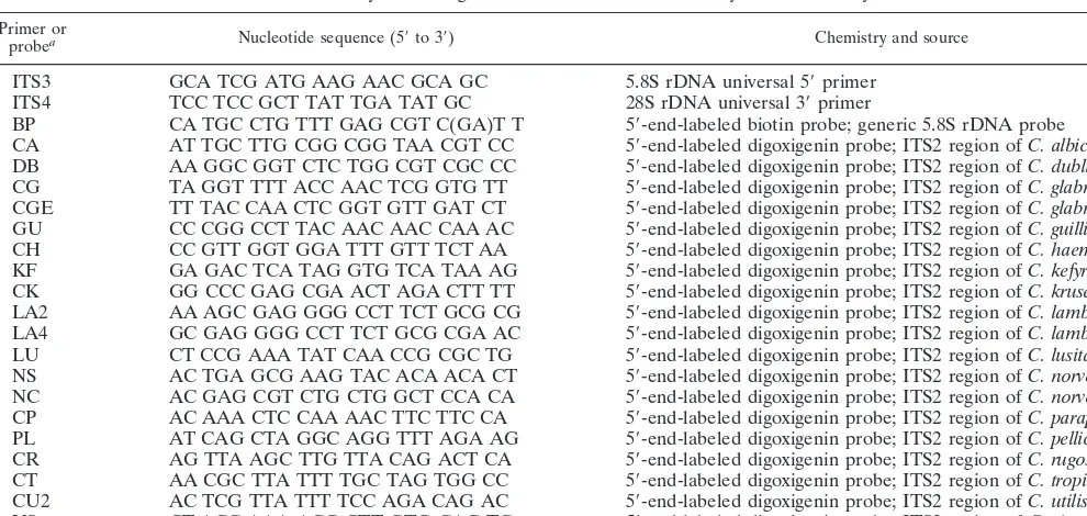
TABLE 1. Synthetic oligonucleotides used in PCR and hybridization analyses Primer or
probea Nucleotide sequence (59to 39) Chemistry and source
ITS3 GCA TCG ATG AAG AAC GCA GC 5.8S rDNA universal 59primer
ITS4 TCC TCC GCT TAT TGA TAT GC 28S rDNA universal 39primer
BP CA TGC CTG TTT GAG CGT C(GA)T T 59-end-labeled biotin probe; generic 5.8S rDNA probe CA AT TGC TTG CGG CGG TAA CGT CC 59-end-labeled digoxigenin probe; ITS2 region of C. albicans DB AA GGC GGT CTC TGG CGT CGC CC 59-end-labeled digoxigenin probe; ITS2 region of C. dubliniensis CG TA GGT TTT ACC AAC TCG GTG TT 59-end-labeled digoxigenin probe; ITS2 region of C. glabrata CGE TT TAC CAA CTC GGT GTT GAT CT 59-end-labeled digoxigenin probe; ITS2 region of C. glabrata GU CC CGG CCT TAC AAC AAC CAA AC 59-end-labeled digoxigenin probe; ITS2 region of C. guilliermondii CH CC GTT GGT GGA TTT GTT TCT AA 59-end-labeled digoxigenin probe; ITS2 region of C. haemulonii KF GA GAC TCA TAG GTG TCA TAA AG 59-end-labeled digoxigenin probe; ITS2 region of C. kefyr CK GG CCC GAG CGA ACT AGA CTT TT 59-end-labeled digoxigenin probe; ITS2 region of C. krusei LA2 AA AGC GAG GGG CCT TCT GCG CG 59-end-labeled digoxigenin probe; ITS2 region of C. lambica LA4 GC GAG GGG CCT TCT GCG CGA AC 59-end-labeled digoxigenin probe; ITS2 region of C. lambica LU CT CCG AAA TAT CAA CCG CGC TG 59-end-labeled digoxigenin probe; ITS2 region of C. lusitaniae NS AC TGA GCG AAG TAC ACA ACA CT 59-end-labeled digoxigenin probe; ITS2 region of C. norvegensis NC AC GAG CGT CTG CTG GCT CCA CA 59-end-labeled digoxigenin probe; ITS2 region of C. norvegica CP AC AAA CTC CAA AAC TTC TTC CA 59-end-labeled digoxigenin probe; ITS2 region of C. parapsilosis PL AT CAG CTA GGC AGG TTT AGA AG 59-end-labeled digoxigenin probe; ITS2 region of C. pelliculosa CR AG TTA AGC TTG TTA CAG ACT CA 59-end-labeled digoxigenin probe; ITS2 region of C. rugosa CT AA CGC TTA TTT TGC TAG TGG CC 59-end-labeled digoxigenin probe; ITS2 region of C. tropicalis CU2 AC TCG TTA TTT TCC AGA CAG AC 59-end-labeled digoxigenin probe; ITS2 region of C. utilis VS CT ACC AAA ACG CTT GTG CAG TC 59-end-labeled digoxigenin probe; ITS2 region of C. viswanathii CZ TC GTT GAC CAG TAT AGT ATT TG 59-end-labeled digoxigenin probe; ITS2 region of C. zeylanoides
aPatents have been issued or are pending for all Candida species probes.
on May 15, 2020 by guest
http://jcm.asm.org/
0.025 (Table 3). Minor cross-reaction was observed with the VS probe against C. tropicalis DNA (0.17560.093); however, the CT probe did not cross-react with C. viswanathii DNA (0.00160.001). Similarly, minor cross-reactions were observed when the CT and CU2 probes were used versus Aspergillus terreus (0.11460.052 and 0.14760.035, respectively), but an A. terreus-specific probe has been developed and has been reported separately (3). Finally, the CK probe demonstrated a minor cross-reaction with C. lambica DNA (0.1146 0.042), but the LA probe gave no cross-reaction with C. krusei DNA (0.00260.003). Therefore, all species could be differentiated by a process of elimination. In addition, compared to the ab-sorbance values for their respective positive controls, the sig-nificantly weaker cross-reactions of the negative controls could be easily discriminated visually.
[image:3.612.54.546.81.435.2]In addition, the ITS2 regions from seven strains of Candida famata were sequenced. Although all of the strains tested were obtained from the ATCC as C. famata or its teleomorph De-baryomyces hansenii, each strain showed sequence heterogene-ity in this region (data not shown), suggesting that this species is a taxonomic complex of more than one species. An ITS2 probe designed for specificity to one of these strains was found to hybridize only with its own DNA and not to the DNA from any of the other four C. famata strains tested. None of the other non-C. famata Candida sp. probes reacted with DNA from the type culture of C. famata (ATCC 36239), indicating
TABLE 2. Microorganisms tested against all probes
Organism Strain Source
Candida albicans B311 CDCaMycology Reference Laboratory, human
Candida dubliniensis CBS 7987 Type culture, human tongue
Candida glabrata Y-65 Type culture, feces
Candida guilliermondii ATCC 6260 Type culture, bronchitis
Candida haemulonii ATCC 22991 Type culture, gut contents of fish
Candida kefyr ATCC 46764 Clinical isolate
Candida krusei CDC 259-75 CDC Mycology Reference Laboratory
Candida lambica ATCC 24750 Type culture, beer
Candida lusitaniae ATCC 34449 Type culture, pig
Candida norvegensis ATCC 22977 Type culture, sputum
Candida norvegica ATCC 36586 Type culture, sputum
Candida parapsilosis ATCC 22019 Type culture, sprue
Candida pelliculosa ATCC 8168 Type culture
Candida rugosa ATCC 10571 Type culture, human feces
Candida tropicalis CDC 38 CDC Mycology Reference Laboratory
Candida utilis ATCC 22023 Type culture, factory
Candida viswanathii ATCC 22981 Type culture, cerebrospinal fluid
Candida zeylanoides ATCC 7351 Type culture, blastomycotic macroglossia
Aspergillus flavus ATCC 11497 Environmental isolate
Aspergillus fumigatus ATCC 36607 Clinical isolate
Aspergillus nidulans ATCC 10074 Unspecified
Aspergillus niger ATCC 16404 Environmental isolate
Aspergillus terreus ATCC 7860 Unspecified
Blastomyces dermatitidis CDC B4478 CDC Mycology Reference Laboratory, dog
Candida catenulata ATCC 18812 Perleche
Candida catenulata ATCC 10565 Type culture, human feces
Candida famata ATCC 36239 Type culture
Cryptococcus humicolus ATCC 14438 Type culture, soil
Cryptococcus humicolus ATCC 38294 Human leg
Cryptococcus neoformans
sero-types A, B, C, D A, 9759-MU-1; B, BIH409; C, K24066TAN;D, 9375 Reference strains from R. Cherniak, GeorgiaState University
Histoplasma capsulatum G217B CDC Mycology Reference Laboratory
Penicillium marneffei CDC B3420 CDC Mycology Reference Laboratory, human
lymph node
Saccharomyces cerevisiae AB972 Baker’s yeast
Stephanoascus ciferrii ATCC 22873 Type culture of Candida ciferri, neck of cow
Trichosporon cutaneum ATCC 34148 Clinical isolate
aCDC, Centers for Disease Control and Prevention.
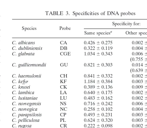
TABLE 3. Specificities of DNA probes
Species Probe Specificity for:
Same speciesa Other species/generab
C. albicans CA 0.42660.275 0.00260.002
C. dubliniensis DB 0.32260.119 0.00460.007
C. glabrata CGE 1.03460.343 0.00660.011 (0.75560.229)b
C. guilliermondii GU 0.82160.303 0.01460.026 (0.63960.172)b
C. haemulonii CH 0.84160.332 0.00260.002
C. kefyr KF 1.18460.384 0.00360.006
C. krusei CK 0.38960.136 0.00960.025
C. lambica LA 0.64060.175 0.00260.002
C. lusitaniae LU 0.48360.162 0.00260.001
C. norvegensis NS 0.71660.242 0.00660.004
C. norvegica NC 0.25860.102 0.00460.004
C. parapsilosis CP 0.49360.231 0.00360.004
C. pelliculosa PL 0.62460.320 0.00360.004
C. rugosa CR 0.22260.098 0.00260.001
C. tropicalis CT 0.87060.354 0.00660.020
C. utilis CU2 1.16160.193 0.00660.024
C. viswanathii VS 0.98860.419 0.00660.028
C. zeylanoides CZ 0.36160.170 0.00160.001 aMean A650value6SD for 22 to 86 test samples.
bNo significant (P,0.05) cross-hybridization of probe with DNAs from 37
other fungi which are listed in Table 2, except for the CGE probe with DNA from
S. cerevisiae and the GU probe with DNA from C. zeylanoides (values in
paren-theses).
on May 15, 2020 by guest
http://jcm.asm.org/
[image:3.612.307.546.477.685.2]that C. famata (type culture) would not be misidentified as another Candida sp. by using these probes.
Multiple strains of several Candida species were tested, and some inherent variability in probe hybridization for strains of the same species was apparent in the range of standard devia-tions of the absorbance values observed (Table 4). However, all strains gave absorbance signals of sufficient strength to allow differentiation of truly positive from truly negative samples, with two exceptions. The CP probe tested against C. parapsilosis Leh-mann group III DNAs (12) and the CH probe for C. haemu-lonii tested against C. haemuhaemu-lonii group 2 (strain 90.00.3593) DNA were significantly less reactive than with the respective positive control strains. These discrepant cases may indicate a finer taxonomic discrimination of isolates by genotypic than phenotypic methods (10–12, 30). Alternatively, combina-tions of probes could be designed for detection of all groups of C. parapsilosis and all groups of C. haemulonii in a clinical setting.
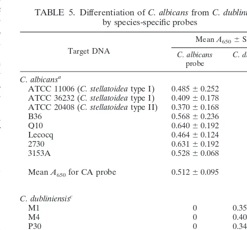
Species-specific probes were also designed to discriminate between two species that have a phenotype in common. A new species, C. dubliniensis, first described by Sullivan et al. (23), is typically identified as C. albicans by routine phenotypic meth-ods. The probes designed in this study readily discriminated C. albicans from C. dubliniensis (Table 5). The CA probe which detected C. albicans DNA did not react with DNA from any C. dubliniensis strain tested, and the DB probe for C. dublini-ensis identification did not hybridize with DNA from any C. al-bicans strain tested. The CA probe also detected both C. stel-latoidea type I and II DNAs and differentiated C. stelstel-latoidea DNA from C. dubliniensis DNA (Table 5).
DISCUSSION
Previous research in this laboratory demonstrated that five Candida species-specific probes could be designed and adapted to a simple PCR-EIA format to detect Candida species DNA (6, 20). This report extends the range of probes to include a test matrix of 18 Candida species that is capable of comple-menting species identification by the API 20C carbohydrate assimilation system. Sixteen of the probes are totally specific and can be used to identify their respective Candida species,
including C. dubliniensis. In addition, C. stellatoidea types I and II (10) can be differentiated from C. dubliniensis by these probes. The C. guilliermondii probe (GU) cross-reacted with C. zeyl-anoides DNA, but the C. zeylzeyl-anoides probe (CZ) did not cross-react with C. guilliermondii DNA, allowing species-specific identification by a process of elimination. Future studies will attempt to design a species-specific probe for each. However, at present, positive identification can still be achieved by a process of elimination by using both probes in the matrix configuration. As designed, all of the probes can be used in the matrix at the same time so that all possible combinations of probes and target DNAs can be tested in a single run. As more probes are added to the matrix, automation and chip array technologies become attractive ways to identify large numbers of different organisms simply and rapidly. The previously pub-lished probe for C. glabrata, CG, was found to cross-react with C. pelliculosa and C. utilis DNAs in this study. Therefore, a new probe, CGE, was designed which eliminated all cross-reactions except for that with S. cerevisiae DNA. Such cross-reactivity should have little impact on clinical diagnosis, however, since it is unlikely, although not impossible, that S. cerevisiae would be found in blood cultures or other normally sterile deep tissue sites. Also, since clinical isolates of both C. glabrata and S. cer-evisiae are innately fluconazole resistant (21, 24, 31), clinical treatment decisions would most likely not be negatively impact-ed by the inability to discriminate C. glabrata from S. cerevisiae. An S. cerevisiae probe has been developed (data not shown), and preliminary testing indicates that it is specific for S. cere-visiae detection. Therefore, this probe could be used in conjunc-tion with the CGE probe to differentiate the two species by a process of elimination.
[image:4.612.50.289.89.255.2]Standardization of DNA extraction for all Candida species is facilitated by using a broth culture method and a commercially available extraction kit. To facilitate prototype testing, isolates were grown overnight to obtain sufficiently large quantities of TABLE 4. Consistency of A650values for isolates
within a given species
Species Probe tested (group)No. of strains Mean A6506SDa
C. guilliermondii GU 5 1.08860.396
C. haemulonii CH 2 (1) 0.79060.122
C. haemulonii CH 1 (2) 0.08860.026b
C. kefyr KF 3 1.29960.270
C. lambica LA 2 0.64060.166
C. lusitaniae LU 2 0.65860.164
C. norvegensis NS 2 0.58860.175
C. parapsilosis CP 3 (I) 0.47760.209
C. parapsilosis CP 3 (II) 0.48960.206
C. parapsilosis CP 2 (III) 0.15860.068b
C. pelliculosa PL 6 0.69360.250
C. rugosa CR 3 0.23460.070
C. tropicalis CT 2 1.24460.083
C. utilis CU2 3 1.10560.399
C. zeylanoides CZ 4 0.48560.128
aMean values for 6 to 22 test samples.
bEach probe listed detected multiple isolates of the same species, except for
the C. haemulonii probe against one C. haemulonii isolate, strain 90.00.3593 (P,
0.001) and for the C. parapsilosis probe against DNA from C. parapsilosis group III isolates (P,0.01).
TABLE 5. Differentiation of C. albicans from C. dubliniensis by species-specific probes
Target DNA
Mean A6506SDb
C. albicans
probe C. dubliniensisprobe C. albicansa
ATCC 11006 (C. stellatoidea type I) 0.48560.252 0 ATCC 36232 (C. stellatoidea type I) 0.40960.178 0 ATCC 20408 (C. stellatoidea type II) 0.37060.168 0
B36 0.56860.236 0
Q10 0.64060.192 0
Lecocq 0.46460.124 0
2730 0.63160.192 0
3153A 0.52860.068 0
Mean A650for CA probe 0.51260.095 0
C. dubliniensisc
M1 0 0.35160.090
M4 0 0.40760.186
P30 0 0.34660.088
1419-2 0 0.40260.132
901013 0 0.39360.101
Mean A650for DB probe 0 0.38060.029
aStrains not obtained from ATCC were obtained from the Centers for Disease
Control and Prevention Mycology Reference Laboratory.
bMean values for 4 to 10 replicates for each isolate.
cStrains were received from S. Lockhart, D. Ahearn, and S. Meyer.
on May 15, 2020 by guest
http://jcm.asm.org/
[image:4.612.307.549.468.693.2]DNA for repeated analyses and probe development. In the clin-ical laboratory setting, sufficient DNA for routine testing, de-rived from primary cultures without subculturing, would further shorten the time required for species identification. Indeed, even species contained in mixed yeast cultures (C. albicans and C. glabrata) have been correctly identified from primary cul-tures in our laboratory by using species-specific probes (20).
All of the currently available commercial tests for species identification, such as the API 20C system, RapID, etc., re-quire subculturing from clinical specimens to obtain pure cul-tures before inoculation of the test panels. Therefore, even if an overnight culture were required prior to PCR-EIA testing, the time to species identification after obtaining a pure culture is still reduced to 7 h rather than a mean of 3.5 days by conven-tional phenotypic identification methods. Also, species identi-fication of unusual species such as C. norvegensis or C. utilis by conventional methods may require up to 4 or 5 weeks (25), whereas the PCR-EIA can identify these species in a single day. Since some species are innately resistant to certain drugs, e.g., C. krusei to fluconazole (16, 18, 29), accurate and timely species identification is important for selection of appropriate-ly targeted therapy.
The recently described species C. dubliniensis was discov-ered when DNA from phenotypically identified “C. albicans” strains did not react with the C. albicans-specific mid-repetitive element Ca3 (2, 23). Molecular differences between C. albicans and C. dubliniensis were confirmed in the current research in that sufficiently significant sequence differences occurred in the ITS2 region to facilitate the development of species-specific probes. Although C. dubliniensis is not currently listed as one of the yeasts identified in the API 20C database, differences be-tween C. albicans and C. dubliniensis in the assimilation of xylose anda-methyl-D-glucoside may prove useful for the phenotypic differentiation of these species (19). In addition, other physical (growth temperature) or chemical (b-glucosidase activity) tests may also help discriminate C. albicans from C. dubliniensis (17, 22).
The C. dubliniensis strains listed in Table 5 were originally obtained in Europe and Australia. C. dubliniensis has also been isolated in North America, as reported by Kleinegger et al. (8) and Boucher et al. (1) in 1996. We confirmed the presence of C. dubliniensis in North America by using our probes to test DNA obtained from oropharyngeal isolates from a population of human immunodeficiency virus-positive persons in the At-lanta, Ga. area (4). To our knowledge, these are the first iso-lates of C. dubliniensis recovered from a human immunodefi-ciency virus-positive population in the United States (4, 19).
This genotype-based identification method has revealed the need for further taxonomic resolution of some species, for ex-ample, C. famata (teleomorph form, D. hansenii). Although the identities of seven C. famata strains tested in this study were confirmed by conventional phenotypic methods (data not shown), the ITS2 probe designed for one of these strains was found to hybridize only with its own DNA and not to the DNA of any of the other four C. famata strains tested. When the sequences of the ITS2 regions of these seven strains were determined and compared, greater differences were observed among these strains than among strains of other Candida species tested. Therefore, the complexity of this taxon is apparent and agrees with the DNA-DNA hybridization studies of Nishikawa et al. (15), who described a low percentage of hybridization between some DNAs from several C. famata strains. Because C. famata appears to be a taxonomic complex, further studies are needed to determine sequences which will allow the detection of clin-ically encountered members of this complex either by redefi-nition of these strains into subspecies or by the use of
combi-nations of probes to simultaneously detect members of the complex. Additional molecular characterization is needed to clarify the taxonomy and identification of strains which appear to be C. famata by phenotypic criteria but differ by genotypic criteria.
Lower absorbance values were obtained for some strains of C. parapsilosis (group III) and C. haemulonii (group 2) than for their positive control strains. However, these values were still significantly greater than those for their respective negative con-trols. Because of the controversy surrounding C. parapsilosis group III and C. haemulonii group 2, the designation of a truly positive cutoff value for these groups awaits taxonomic resolu-tion. The clinical significance of C. parapsilosis group III and C. haemulonii group 2 is not known, nor are the true incidence and prevalence of these groups. However, the CP probe cor-rectly identified all clinical blood isolates of C. parapsilosis in a previous study (20). Therefore, it is likely that the CP probe, at least, can identify clinically relevant strains. The same is yet to be determined for the CH probe. In addition, combinations of probes, used in one reaction mixture, to identify all groups of C. parapsilosis and all groups of C. haemulonii could be de-signed if such a need were identified in the clinical setting.
The present method of sample preparation and PCR-EIA is amenable to automation, and the entire panel of 18 different probes can be tested against an unknown yeast in a simple microtitration plate format. Greater numbers of isolates will be tested in prospective clinical studies to validate the specificity of each probe. These probes were designed to detect 1 ng of target DNA recovered from Candida sp. cultures, and the limit of their sensitivity has not yet been determined in clinical samples. Previous studies using positive blood culture bottles suggest that these probes may be useful for the direct identi-fication of Candida species from primary cultures (20). Minor probe cross-reactions should not be problematic, since a pro-cess of elimination with specific, non-cross-reacting, paired probes allows specific differentiation (e.g., the CK probe and C. lambica DNA). When sensitivity limits become an issue, such as in clinical samples containing unknown quantities of DNA, then a matrix of probes will differentiate truly positive samples from truly negative samples. Ultimately, once a suffi-cient panel of medically important Candida and/or yeast spe-cies has been constructed, the ideal assay will consist of probes attached to a nylon membrane or to wells of a microtiter plate, a single universal PCR using premixed PCR reagents, and the colorimetric development of the matrix array in a single assay. Commercially prepared, premixed PCR reagents are available, and prototype assays with such configurations have already been examined in our laboratory (27) and continue to be im-proved to optimize the speed and simplicity of species-specific yeast identification.
ACKNOWLEDGMENTS
This research was supported in part through an Emerging Infectious Diseases Training Fellowship from the Association of State and Ter-ritorial Public Health Laboratory Directors for C.M.E. and also by an appointment to the Research Participation Program at the CDC, Na-tional Center for Infectious Diseases, Division of Bacterial and My-cotic Diseases, administered by the Oak Ridge Institute for Science and Education through an interagency agreement between the U.S. Department of Energy and CDC.
We thank J. H. Shin, Chonnam University Medical School, Kwangju, Korea, for blood culture isolates of C. pelliculosa and C. guilliermondii; R. Cherniak, Georgia State University, Atlanta, for isolates of C.
neo-formans serotypes A, B, C, and D; P. F. Lehmann, Medical College of
Ohio, Toledo, for isolates of C. parapsilosis groups I, II, and III; D. Ahearn and S. Meyer, Georgia State University, Atlanta, for isolates of
C. haemulonii and C. dubliniensis; S. Lockhart, University of Iowa,
on May 15, 2020 by guest
http://jcm.asm.org/
Iowa City, for isolates of C. dubliniensis; L. de Aguirre, Instituto Investi-gaciones Veterinarias, Maracay, Venezuela, for DNA extracted from filamentous fungi; and B. A. Lasker, CDC, Atlanta, Ga., for DNA ex-tracted from dimorphic fungi and S. cerevisiae.
REFERENCES
1. Boucher, H., S. Mercure, S. Montplaisir, and G. Lemay. 1996. A novel group I intron in Candida dubliniensis is homologous to a Candida albicans intron. Gene 180:189–196.
2. Coleman, D. C., D. J. Sullivan, D. E. Bennett, G. P. Moran, H. J. Barry, and D. B. Shanley.1997. Candidiasis: the emergence of a novel species, Candida
dubliniensis. AIDS 11:557–567.
3. de Aguirre, L. A., H. Vaishnav, J. M. Westerman, E. Reiss, and C. J. Morrison.1997. Rapid differentiation of Aspergillus species from other fila-mentous fungi and yeasts using species-specific DNA probes, abstr. C234, p. 161. In Abstracts of the 97th General Meeting of the American Society for Microbiology 1997. American Society for Microbiology, Washington, D.C. 4. Elie, C. M., B. A. Lasker, L. W. Mayer, W. R. Pruitt, G. Smith, D. Rimland,
L. Gallagher, E. Reiss, C. J. Morrison, and M. M. McNeil.1998. Rapid differentiation of Candida dubliniensis from atypical C. albicans isolates using species-specific DNA probes, abstr. C290, p. 179. In Abstracts of the 98th General Meeting of the American Society for Microbiology 1998. American Society for Microbiology, Washington, D.C.
5. Fridkin, S. K., and W. R. Jarvis. 1996. Epidemiology of nosocomial fungal infections. Clin. Microbiol. Rev. 9:499–511.
6. Fujita, S.-I., B. A. Lasker, T. J. Lott, E. Reiss, and C. J. Morrison. 1995. Microtitration plate enzyme immunoassay to detect PCR-amplified DNA from Candida species in blood. J. Clin. Microbiol. 33:962–967.
7. Jarvis, W. R. 1995. Epidemiology of nosocomial fungal infections, with emphasis on Candida species. Clin. Infect. Dis. 20:1526–1530.
8. Kleinegger, C. L., S. R. Lockhart, K. Vargas, and D. R. Soll. 1996. Fre-quency, intensity, species, and strains of oral Candida vary as a function of host age. J. Clin. Microbiol. 34:2246–2254.
9. Kwok, S., and R. Higuchi. 1989. Avoiding false positives with PCR. Nature (London) 339:237–238.
10. Kwon-Chung, K. J., W. S. Riggsby, R. A. Uphoff, J. B. Hicks, W. L. Whelan, E. Reiss, B. B. Magee, and B. L. Wickes.1989. Genetic differences between type I and type II Candida stellatoidea. Infect. Immun. 57:527–532. 11. Lehmann, P. F., L.-C. Wu, W. R. Pruitt, S. A. Meyer, and D. G. Ahearn. 1993.
Unrelatedness of groups of yeasts within the Candida haemulonii complex. J. Clin. Microbiol. 31:1683–1687.
12. Lin, D., L.-C. Wu, M. G. Rinaldi, and P. F. Lehmann. 1995. Three distinct genotypes within Candida parapsilosis from clinical sources. J. Clin. Micro-biol. 33:1815–1821.
13. Lott, T. J., B. M. Burns, R. Zancope-Oliveira, C. M. Elie, and E. Reiss. 1998. Sequence analysis of the internal transcribed spacer 2 (ITS2) from yeast spe-cies within the genus Candida. Curr. Microbiol. 36:63–69.
14. Lott, T. J., R. Kuykendall, and E. Reiss. 1993. Nucleotide sequence analysis of the 5.8S rDNA and adjacent ITS2 region of Candida albicans and related species. Yeast 2:1199–1206.
15. Nishikawa, H., H. Tomomatsu, T. Sugita, R. Ikeda, and T. Shinoda. 1996. Taxonomic position of clinical isolates of Candida famata. J. Med. Vet. Mycol. 34:411–419.
16. Pfaller, M. A. 1995. Nosocomial fungal infections: epidemiology of candidi-asis. J. Hosp. Infect. 30(Suppl.):329–338.
17. Pinjon, E., D. Sullivan, I. Salkin, D. Shanley, and D. Coleman. 1998. Simple, inexpensive, reliable method for differentiation of Candida dubliniensis from
Candida albicans. J. Clin. Microbiol. 36:2093–2095.
18. Rex, J. H., M. A. Pfaller, A. L. Barry, P. W. Nelson, and C. D. Webb for the NIAID Mycoses Study Group, and The Candidemia Study Group.1995. Antifungal susceptibility testing of isolates from a randomized multicenter trial of fluconazole versus amphotericin B as treatment of nonneutropenic pa-tients with candidemia. Antimicrob. Agents Chemother. 39:40–44. 19. Salkin, I. F., W. R. Pruitt, A. A. Padhye, D. Sullivan, D. Coleman, and D. H.
Pincus.1998. Distinctive carbohydrate assimilation profiles used to identify the first clinical isolates of Candida dubliniensis recovered in the United States. J. Clin. Microbiol. 36:1467. (Letter.)
20. Shin, J. H., F. S. Nolte, and C. J. Morrison. 1997. Rapid identification of
Candida species in blood culture by a clinically useful polymerase chain
reaction method. J. Clin. Microbiol. 35:1454–1459.
21. Sobel, J. D., J. Vazquez, M. Lynch, C. Meriwether, and M. J. Zervos. 1993. Vaginitis due to Saccharomyces cerevisiae: epidemiology, clinical aspects, and therapy. Clin. Infect. Dis. 16:93–99.
22. Sullivan, D., and D. Coleman. 1998. Candida dubliniensis: characteristics and identification. J. Clin. Microbiol. 36:329–334.
23. Sullivan, D. J., T. J. Westerneng, K. A. Haynes, D. E. Bennett, and D. C. Coleman.1995. Candida dubliniensis sp. nov.: phenotypic and molec-ular characterization of a novel species associated with oral candidosis in HIV-infected individuals. Microbiology 141:1507–1521.
24. Tiballi, R. N., J. E. Spiegel, L. T. Zarins, and C. A. Kauffman. 1995.
Sac-charomyces cerevisiae infections and antifungal susceptibility studies by
col-orimetric and broth macrodilution methods. Diagn. Microbiol. Infect. Dis. 23:135–140.
25. Warren, N. G., and K. C. Hazen. 1995. Candida, Cryptococcus, and other yeasts of medical importance, p. 723–737. In P. R. Murray, E. J. Baron, M. A. Pfaller, F. C. Tenover, and R. H. Yolken (ed.), Manual of clinical microbi-ology, 6th ed. American Society for Microbimicrobi-ology, Washington, D.C. 26. Wenzel, R. P. 1995. Nosocomial candidemia: risk factors and attributable
mortality. Clin. Infect. Dis. 20:1531–1534.
27. Westerman, J. M., C. M. Elie, and C. J. Morrison. 1998. Improved identi-fication of Candida species using biotinylated species-specific DNA probes, abstr. J107, p. 481. In Abstracts of the 38th Interscience Conference on Antimicrobial Agents and Chemotherapy. American Society for Microbiol-ogy, Washington, D.C.
28. White, T. J., T. D. Burns, S. B. Lee, and J. W. Taylor. 1990. Amplification and direct sequencing of fungal ribosomal RNA genes for phylogenetics, p. 315–322. In M. A. Innis, D. H. Gelfand, J. J. Sninsky, and T. J. White (ed.), PCR protocols. Academic Press, San Diego, Calif.
29. Wingard, J. R. 1995. Importance of Candida species other than C. albicans as pathogens in oncology patients. Clin. Infect. Dis. 20:115–125.
30. Zeng, S., L.-C. Wu, and P. F. Lehmann. 1996. Random amplified polymor-phic DNA analysis of culture collection strains of Candida species. J. Med. Vet. Mycol. 34:293–297.
31. Zerva, L., R. J. Hollis, and M. A. Pfaller. 1996. In vitro susceptibility testing and DNA typing of Saccharomyces cerevisiae clinical isolates. J. Clin. Micro-biol. 34:3031–3034.