Dendritic cells and the intestinal bacterial flora:
a role for localized mucosal immune responses
Holm H. Uhlig, Fiona Powrie
J Clin Invest.
2003;
112(5)
:648-651.
https://doi.org/10.1172/JCI19545
.
Mammals coexist in an overall symbiotic relationship with a complex array of commensal
bacterial flora that colonizes the gastrointestinal tract. These intestinal bacteria interface
with cells of the mucosal immune system, including DCs. Here we discuss mechanisms of
interaction between intestinal bacteria and DCs and the role of localized gastrointestinal
immune responses.
Commentary
Find the latest version:
prokaryotes; therefore gut flora are not the source. A dietary source can be questioned, as it is unlikely that the Hsp110 would be recovered in a struc-turally intact form in the large intes-tine. And so the most likely source is the gut epithelium itself. Yet, Hsp110 does not possess a signal peptide and so cannot be secreted from IECs via the classical secretory pathway. Non-canonical secretion, as seen, for exam-ple, with FGF-2, is a possibility (14). However, this release pathway, and the analogous pathway in yeast, appear to transport very few proteins. Perhaps, then, Hsp110 is released into the intes-tinal lumen as a consequence of epithe-lial renewal, particularly in response to IEL activation (15). If this is the case, though, it might be expected that other abundant cytosolic chaperone proteins, such as Hsp90 and Hsp70, would be recovered in the intestinal contents as well. Nonetheless, perhaps inappropri-ately elevated levels of Hsp110 in the intestinal lumen serve as an immuno-logical “trigger” leading to aberrant induction of CD1d expression, a subse-quent activation of NK T cells, and
IL-13–elicited destruction of IECs (Fig-ure 1) (16). Whatever the mechanism, one is left to ponder the fascinating mys-tery of why IECs might (uniquely?) release Hsp110 into the gut lumen. Insights into the precise cellular source of the gut-lumen Hsp110 and the mecha-nism of its release will provide all-impor-tant clues to this new riddle in the ever-expanding world of Hsp110 function.
1. Fiocchi, C. 1998. Inflammatory bowel disease: eti-ology and pathogenesis. Gastroenterology.
115:182–205.
2. Blumberg, R.S. 2001. Characterization of CD1d in mucosal immune function: an immunotherapeutic target for inflammatory bowel disease. Keio J. Med.
50:39–44.
3. Mizoguchi, A., Mizoguchi, E., Takedatsu, H., Blum-berg, R.S., and Bhan, A.K. 2002. Chronic intestinal inflammatory condition generates IL-10-producing regulatory B cell subset characterized by CD1d upregulation. Immunity.16:219–230.
4. Page, M.J., et al. 2000. Cd1d-restricted cellular lysis by peripheral blood lymphocytes: relevance to the inflammatory bowel diseases. J. Surg. Res.
92:214–221.
5. Bendelac, A., et al. 1995. CD1 recognition by mouse NK1+ T lymphocytes. Science.12:863–865. 6. Mendiratta, S.K., et al. 1997. CD1d1 mutant mice
are deficient in natural T cells that promptly pro-duce IL-4. Immunity.6:467–477.
7. Colgan, S.P., Hershberg, R.M., Furuta, G.T., and Blumberg, R.S. 1999. Ligation of intestinal epithe-lial CD1d induces bioactive IL-10: critical role of the cytoplasmic tail in autocrine signaling. Proc. Natl.
Acad. Sci. U. S. A.96:13938–13943.
8. Mowat, A.M., and Weiner, H.L. 1999. Oral tolerance: physiological basis and clinical applications. In
Mucosal immunology. P.L. Ogra et al., editors. Acade-mic Press Inc. San Diego, California, USA. 587–618. 9. Colgan, S.P., et al. 2003. Intestinal heat shock pro-tein 110 regulates expression of CD1d on intestin-al epitheliintestin-al cells. J. Clin. Invest. 112:745–754. doi:10.1172/JCI200317241.
10. Lee-Yoon, D., Easton, D., Murawski, M., Burd, R., and Subjeck, J.R. 1995. Identification of a major subfamily of large hsp70-like proteins through the cloning of the mammalian 110-kDa heat shock pro-tein. J. Biol. Chem.270:15725–15733.
11. Easton, D.P., Kaneko, Y., and Subjeck, J.R. 2000. The hsp110 and Grp1 70 stress proteins: newly recog-nized relatives of the Hsp70s. Cell Stress Chaperones.
5:276–290.
12. Anderson, K.M., and Srivastava, P.K. 2000. Heat, heat shock, heat shock proteins and death: a central link in innate and adaptive immune responses.
Immunol. Lett.74:35–39.
13. Srivastava, P.K., and Amato, R.J. 2001. Heat shock proteins: the ‘Swiss Army Knife’ vaccines against cancers and infectious agents. Vaccine.
19:2590–2597.
14. Wakisaka, N., Murono, S., Yoshizaki, T., Furukawa, M., and Pagano, J.S. 2002. Epstein-Barr virus latent membrane protein 1 induces and causes release of fibroblast growth factor-2. Can-cer Res.62:6337–6344.
15. Guy-Grand, D., DiSanto, J.P., Henchoz, P., Malassis-Seris, M., and Vassalli, P. 1998. Small bowel enteropathy: role of intraepithelial lymphocytes and of cytokines (IL-12, IFN-gamma, TNF) in the induc-tion of epithelial cell death and renewal. Eur. J. Immunol.28:730–744.
16. Bouma, G., and Strober, W. 2003. The immunolog-ical and genetic basis of inflammatory bowel dis-ease. Nat. Rev. Immunol.3:621–633.
Dendritic cells and the intestinal
bacterial flora: a role for localized
mucosal immune responses
Holm H. Uhlig and Fiona Powrie
Sir William Dunn School of Pathology, University of Oxford, Oxford, United Kingdom Mammals coexist in an overall symbiotic relationship with a complex array of commensal bacterial flora that colonizes the gastrointestinal tract. These intestinal bacteria interface with cells of the mucosal immune system, including DCs (see the related article beginning on page 693). Here we discuss mechanisms of interaction between intestinal bacteria and DCs and the role of localized gastrointestinal immune responses.
J. Clin. Invest.112:648–651 (2003). doi:10.1172/JCI200319545.
Address correspondence to: F. Powrie, Sir William Dunn School of Pathology, University of Oxford, South Parks Road, Oxford OX1 3RE, United Kingdom. Phone: 44-1865-285494; Fax: 44-1865-275591; E-mail: fiona.powrie@pathology.ox.ac.uk.
Conflict of interest: The authors have declared that no conflict of interest exists.
Nonstandard abbreviations used:
gastrointestinal tract (GIT); inflammatory bowel disease (IBD); nucleotide-binding oligomerization domain 2 (NOD2).
Inflammatory bowel disease is associated with a dysregulated immune response to intestinal bacterial flora
Through processes of evolutionary and individual adaptation, mammals coexist with an estimated 300 to 500 different species of commensal bacte-ria that colonize the gastrointestinal tract (GIT) in an overall symbiotic
rela-tionship (1, 2). The presence of intes-tinal bacteria plays an important role in host metabolism, the development of the intestinal epithelium, and the intestinal immune system, and it also protects the host against rapid colo-nization by intestinal pathogens (1, 2). To allow sufficient defense against potential pathogens but restrict the immune response to nonpathogenic resident commensal bacteria, the mucosal immune system needs to be tightly regulated.
Recently it was found that a subset of patients with Crohn disease have loss-of-function mutations in the gene that encodes the nucleotide-binding oligomerization domain 2 (NOD2) protein (6). NOD2is a patho-gen-recognition receptor that recog-nizes muramyl dipeptide derived from bacterial peptidoglycans (7). Although it is not understood how mutations in the NOD2pathway give rise to IBD, these findings suggest that alterations in the recognition of intestinal bacteria can contribute to chronic inflammation.
Studies of IBD models in rodents provide compelling evidence that the
bacterial flora plays a key role in the pathogenesis of the disease, as chronic intestinal inflammation fails to devel-op under germfree conditions (8). However, precisely how commensal bacteria in the intestine interface local-ly with cells of the immune system to initiate and perpetuate intestinal inflammation remains unclear.
Intestinal bacterial microflora induces localized IL-23 secretion by DCs
The proinflammatory cytokine IL-12 has been implicated in the pathogene-sis of IBD in a number of mouse mod-els and also in the
[image:3.576.94.502.56.373.2]immunopathogen-esis of Crohn disease (3, 8). In order to study IL-12 production in situ under physiological conditions, Becker and coworkers (9) developed a transgenic mouse expressing a reporter gene under the control of the IL-12p40 sub-unit promoter (see their article in this issue of the JCI). Somewhat unexpect-edly, they found that p40 transcription is restricted primarily to the terminal ileum, where it is produced by a subset of DCs. Recently it has become clear that p40 is a component not only of IL-12 (p40/p35) but also of the Th1-inducing cytokine IL-23 (p40/p19) (10). Strikingly, the elevated IL-12p40 in the ileum, observed by Becker et al.,
Figure 1
was accompanied by p19 but not p35 mRNA and increased IL-23 protein complex formation. IL-23 was pro-duced in response to the bacterial flora, as it was not detectable in the ileum of mice housed under germfree conditions. Indeed, using an elegant combination of immunohistology and 16S-rRNA fluorescence in situ hybrid-ization, the authors were able to show that some of the DCs with active p40 transcription contained intracellular nondegraded bacteria.
Restriction of DC IL-23 production to the terminal ileum and not the
proximal small intestine or the colon suggests a specific activation state of CD11c+cells in the former site. A key question is whether this represents the consequence of a hitherto unappreci-ated active sampling of commensal bacteria by DCs in the ileum, or is the result of selective colonization and invasion of a specific pathogen in the terminal ileum that leads to a local host immune response involving IL-23 secretion. The finding that bac-teria-induced DC activation resulted in IL-23 as opposed to IL-12 produc-tion raises the possibility that IL-23 is
an important mediator in intestinal inflammation, as has recently been reported in a model of cerebral inflam-mation (11). IL-23 may also play an important role in host defense against invading bacteria as a consequence of its ability to maintain Th1 cell memo-ry responses by acting directly on CD4+memory lymphocytes (10). Fur-thermore, IL-23 can activate DCs and macrophages via autocrine mecha-nisms (12). However, understanding of the biology of IL-23 is at an early stage, and further work is required to eluci-date its role in host defense and intes-tinal inflammation.
The results of the study reported by Becker et al. (9) raise the question of how intestinal DCs come into contact with bacterial antigens. Several routes have been proposed, and these may vary depending on the nature of the bacterium (Figure 1) (13). Obviously, in order to prevent systemic infection, there is a clear need for the immune system to respond to bacteria that translocate from the lumen toward the lamina propria. Evidence also suggests that there are more active modes of antigen sampling involving M cells or intra- and transepithelial DCs. Howev-er, the reason why the immune system bothers to actively sample intestinal antigens from the lumen is less clear. Such a mechanism might aid sustained antigen delivery for T cell–dependent and T cell–independent IgA produc-tion (14). Alternatively, the active sam-pling of low amounts of intestinal anti-gens may be involved in inducing and maintaining T cell tolerance toward the intestinal bacterial flora.
Localized mucosal immune
responses as a result of differential colonization and pathogenicity
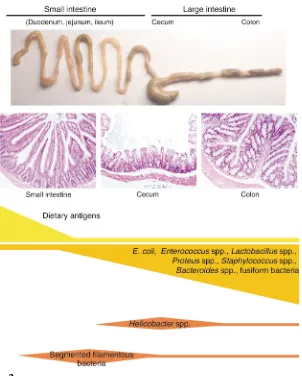
[image:4.576.70.372.51.427.2]It is tempting to speculate, as do Beck-er et al. (9), that the distinct elevation of IL-23 expression in the ileum reflects an increased susceptibility to inflamma-tion in the terminal ileum, a site that is affected in patients with Crohn disease (3). However, this simple model with a dominant IL-23 response in the termi-nal ileum is difficult to reconcile with the finding that most mouse models of intestinal inflammation express domi-nant pathology in the cecum and colon, but not in the terminal ileum (8).
Figure 2
The GIT provides distinct niches for colonization of commensal bacteria, as indicated by qualitative and quanti-tative differences in the bacterial flora throughout the GIT (Figure 2) (1, 15). The anatomical architecture in differ-ent parts of the GIT reflects its func-tional role in digestion and also in host defense (16). Thus, there are clear differences not only in the epithelial intestinal architecture, but also in the density of cells that are closely associ-ated with the innate and adaptive immune response, such as goblet-pro-ducing cells, Paneth cells, DCs, and B and T lymphocytes. Furthermore, regional differences are found in the presence and density of mucosal lym-phoid structures such as Peyer’s or cecal patches, or isolated lymphoid follicles (16). It is likely that the lym-phoid tissue throughout the GIT plays an important role in localized immune responses to bacteria that populate the respective compartment. Indeed, the development of the local-ized mucosa-associated immune sys-tem is only in part genetically deter-mined; it is also functionally dependent on the presence of the bac-terial microflora (17). In addition, dif-ferential complementary determining region-3 T cell receptor usage among T cells in different regions of the colon provides further support for localized T cell immune responses within the large intestine (18). Interestingly, in HLA-B27 transgenic rats, the bacterial load in the cecum determines the severity of mucosal inflammation, and the development of colitis in T cell receptor-α mutant mice can be pre-vented by removal of parts of the cecum. These results suggest that the manipulation of bacteria and/or cor-responding mucosa-associated lym-phoid tissue in specific intestinal com-partments can influence the induction of intestinal immunopathology (19, 20). Localized innate and adaptive
mucosal immune responses may pro-vide an efficient response to the respective flora but restrict extensive immunopathology. In this regard, the IL-12p40 promoter transgenic mice produced by Becker and coworkers (9) will be an excellent tool to study the interaction between particular bacte-ria and the host immune system and how this influences the localization of the immune response.
A dilemma for those using model systems in mice to study interactions between the intestinal bacterial flora and the immune system is that, despite increasing control of host genetics, afforded by the use of inbred mice with defined genetic alterations, our knowledge about the composition and function of the corresponding intestinal bacterial flora is limited to just a few bacteria. Because of the great variety of housing conditions, there may be large variations within the bac-terial flora that might influence the activation state of the immune system and the interpretation of experiments in different laboratories. The further identification of distinct intestinal commensal and pathogenic bacteria and their ability to colonize and invade distinct compartments of the GIT will afford a better understanding of the interplay among different bacteria and between bacteria and the host in phys-iology and disease.
Acknowledgments
Holm H. Uhlig is supported by the European Society of Clinical Micro-biology and Infectious Diseases and the Deutsche Forschungsgemein-schaft. Fiona Powrie is supported by the Wellcome Trust.
1. Hooper, L.V., and Gordon, J.I. 2001. Commensal host-bacterial relationships in the gut. Science.
292:1115–1118.
2. Guarner, F., and Malagelada, J.R. 2003. Gut flora in health and disease. Lancet. 361:512–519. 3. Shanahan, F. 2002. Crohn’s disease. Lancet.
359:62–69.
4. Swidsinski, A., et al. 2002. Mucosal flora in inflammatory bowel disease. Gastroenterology.
122:44–54.
5. Linskens, R.K., Huijsdens, X.W., Savelkoul, P.H., Vandenbroucke-Grauls, C.M., and Meuwissen, S.G. 2001. The bacterial flora in inflammatory bowel disease: current insights in pathogenesis and the influence of antibiotics and probiotics.
Scand. J. Gastroenterol. Suppl. 234:29–40. 6. Ogura, Y., et al. 2001. A frameshift mutation in
NOD2 associated with susceptibility to Crohn’s disease. Nature.411:603–606.
7. Inohara, N., et al. 2003. Host recognition of bac-terial muramyl dipeptide mediated through NOD2. Implications for Crohn’s disease. J. Biol. Chem.278:5509–5512.
8. Strober, W., Fuss, I.J., and Blumberg, R.S. 2002. The immunology of mucosal models of inflam-mation. Annu. Rev. Immunol.20:495–549. 9. Becker, C., et al. 2003. Constitutive p40promoter
activity and IL-23 production in the terminal ileum mediated by dendritic cells. J. Clin. Invest.
112:693–706. doi:10.1172/JCI200317464. 10. Trinchieri, G. 2003. Interleukin-12 and the
regu-lation of innate resistance and adaptive immuni-ty. Nat. Rev. Immunol.3:133–146.
11. Cua, D.J., et al. 2003. Interleukin-23 rather than interleukin-12 is the critical cytokine for autoim-mune inflammation of the brain. Nature.
421:744–748.
12. Puccetti, P., Belladonna, M.L., and Grohmann, U. 2002. Effects of IL-12 and IL-23 on antigen-pre-senting cells at the interface between innate and adaptive immunity. Crit. Rev. Immunol.
22:373–390.
13. Didierlaurent, A., Sirard, J.C., Kraehenbuhl, J.P., and Neutra, M.R. 2002. How the gut senses its content. Cell. Microbiol.4:61–72.
14. Macpherson, A.J., et al. 2000. A primitive T cell-independent mechanism of intestinal mucosal IgA responses to commensal bacteria. Science.
288:2222–2226.
15. Jiang, H.Q., Bos, N.A., and Cebra, J.J. 2001. Tim-ing, localization, and persistence of colonization by segmented filamentous bacteria in the neona-tal mouse gut depend on immune status of mothers and pups. Infect. Immun.69:3611–3617. 16. Mowat, A.M. 2003. Anatomical basis of tolerance and immunity to intestinal antigens. Nat. Rev. Immunol.3:331–341.
17. Yamanaka, T., et al. 2003. Microbial colonization drives lymphocyte accumulation and differentia-tion in the follicle-associated epithelium of Peyer’s patches. J. Immunol.170:816–822. 18. May, E., et al. 2002. Regional variation of the
alphabeta T cell repertoire in the colon of healthy individuals and patients with Crohn’s disease.
Hum. Immunol.63:467–480.
19. Rath, H.C., et al. 1999. Varying cecal bacterial loads influences colitis and gastritis in HLA-B27 transgenic rats. Gastroenterology.116:310–319. 20. Mizoguchi, A., Mizoguchi, E., Chiba, C., and Bhan, A.K. 1996. Role of appendix in the devel-opment of inflammatory bowel disease in TCR-alpha mutant mice. J. Exp. Med.184:707–715. 21. Rescigno, M., et al. 2001. Dendritic cells express
tight junction proteins and penetrate gut epithe-lial monolayers to sample bacteria. Nat. Immunol.