INTRODUCTION
The genesis of polarity* in the early embryo is crucial to development because it determines the body plan of the whole animal. Although in many species the polarity of the embryo takes its roots from the spatial patterning of the egg, for a long time we have thought of mammals as an exception to this model (for a review, see Gurdon, 1992). It was generally believed that the mammalian body was not shaped according to developmental cues laid out within the egg or early embryo. Rather, the cells within an early embryo were considered to be entirely naïve and to decide their fate through interactions between each other. Moreover, embryonic polarity was assumed to be established only at or shortly before gastrulation, possibly due to interactions between the embryo and the uterus of the mother upon implantation (for a review, see Beddington and Robertson, 1998). So what encouraged the idea that
mammalian embryos develop their polarity in this unique way and does current knowledge justify retaining this view? First, the body axes in the mouse only become morphologically apparent several days after implantation. Specifically, it is only when gastrulation is imminent that the anteroposterior axis can be identified unequivocally. At this stage, the mouse embryo is approaching the end of its first week of life – one third of its entire gestation – a significantly later stage than when the axes emerge in other animals. However, as it now appears, asymmetric gene expression patterns predicting the anteroposterior axis arise before the morphological changes are evident. Moreover we do not yet know whether these expression patterns represent the earliest molecular asymmetries (for reviews, see Beddington and Robertson, 1999; Lu et al., 2001). Second, in organisms as diverse as C. elegans (for reviews, see Bowerman and Shelton, 1999; Golden, 2000; Gotta and Ahringer, 2001), Drosophila (Riechmann and Ephrussi, 2001), ascidians (Roegiers et al., 1999; Jeffery, 2001), Xenopus (King et al., 1999) or zebrafish (Solnica-Krezel, 1999), the establishment of polarity and cell Printed in Great Britain © The Company of Biologists Limited 2002
DEV7930
Although in most species the polarity of the embryo takes its roots from the spatial patterning of the egg, mammals were viewed as an exception. This was because the anteroposterior polarity of the mouse embryo could not be seen until gastrulation, and no developmental cues were known that could define polarity at earlier stages. Why should we now re-consider this view? While mechanisms of axis formation in mammals could, in principle, be unique, the evolutionary conservation of numerous other developmental processes raises the question of why mammals would have evolved a different way or timing of organising their embryonic polarity. Indeed, recent evidence shows that well before the onset of gastrulation, the mouse embryo initiates asymmetric patterns of gene expression in its visceral endoderm. Although this extra-embryonic tissue does not contribute to the body itself, it is involved in axis formation. Other recent work has revealed that spatial distribution of cells in the visceral endoderm can be traced back to polarity present at the blastocyst stage. These insights have raised the possibility that
embryonic polarity might also originate early during development of mammalian embryos. Indeed it now appears that there are at least two spatial cues that operate in the mouse egg to shape polarity of the blastocyst. One of these is at the animal pole, which is defined by the site of female meiosis, and another is associated with the position of sperm entry. In this review I discuss these recent findings, which have led to the recognition that mouse embryos initiate development of their polarity at the earliest stages of their life. This novel perspective raises questions about the nature of cellular and molecular mechanisms that could convert developmental cues in the zygote to axes of the blastocyst, and hence into polarity of the post-implantation embryo. It also brings to light the need to understand how such mechanisms could enable early mouse development to be so regulative.
Key words: Mouse, Embryo, Polarity, Axis, Cleavage, Fertilisation, Sperm
SUMMARY
REVIEW ARTICLE
Patterning of the embryo: the first spatial decisions in the life of a mouse
Magdalena Zernicka-Goetz
Wellcome/CRC Institute and Department of Genetics, Tennis Court Road, Cambridge CB2 1QR, UK
e-mail: [email protected]
Accepted 16 November 2001
fate is achieved by the programmed distribution of morphogenetic determinants; no such factors have yet been identified in mammalian embryos. Although there are reports of asymmetrically distributed proteins that appear to be inherited by specific lineages during cleavage in some mammalian eggs (Dalcq, 1957; Antczak and Van Blerkom, 1997), we still cannot judge whether these have any role in deciding cell fate. But the third and principal reason for considering mammalian embryos to be unique is the extensive developmental plasticity they display, particularly at pre-implantation stages. Thus, they are able to recover from experimental perturbation that would disrupt early patterning in other organisms. If, for example, cells of early mouse embryos are removed or re-positioned, further development can still occur (for reviews, see McLaren, 1976; Papaioannou and Ebert, 1986). Because such regulative ability and early patterning were generally considered mutually exclusive properties, it was concluded that polarity was imposed upon the embryo, rather than being intrinsic to it.
But why should developmental plasticity of an embryo argue conclusively against a role for early patterning? In most, if not all, organisms, early patterning and regulative ability seem actually to peacefully co-exist. In C. elegans, for example, the polarity of which was initially considered to be entirely dependent on early patterning (a so-called ‘mosaic’ embryo), it is now known that some regulative capacity allows cells to shape their fate in response to extrinsic information (for a review, see Labouesse and Mango, 1999). Conversely other organisms, such as sea urchins, which have substantial regulative capacity, appear to have their patterning established at early stages (Horstadius, 1973; Cameron et al., 1989; Davidson, 1989; McCain and McClay, 1994; Angerer and Angerer, 1999). Even the frog embryo, which was once regarded as an archetype of regulative development, has been revealed to be under maternal influence in specifying cell fate (for a review, see Sullivan et al., 1999). Similarly, spatial patterning and regulation both play their part in establishing polarity in chick embryos (for a review, see Bachvarova, 1999). Such interplay between early patterning and regulation could specify embryonic polarity in a way that is less determinative and endows the embryo with developmental flexibility.
There is some risk that emphasising the regulative capacity of mammalian embryos might have inadvertently delayed understanding of whether development of their polarity involves any early (pre-implantation) patterning. When we now re-consider the experimental manipulations of mouse embryos reported from the 1960s to the 1980s, we find that they were carried out without regard to the polarity of the egg or early embryo. Those early studies on cell fate and potency focussed, instead, on the analysis of cellular differentiation, specifically into the two embryonic lineages: inner cell mass (ICM) and trophectoderm (e.g. Tarkowski and Wroblewska, 1967; Kelly, 1977; Rossant, 1976; Balakier and Pedersen, 1982; Pedersen et al., 1986; Fleming, 1987). Consequently, although they were invaluable in demonstrating that cells could contribute to both early lineages, they were not designed to address issues relating to embryo form and pattern. This is because the landmarks for early embryo polarity were not recognised at that time, even though the possibility of orderly allocation during cleavage could not be totally dismissed (Graham, 1971).
Could it therefore be that embryonic polarity originates very
early in the development also of mammalian embryos? To address this question I will review what we currently know about when and how patterning develops in the mouse embryo by tracing the chain of events from gastrulation back to early developmental processes. I will first concentrate on describing what we have recently learned about the links between embryonic polarity after implantation and polarity of the blastocyst before it implants. Next, I will examine links between patterning of the blastocyst, and the earliest stages of the embryo’s life, fertilisation and cleavage. In doing so I wish to ask whether the axes of the mouse embryo can be traced back as far as the egg itself. Finally, I will discuss what we still have to learn in order to account for these events at the cellular and molecular levels, and how this may help us to reconcile an early onset of patterning in mouse embryos with their regulative ability. My hope for this review is to provide a contemporary view on the origins of mouse polarity that accommodates both the recent insights and the classical observations.
Does polarity developed before implantation anticipate future body pattern?
Lewis Wolpert captivated our attention with the concept that ‘It is not birth, marriage, or death, but gastrulation, which is truly the most important time in our life’ (Slack, 1983). Perhaps gastrulation was endowed with such importance because it implements the body plan, and it is so well conserved in evolution. However, as will be assessed in this review, the general strategy whereby the body layout is first drafted also now appears to have been strongly conserved. The major body axis of the mammalian embryo is not initiated, as was previously thought, at the onset of gastrulation, but can be recognised well before this process begins (for a review, see Lu et al., 2001). This leads to questions of when the mammalian embryo first becomes asymmetric and of the nature of cues that trigger this process. If the mouse embryo, within the first days of its life, develops patterning that anticipates its future body plan, should we consider this to be of equal significance to gastrulation or marriage, then if not at least to engagement?
Defining polarity before and after implantation
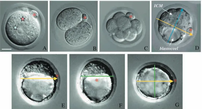
blastocyst is slightly flattened, giving it a shape that lies somewhere between that of an American football and a European one, with its longer dimension being a polarised axis of bilateral symmetry (Fig. 1E-G). However, before I discuss further how blastocyst polarity relates to preceding events that establish asymmetry of the zygote, I would like to follow what happens to blastocyst polarity as the embryo implants.
Implantation, its symmetries and asymmetries
Implantation remains something of an enigma: it cannot be observed in vivo or mimicked in vitro. Consequently, our view of it must still be supported by the power of our imagination, in order to provide continuity to a number of static pictures. Implantation not only establishes direct interaction of the embryo with the tissues of the mother that are crucial for development, but also provides the first chance for the uterine environment to influence embryo polarity. Are the asymmetries that become obvious after implantation imposed on an embryo by this process or are they present earlier, defined within the polarity of the embryo before it touches the uterus? Alternatively, do these two influences interact to pattern the later embryo?
Just before it implants, the blastocyst is composed of three different tissues: two of them are referred to as ‘extra-embryonic’ (trophectoderm and primitive endoderm), because they do not contribute any descendants to the future body; and the third one is termed ‘embryonic’ (the core cells of the ICM) (Fig. 2A-C). There are two distinct types of trophectoderm: the mural trophectoderm, which surrounds the blastocyst cavity, and the polar trophectoderm, which covers the entire ICM. Primitive endoderm differentiates on the surface of the ICM and is destined to contribute to parietal and visceral endoderm. Finally, the remaining core cells of the ICM will develop into the epiblast, the progenitor tissue for the whole future body.
When the mouse embryo implants, it changes its form and size rather dramatically. Proliferating polar trophectoderm yields the extra-embryonic ectoderm that seems to ‘push’ the proliferating ICM complex into the blastocyst cavity (Copp, 1979). This inward growth, which is characteristic of rodent embryos, transforms the embryo into an elongated cylindrical structure (so called, egg cylinder). Thus formed, the egg cylinder has a proximodistal axis about which the embryo is regarded to be largely radially symmetrical until just before gastrulation, when its anteroposterior orientation emerges (Huber, 1915). So what is the consequence of the bilateral symmetry that has been observed before implantation?
[image:3.612.118.507.72.283.2]the asymmetrical properties of the implanting blastocyst (reviewed by Tam et al., 2001). This stimulating concept was subsequently partially re-addressed by Gardner et al. (Gardner et al., 1992). However, although their findings confirmed that the anteroposterior axis was oriented in alignment with the tilt of the ectoplacental cone, they were contrary to Smith’s prediction that the direction of this tilt would foretell the
polarity of the anteroposterior axis. Rather, they found that the direction of tilt of the ectoplacental cone/egg cylinder interface was associated as often with the anterior as it was with the posterior of the definitive embryo.
At present, it is still impossible to reach a firm conclusion about the significance of the various asymmetries observed during implantation, because it is not clear exactly how they relate to each other. For example, we do not know whether the post-implantation tilt described by Smith (Smith, 1985) and studied by Gardner et al. (Gardner et al., 1992) bears any relationship to the tilt of the ICM surface in embryos that are a few days younger. It also remains unknown whether the asymmetry of the implanting blastocyst bears any relationship to either the axis of bilateral symmetry (animal-vegetal axis) of the pre-implantation blastocyst or indeed to any later axis. Accordingly, these various asymmetries at different peri-implantation stages highlight the need for experiments that might link them to the polarity of the embryo before and after implantation.
Following cell fate shows that polarity of the pre-implantation blastocyst prefigures polarity of the post-implantation egg cylinder
[image:4.612.45.295.72.421.2]Insight into whether asymmetries present in the pre-implantation blastocyst predict any aspect of polarity of the post-implantation embryo was gained only recently through lineage tracing studies (Weber et al., 1999). These required development of an approach for marking individual early embryonic cells in an enduring way, so that their progeny could be followed beyond implantation, when dramatic growth of the embryo begins (Zernicka-Goetz et al., 1997; Zernicka-Goetz,
Fig. 2. Relationship between lineages of the pre-implantation blastocyst and post-implantation egg cylinder with summary showing asymmetric distribution of visceral endoderm cells at E5.5 and E6.5. (A-C) Core of the ICM in the blastocyst (A, blue) contributes to the epiblast of the egg cylinder at E5.5 (B) and E6.5 (C). Primitive endoderm in the blastocyst (A, yellow) contributes to parietal endoderm (not shown) and visceral endoderm of the egg cylinder (B,C). Polar trophectoderm (A, green) develops into extra-embryonic ectoderm of the egg cylinder (B,C). Colour has been added to DIC images of embryos to indicate lineage relationships between cells. (D) Schematic representation (box-whisker plots) of findings of Weber et al. (Weber et al., 1999) tracing the fate of ICM cells near the polar body (as indicated by the green star) (N/PB, blue) and away from it (A/PB, red) during development from blastocyst to egg cylinder stages. Bar indicates median; the lower and upper limit of the boxes and their whiskers illustrate 25%, 75% and entire range of distributions, respectively. N/PB descendants in visceral endoderm tend to be distributed more distally (embryonic), and A/PB
descendants are distributed more proximally (extra-embryonic). This reciprocal fate of visceral endoderm descendants is already apparent at E5.5 and accentuates with development to E6.5.
A B
Em
Ab
A P
Em
[image:4.612.333.533.429.593.2]Ab “AP axis” ?
Fig. 3. Schematic model based on Smith’s observations and
conclusions on relationship between asymmetry of the blastocyst and early post-implantation egg cylinder (Smith, 1980; Smith 1985; Gardner et al., 1992) and further modified to emphasise unresolved issues. (A) Implanting blastocyst oriented with its embryonic-abembryonic axis parallel to the mesometrial-antimesometrial axis of the uterus. Smith observed the upward tilting of the polar
1999; Weber et al., 1999). Such an approach enabled tracing the lineages of ICM cells located on either end of the animal-vegetal axis of the blastocyst until the anteroposterior axis became clearly visible (Weber et al., 1999). For two reasons, this study concentrated on the precursor cells of visceral endoderm (the embryonic tissue that surrounds both epiblast and extra-embryonic ectoderm; Fig. 2). First, the ICM descendants in visceral endoderm tend to grow in coherent clones (Gardner, 1984), whereas ICM descendants destined to become epiblast mix during egg cylinder formation (Beddington et al., 1989; Lawson et al., 1991; Gardner and Cockroft, 1998). Such clonal coherence might enable the cells of the visceral endoderm to maintain and carry extant positional information onwards to later stages. Second, the visceral endoderm patterns the cells of the epiblast (for a review, see Beddington and Robertson, 1999; Thomas and Beddington, 1996).
The lineage tracing study of Weber and colleagues (Weber et al., 1999) marked surface cells of the ICM located either near the polar body (N/PB), which marks the animal pole, or away from the polar body (A/PB), at the vegetal pole, and then examined their fate in the egg cylinder (both the day before primitive streak formation and the day of its formation). This work revealed that when the ICM and polar trophectoderm complex elongats to form the egg cylinder, the distribution of labelled visceral endoderm cells is not symmetrical but polarised in relation to the polarity of the blastocyst. Specifically, visceral endoderm descendants of N/PB cells tend to become located progressively more distally as the egg cylinder grows, in comparison with descendants of A/PB cells, which tend to occupy more proximal positions (Fig. 2D).
The mechanistic basis for such polarised egg cylinder growth is still not clear. However, several general conclusions can be drawn. Visceral endoderm clones in the extra-embryonic part of the egg cylinder were often coherent, whereas clones in the embryonic part were dispersed and usually consisted of small groups of scattered cells (Fig. 4). This indicates that the nature or extent of cell displacement in these two parts of the embryo differs. The larger and more coherent clones found in the extra-embryonic portion of the mouse egg cylinder typically displayed a diagonal orientation, extending from anterior-proximal to more posterior-distal regions. This also suggests that egg cylinder growth might differ on the prospective anterior and posterior sides. In the distal (embryonic) part of the egg cylinder, the characteristic distribution of labelled cells was strikingly reminiscent of the ‘polonaise’ movements first described by Graper (Graper, 1929) in the chick embryo and named after the choreography of a traditional Polish dance. A detailed view of these movements has recently been captured by Claudio Stern’s group in time lapse studies of chick embryos. Their film also beautifully illustrates how hypoblast cells (the tissue corresponding to mouse visceral endoderm) move along the future path of the primitive streak, thus predicting the anteroposterior axis (Foley et al., 2000). Interestingly, the orientation of the extra-embryonic visceral endoderm clones in mouse embryos is consistent with the polonaise distribution in the embryonic part, which suggests a common underlying mechanism (Fig. 4). Indeed, the whole visceral endoderm layer would seem to be participating in these characteristic movements, which precede the onset of gastrulation (defined by primitive streak formation), as in the chick embryo.
To summarise, tracing the fate of ICM cells reveals a relationship between the polarised organisation of the blastocyst and the post-implantation egg cylinder, as the distribution of labelled visceral endoderm cells is asymmetric along the proximal-distal axis of the egg cylinder. Moreover, the clonal patterns of labelled cells suggest that polarised displacements of visceral endoderm precede gastrulation. This would encompass the shifts in cell positions along the future anteroposterior axis that were originally discovered by Rosa Beddington’s group (Thomas et al., 1998) and corroborated by others (Weber et al., 1999; Kimura et al., 2000; Perea-Gomez et al., 2001). Thus, asymmetric cell displacements occurring between implantation and gastrulation appear to be one part of the specification of the anteroposterior axis in the mouse.
Molecular aspects of the polarised behaviour of visceral endoderm cells
[image:5.612.59.291.424.584.2]The asymmetric displacements of the visceral endoderm cells are accompanied by dynamic changes in patterns of gene expression. To what extent do these changes in gene expression support a role for such displacements in the development of anteroposterior polarity? The initial report by Thomas et al. (Thomas et al., 1998) showed that the domain of Hex expression in visceral endoderm shifts from the distal tip of the E5.5 embryo to the anterior by E6.5. Recent studies found that another gene, cerberus-like (Belo et al., 1997; Shawlot et al., 1998; Biben et al., 1998; Stanley et al., 2000), undergoes a similar transition in its expression pattern. The anterior shift of such distally expressed genes fails to occur in animals mutant for several other genes. For example, loss of expression Fig. 4. Distribution of GFP-labelled descendants in the visceral
of cripto results in forebrain markers appearing at the distal tip of the egg cylinder, instead of at its future anterior, which leads to defective anterior development (Ding et al., 1998). A similar phenotype has been reported for Otx2 mutants (Kimura et al., 2000; Perea-Gomez et al., 2001). This points to a role for these genes in the displacement of cells towards the anterior of the embryo and the importance of this displacement in transforming the proximodistal polarity of the egg cylinder to an anteroposterior one.
The localised expression of Hex has focused attention on the small group of cells that initially occupy the distal tip of the embryonic region of the egg cylinder and then shift to its anterior margin just before gastrulation (Fig. 5). This anterior visceral endoderm, or AVE, has been singled out for further analysis of its role in anterior development. Although studies of the AVE have focused on induction of the head region, they also shed light on development of anteroposterior polarity as a whole. So what originally promoted the idea that the AVE acts as an anterior inducing centre? It was particularly revealing that ablation of the AVE led to embryos that lack head structures (Thomas and Beddington, 1996). Additionally, several transcription factors expressed in the AVE, such as Otx2 (Simeone et al., 1993; Ang et al., 1994), Goosecoid (Blum et al., 1992) and HNF3β (Sasaki and Hogan, 1993; Weinstein et al., 1994), are also expressed in the node, which is the organiser at later developmental stages. Subsequent
genetic evidence has shown that embryos lacking Otx2 (Acampora et al., 1995; Matsuo et al., 1995; Ang et al., 1996) or Lim1 (Shawlot and Behringer, 1995) function have a normal trunk and tail but lack head structures. In both cases, these anterior defects can be rescued, at least partially, in chimaeras with wild-type gene function in the visceral endoderm [Otx2 (Rhinn et al., 1998; Rhinn et al., 1999) and Lim1 (Shawlot et al., 1999)]. Similarly, although a homozygous deletion of nodal leads to strong morphological defects (Conlon et al., 1994), overall anteroposterior pattern is rescued in chimaeric embryos with wild-type nodal function in the visceral endoderm (Varlet et al., 1997). Taken together, these findings demonstrate a role for the visceral endoderm, and perhaps specifically the AVE, in anteroposterior patterning.
However, it now appears that while the AVE is important for anterior development, it is not sufficient. Specifically, mutant embryos that lack expression of Wnt3 (Liu et al., 1999) or both chordin and noggin (Bachiller et al., 2000) form the AVE, yet their head development is impaired. In addition, embryonic manipulations suggest that the AVE may not act as a direct anterior-inducing centre. This is because the AVE on its own fails to induce the anterior nervous system when grafted to a lateral region of the egg cylinder, but is able to do so when combined with signals from the tip of the early primitive streak and the anterior epiblast (Tam and Steiner, 1999). Consistent with this, genetic studies indicate that the anterior definitive endoderm, a derivative of epiblast, is required in addition to the AVE in the process of head induction (Shawlot et al., 1999; Martinez-Barbera et al., 2000; Mukhopadhyay et al., 2001).
Concomitant with the onset of anterior-specific gene expression in the AVE, posterior patterns of gene expression are also becoming established. Again, it is an extra-embryonic tissue, in this case extra-embryonic ectoderm, that patterns the cells of adjacent epiblast. Hence, posterior character first develops in the proximal epiblast, induced by BMP4 expressed in the extra-embryonic ectoderm (for a review, see Behringer et al., 2000; Lawson et al., 1999). Next, simultaneous with the distal to anterior displacement of Hex expression in AVE, the activity of proximally expressed genes shifts towards the future posterior of the epiblast, where the primitive streak will form (Fig. 5). It seems that there is a relationship between these changes in the posterior and the anterior patterns of gene expression: if one fails, so does the other. In mutants where the expression domain of the AVE genes does not shift, the expression of brachyury and Wnt3 fails to localise to the posterior (Kinder et al., 2001). However, whereas lineage studies indicate that changes in visceral endoderm gene expression reflect the movement of cells anteriorly (Thomas et al., 1998; Weber et al., 1999), the nature of the proximal-to-posterior shift in gene expression in the epiblast is unclear. Thus, posterior localisation of gene expression could represent a posterior displacement of epiblast before the onset of gastrulation, mirroring the anterior displacement of AVE. However, it also might reflect still poorly understood changes from radial to bilateral symmetry as the egg cylinder approaches gastrulation (Snell and Stevens, 1966). Alternatively, it could represent the anterior movement of the AVE, which acts to restrict posterior identity by silencing expression of these genes in the anterior (but not the posterior) of the embryo. This possibility has been expressed in a model (Fig. 5) in which the role of the AVE is not to induce anterior
Distal extra-embryonic ectoderm induction
Proximal/posterior epiblast gene expression pattern Distal/anterior visceral endoderm
E5.5 E6.0 E6.5
A P
Proximal
[image:6.612.64.271.75.240.2]Distal
Fig. 5. Model of visceral endoderm cell movements and
structures, but rather to set the stage for anterior patterning by preventing development of posterior character in the adjacent epiblast (Kimura et al., 2000; Kimura et al., 2001; Perea-Gomez et al., 2001). Another part of the role of the visceral endoderm might be to direct movements of adjacent epiblast cells away from the posteriorising signals present in the primitive streak as observed in chick embryos (for a review, see Stern, 2001; Foley et al., 2000). Thus, it is possible that not only the local ‘contents’ of the AVE itself, but a more global property of the visceral endoderm is important in the development of the anteroposterior patterning.
In summary, the patterns of gene expression described above are consistent with polarised expansion of the egg cylinder when it develops from the blastocyst stage, as revealed by cell lineage studies. Yet they do not tell us how polarity of the blastocyst translates into such asymmetric gene expression. In part, this might be because the studies on gene expression patterns so far have concentrated on changes between 5.5 and 6.5 days of development, owing to the fact that the 5.5-day-old embryo is the earliest post-implantation stage that can be recovered efficiently. Consequently, we still do not know the identities of genes upstream of those expressed at E5.5 and thus cannot exclude the possibility that these are preceded by other asymmetries in the pre-implantation embryo itself. At present, we also cannot rule out a modulating effect of the uterus on the patterning of the implanting embryo. For example, it can be speculated that initial embryonic polarity could act by predisposing specific regions of the blastocyst to adhere to the uterus, which in turn could induce asymmetric behaviour of the embryo. In this way, intrinsic and extrinsic influences could interact and the asymmetries within the embryo itself might be enhanced by implantation. It can therefore be concluded that positional information in either the embryo itself or in both embryo and uterus predicts the anteroposterior axis.
Moving forwards
Both the asymmetric proximodistal distribution of ICM descendants in the visceral endoderm and the directionality of clonal spreading link the polarity of the blastocyst and that of the post-implantation embryo. However, they do not reveal which features of the embryo trigger directional displacements of visceral endoderm to bring cells to locations of the egg cylinder at which they can adopt specific fates. To address this, it seems necessary to resolve several other issues related to the development of the embryo between the blastocyst and egg cylinder stages. Visceral endoderm descendants of N/PB or A/PB cells do not exclusively occupy future anterior or posterior positions, nor do they consistently occupy the left and right aspects of the egg cylinder in a symmetrical fashion (Fig. 4) (Weber et al., 1999) (R. Pedersen, R. Weber and M. Z.-G., unpublished). Thus, the relationship between the animal-vegetal axis of the blastocyst and the future anteroposterior axis is not a simple one. This justifies caution in deciding exactly how blastocyst polarity predicts polarity of the post-implantation embryo. Indeed it still has to be determined whether the precise positioning of the anteroposterior axis could also be influenced by other asymmetries in the blastocyst, or the implantation site, that are known or yet to be identified. There is also a need to understand the mechanism that mediates the displacement of visceral endoderm cells and to determine whether this is a consequence, for example, of
differential cell division or cell migration, or a reflection of any other transformations in the egg cylinder as it elongates. It may then become clearer to what extent these movements follow intrinsic polarised information within the blastocyst or the degree to which asymmetry of implantation has an effect. Finally, the discovery that axial organisation of the post-implantation embryo is already anticipated within the blastocyst structure naturally invites the question ‘how does the blastocyst acquire its polarity?’.
How does the polarised architecture of the blastocyst develop?
In view of the relationship between polarity of the blastocyst and that of the later embryo, it now seems premature to dismiss early patterning in guiding development of mammalian embryos. Indeed recent studies suggest there are at least two developmental cues that operate in the mouse egg to direct axial organisation of the blastocyst. It has thus emerged that both animal-vegetal and embryonic-abembryonic axes of blastocyst symmetry have their roots in the very earliest stages of embryonic development.
Early developmental cues and blastocyst polarity
The first cue for developmental patterning in the mouse egg appears to be at its animal pole. This is defined by the second meiotic division where the polar body provides a persistent landmark that later becomes aligned with the plane of bilateral symmetry of the blastocyst (Fig. 1). Although the position of the polar body varies along this plane, it has been found to have a pronounced tendency to be located near the boundary between the embryonic and abembryonic regions (Gardner, 1997). Thus, the persistence of the polar body tethered to the surface of the embryo provides the link between a point on the surface of the animal pole of the zygote and its location at the blastocyst stage. Could this indicate a spatial relationship between the organisation of the egg and blastocyst that reflects the entire animal-vegetal axis? Tracing the lineages of early blastomeres has shown that not only the cortical surface but also the cytoplasmic contents of the animal pole are conveyed to the region of the blastocyst that lies adjacent to the polar body (Ciemerych et al., 2000). Similarly, the cytoplasmic contents of the vegetal pole are conveyed to the opposite region of the blastocyst. As a result, animal and vegetal pole-derived cells come to lie on opposite ends of an axis with polarity that persists from early cleavage stages.
sister; thus, contributing to the well-known asynchrony of division of early blastomeres (Bennett, 1982; Piotrowska and Zernicka-Goetz, 2001). Third, in most embryos (81%) in which the SEP marker was located at the first cleavage plane it was later found to mark the boundary between embryonic and abembryonic regions of the blastocyst (Piotrowska and Zernicka-Goetz, 2001). Consequently, the location of the SEP marker at this boundary indicates that the position of the first cleavage plane separates the future embryonic and abembryonic parts of the blastocyst. These results together suggest that either sperm entry itself defines a positional cue that affects the orientation and timing of the early cleavages or occurs preferentially at the site of such a cue. Ultimately, this will fashion the alignment of the embryonic-abembryonic axis of the blastocyst. The relationship between the first cleavage plane and the embryonic-abembryonic axis has also been observed in another study (Gardner, 2001). In this case oil droplets were used to mark regions of the zona pellucida in relation to underlying features of two-cell stage mouse embryos. These were subsequently assessed with respect to the axial organisation of the blastocyst, when it was also found that the orientation of the first cleavage predicts the embryonic-abembryonic boundary.
These findings were unexpected. Not only was there no real weight given to the idea that sperm entry position might relate to spatial development of the mouse embryo, but also the first cleavage was thought to be oriented randomly about the animal-vegetal axis. Moreover, it was not thought that the embryonic-abembryonic axis of the mouse embryo might be anticipated at the earliest possible stage of zygotic development, at the time of its initial cleavage. Finally, these findings have led to the surprising prediction that blastomeres arising from the first cleavage division would follow different destinies: one contributing mainly to embryonic and the other to abembryonic parts of the blastocyst. Such a degree of ‘pre-patterning’ was not expected because, at first sight, it seemed to conflict with the equality of potential and therefore naïvety of early mouse blastomeres. A model has been put forward, however, that accommodates the plasticity of the two-cell stage blastomeres (their ability to contribute to both ICM and
trophectoderm lineages) with the partitioning of the embryo into future embryonic and abembryonic parts by the first cleavage. In this model, one two-cell blastomere has been predicted to contribute descendants to mainly mural trophectoderm and to the surface ICM cells that can form primitive endoderm, while the other blastomere has been predicted to contribute descendants to mainly polar trophectoderm and the core ICM cells that would become primarily epiblast (Piotrowska and Zernicka-Goetz, 2001; Gardner, 2001). Could support for this hypothesis be provided by tracing blastomere lineages of the intact two-cell embryo?
Two-cell embryos have polarity predicting the embryonic-abembryonic axis
Previous lineage tracing of two-cell blastomeres by intracellular injection of horseradish peroxidase confirmed that both cells contribute to ICM and trophectoderm, but did not reveal any tendency of such cells to contribute preferentially to either embryonic or abembryonic regions of the blastocyst (Balakier and Pedersen, 1982; Gardner, 1997). This outcome seemed therefore to support the conclusion that blastocyst polarity does not emerge from spatial patterning in the earlier embryo. However, there is a certain risk attached to such a definitive interpretation. This is because those studies did not consider the possibility that intracellular microinjection of markers could delay cell division, thus altering division order and affecting cell fate (Dyce et al., 1987).
[image:8.612.163.561.525.718.2]Thus, the question of whether fate of blastomeres is distinguishable at the two-cell stage has been re-investigated using an alternative approach. In this, fluorescent markers were applied externally to cells, that is without physically disturbing them, and confocal imaging was used to analyse individual sections of the resulting blastocyst (Piotrowska et al., 2001). The aim of this work was to examine the contribution of each of the two blastomeres to three parts of the blastocyst, an embryonic part, an abembryonic part and the boundary zone between them (Fig. 6). The boundary zone was defined as an approximately one-cell deep layer of ICM adjacent to the interior margin of the blastocoel from which primitive endoderm would be expected
Fig. 6. Distinguishable fates of blastomeres of the two-cell embryo. Confocal sections of a blastocyst showing that the clonal border between progeny of the two-cell blastomeres corresponds with a plane separating the embryonic and abembryonic parts. Blastomeres were labelled at the two-cell stage with dyes of different colours (Piotrowska et al., 2001). The boundary zone is marked with broken red lines and the border of the blastocoel was traced on a central section and is shown projected onto each of the other sections as a broken white line. (A-H) Individual optical sections at
to arise. This study demonstrated that, as hypothesised, two-cell blastomeres have a strong tendency to inhabit either the embryonic or abembryonic parts of the blastocyst (Fig. 6). Moreover, the blastomere that contributes preferentially to the abembryonic part also donates significantly more cells to the boundary zone. This work therefore provides support for the recent model proposing that destinies of blastomeres could already be distinguished at the two-cell stage (Piotrowska and Zernicka-Goetz, 2001; Gardner, 2001).
This cell lineage study has also provided an opportunity to determine whether either the earlier or later dividing two-cell blastomere colonised particular parts of the blastocyst. The possibility that the order of blastomere division might predict the final positioning of descendants within the blastocyst had been suggested previously (Graham and Deussen, 1978). But previous conclusions seemed somewhat conflicting. Whereas some studies showed that the earlier dividing blastomeres made a disproportionate contribution to the ICM (Kelly et al., 1978; Graham and Deussen, 1978; Spindle, 1982; Surani and Barton, 1984; Garbutt et al., 1987a), another showed that they had a tendency to be associated with the nascent blastocoel and thus the abembryonic region of the blastocyst (Garbutt et al., 1987b). As a result, the significance of the order of division for embryonic polarity remained unsettled. Understandably, curiosity about this issue was re-awakened by the observation that the earlier dividing two-cell blastomere is the one inheriting the part of the egg surface that has been penetrated by the sperm. Indeed, it has been observed that the earlier dividing blastomere is the one that contributes descendants preferentially to the embryonic part of the blastocyst (Piotrowska et al., 2001). This indicates that not only the orientation, but also polarity of the embryonic-abembryonic axis of the blastocyst can already be predicted by the early cleavage pattern (Fig. 7). Because this polarity typically manifests itself as the earlier division of the two-cell blastomere that acquired the part of the egg where the sperm entered, embryonic-abembryonic asymmetry can ultimately be traced back to events seen at fertilisation.
Relationship between animal-vegetal and embryonic-abembryonic axes
Understanding the fate of two-cell blastomeres also provides
insight into the sources of variation in the way the blastocyst is organised that had been detected, but not explained, by previous studies. This is because the distribution of the progeny of the two blastomeres revealed that the clonal border between them, which reflects the first cleavage plane, was often not perfectly parallel to the embryonic-abembryonic boundary (Piotrowska et al., 2001). Instead it usually showed some angular displacement (a tilt) from this boundary. Consequently, the boundary zone was generally occupied by cells derived from both two-cell blastomeres, although the greater contribution was from the later-dividing one. As the location of the polar body reflects a point upon the first cleavage plane, its position should vary with respect to the embryonic-abembryonic axis, as does the cleavage plane itself. This might now explain the variability in the location of the polar body observed previously (Gardner, 1997; Ciemerych et al., 2000). Similarly, the variation between the orientation of first cleavage plane (as identified by sperm entry position, or as marked on the overlying zone pellucida) and orientation of the embryonic-abembryonic boundary can also be understood from this perspective.
Interestingly, even in embryos that display little tilt between the clonal border of the progeny of two-cell blastomeres and the embryonic-abembryonic boundary, the surface ICM cells from the ends of the animal-vegetal axis are often derived from different two-cell stage progenitors (Fig. 6). Could this have consequences for subsequent development? Certain cells within this region follow different fates, depending on their position on the animal-vegetal axis (Weber et al., 1999). However, although it is quite tempting to think that different behaviour of cells located at opposite ends of this axis might reflect their origins from distinct two-cell blastomeres, gaining insight into this would require further careful experiments.
[image:9.612.172.574.74.174.2]It is intriguing that the sperm entry position, the plane of first cleavage and the boundary between embryonic and abembryonic regions are related to each other, but variably so. At this stage, there are several possibilities that can account for such variability. First, there could be as yet unknown factor(s) in the egg that influence the orientation of the embryonic-abembryonic axis and that also have a specific spatial relationship to the first cleavage plane [see fig. 5 in Piotrowska et al. (Piotrowska et al., 2001)]. Second, variation in the Fig. 7. The embryonic part of the
blastocyst tends to be derived from the first blastomere to divide at the two-cell stage. (A) Egg shortly after fertilisation showing the fertilisation cone (fc) with sperm tail coloured in yellow and fluorescent bead marking the sperm entry point (SEP) shown in green. Second polar body marks the animal
position of the sperm entry along the egg’s animal-vegetal circumference might contribute to variability in the orientation of the embryonic-abembryonic axis. Third, stochastic factors could influence the position where the blastocyst cavity forms with respect to the first cleavage plane. Currently it is difficult to distinguish between these alternatives, or eliminate any others, because of lack of basic information on the molecular and cellular nature of polarity at the onset of mammalian development.
What is the molecular and cellular basis for early patterning of the mouse embryo?
The polar body at the animal pole and the SEP at the egg surface provide markers that enable recognition of orderly cell deployment during the first few days of mouse embryo development. But what is it at the animal pole that predicts spatial patterning, and what is the molecular nature of the positional cue associated with the SEP? Also the mechanisms by which these positional cues achieve their effects on the early patterning of the embryo remain unknown. It seems likely that the animal pole of the egg has its origin in the process of oocyte maturation. Specifically this is when, after germinal vesicle breakdown, the meiotic spindle that forms in a relatively central position of the oocyte moves to its cortex. Does the spindle move to the nearest region of the cortex as one study suggests (Verhlac et al., 2000), or is it responding to some inconspicuous asymmetrically distributed ‘factor’? There are descriptions of various properties that distinguish the animal pole of the egg from its vegetal counterpart, but whether these have significance for polarity of the oocyte or zygote is also unknown. For example, the properties of the egg surface overlying the meiotic spindle (i.e. the animal pole) are different from those around the rest of the egg surface (Eager et al., 1976; Nicosia et al., 1977; Phillips and Shalgi, 1980; Wolf and Ziomek, 1983; Maro et al., 1984; Maro et al., 1986; Longo and Chen, 1985; Calarco, 1991). Some of these surface asymmetries could explain why the sperm penetrates the mouse oocyte preferentially at regions other than its animal pole (Evans et al., 2000). It is striking that phytohemaglutinin-coated beads used to mark the egg surface where the sperm entered tended to retain their position relative to other surface markers from egg to blastocyst stages. Martin Johnson has pointed out that this could indicate the presence of some ‘solid islands (of surface matrix) in a sea of otherwise fluid lipids’ (Johnson, 2001). Perhaps these islands or even continents are linked through some underlying cortical cytoskeletal structure. Whatever their structure, some surface features of the egg appear to be preserved throughout these developmental stages.
The vegetal pole, and thus animal-vegetal polarity, also becomes evident within the egg. For example, pulsatile waves of Ca2+ release that occur in response to fertilisation are initiated near the sperm entry point, then move towards the vegetal pole (Kline et al., 1999; Deguchi et al., 2000). There is also an asymmetric distribution of mitochondria (Van Blerkom and Runner, 1984; Calarco, 1995) along the animal-vegetal axis. Although no maternal transcripts have yet been found to be asymmetrically distributed in mouse eggs, as occurs in other systems, location of certain proteins, including leptin and Stat3, correlates with the animal-vegetal axis in the egg and later embryo (Antczak and Van Blerkom, 1997).
Nevertheless the functional significance of these findings still remains unclear.
The events triggered by fertilisation in the mouse zygote are equally poorly understood. In organisms such as C. elegans and Xenopus, sperm entry stimulates cytoskeletal reorganisation that redistributes cytoplasmic constituents within the egg so they will be asymmetrically inherited. In Xenopus, this is accomplished by cortical rotation in response to sperm entry (for a review, see Gerhart, 1991; Vincent and Gerhart, 1987), which changes the relative distribution of maternal factors such as dishevelled protein (Miller et al., 1999). In C. elegans, sperm entry triggers cytoplasmic fluxes that establish asymmetric distribution of molecules, which are important in the subsequent development of embryonic polarity such as Par proteins (for a review, see Bowerman and Shelton, 1999; Goldstein and Hird, 1996; Wallenfang and Seydoux, 2000; Kemphues, 2000). To date there are no reports, however, showing that the mouse egg acquires an asymmetric distribution of its molecular components in response to fertilisation. Where egg constituents have been found to redistribute in response to sperm entry, this has often been linked to the influence of sperm components, such as the centrosome, on the egg cytoskeleton. Although no sperm centriole has been observed in the mouse egg, sperm penetration does bring about cytoskeletal re-organisation (for a review, see Maro, 1985). This includes formation of the fertilisation cone, which is associated with local changes in actin filament distributions that overlie the nascent male chromatin. Thus, the possibility cannot be discounted that sperm entry is responsible for cytoskeletal reorganisation and subsequent events in mouse eggs as it is in other animals. However, we also cannot at present exclude that the position of sperm entry around the radial circumference of the egg could be influenced by some feature of the oocyte itself. One way to gain insight into the role of the SEP versus the oocyte in setting up the first cleavage plane is to study blastocyst patterning in the absence of the SEP, namely, in parthenogenetically activated eggs. Such experiments are currently in progress in my laboratory.
Regulation and its relationship to early patterning
It thus appears there is an underlying order in early mouse development that has previously been unappreciated. However, as for many other developmental concepts, the idea of early patterning in mammalian development is not new. Among its proponents were Dalcq and colleagues, who argued that spatial patterning of the early mammalian embryo is reflected in the polarised distribution of constituents (Dalcq, 1957; Mulnard, 1965) (reviewed by Denker, 1983). They proposed that such constituents were partitioned to different blastomeres by cleavage divisions as in other model systems with visible constituents. This proposal was based on histochemical analysis of acid phosphatases and other components that could be interpreted as representing cytoplasmic determinants. However, there was no experimental evidence for any role of such components in the subsequent differentiation of the blastomeres, and studies to establish the lineage history of early embryonic cells expressing such markers were lacking. Rather, this hypothesis appeared to be so much at odds with the results of subsequent work showing the plasticity of mammalian embryos that it was not embraced, but set aside.
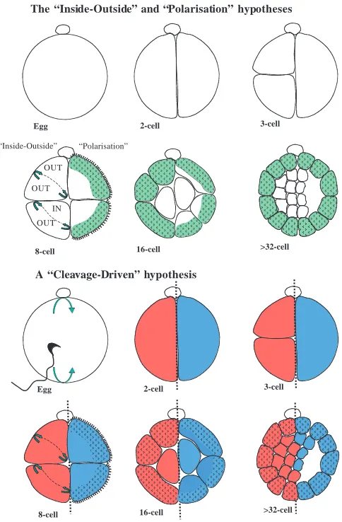
Nevertheless, it could not be excluded that the preimplantation embryo might possess some inherent organisation (‘cryptic preformation’) that is overridden by its regulative ability following experimental perturbation (Graham, 1971). So what has the regulative ability of mouse embryos taught us and how can this accommodate the existence of such early patterning? Regulative capacity was first identified in rabbit and mouse embryos, and subsequently in other species, suggesting that it is characteristic of all eutherian mammals (reviewed by Papaioannou and Ebert, 1986). It was first observed that if single two-cell stage blastomeres are separated, each can fully participate in embryogenesis and has complete developmental potential (Seidel, 1952; Tarkowski, 1959a; Tarkowski, 1959b; Papaioannou et al., 1989). Tarkowski and Wroblewska found that such capacity became limited during cleavage and that isolated, individual four- or eight-cell mouse blastomeres formed mainly trophectoderm rather than a normal blastocyst (Tarkowski and Wroblewska, 1967). This limitation did not however arise from lack of differentiative capacity, because all four-cell mouse blastomeres and at least some eight-cell, 16-cell and even ICM 16-cells maintain the ability to contribute to both ICM and trophectoderm lineages when combined with cells from other embryos to produce chimeras (Rossant, 1975; Rossant, 1976; Kelly, 1977; Kelly et al., 1978; Rossant and Lis, 1979; Rossant and Vijh, 1980; Tarkowski et al., 2001). Such studies revealing the plasticity of mammalian embryos led to the development of the key concept that the position of a blastomere within the embryo is important for its future destiny. To explain diversification of fate by seemingly identical, naïve early blastomeres, two major hypothesis were put forward. In the ‘inside-outside’ hypothesis, Tarkowski and Wroblewska proposed that the fate of blastomeres was a consequence of their having been exposed to different environments (Tarkowski and Wroblewska, 1967): blastomeres that come to lie inside the embryo, being surrounded by other cells, will differentiate into the cells of the ICM, and conversely those cells that stay outside will face the external environment and, thus, develop as trophectoderm cells. The inside-outside hypothesis accounts for the fact that isolated four- to eight-cell
blastomeres form mainly trophectoderm. This is because each cell has been reduced in size as a consequence of cleavage, so it no longer has enough mass to enclose a group of inner cells by the onset of trophectoderm differentiation. It was subsequently observed that in response to their location during cleavage, outer cells differentiate their surface features and cytoskeleton to become polarised in an outside-inside direction. Once acquired, such cellular polarity will endow progeny cells with different properties, depending upon the orientation of cleavage. Accordingly, the ‘polarisation’ hypothesis proposes that differences between blastomeres arise because of such cellular polarisation (Johnson and Ziomek, 1981; Johnson et al., 1981). This hypothesis accounts for the observed trophectoderm fate of cells that inherit the surface features, which differ in their cytoplasmic or cytocortical inheritance from those that lack such features. Both the inside-outside and the polarisation hypotheses thus attribute the ‘decision’ to differentiate into trophectoderm versus ICM to environmental information, although the factor(s) that would trigger such a decision remain unknown.
However, by focussing on the differentiation of ICM and trophectoderm, the earlier experimental studies and the ensuing hypotheses did not explore the question of why the ICM and blastocyst cavity form where they do, at opposite poles, thus defining the embryonic-abembryonic axis. This is a question not about the differentiative capacity of each blastomere, but about the organisation of the embryo as a whole. Now that we have more information about spatial development of the intact embryo, we need to accommodate it into a new hypothesis of how overall embryo form and pattern are initiated. The model proposing that the first cleavage division separates the egg into halves that follow different destinies (Piotrowska and Zernicka-Goetz, 2001; Gardner, 2001), together with lineage tracing studies (Piotrowska et al., 2001) provide the basis for such a hypothesis. This ‘cleavage-driven’ hypothesis proposes that blastocyst polarity arises from asymmetries generated during early cleavage of the embryo (Fig. 8). It predicts that either composition or behaviour of blastomeres differ along the future axes of the embryo, resulting in the overall polarised form of the blastocyst. This hypothesis does not therefore preclude a role for other information in guiding the differentiation into ICM and trophectoderm. This is because it postulates that the boundary between embryonic and abembryonic parts is not immediately adjacent to the interior margin of the blastocoel (blastocoel roof), but lies deeper within the ICM. Consequently, each blastomere will be expected typically to form both ICM and trophectoderm derivatives and this could proceed according to the previously proposed ‘inside-outside’ or ‘polarisation’ mechanisms. Thus, this ‘cleavage-driven’ hypothesis takes into account a role for early patterning information derived from the zygote, and also is actually compatible with existing models accounting for the differentiation of ICM and trophectoderm lineages.
Moving towards a reconcilation of regulation and early patterning
landmarks of mouse polarity that enable us to ask whether potential ‘spatial information’ is localised to specific polar
regions of the egg. The studies performed to address this question have thus far concentrated on the animal-vegetal axis of the egg. They showed that removal of cytoplasmic and cortical components either of the animal or vegetal pole of the egg (Zernicka-Goetz, 1998) or polar cytoplasm at the two- or eight-cell stages (Ciemerych et al., 2000) was compatible with full-term development. How could such regulative ability, which includes even the capacity to recuperate from loss or redistribution of egg poles, be reconciled with early patterning? Taken at face value, these findings would seem inconsistent with a role for spatial patterning in the egg or early embryo. But are they?
A solution to this dilemma may become clear once we understand better the mechanisms by which polarity is established in normal development and learn what actually occurs when an embryo recovers from experimental manipulations. At present there are several possibilities that could account for regulative responses in the context of normal development. First, regulative ability might mean that when the initial polarity of the egg is perturbed, it becomes re-established. For this to occur, positional information would have to be preserved at least to some extent. There could be, for example, a morphogenetic gradient in the egg or embryo that relies on the relative and not the absolute concentration of a morphogen. In this case, even after one pole is removed, the gradient could persist and still be able to direct proper development. It is also possible that either remaining pole of the egg is able to define polarity because it contains information that identifies it as one end of an axis. Thus, in the experimental manipulations described above, polarity would have been disturbed but not really destroyed. Second, regulative development might indicate that when intrinsic polarity is disturbed or destroyed, other asymmetries in the embryo itself or its environment can be adopted as spatial cues to re-establish polarity. In such event, the axes of the regulating embryo might not have the original orientation as in the unperturbed embryo, but might nevertheless arise out of the tendency to polarise using whatever cues might be available in those altered circumstances. A third possibility is, of course, that polarity had not actually been disturbed in experiments that cut away poles of the egg or embryos (Zernicka-Goetz, 1998; Ciemerych et al., 2000). This would be the case if polarised components were sequestered not into the animal or vegetal poles, but resided in other regions, such as at the sides of the egg or embryo, or if they were spatially distributed according to some other positional cue. Alternatively, if the polarity of an embryo were defined through a polarised component such as the cytoskeleton, then it could persist even in a fragment of the embryo. Finally, even in normal development, mammalian embryonic axes could be established by a combination of intrinsic positional cues in the zygote and by the interactions of cells with each other or with their environment. Such a mechanism would also explain the great versatility of the early mammalian embryo. Overall, the regulative aspects of development might provide safeguards that, as in other biological processes, ensure a robust, failsafe system.
In conclusion, even though the mouse embryo shows great flexibility, it appears that its polarity is developing at the very beginning of embryonic life. Consequently, early patterning and regulative ability are interwoven, rather than being
OUT OUT
OUT IN
“Inside-Outside” “Polarisation”
Egg 2-cell 3-cell
8-cell 16-cell >32-cell
Egg 2-cell 3-cell
8-cell 16-cell >32-cell
The “Inside-Outside” and “Polarisation” hypotheses
[image:12.612.48.290.72.441.2]A “Cleavage-Driven” hypothesis
Fig. 8. Decision-making in the early mouse embryo. The prevailing models (upper panels) over the past 20-30 years have acknowledged a differentiation event that discriminates ICM (white) from trophectoderm (pale green) lineages. In the ‘inside-outside’
mutually exclusive. This is perhaps what enables embryos to endure the traumas that we impose on them as well as to accommodate naturally occurring variability. One of the benefits of understanding in molecular terms how the embryo becomes polarised at its earliest stages will be insight into how these regulative processes actually occur. Finally, the recent findings showing a link between earlier and later axial development of the mouse embryo imply that mammals may not be so exceptional after all in how and when their polarity is acquired. But still we wait to learn how far the two-century-old statement of Geoffroy Hilaire (Geoffroy Saint-Hilaire, 1807) applies to mammals: ‘Nature works constantly with the same materials. She is ingenious to vary only the forms. As if, in fact, she were restricted to the same primitive ideas, one sees her tend always to cause the same elements to reappear, in the same number, in the same circumstances, and with the same connections’.
I apologise to all those authors whose original work I could not cite owing to limitation of space. I appreciate invaluable discussions with Chris Graham about patterning in early mouse development, and I also thank David Glover, Roger Pedersen and Peter Lawrence for kindly commenting on the draft of this manuscript. I am grateful to Lister Foundation for Preventive Medicine and Wellcome Trust for support, and I acknowledge my yet unborn daughter for keeping me company while I was writing this review.
REFERENCES
Acampora, D., Mazan, S., Lallemand, Y., Avantaggiato, V., Maury, M., Simeone, A. and Brulet, P. (1995). Forebrain and midbrain regions are
deleted in Otx2–/– mutants due to a defective anterior neuroectoderm
specification during gastrulation. Development 121, 3279-3290.
Ang, S. L., Conlon, R. A., Jin, O. and Rossant, J. (1994). Positive and
negative signals from mesoderm regulate the expression of mouse Otx2 in ectoderm explants. Development 120, 2979-2989.
Ang, S. L., Jin, O., Rhinn, M., Daigle, N., Stevenson, L. and Rossant, J.
(1996). A targeted mouse Otx2 mutation leads to severe defects in gastrulation and formation of axial mesoderm and to deletion of rostral brain. Development 122, 243-252.
Angerer, L. M. and Angerer, R. C. (1999). Regulative development of the
sea urchin embryo: signalling cascades and morphogen gradients. Semin.
Cell Dev. Biol. 10, 327-334.
Antczak, M. and Van Blerkom, J. (1997). Oocyte influences on early
development: the regulatory proteins leptin and STAT3 are polarized in mouse and human oocytes and differentially distributed within the cells of the preimplantation stage embryo. Mol. Hum. Reprod. 3, 1067-1086.
Bachiller, D., Klingensmith, J., Kemp, C., Belo, J. A., Anderson, R. M., May, S. R., McMahon, J. A., McMahon, A. P., Harland, R. M., Rossant, J. and De Robertis, E. M. (2000). The organizer factors Chordin and
Noggin are required for mouse forebrain development. Nature 403, 658-661.
Bachvarova, R. F. (1999). Establishment of anterior-posterior polarity in avian
embryos. Curr. Opin. Genet. Dev. 9, 411-416.
Balakier, H. and Pedersen, R. A. (1982). Allocation of cells to inner cell
mass and trophectoderm lineages in preimplantation mouse embryos. Dev.
Biol. 90, 352-362.
Beddington, R. S., Morgernstern, J., Land, H. and Hogan, A. (1989). An
in situ transgenic enzyme marker for the midgestation mouse embryo and the visualization of inner cell mass clones during early organogenesis.
Development 106, 37-46.
Beddington, R. S. and Robertson, E. J. (1998). Anterior patterning in mouse. Trends Genet. 14, 277-284.
Beddington, R. S. and Robertson, E. J. (1999). Axis development and early
asymmetry in mammals. Cell 96, 195-209.
Behringer, R. R., Wakamiya, M., Tsang, T. E. and Tam, P. P. L. (2000). A
flattened mouse embryo: leveling the playing field. Genesis 28, 23-30.
Belo, J. A., Bouwmeester, T., Leyns, L., Kertesz, N., Gallo, M., Follettie, M. and De Robertis, E. M. (1997). Cerberus-like is a secreted factor with
neutralizing activity expressed in the anterior primitive endoderm of the mouse gastrula. Development 68, 45-57.
Bennett, J. (1982). Sperm entry point is related to early division of mouse
blastomeres. J. Cell Biol. 95, 163a.
Biben, C., Stanley, E., Fabri, L., Kotecha, S., Rhinn, M., Drinkwater, C. et al. (1998). Murine cerberus homologue mCer-1: a candidate anterior
patterning molecule. Dev. Biol. 194, 135-151.
Blum, M., Gaunt, S. J., Cho, K. W., Steinbeisser, H., Blumberg, B., Bittner, D. and De Robertis, E. M. (1992). Gastrulation in the mouse: the role of
the homeobox gene goosecoid. Cell 69, 1097-1106.
Bowerman, B. and Shelton, C. A. (1999). Cell polarity in the early
Caenorhabditis elegans embryo. Curr. Opin. Genet. Dev. 9, 390-395.
Calarco, P. G. (1991). Fertilization of the mouse oocyte. J. Electron Microsc. Tech. 17, 401-411.
Calarco, P. G. (1995). Polarization of mitochondria in the unfertilized mouse
oocyte. Dev. Genet. 16, 36-43.
Cameron, R. A., Fraser, S. E., Britten, R. J. and Davidson, E. H. (1989).
The oral-aboral axis of a sea urchin embryo is specified by first cleavage.
Development 106, 641-647.
Ciemerych, M. A., Mesnard, D. and Zernicka-Goetz, M. (2000). Animal
and vegetal poles of the mouse egg predict the polarity of the embryonic axis, yet are nonessential for development. Development 127, 3467-3474.
Conlon, F. L., Lyons, K. M., Takaesu, N., Barth, K. S., Kispert, A., Herrmann, B. and Robertson, E. J. (1994). A primary requirement for
nodal in the formation and maintenance of the primitive streak in the mouse.
Development 120, 1919-1928.
Copp, A. J. (1979). Interaction between inner cell mass and trophectoderm of
the mouse blastocyst. II. The fate of the polar trophectoderm. J. Embryol.
Exp. Morphol. 51, 109-120.
Dalcq, A. M. (1957). Introduction to General Embryology. London: Oxford
University Press.
Davidson, E. H. (1989). Lineage-specific gene expression and the regulative
capacities of the sea urchin embryo: a proposed mechanism. Development
105, 421-445.
Denker, H. W. (1983). Cell lineage, determination and differentiation in
earliest developmental stages in mammals. Bibl. Anat. 24, 22-58.
Deguchi, R., Shirakawa, H., Oda, S., Mohri, T. and Miyazaki, S. (2000).
Spatiotemporal analysis of Ca2+waves in relation to the sperm entry site
and animal-vegetal axis during Ca2+oscillations in fertilized mouse eggs.
Dev. Biol. 218, 299-313.
Ding, J., Yang, L., Yan, Y. T., Chen, A., Desai, N., Wynshaw-Boris, A. and Shen, M. M. (1998). Cripto is required for correct orientation of the
anterior-posterior axis in the mouse embryo. Nature 395, 702-707.
Dyce, J., George, M., Goodall, H. and Fleming, T. P. (1987). Do
trophectoderm and inner cell mass cells in the mouse blastocyst maintain discrete lineages? Development 100, 685-698.
Eager, D. D., Johnson, M. H. and Thurley, K. W. (1976). Ultrastructural
studies on the surface membrane of the mouse egg. J. Cell Sci. 22, 345-353.
Evans, J. P., Foster, J. A., McAvey, B. A., Gerton, G. L., Kopf, G. S. and Schultz, R. M. (2000). Effects of perturbation of cell polarity on molecular
markers of sperm-egg binding sites on mouse eggs. Biol. Reprod. 62, 76-84.
Fleming, T. P. (1987). A quantitative analysis of cell allocation to
trophectoderm and inner cell mass in the mouse blastocyst. Dev. Biol. 119, 520-531.
Foley, A. C., Skromne, I. and Stern, C. D. (2000). Reconciling different
models of forebrain induction and patterning: a dual role for the hypoblast.
Development 127, 3839-3854.
Garbutt, C. L., Johnson, M. H. and George, M. A. (1997a). When and how
does cell division order influence cell allocation to the inner cell mass of the mouse blastocyst? Development 100, 325-332.
Garbutt, C. L., Chisholm, J. C. and Johnson, M. H. (1997b). The
establishment of the embryonic-abembryonic axis in the mouse embryo.
Development 100, 125-134.
Gardner, R. L. (1984). An in situ cell marker for clonal analysis of
development of the extraembryonic endoderm in the mouse. J. Embryol.
Exp. Morphol. 80, 251-288.
Gardner, R. L. (1997). The early blastocyst is bilaterally symmetrical and its
axis of symmetry is aligned with the animal-vegetal axis of the zygote in the mouse. Development 124, 289-301.
Gardner, R. L. (2001). Specification of embryonic axes begins before
cleavage in normal mouse development. Development 128, 839-847.
Gardner, R. L. and Cockroft, D. L. (1998). Complete dissipation of coherent
clonal growth occurs before gastrulation in mouse epiblast. Development