Harnessing FOXP3
+
regulatory T cells for
transplantation tolerance
Herman Waldmann, … , Duncan Howie, Stephen Cobbold
J Clin Invest.
2014;
124(4)
:1439-1445.
https://doi.org/10.1172/JCI67226
.
Early demonstrations that mice could be tolerized to transplanted tissues with short courses
of immunosuppressive therapy and that with regard to tolerance to self, CD4
+FOXP3
+regulatory T cells (Tregs) appeared to play a critical role, have catalyzed strategies to
harness FOXP3-dependent processes to control rejection in human transplantation. This
review seeks to examine the scientific underpinning for this new approach to finesse
immunosuppression.
Review
Find the latest version:
Harnessing FOXP3
+
regulatory T cells
for transplantation tolerance
Herman Waldmann, Robert Hilbrands, Duncan Howie, and Stephen Cobbold
Sir William Dunn School of Pathology, University of Oxford, Oxford, United Kingdom.
Early demonstrations that mice could be tolerized to transplanted tissues with short courses of
immunosup-pressive therapy and that with regard to tolerance to self, CD4
+FOXP3
+regulatory T cells (Tregs) appeared
to play a critical role, have catalyzed strategies to harness FOXP3-dependent processes to control rejection in
human transplantation. This review seeks to examine the scientific underpinning for this new approach to
finesse immunosuppression.
Introduction
The need for transplantation of histoincompatible cells and tis-sues provides a perpetual challenge to the field of immunology: that of overcoming rejection. From the time that Medawar and colleagues demonstrated that transplantation tolerance could be acquired, there has been the hope that tolerance mechanisms could eventually be harnessed to minimize, if not eliminate, the use of long-term immunosuppression.
The need to explain the diversity of lymphocyte receptors for for-eign antigens begat the clonal selection theory and notions that self-tolerance could be fully explained by clonal deletion at an early stage of lymphocyte development. This thinking provided a high bar for ideas on translation based on targeting immature lympho-cytes. The later discovery in adult rodents that tolerance to foreign proteins and tissues could be achieved following short-term immu-nosuppression with immunosuppressive drugs (1) and certain antilymphocyte monoclonal antibodies (2, 3) led, in turn, to the finding that such tolerance was often dominant and suppressive and largely mediated through CD4+ regulatory T cells (Tregs) (4).
Converging with parallel studies on self-tolerance (5–7), the major Treg responsible for maintaining allograft tolerance proved to be CD4+CD25+FOXP3+ (4, 8, 9). The longevity of tolerance seen in
these experimental models was shown to be dependent on con-stant vigilance by FOXP3+ Tregs (8) and constant recruitment of
new Tregs (infectious tolerance) (8), both processes dependent on a constant supply of graft antigens (10). In addition, linked sup-pression meant that induced tolerance to one set of antigens could be extended to others coexisting in the same tissue, without the need for further therapeutic intervention (11, 12).
The ability to control inflammation can also be achieved with FOXP3-negative CD4 T cells that are often referred to as Tr1 or Tr1-like cells (13–16). Although offering great potential, a proper account of such cells would be outside the scope of this short review, and the reader is referred to excellent reviews on the topic (17–19).
Overall, the findings on CD4+ Tregs, albeit largely derived
from rodents, have provided a new optimism for the therapeu-tic induction of operational tolerance, because unlike strategies based on clonal deletion, one would not need to permanently inactivate all potentially destructive alloreactive T cells. The finding that self-tolerance in humans is also dependent on
FOXP3+ Tregs (20–23) suggests that extrapolation from rodent
studies to humans is not inappropriate
However, harnessing Tregs, although attractive as a concept, still provides a significant challenge, as this heterogeneous cell popula-tion (24) would need to control a broad spectrum of unpredict-able inflammatory responses. The challenge in transplantation is to recruit sufficient numbers of the appropriate Tregs to cover the breadth of tissue-damaging mechanisms evoked.
Tregs and their roles in transplantation tolerance
The FOXP3+ Tregs that populate the peripheral immune system
comprise a set that develops in the thymus (tTregs) (25) and a minority that is induced in the periphery (pTregs) under the influ-ence of TGF-β and mTOR inhibition(26–32). The T cell receptor repertoires of the populations differ, with the latter showing a pat-tern more similar to that of conventional T cells. These features have been interpreted to indicate that tTregs may be preoccupied with ensuring tolerance to self-antigens, while pTregs operate to moderate responses to certain “foreign” antigens that might be found in the gut microbiome or in the fetus during pregnancy (33). Given the potential cross-reactivity of the T cell receptor repertoire, a proportion of tTregs would be expected to exhibit alloreactivity exploitable for tolerance induction (34, 35). The conventional repertoire of pTregs also points to a real prospect for selective tolerizing vaccinations to foreign graft antigens (36).
That tTregs can contribute to transplantation tolerance is illus-trated in certain mouse strains that naturally fail to reject allo-grafts and in which depletion of Tregs then exposes their potential to reject (37, 38). Furthermore, under conditions of lymphopenia, tTregs interfere with the homeostatic expansion of T cells that are competent to reject grafts (39). This is so even if the Tregs are derived from mice in which tolerance by clonal deletion has been enabled through hemopoietic chimerism (40). This tells us that Tregs need not exhibit allospecificity to control rejection in the context of homeostatic expansion and that they perhaps exploit their self-reactive repertoire for that purpose.
The best evidence that pTregs can be harnessed within the host to elicit transplantation tolerance came from studies in TCR trans-genic mice using coreceptor blockade with anti-CD4 antibodies (41). Such mice carrying just one homogenous TCR generate no tTregs, yet can be tolerized to grafts carrying the nominal antigen. Tolerance is accompanied by induction of FOXP3+ pTregs.
How-ever, where TGF-β was prevented from signaling to conventional T cells or where mice lacked a functional FOXP3 gene, tolerance
Conflict of interest: Herman Waldmann and Stephen Cobbold receive royalties from the University of Cambridge on sales of Lemtrada (alemtuzumab).
review
could not be achieved (9, 41, 42). Tolerance through generation of pTregs has more recently been demonstrated with antigen alone in ingenious vaccination protocols (36, 43).
The preoccupation with lineages may, however, obscure an important aspect of FOXP3 expression. The fact that CD4 T cells can transiently or “promiscuously” express FOXP3 may also be relevant to therapeutic induction of tolerance. It has long been known that ectopic FOXP3 expression can turn down inflamma-tory cytokine production and damaging effector functions includ-ing graft rejection (9, 44–46). On this basis, treatments that turn on FOXP3 expression and function, even transiently, may help ensure a temporary ceasefire, perhaps paving the way for stable regulation to evolve over time.
These studies teach us that both tTregs and pTregs can suppress rejection responses, even if they do not necessarily use the exact same mechanisms and TCR specificities. Consequently, where pos-sible, our therapies should exploit both types of Tregs and even early (less stable) stages of their development.
One major gap in our knowledge of Tregs is how homeostasis of tTreg and pTreg populations is maintained, both in relation to each other and to other lymphocyte subsets (47, 48). If our ultimate goal is to exploit antigen-specific Tregs, then we need to understand the nature of the Treg “niche” and how the pTregs might be given a competitive advantage over effector cells and tTregs.
Tregs as lineages
Much of the current thinking on the exploitation of Tregs is predi-cated on both types functioning as stable lineages. From a clinical perspective, such exploitation can be approached from two dif-ferent angles. First, Tregs might be generated in large numbers ex vivo and administered as a cell product (49). Alternatively, proto-cols might be designed to enhance the generation of stable Treg lineages within the patient that would render these cells more amenable to standard pharmaceutical approaches. The Tregs that develop in vivo would, of course, be influenced and shaped in their development by graft exposure in a contextual way that might not be easily simulated by Tregs generated ex vivo.
The importance of the transcription factor FOXP3
It is undeniable that FOXP3 plays a key role in ensuring immune homeostasis and tolerance. FOXP3 is essential for the development and suppressor function of Tregs, since its loss or even disruption results in overt lymphoproliferative disease, autoimmunity, and graft rejection (44, 50, 51). But clearly, there is more to Tregs than simple FOXP3 expression. They are characterized by specific netic changes that define their lineage commitment. These epige-netic changes can be established independently of FOXP3, indicat-ing that FOXP3 is a rather late-actindicat-ing transcription factor in Treg lineage commitment (52, 53). Furthermore, expression of FOXP3 can occur transiently in nonregulatory cells (“promiscuous” FOXP3 expression) that fail to undergo Treg-specific epigenetic changes and lineage commitment, but can also be lost transiently in committed Tregs characterized by their epigenetic signature (“ex-Tregs”) (54–57). These findings have important implications for the way we regard the stability and plasticity of Tregs in relation to FOXP3 expres-sion (58, 59). It suggests that FOXP3 protein, though essential for suppressor function, does not unequivocally reflect the epigenome associated with committed Tregs. From the point of view of therapy, the issues of functional stability and derivation of undesirable proin-flammatory revertants need to be rigorously controlled (60).
How is lineage stability acquired?
TCR signaling. Despite the fact that the epigenetic signature is the best available indicator of lineage stability to date, the signals required for its establishment remain poorly understood. The intensity and duration of TCR signaling are critical for acquisition of the Treg-specific epigenetic signature (52). This is supported by the well-known finding that CD4 T cells expressing nuclear FOXP3 following short-term TGF-β–dependent induction in vitro rarely express Treg-specific epigenetic changes and, consequently, generate unstable FOXP3+ cells that are able to revert to
proin-flammatory functions (61). In the case of tTregs, lineage-specific epigenetic marks are installed very early on in thymic develop-ment, even before FOXP3 is expressed (53, 62). Since proliferation seems of little importance, it would seem that an active mecha-nism is involved in DNA demethylation of the developing Treg, as has previously been suggested for the stabilization of Il2 gene expression (62, 63).
Metabolic requirements. Increasing evidence suggest that metabol-ic changes play an important role in regulating FOXP3 expression and lineage commitment, at least for pTregs. Tregs and effector T cells require different metabolic programs to function. Actively proliferating effector T cells express high levels of the cell surface glucose transporter GLUT1 and engage the relatively energy- inefficient aerobic glycolysis and glutaminolysis pathways. Despite yielding low amounts of ATP, aerobic glycolysis provides precur-sors for nucleotide synthesis via the pentose phosphate shunt and fatty acids via the metabolism of citrate, both of which are required for a cell to increase biomass during division. This mode of metabolism is common to tumor cells and has been termed the “Warburg effect.” In contrast to Th1, Th2, and Th17 cells, which have high plasma membrane expression of the GLUT1 glucose transporter and actively engage glycolysis, Tregs have little GLUT1 and use oxidative phosphorylation via lipid oxidation as their pri-mary energy source (64, 65). There is an intricate link between con-trol of the AKT/mTOR/HIF1a axis and the source of ATP that a cell uses (29). At present, it is unclear whether the signals, which induce the Treg or T effector lineage, are controlled by metabol-ic pathway engagement, as has been described for CD8 T cells (66–68), or whether the divergent metabolism is a consequence of the lineage decision.
function and survival by enhancing lipid metabolism and inhibi-tion of TORC2 signaling (29). It is still unclear, though, whether the engagement of glycolysis downstream of HIF1a activation or the activation of lipid metabolism by inhibition of TORC2 is a prerequisite for these lineage decisions or merely a result of differ-ent fuel demands. The implications of these studies are that there may be a number of metabolic targets for skewing T cells toward Treg development.
Tregs and their role in tolerated tissues
The demonstrations of linked suppression (11, 12) and FOXP3+
Tregs in tolerated grafts suggested (72, 73) that Tregs operate within tissues to protect them against immune attack. Direct evi-dence for this has been provided by retransplantation of tolerated grafts into recipients with no adaptive immune system, in which ablation of CD25+ (73) or FOXP3+ T cells (8) resulted in graft
rejection by residual lymphocytes within those grafts. This tells us that even though the tolerated tissue contains T cells capa-ble of rejecting it, they are constrained from doing so by Tregs within them. It seems unlikely that Tregs fulfill this role as the sole arbiters of suppression. Rather, we imagine that they initiate a cascade of antiinflammatory behavior to which many different tissue components contribute.
We have previously referred to the tolerogenic microenviron-ment that is maintained by Tregs within a tolerated tissue as a form of “induced immune privilege” (73). A classic example of tissue-restricted immune privilege is the maintenance of the semi-allogeneic fetus in pregnancy, in which a role for Tregs has also been implicated (74, 75). One critical component in maintaining tolerance to the murine fetus is the expression in the placenta of the tryptophan catabolizing enzyme indeoleamine 2,3 dioxyge-nase (IDO). Tryptophan is an amino acid essential for T cell pro-liferation and effector cell differentiation. Blocking IDO-mediated tryptophan depletion by administration of the inhibitor 1-MT induced rejection of allogeneic, but not syngeneic, fetuses (76). Redundancies in pathways of amino acid catabolism may explain why a role for IDO in pregnancy has not been a universal finding (77, 78). T cells can sense a lack of tryptophan via GCN2 and the integrated stress response pathway, which suppresses their prolif-eration, and this has been claimed to enhance their differentiation into FOXP3-expressing pTregs (79). Mast cells can also deplete tryptophan by expressing tryptophan hydroxylase (TPH1), and thus also contribute to the tolerogenic milieu (80).
Tryptophan is the least abundant of the essential amino acids and presumably the easiest to deplete, yet IDO- and GCN2-knockout mice were fully permissive for the induction of tol-erance to allogeneic skin grafts by coreceptor blockade (ref. 81 and our unpublished observations). We hypothesized that the principle of nutrient depletion maintaining tolerance might also extend to other essential amino acids and pathways of nutrient sensing, perhaps in a redundant fashion. We observed in a num-ber of in vitro and in vivo systems that Tregs were associated with an increased expression of a number of enzymes that could cat-abolize or consume each of the nine different essential amino acids (77, 82). Depletion of any one of the essential amino acids was found to block T cell proliferation and to synergize with TGF-β for the further induction of FOXP3-expressing Tregs. Both of these features were dependent on amino acid sensing via the RAGulator/mTORC1 pathway (our unpublished observa-tions) rather than on GCN2 (77).
The mTORC1 complex acts as a major integrator of nutrient sensing and growth factor signaling in all eukaryotic cells, which in turn leads to coordination of protein translation, cell prolif-eration, and metabolism. It is also involved in the response to hypoxia, principally by the sensing of intracellular AMP/ADP to ATP ratios via AMP kinase. It seems that this pathway can also be used to sense extracellular adenosine (83). Inflammation and cell death are associated with the release of ATP, which can signal via the P2X family of receptors to activate both T cells and APCs, but this can be antagonized by the expression of the cell surface ectoenzymes CD39 and CD73 that convert ATP into adenosine (84). TGF-β, operating in the local microenvironment, is a power-ful stimulator of CD39 and CD73 expression (85). Adenosine can signal via the adenosine G protein–coupled family of receptors or be taken up directly via adenosine transporters, both of which lead to an increase in intracellular AMP, the activation of AMPK, and, as with amino acid depletion, the inhibition of mTOR, favoring FOXP3 expression (86).
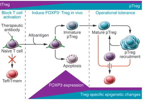
[image:4.585.301.544.83.257.2]We propose, therefore, that the tolerogenic microenvironment is one in which the availability of nutrients is tightly controlled, requiring any T cell that enters a tolerated tissue to adapt its metabolism to use either those external nutrients available or to rely on autophagy and salvage pathways to recycle intracellular
Figure 1
Therapeutic intervention aimed at inducing transplantation tolerance can be thought of in three stages. The first (left) is concerned with cre-ating a ceasefire, in which inflammation and danger signals are mini-mized. In the example given, short-term inhibition of T cell activation is achieved with antibodies that block T cell coreceptors and costimula-tory molecules such as CD4, CD8, and CD40L. Under these nonde-pleting conditions, in the second stage (middle), both self-reactive and alloreactive FOXP3+ natural Tregs (tTreg) are recruited and expanded.
review
components that provide sufficient energy to function and sur-vive. Evidence is growing that T cell differentiation and metabo-lism are inherently coupled in determining the development of memory, effector, or regulatory T cells (87, 88).
Toward therapeutic applications
Administration of Tregs from without. The discovery of Tregs has spawned great interest in the use of in vitro–expanded, person-alized Treg therapy (19, 89–97). Undoubtedly, this will provide important proof-of-principle discoveries, but routine clinical application, even with evidence of efficacy, may still be a signifi-cant challenge. The constraints on the application of Treg therapy are scientific, logistical, and commercial. We still know little about the mechanisms of suppression, the factors affecting stability of their antiinflammatory functions (98), or about Treg heterogene-ity and context-related requirements, and we lack well-validated biomarker handles to control the quality of the therapeutic
prod-ucts. The availability of humanized mouse models to test efficacy is certainly a significant step in this field (93, 99, 100). With regard to logistics, the need to generate purified cells to GMP standards is itself no small challenge. Finally, as for all personalized cell thera-pies, the numerous barriers to commercialization certainly need to be overcome.
Enhancing Tregs from within. Given these considerations, what principles can be exploited to favor the regulation and dominant tolerance generated within the host?
There are relatively few strategies proposed for selectively expanding endogenous Tregs. One based on selective vaccination has already been reported (36, 43). The other, based on the known ability of IL-2 to stabilize and expand Tregs, uses IL-2 or IL-2-anti– IL-2 antibody complexes to act on endogenous Tregs (101–103). The latter would be clinically attractive if one could generate a druggable form of IL-2 that would signal only to Tregs and not to proinflammatory effector T cells.
Murine models have provided a number of antibody-based pro-tocols based on short-term treatments that appear to favor Tregs. First, let us consider the situation of tolerogenic protocols that block coreceptor and/or costimulatory signals. Regulation seems to be favored when the following three (somewhat overlapping) stages are completed (Figure 1).
First, a “ceasefire” from aggression must be established for long enough to ensure no proinflammatory “sniper” activity while tol-erance mechanisms are induced. As a consequence of the ceasefire, some effectors undergo apoptosis, and this, together with tissue-healing events, encourages the production of active TGF-β, inhibi-tion of mTOR and proinflammatory cytokines, and the extincinhibi-tion of danger signals. Many of the situations in which mTOR is inhib-ited can locally antagonize destructive immune responses.
The second stage is based on the conversion of naive CD4+
T cells into pTregs. It is here that the molecular mechanisms involved in FOXP3 expression, outlined above, are integrated. Although transient expression of FOXP3 may be insufficient to establish stable Tregs, it may contribute to the restraint (9, 45, 46), and even apoptosis (104), of potential effector cells and thus support the drug-initiated ceasefire. This buys the necessary time (105) for some FOXP3-converted cells to undergo the epi-genetic changes that eventually stabilize them (52). Tregs with specificity for antigen are equipped with metabolic characteris-tics that provide them with the capacity to gain relative benefit from available antigen stimulation and nutrients, in which con-ventional cells may be compromised by the therapeutic agents or the metabolic environment (i.e., EAA depletion and mTOR inhibition), thereby allowing preferential expansion of Tregs and consolidation of the immune-privileged microenvironment they impose in the tissues.
Finally, we propose that tolerance enters an autonomous phase not requiring further maintenance drug therapy. As the graft heals, its immunogenic power will diminish, as will the impact of direct allorecognition. The “vaccination” of pTregs, which see donor peptides indirectly presented on host MHCs, will on the contrary increase, so that by the time therapeutic agent levels disappear, regulation and infectious tolerance have merged as the dominant processes (12). Of the many induced CD4+ T cells that express
FOXP3 en route to tolerance, only a minority will have become committed Treg lineage cells, but a combination of their numbers and strategic placement (draining lymphoid tissue and the graft itself) should allow them to have long-term dominant
(antigen-Figure 2
[image:5.585.45.285.82.357.2]specific) control on residual potential effectors of immunity. There is no doubt that donor antigens continuously supplied by the engrafted tissue are critical to maintain active Treg-mediated tolerance (10). Such antigens can be considered the “booster” doses often needed in conventional vaccines. This, together with the finding that Treg depletion after tolerance induction reverses the tolerant state (8), provides compelling evidence that continued vigilance by Tregs as well as infectious tolerance are both sufficient and essential for long-term graft survival.
Of course, in patients, a single set of generic principles may not always be appropriate for generating tolerance, but they may still be conducive to drug minimization strategies. Prior priming need not preclude amplification of tolerance mechanisms if sufficient regulation can be generated (106, 107).
The above is clearly an oversimplified scheme, but it is consistent with much of the data derived from experimental tolerance studies in rodents using CD4 (plus CD8), CD40L, and CD3 antibodies as agents for tolerance induction. The ceasefire created by these treat-ments relies on signaling blockade rather than T cell depletion and subsequent lymphopenia.
Unfortunately, many of the agents that have proven most effec-tive in generating dominant tolerance discussed above have not yet emerged as licensed drugs available for clinical trials. This is partly because some have exhibited undesirable side effects, partly because there has not been any easy route to establish-ing definitive trials to enable drug approval, and, perhaps most poignant, because there is uncertainty about tolerance-promot-ing agents as commercial products. The harsh reality may be that we need to build on those drugs that are currently commercially
available (e.g., ATG, alemtuzumab, abatacept) to determine how to incorporate them into tolerogenic protocols.
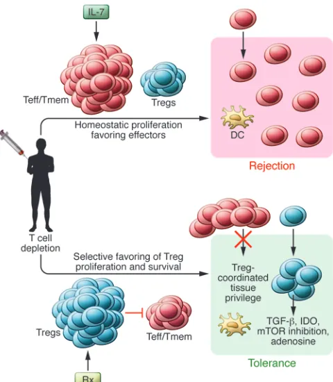
Given the limited repertoire of licensed drugs, induction strat-egies based on lymphocyte depletion provide immediate oppor-tunities. The major disadvantage of lymphocyte depletion is the homeostatic expansion of host lymphocytes and immune recon-stitution favoring memory and effector T cells that provide a bar-rier to tolerance (108). With that in mind, a modification of the tolerance-inducing principles espoused above should aim to ena-ble Tregs to emerge as a dominant repopulating element, aena-ble to override any expanding effector and memory T cells. Treatments might also take advantage of the range of transiently expressed factors that are known to contribute to induction of pTregs (Fig-ure 2). We are confident that the rapidly expanding knowledge of the molecular mechanisms orchestrating Treg development will facilitate the early application of such protocols, which we coin with the acronym PARIS (physician-aided reconstitution of the immune system).
Acknowledgments
H. Waldmann, S. Cobbold, and D. Howie were supported by an ERC Advanced Investigator grant (to H. Waldmann), and R. Hilbrands was supported by a long-term fellowship from the European Molecular Biology Organization (EMBO).
Address correspondence to: Herman Waldmann, University of Oxford, Sir William Dunn Sch Pathol, South Parks Rd., Oxford OX1 3RE, United Kingdom. Phone: 44.1865.275503; Fax: 44.1865.275505; E-mail: herman.waldmann@path.ox.ac.uk.
1. Hall BM, Jelbart ME, Gurley KE, Dorsch SE. Specific unresponsiveness in rats with prolonged cardiac allograft survival after treatment with cyclospo-rine. Mediation of specific suppression by T helper/ inducer cells. J Exp Med. 1985;162(5):1683–1694. 2. Benjamin RJ, Waldmann H. Induction of
toler-ance by monoclonal antibody therapy. Nature. 1986;320(6061):4z 49–451.
3. Qin SX, Cobbold S, Benjamin R, Waldmann H. Induction of classical transplantation tolerance in the adult. J Exp Med. 1989;169(3):779–794. 4. Qin S, et al. “Infectious” transplantation tolerance.
Science. 1993;259(5097):974–977.
5. Mason DW. Subsets of CD4+ T cells and their roles in autoimmunity. Philos Trans R Soc Lond B Biol Sci. 1993;342(1299):51–56.
6. Sakaguchi S, Sakaguchi N, Asano M, Itoh M, Toda M. Immunologic self-tolerance maintained by acti-vated T cells expressing IL-2 receptor alpha-chains (CD25). Breakdown of a single mechanism of self-tolerance causes various autoimmune diseases.
J Immunol. 1995;155(3):1151–1164.
7. Kim J, et al. Cutting edge: depletion of Foxp3+ cells leads to induction of autoimmunity by specific ablation of regulatory T cells in genetically tar-geted mice. J Immunol. 2009;183(12):7631–7634. 8. Kendal AR, et al. Sustained suppression by Foxp3+
regulatory T cells is vital for infectious transplanta-tion tolerance. J Exp Med. 2011;208(10):2043–2053. 9. Regateiro FS, et al. Foxp3 expression is required
for the induction of therapeutic tissue tolerance.
J Immunol. 2012;189(8):3947–3956.
10. Cobbold SP, Adams E, Marshall SE, Davies JD, Waldmann H. Mechanisms of peripheral tolerance and suppression induced by monoclonal antibod-ies to CD4 and CD8. Immunol Rev. 1996;149:5–33. 11. Davies JD, Leong LY, Mellor A, Cobbold SP, Wald-mann H. T cell suppression in transplantation tolerance through linked recognition. J Immunol.
1996;156(10):3602–3607.
12. Wise MP, Bemelman F, Cobbold SP, Waldmann H. Linked suppression of skin graft rejection can operate through indirect recognition. J Immunol. 1998;161(11):5813–5816.
13. Groux H, et al. A CD4+ T-cell subset inhibits anti-gen-specific T-cell responses and prevents colitis.
Nature. 1997;389(6652):737–742.
14. Passerini L, et al. Functional type 1 regulatory T cells develop regardless of FOXP3 mutations in patients with IPEX syndrome. Eur J Immunol. 2011;41(4):1120–1131.
15. Oliveira VG, Agua-Doce A, Curotto de Lafaille MA, Lafaille JJ, Graca L. Adjuvant facilitates toler-ance induction to factor VIII in hemophilic mice through a Foxp3-independent mechanism that relies on IL-10. Blood. 2013;121(19):3936–3945. 16. Sundstedt A, O’Neill EJ, Nicolson KS, Wraith DC.
Role for IL-10 in suppression mediated by pep-tide-induced regulatory T cells in vivo. J Immunol. 2003;170(3):1240–1248.
17. Ng TH, Britton GJ, Hill EV, Verhagen J, Burton BR, Wraith DC. Regulation of adaptive immunity; the role of interleukin-10. Front Immunol. 2013;4:129. 18. Gregori S, Goudy KS, Roncarolo MG. The cellular
and molecular mechanisms of immuno-suppression by human type 1 regulatory T cells. Front Immunol. 2012;3:30.
19. Allan SE, et al. CD4+ T-regulatory cells: toward therapy for human diseases. Immunol Rev. 2008; 223:391–421.
20. Bennett CL, et al. The immune dysregulation, poly-endocrinopathy, enteropathy, X-linked syndrome (IPEX) is caused by mutations of FOXP3. Nat Genet. 2001;27(1):20–21.
21. Wildin RS, et al. X-linked neonatal diabetes melli-tus, enteropathy and endocrinopathy syndrome is the human equivalent of mouse scurfy. Nat Genet. 2001;27(1):18–20.
22. Gambineri E, Torgerson TR, Ochs HD. Immune dysregulation, polyendocrinopathy, enteropathy, and X-linked inheritance (IPEX), a syndrome of systemic autoimmunity caused by mutations of FOXP3, a critical regulator of T-cell homeostasis.
Curr Opin Rheumatol. 2003;15(4):430–435. 23. Sakaguchi S. The origin of FOXP3-expressing
CD4+ regulatory T cells: thymus or periphery. J Clin Invest. 2003;112(9):1310–1312.
24. Chaudhry A, Rudensky AY. Control of inflamma-tion by integrainflamma-tion of environmental cues by regu-latory T cells. J Clin Invest. 2013;123(3):939–944. 25. Sakaguchi S. Naturally arising Foxp3-expressing
CD25+CD4+ regulatory T cells in
immunologi-cal tolerance to self and non-self. Nat Immunol. 2005;6(4):345–352.
26. Chen W, Wahl SM. TGF-beta: the missing link in CD4+CD25+ regulatory T cell-mediated
immunosuppression. Cytokine Growth Factor Rev. 2003;14(2):85–89.
27. Sauer S, et al. T cell receptor signaling controls Foxp3 expression via PI3K, Akt, and mTOR.
Proc Natl Acad Sci U S A. 2008;105(22):7797–7802. 28. Powell JD, Heikamp EB, Pollizzi KN, Waickman AT.
A modified model of T-cell differentiation based on mTOR activity and metabolism [published online ahead of print October 7, 2013]. Cold Spring Harb Symp Quant Biol. doi:10.1101/sqb.2013.78.020214. 29. Zeng H, Yang K, Cloer C, Neale G, Vogel P, Chi H.
mTORC1 couples immune signals and metabolic programming to establish T(reg)-cell function.
Nature. 2013;499(7459):485–490.
30. Park Y, et al. TSC1 regulates the balance between effector and regulatory T cells. J Clin Invest. 2013; 123(12):5165–5178.
review
32. Liu G, Yang K, Burns S, Shrestha S, Chi H. The S1P(1)-mTOR axis directs the reciprocal differ-entiation of T(H)1 and T(reg) cells. Nat Immunol. 2010;11(11):1047–1056.
33. Josefowicz SZ, et al. Extrathymically generated regulatory T cells control mucosal TH2 inflamma-tion. Nature. 2012;482(7385):395–399.
34. Tawara I, et al. A crucial role for host APCs in the induction of donor CD4+CD25+ regulatory T
cell-mediated suppression of experimental graft-versus-host disease. J Immunol. 2010;185(7):3866–3872. 35. Tsang JY, et al. The potency of allospecific Tregs cells
appears to correlate with T cell receptor functional avidity. Am J Transplant. 2011;11(8):1610–1620. 36. von Boehmer H, Daniel C. Therapeutic
opportuni-ties for manipulating T(Reg) cells in autoimmunity and cancer. Nat Rev Drug Discov. 2013;12(1):51–63. 37. Miyajima M, et al. Early acceptance of renal
allografts in mice is dependent on foxp3(+) cells.
Am J Pathol. 2011;178(4):1635–1645.
38. Benghiat FS, et al. Critical influence of natural reg-ulatory CD25+ T cells on the fate of allografts in the absence of immunosuppression. Transplantation. 2005;79(6):648–654.
39. Neujahr DC, et al. Accelerated memory cell homeo-stasis during T cell depletion and approaches to overcome it. J Immunol. 2006;176(8):4632–4639. 40. Graca L, Le Moine A, Lin CY, Fairchild PJ,
Cob-bold SP, Waldmann H. Donor-specific trans-plantation tolerance: the paradoxical behavior of CD4+CD25+ T cells. Proc Natl Acad Sci U S A.
2004;101(27):10122–10126.
41. Cobbold SP, et al. Induction of foxP3+ regulatory
T cells in the periphery of T cell receptor trans-genic mice tolerized to transplants. J Immunol. 2004;172(10):6003–6010.
42. Daley SR, Ma J, Adams E, Cobbold SP, Waldmann H. A key role for TGF-beta signaling to T cells in the long-term acceptance of allografts. J Immunol. 2007;179(6):3648–3654.
43. Verginis P, McLaughlin KA, Wucherpfennig KW, von Boehmer H, Apostolou I. Induction of anti-gen-specific regulatory T cells in wild-type mice: visualization and targets of suppression. Proc Natl Acad Sci U S A. 2008;105(9):3479–3484.
44. Hori S, Nomura T, Sakaguchi S. Control of regula-tory T cell development by the transcription factor Foxp3. Science. 2003;299(5609):1057–1061. 45. Andersen KG, Butcher T, Betz AG. Specific
immu-nosuppression with inducible Foxp3-transduced polyclonal T cells. PLoS Biol. 2008;6(11):e276. 46. McMurchy AN, et al. A novel function for FOXP3 in
humans: intrinsic regulation of conventional T cells.
Blood. 2013;121(8):1265–1275.
47. Huang YJ, et al. Induced and thymus-derived Foxp3 regulatory T cells share a common niche.
Eur J Immunol. 2014;44(2):460–468.
48. Liston A, Gray DH. Homeostatic control of reg-ulatory T cell diversity [published online ahead of print January 31, 2014]. Nat Rev Immunol. doi:10.1038/nri3605.
49. Roncarolo MG, Battaglia M. Regulatory T-cell immunotherapy for tolerance to self antigens and alloantigens in humans. Nat Rev Immunol. 2007;7(8):585–598.
50. Williams LM, Rudensky AY. Maintenance of the Foxp3-dependent developmental program in mature regulatory T cells requires continued expres-sion of Foxp3. Nat Immunol. 2007;8(3):277–284. 51. Wan YY, Flavell RA. Regulatory T-cell functions
are subverted and converted owing to attenuated Foxp3 expression. Nature. 2007;445(7129):766–770. 52. Ohkura N, et al. T cell receptor stimulation-in-duced epigenetic changes and Foxp3 expression are independent and complementary events required for Treg cell development. Immunity. 2012; 37(5):785–799.
53. Samstein RM, et al. Foxp3 exploits a pre-existent
enhancer landscape for regulatory T cell lineage specification. Cell. 2012;151(1):153–166. 54. Komatsu N, Mariotti-Ferrandiz ME, Wang Y,
Malissen B, Waldmann H, Hori S. Heterogeneity of natural Foxp3+ T cells: a committed regulatory
T-cell lineage and an uncommitted minor popu-lation retaining plasticity. Proc Natl Acad Sci U S A. 2009;106(6):1903–1908.
55. Miyao T, et al. Plasticity of Foxp3(+) T cells reflects promiscuous Foxp3 expression in conventional T cells but not reprogramming of regulatory T cells.
Immunity. 2012;36(2):262–275.
56. Rubtsov YP, et al. Stability of the regulatory T cell lineage in vivo. Science. 2010;329(5999):1667–1671. 57. Bailey-Bucktrout SL, et al. Self-antigen-driven
activation induces instability of regulatory T cells during an inflammatory autoimmune response.
Immunity. 2013;39(5):949–962.
58. Sakaguchi S, Vignali DA, Rudensky AY, Niec RE, Waldmann H. The plasticity and stability of regula-tory T cells. Nat Rev Immunol. 2013;13(6):461–467. 59. Hori S. Regulatory T cell plasticity: beyond the con-troversies. Trends Immunol. 2011;32(7):295–300. 60. Chong AS, Alegre ML. The impact of infection
and tissue damage in solid-organ transplantation.
Nat Rev Immunol. 2012;12(6):459–471.
61. Floess S, et al. Epigenetic control of the foxp3 locus in regulatory T cells. PLoS Biol. 2007;5(2):e38. 62. Toker A, et al. Active demethylation of the Foxp3
locus leads to the generation of stable regula-tory T cells within the thymus. J Immunol. 2013; 190(7):3180–3188.
63. Bruniquel D, Schwartz RH. Selective, stable demethylation of the interleukin-2 gene enhances transcription by an active process. Nat Immunol. 2003;4(3):235–240.
64. Michalek RD, et al. Cutting edge: distinct glycolytic and lipid oxidative metabolic programs are essen-tial for effector and regulatory CD4+ T cell subsets.
J Immunol. 2011;186(6):3299–3303.
65. Shi LZ, et al. HIF1alpha-dependent glycolytic path-way orchestrates a metabolic checkpoint for the differentiation of TH17 and Treg cells. J Exp Med. 2011;208(7):1367–1376.
66. Chang CH, et al. Posttranscriptional control of T cell effector function by aerobic glycolysis. Cell. 2013;153(6):1239–1251.
67. Gubser PM, et al. Rapid effector function of memory CD8(+) T cells requires an immediate-early glyco-lytic switch. Nat Immunol. 2013;14(10):1064–1072. 68. van der Windt GJ, et al. Mitochondrial respiratory
capacity is a critical regulator of CD8+ T cell
mem-ory development. Immunity. 2012;36(1):68–78. 69. Dang EV, et al. Control of T(H)17/T(reg)
bal-ance by hypoxia-inducible factor 1. Cell. 2011; 146(5):772–784.
70. Fassett MS, Jiang W, D’Alise AM, Mathis D, Benoist C. Nuclear receptor Nr4a1 modulates both regula-tory T-cell (Treg) differentiation and clonal deletion.
Proc Natl Acad Sci U S A. 2012;109(10):3891–3896. 71. Waickman AT, Powell JD. mTOR, metabolism, and
the regulation of T-cell differentiation and func-tion. Immunol Rev. 2012;249(1):43–58.
72. Graca L, Cobbold SP, Waldmann H. Identifica-tion of regulatory T cells in tolerated allografts.
J Exp Med. 2002;195(12):1641–1646.
73. Cobbold SP, et al. Immune privilege induced by regulatory T cells in transplantation tolerance.
Immunol Rev. 2006;213:239–255.
74. Samstein RM, Josefowicz SZ, Arvey A, Treuting PM, Rudensky AY. Extrathymic generation of reg-ulatory T cells in placental mammals mitigates maternal-fetal conflict. Cell. 2012;150(1):29–38. 75. Andersen KG, Nissen JK, Betz AG. Comparative
genomics reveals key gain-of-function events in Foxp3 during regulatory T cell evolution. Front Immunol. 2012;3:113.
76. Munn DH, et al. Prevention of allogeneic fetal
rejection by tryptophan catabolism. Science. 1998; 281(5380):1191–1193.
77. Cobbold SP, et al. Infectious tolerance via the consumption of essential amino acids and mTOR signaling. Proc Natl Acad Sci U S A. 2009; 106(29):12055–12060.
78. Metz R, Duhadaway JB, Kamasani U, Lau-ry-Kleintop L, Muller AJ, Prendergast GC. Novel tryptophan catabolic enzyme IDO2 is the preferred biochemical target of the antitumor indoleamine 2,3-dioxygenase inhibitory compound D-1-meth-yl-tryptophan. Cancer Res. 2007;67(15):7082–7087. 79. Fallarino F, et al. The combined effects of tryp-tophan starvation and tryptryp-tophan catabolites down-regulate T cell receptor zeta-chain and induce a regulatory phenotype in naive T cells. J Immunol. 2006;176(11):6752–6761.
80. Nowak EC, et al. Tryptophan hydroxylase-1 regulates immune tolerance and inflammation. J Exp Med. 2012;209(11):2127–2135.
81. Cobbold SP, Adams E, Nolan KF, Regateiro FS, Waldmann H. Connecting the mechanisms of T-cell regulation: dendritic cells as the missing link.
Immunol Rev. 2010;236:203–218.
82. Cobbold SP, Adams E, Waldmann H. Biomarkers of transplantation tolerance: more hopeful than helpful? Front Immunol. 2011;2:9.
83. Rolf J, Zarrouk M, Finlay DK, Foretz M, Viol-let B, Cantrell DA. AMPKα1: a glucose sensor that controls CD8 T-cell memory. Eur J Immunol. 2013;43(4):889–896.
84. Deaglio S, et al. Adenosine generation catalyzed by CD39 and CD73 expressed on regulatory T cells mediates immune suppression. J Exp Med. 2007;204(6):1257–1265.
85. Regateiro FS, et al. Generation of anti-inflamma-tory adenosine by leukocytes is regulated by TGF-β.
Eur J Immunol. 2011;41(10):2955–2965.
86. Delgoffe GM, et al. The mTOR kinase differentially regulates effector and regulatory T cell lineage commitment. Immunity. 2009;30(6):832–844. 87. Cobbold SP. The mTOR pathway and
inte-grating immune regulation [published online ahead of print August 19, 2013]. Immunology. doi:10.1111/imm.12162.
88. Heikamp EB, Powell JD. Sensing the immune microenvironment to coordinate T cell metabo-lism, differentiation and function. Semin Immunol. 2012;24(6):414–420.
89. Sawitzki B, et al. Prevention of graft-versus-host disease by adoptive T regulatory therapy is associ-ated with active repression of peripheral blood toll-like receptor 5 mRNA expression. Biol Blood Marrow Transplant. 2014;20(2):173–182.
90. Hippen KL, et al. Massive ex vivo expansion of human natural regulatory T cells (T(regs)) with minimal loss of in vivo functional activity. Sci Transl Med. 2011;3(83):83ra41.
91. Tang Q, Bluestone JA. Regulatory T-cell therapy in transplantation: moving to the clinic. Cold Spring Harb Perspect Med. 2013;3(11):a015552.
92. McMurchy AN, Bushell A, Levings MK, Wood KJ. Moving to tolerance: clinical application of T regu-latory cells. Semin Immunol. 2011;23(4):304–313. 93. Wu DC, et al. Ex vivo expanded human regulatory
T cells can prolong survival of a human islet allog-raft in a humanized mouse model. Transplantation. 2013;96(8):707–716.
94. Putnam AL, et al. Clinical grade manufactur-ing of human alloantigen-reactive regulatory T cells for use in transplantation. Am J Transplant. 2013;13(11):3010–3020.
95. Edinger M. Regulatory T cells for the prevention of graft-versus-host disease: professionals defeat amateurs. Eur J Immunol. 2009;39(11):2966–2968. 96. Di Ianni M, et al. Tregs prevent GVHD and promote
97. Martelli MF, et al. “Designed” grafts for HLA-hap-loidentical stem cell transplantation. Blood. 2014;123(7):967–973.
98. Hansmann L, et al. Dominant Th2 differentiation of human regulatory T cells upon loss of FOXP3 expression. J Immunol. 2012;188(3):1275–1282. 99. Sagoo P, Ali N, Garg G, Nestle FO, Lechler RI,
Lom-bardi G. Human regulatory T cells with alloantigen specificity are more potent inhibitors of alloim-mune skin graft damage than polyclonal regula-tory T cells. Sci Transl Med. 2011;3(83):83ra42. 100. Nadig SN, et al. In vivo prevention of transplant
arteriosclerosis by ex vivo-expanded human regu-latory T cells. Nat Med. 2010;16(7):809–813. 101. Webster KE, et al. In vivo expansion of T reg cells with
IL-2-mAb complexes: induction of resistance to EAE and long-term acceptance of islet allografts without immunosuppression. J Exp Med. 2009;206(4):751–760. 102. Boyman O, Sprent J. The role of interleukin-2 during homeostasis and activation of the immune system.
Nat Rev Immunol. 2012;12(3):180–190.
103. Matsuoka K, et al. Low-dose interleukin-2 therapy restores regulatory T cell homeostasis in patients with chronic graft-versus-host disease. Sci Transl Med. 2013;5(179):179ra143.
104. Tai X, et al. Foxp3 transcription factor is proap-optotic and lethal to developing regulatory T cells unless counterbalanced by cytokine survival signals.
Immunity. 2013;38(6):1116–1128.
105. Scully R, Qin S, Cobbold S, Waldmann H.
Mecha-nisms in CD4 antibody-mediated transplantation tolerance: kinetics of induction, antigen depen-dency and role of regulatory T cells. Eur J Immunol. 1994;24(10):2383–2392.
106. Marshall SE, Cobbold SP, Davies JD, Martin GM, Phillips JM, Waldmann H. Tolerance and suppres-sion in a primed immune system. Transplantation. 1996;62(11):1614–1621.
107. Siepert A, et al. Permanent CNI treatment for pre-vention of renal allograft rejection in sensitized hosts can be replaced by regulatory T cells. Am J Transplant. 2012;12(9):2384–2394.