DEVELOPMENT OF WOUND HEALING HERBAL
FORMULATION
“HERBAL WOUND GUARD”
Rajput Rekha T.*, Gohil Kashmira J.**, Singh Poonam** and Singh Surendra**
Anand College of Pharmacy, Keetham, Agra-282007.
AbstractThe present study was to evaluate the wound healing activity of developed polyherbal formulation in the form of ointment by using hydroalcoholic extracts of leaves of Ficus religiosa, Mentha arvensis and roots of Rauwolfia serpentina with excipients. The phytochemical studies revealed the presence of alkaloids, phytosterol, tannins, flavonoids and protein. The developed polyherbal ointment was examined for wound healing activity in two types of wound models on rats; the excision and incision wound model. The polyherbal formulation was applied once daily until complete healing of wound and it was noted that the period of epithelization significantly (P < 0.05) reduced when compared with vehicle control group (15 ±0.34) which was nearly comparable with standard drug group. The tensile strength of healed wound was significantly increased (P < 0.05) as compared to vehicle control group. Hence the developed polyherbal formulation showed significant (P < 0.05) wound healing activity as compared to control group. The obtained results were nearly compared with standard drug group betadine ointment in terms of contractibility, wound closure time and tensile strength.
Index Terms- Polyherbal formulation, Ointment, Hydroalcoholic extracts.
I.INTRODUCTION
Wound is a physical trauma where the skin is torn, cut or punctured. On exposure to air, on microorganism enter the wound which leads to wound contamination and finally development of infection1. Dermal wound is a common pathologic condition and may be defined as any break in the integrity of the skin. It is associated with high degree of morbidity due to blood loss, pain, edema, inflammation and loss of functionality. Cut wound are characterized by migration and proliferation of fibroblasts, endothelial cells, deposition of connective tissue, angiogenesis, re-epithelization and finally contraction of wound2. Presently the scientists are keen to evaluate drugs from plant origin. It is due to their specific healing properties, healing action and non-toxic effects. Several plants and their products are used in folk medicine to treat wound, and the plants like Ginsieng, Sunflower, Brahmi etc. have been reported to promote the healing3.
The leaves of the Ficus religiosa belonging to family Moraceae have purgative properties and are also recommended for wounds and skin infection4, anticancer activity5, antiulcer activity6, anti-diabetic activity7. The leaves of Mentha arvensis, Laminaceae
known as “Pudina” acrid, aromatic, thermogenic, stimulant, anodye, deodorant, antiseptic, vulnerary, anthelmintic, carminative, digestive, stomachic, antiemetic, cardiotonic, expectorant, diuretics, depurative, dentifrice, antispasmodic, febrifuge and contraceptive. They are useful in vitiated condition of vata, arthralgia, halitosis, indolent ulcers, wounds, cuts, helminthiasis, dyspepsia, flatulence, colic, peptic ulcer, vomiting, diarrhea, cardiac debility, cough, asthma, bronchitis, strangury, skin disease, amenorrhoea, fever and general weakness8. It is also have antimicrobial activity9, antiulcer activity10, antifungal activity11, anti-bacterial activity12. Roots of Rauwolfia serpentina, Apocynaceae, are bitter, acrid, laxative, anthelmintic, thermogenic, diuretic and possess sedative properties. It is highly reputed for hypertension and is useful in strangury, fever, wound, colic, insomnia, epilepsy, giddiness, dyspepsia and vitiated conditions of kapha and vata. The decoction of the root is used to increase uterine contractions13. Roots of the plant also used as antidiabetic 14 and antidiarrhoeal drug15.
The objective of the present study was to investigate wound healing activity of developed polyherbal formulation in form of suspension by using combined herbal extracts.
II. MATERIALS AND METHODS Plant Material:
Leaves of Ficus religiosa, Mentha arvensis and roots of Rauwolfia serpentina were procured from local market and same were authenticated by Dr. Seema Bhadhauria, Head of Department of Botany, R.B.S. College, Agra and sample specimen (Voucher No. of the Specimen: RBSC/2014/196) were deposited in the herbarium of the Department of Pharmacognosy, Anand College of Pharmacy, Agra for future reference.
Extraction:
Two hundred gram each, standardized powder of leaves of Ficus religiosa, Mentha arvensis and roots of Rauwolfia serpentina in the ratio of 1:2:1 were subjected to extraction with 60% hydroalcohol by maceration for seven days at room temperature16. After extraction, the extract was filtered and concentrated at room temperature. The extract was subjected to qualitative chemical tests adopting standard procedure.17, 18.
Development of Polyherbal Formulation (PHF):
aqueous phase with constant stirring. Remove from the heat and stir the mixture until it congeals. The ingredients and their quantity were used to prepare formulation or ointment specified in Table I.
Table I: Formula for Formulation Sr.
No.
Name of Ingredients
Quantity in percent
Quantity Taken
1 Hydroalcoholic extract
6% 1.5g
2 Sodium Lauryl Sulphate
1% 0.25g
3 Propylene Glycol 12% 3.0ml 4 Stearyl Alcohol 25% 6.25g 5 White Petrolatum 25% 6.25g 6 Purified Water 31% 7.75ml
Standardization of polyherbal formulation (PHF):
Standardization of developed polyherbal formulation in the form of ointment was done by using different organoleptic characters (color, odor and taste) as well as physicochemical parameters like pH, loss on drying, speadability, diffusion study, skin irritation study20. The results of the studies were shown in Table II.
pH: The pH of the formulation was recorded by using a digital pH meter of Systronic, Ahmedabad. Weighted quantity of the sample was dissolved in distilled water and stored for two hours. The measurement of pH was done in triplicate and average values were considered.
Spread-ability: The spread ability was expressed in terms of times in seconds taken by two slides to slip off from ointment placed in between the slides under the direction of certain load. Spread ability was calculated by using the formula.
S= (M×L÷T) Where,
S=Spreadability
M=Weight tied to upper slide L=Length of glass slides
T=Time taken to separate the slide
Diffusion study:
The diffusion study was carried by preparing agar nutrient medium of known concentration. It was poured into a petridish and allowed to set. A hole was bored at the center of the petridish and the prepared formulation was placed in it. The time taken for the ointment to get diffused was noted.
Skin irritation study:
[image:2.612.50.294.108.248.2]Healthy rabbits were selected and were shaved in two different areas of the dorsal side, each about 500mm. The rabbit was kept in rabbit holder and the first area was kept as control, to which emulsifying ointment base was applied, the second area was treated with polyherbal ointment. After 48 hrs the skin was observed and compared with control.
Table II: Physicochemical properties of the formulation Sr.
No.
Physicochemical parameters
Formulation
1 Color Light skin color
2 Odor Sweetish with bitter
characteristic 3 Loss on drying 9.6 % w/w
4 pH 7.0
5 Spreadability (seconds) 18 6 Diffusion study 0.68 cm
7 Skin irritation study No skin irritation was observed
CHROMATOGRAPHY STUDIES:
Chromatography study through Thin Layer Chromatography was done to evaluate the developed formulation for the presence of different chemical constituents which is responsible for wound healing activity and compare it with extracts. The obtained results were shown in Figure 1 and Table III.
Experimental condition:
TLC glass plate: Silica gel G
Solvent system: Chloroform: Acetone (9.5: 0.5)
Figure 1: Thin Layer Chromatography Profile of Extract and Formulation
λ-max -366nm
λ-max -254nm
Table III: Results of TLC Profile of Extracts and Formulation
Name of
compounds Rf values
Presence of
possibility of
[image:2.612.384.544.302.475.2] [image:2.612.149.569.307.730.2]compounds
1.Extracts and Formulation
0.285
Myrecetin hexaacetate 0.1 Dimethoxycinnamic acid 0.20 Trimethoxycinnamic acid 0.30
Catechin Penta acetate 0.40 Khellin
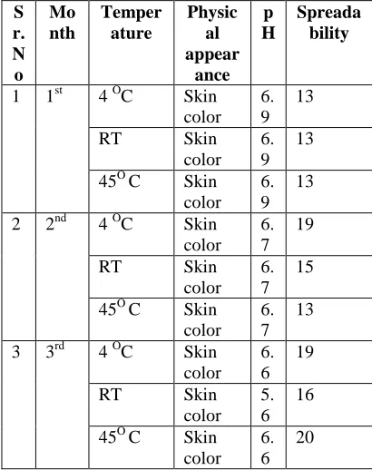
STABILITIES STUDIES:
The purpose of it to find out the quality of product during storage at Room temperature, 4 0C and 45 0C due to environmental factor‘s effect.
Method:
[image:3.612.29.307.58.166.2] [image:3.612.99.304.302.561.2]The formulation means ointment was packed in white colored glass bottle, which are tightly capped. They were then stored for three months and evaluated for various parameters at Room temperature, 4 0C and 45 0C. The obtained results were shown in Table IV.
Table IV: Stability data of the formulation (Ointment) S r. N o Mo nth Temper ature Physic al appear ance p H Spreada bility
1 1st 4 OC Skin color
6. 9
13
RT Skin
color 6. 9
13
45O C Skin color
6. 9
13
2 2nd 4 OC Skin color
6. 7
19
RT Skin
color 6. 7
15
45O C Skin color
6. 7
13
3 3rd 4 OC Skin color
6. 6
19
RT Skin
color 5. 6
16
45O C Skin color
6. 6
20
WOUND HEALING ACTIVITY: Toxicity studies:
Toxicity studies were done as per the Organization for Economic Co-operation and Development (OECD), revised 423 guidelines21. Albino mice were kept overnight fasting prior to extract administered in suspension form. A total 6 animals of two groups three in each group received formulation from 300 mg/kg to 2000 mg/kg body weight. After each administration of dose food was withheld for further 3-4 hours. Animals were observed individually daily for a period of 14 days. Based on these studies the doses were selected for the evaluation of wound healing activities.The LD50 of the extracts falls under the class for values
with no signs of acute toxicity till 2000 mg/kg body weight, so that 1/10th was taken as effective therapeutic doses for
formulation. The selected doses for formulation were 200 mg/kg body weight.
Administrations:
Experimental animals were grouped into three, six each and were treated as follow. Group I treated with simple petroleum jelly (Vaseline) served as control or vehicle control group, Group II rats were treated with standard drug betadine serve as positive control or standard group, Group III animals treated with polyherbal formulation (PHF) in the form of ointment serve as treatment or formulation group.
Excision wound model:
An impression was made on the dorsal thoracic region 1cm away from the vertebral column and 5 cm away from the ear of the anesthetized rat. Skin was excised to full thickness to obtain a wound area of about 500 mm2. The ointment was applied once daily until complete healing of wound and the wound area was measured on a millimeter scale graph paper on alternate days. The percentage of wound healing was calculated. Falling of scar was taken as the endpoint for complete epithelization and the day taken for this was considered as period of epithelization 21. The obtained results were shown in Table V.
Incision wound model:
Two paravertebral straight incisions of 6 cm were made on either sides of the vertebral column. Homeostasis was achieved by blotting the wound with a cotton swab dipped in saline and the wound was closed by means of interrupted sutures at equidistance 1 cm apart. Animals were treated daily with formulation, as mentioned above under excision wound model from initial days to 14th post wounding day. The tensile strength in each group is determined on the 15th day by continuous, constant water flow 20. The results were shown in Table VI.
Statistical analysis:
Data were analysed using Graphpad Prism Software version 2.01 (GraphPad Sofware, La Jolla, USA). All the values were expressed as mean ± standard error of mean (SEM). The significance of difference between two groups for antidiaarhoeal activity was analysed using one-way analysis of variance (ANOVA) followed by post hoc Dunnet’s tests. For statistical analysis, P < 0.001 was considered statistically significant.
III. RESULTS
Preliminary phytochemical screening of hydroalcoholic extracts:
The hydroalcoholic extracts was subjected to qualitative method of preliminary phytochemical analysis that showed the presence of tannins, saponins, flavonoids, triterpenoid, alkaloids, glycosides and carbohydrates.
Wound healing activity:
In excision wound model, the fifty percent closure of wound area for the polyherbal ointment was found to be 5.2 ± 0.17, increase significantly (P < 0.05; F=80) when compared with vehicle control group (8.0 ± 0.90) as well as standard drug group(P < 0.05; F=73.84). The obtained data showed that the rate of wound contraction was significantly higher in the animals treated with developed formulation when compared with vehicle control and it was nearly comparable with standard drug group. The ointment exhibited significantly decreased (P < 0.01, P < 0.05; F=0.161) period of epithelization compared to control group.
Sr. No. Name of groups
50% wound
contraction in days
Periods of
epithelization in days
1 Control 8.0±0.90 15.0±0.34 2 Standard 4.6±0.98 9.0±0.98** 3 Formulation 5.2±0.17 11±0.15** All values are mean ± SEM, n=6,*p<0.05 indicates significant compared to the control.
In incision wound model, significantly increase in tensile strength of healed wounds of polyherbal ointment 298 ± 7.90 when compared with vehicle group 200 ± 9.19 and this results was nearly compare with standard drug group 345 ± 6.89.
Table VI: Effects of the polyherbal formulation in excision wound model
Sr No.
Name of groups Tensile strength
1 Control 200±9.19
2 Standard 345±6.89
3 Formulation 298±7.90**
All values are mean ± SEM, n=6,*p<0.05 indicates significant compared to the control.
IV. DISCUSSION
Wound healing is a complex process. That involves a chain of biochemical and cellular processes. These processes are mainly classified into three phases- inflammation, proliferation and remodeling. The inflammatory cells promote migration and proliferation of endothelial cells, leading to neovascularization. The proliferative phase is characterized by angiogenesis, collagen deposition, granulation tissue formation, epithelization and wound contraction. Finally the fibroblasts grow and form extracellular matrix as part of tissue remodelin22. Healing is delayed and the ability to accelerate the wound healing is highly desirable19.
In the present study the phytochemical investigation of extracts showed the presence of triterpenoids, saponins, alkaloids, carbohydrates and flavonoids. The literature survey reveals the presence of several phytocostituents like tritepenoids, saponins, alkaloids and flavonoids are known to promote wound healing process due to their antioxidant, anti microbial activities23 and astringent property24. Furthermore the wound healing effect can be attributed to free radical scavenging activity of flavonoids and triterpenoids. Both these type of phytoconstituents are known to reduce lipid peroxidation, not only by preventing or slowing the onset of cell necrosis, but also improving vascularity. Hence any drug that inhibits lipid peroxidation is believed to increase the viability of collagen fibrils, which in turn results in increase in the strength of collagen fiber by increasing the circulation, preventing the cell damage and promoting the DNA synthesis25. The increase in tensile strength may be due to the promotion of collagen formation which significantly contributes to better and effective healing.
REFERENCES
1. S.M. Patil, G.N. Sapkale, M.B. Patil and C.K. Sompure, “Formulation and wound healing activity of seed extract of Annona
reticulat”, Indian Drugs 2010; 47(1): 48-51.
2. B. Yaduvanshi, R. Mathur, S.R. Mathur, T. Velpandian, “Evaluation of wound healing potential of topical formulation of leaf juice of Tridax procumbens L. in mice”, Indian J Pharm Sci 2011; 73( 3): 303-306.
3. R.S. Bagali, V.P. Rasal, A.R. Tekade and R.H. Kale, “Wound healing effect of Artemisia pallens Wall.” Indian Drugs 2006; 43(12): 981-984.
4. R.V. Nair, “ Indian Medicinal Plants- a compendium of 500 species
(Ficus religiosa Linn., Warrier PK, Nambiar VPK, Ramankutty C
editors)”, Volume-III, Hyderabad: Orient Longman Private Limited, 1995, pp. 38-40.
5. V. Gulecha and S. Thangavel, “Anticancer activity of Tephrosia
purpurea and Ficus religiosa using MCF 7 cell line”, Asian Pacific
J Tropical Medicine 2011: 526-529.
6. B. Divya and V. Planvel, “Study of anti ulcer activity of Ficus religiosa leaf extract on experimentally induced gastric ulcers in rats”, Inter J Pharm Chem Sci 2012; 1( 3): 713-715.
7. S. Chaudhary, A.P. Pathak and S. Khare “Evaluation of antidiabetic activity of leaves and fruits of Ficus religiosa”, Inter J Pharm Life Sci 2011; 2(12): 1325-1326.
8. R.V. Nair, “Indian Medicinal Plants- a compendium of 500 species.
(Mentha arvensis Linn, Warrier PK, Nambiar VPK, Ramankutty C
editors)”, Volume-IV. Hyderabad: Orient Longman Private Limited; 1995. p. 15-17.
9. R. Mickiene, M. Sugandhi and G.M. Bai, “Antimicrobial activity of Mentha arvensis and Zingiber officinalis R. essential oils”, Biologija
2011; 57 (2): 92-97.
10. R.L. Londonkar, P.V. Poddar, “Studies on activity of various extracts
of Mentha arvensis against drug induced gastric ulcer in mammals”,
World J of Gastrointestinaloncol 2009; 1(1): 82-88.
11. K.K. Santosh and H.D. Coutinho, “Anti-candida activity of Mentha
arvensis and Turnera ulmifolia”, J Med and Food 2012; 15(3):
322-324.
12. B. Rachel, M. Sugandhi and G.M. Bai, “Antimicrobial activity of
Mentha arvensis”, J Adv Lab Res Bio 2011: 8-11.
13. R.V. Nair, “Indian Medicinal Plants- a compendium of 500 species (Rauvolfia serpentine (Linn.) Benth. Ex Kurz Warrier PK, Nambiar VPK, Ramankutty C editors), Volume-IV. Hyderabad: Orient Longman Private Limited; 1995. p. 409.
14. M.B. Azmi and S. Qureshi, “Methanolic root extract of Rauwolfia serpentina improves the glucose tolerance in wistar mice”, J Food and Drug Analysis 2012; 20(12): 484-488.
15. M.I. Ezeja, K.G. Madubuike and D.C. Ifenkwe, “Antidiarrhoeal activity of leaf of methanolic extract of Rauwolfia serpentine”,
Asian Pacific J Tropical Biomed 2012; 2 (6): 430-432.
16. R.M. Mehta, “Pharmaceutics”, Extraction Processes. 1st ed. Delhi: Vallabh Prakashan; 2000.p.135.
17. C. Nisha, J. Balagi, S. Venkatraman and K.L. Madhumati, “Pharmacognostic and preliminary phytochemical screening of root and rhizomes of Corallocarpus epigaeus”, Inter J of Pharm and Biomed Res 2010; 1(1): 24-27.
18. D.L. Chothani and N.M. Patel, “Preliminary phytochemical screening, pharmacognostic and physicochemical evaluation of leaf
of Gmelina arborea”, Asian J of Tropical Biomed 2012: S
1333-S1337.
19. N.R.N. Jagdish and R. Mohamood, “Wound healing effect of Echinops echinatus Roxb. Roots”, Indian Drugs 2009; 46(4): 342-346.
20. A.N. Kavitha, V. Deepthi and N. Naira, “Design, formulation and evaluation of a polyherbal ointment for its wound healing activity”, Pharmacophore 2013; 4(5): 175-180.
21. OECD/OCDE, guidelines for the testing of chemicals. Revised draft guidelines 423: Acute Oral Toxicity-Acute Toxic Class Method, Revised Document 2000 Oct.
[image:4.612.30.303.57.131.2]23. K.S. Purna, P.R. Neelakanta and M. Babu, “Investigations on wound healing by using amphibian skin”, Ind. J Exp. Bio 1995; 33: 673-676.
24. S.M. Vidhya, V. Krishna, B.K. Manjunatha, S.D. Singh and K.L. Mankani, “Wound healing activity of Leucuas hirata”, Indian J Pharm Sci 2006: 380.
25. S.D. Joshi, M.B. Arvind, K. Ashok, V.P. Veerapur and C.S. Shastry, “Wound healing activity of Dodonea viscose leaves”, Indian Drugs
2003; 9: 549.
AUTHORS
Rajput Rekha T.*, Gohil Kashmira J.**, Singh
Poonam** and Singh Surendra**
Anand College of Pharmacy, Keetham, Agra-282007.
Address:
1. First author OR Corresponding author:
Rajput Rekha Tarasingh Assistant Professor,
Anand College of Pharmacy, Keetham, Agra.
Or Contact address:
15, Kaverikunj, Phase-II, Near Rashminagar circle, In front of Shiva temple, Kamlanagar, Agra- 282002. Phone No.: 09837090955
E-mail ID: rekhamsingh2006@yahoo.co.in,
rekha.rajput@sgei.org
2. Second author Gohil Kashmira J.
Director,
Anand College of Pharmacy, NH-2, Mathura-Agra road, Keetham, Agra.
Phone No.:07055503366 E-mail ID: directoracp@sgei.org
3. Singh Poonam
B. Pharm final year students
Anand College of Pharmacy, NH-2, Mathura-Agra road, Keetham, Agra.
E-mail ID: Poonam.acp@sgei.org
4. Singh Surendra
B. Pharm final year students
Anand College of Pharmacy, NH-2, Mathura-Agra road, Keetham, Agra.