INTRODUCTION
Limb development is an outstanding model for investigating pattern formation in vertebrate embryos. Extensive work, mostly on developing chicken wings, has shown that sonic hedgehog (Shh) produced by the polarising region, as discovered by Saunders and Gasseling (Saunders and Gasseling, 1968), at the posterior margin (i.e. that nearer the tail) of the bud is pivotal in digit patterning (Riddle et al., 1993; Yang et al., 1997). The current model for the chicken wing is that Shh diffuses from the polarising region, setting up a concentration gradient across neighbouring cells. Shh modulates the processing of Gli2 and Gli3, which are bifunctional transcriptional effectors of Shh signalling, into transcriptional activators and repressors. High levels of Shh near the polarising region lead to low levels of Gli repressor activity, especially of Gli3, but to high levels of Gli activator activity. Anteriorly, where Shh levels are low, Gli repressors predominate. The ratio of Gli2/3 activator to Gli2/3 repressor is then thought to provide positional information that results in appropriate digits developing in their proper positions (Tickle, 2006). Growth of the field of cells responding to Shh signalling is integrated with patterning to ensure that the correct number of digits form, and Shh signalling has been shown to control the expression of genes encoding cell cycle regulators in the chicken wing bud (Towers et al., 2008).
The classical chicken mutant talpid3, in which many morphologically identical digits develop, provides a useful model for exploring digit patterning. The talpid3 mutation arose spontaneously in a flock (P. Hunton, MSc Thesis, University of London, 1960). Of the other talpid chicken mutants with similar phenotypes, the talpid1mutant (Cole, 1942) is now extinct; the talpid2mutant (Abbott et al., 1959) is maintained in the USA, but it is not known whether it is allelic with talpid3. Donald Ede carried out extensive embryological and cellular analyses on the talpid3 chicken mutant, documenting its abnormalities, in addition to polydactyly, and alterations in cell behaviour (Ede and Kelly, 1964a; Ede and Kelly, 1964b; Ede and Agerbak, 1968). It subsequently emerged that the response to Shh signalling in talpid3 embryos is abnormal. In talpid3wing buds, Shh-responsive 5⬘Hoxd genes, which are normally posteriorly expressed, are expressed all across the anterior-posterior axis (Izpisúa-Belmonte et al., 1992), whereas the expression of other genes, including Ptch1and Gli1, is lost (Lewis et al., 1999). Furthermore, the development of all the regions affected in the mutant depends on hedgehog (Hh) signalling, including the face and neural tube (Lewis et al., 1999; Buxton et al., 2004; Davey et al., 2006). Some aspects of the phenotype represent gain of Hh function (e.g. polydactyly), whereas others represent loss of Hh function (e.g. dorsalisation of the neural tube) (Davey et al., 2006). This complex phenotype can be understood in terms of the loss of both Gli repressor activity (limb polydactyly) and Gli activator activity (neural tube dorsalisation). Gli3–/–mouse mutants are polydactylous, whereas Gli2–/–mouse mutants have a dorsalised neural tube (Vortkamp et al., 1992; Matise et al., 1998).
The gene affected in the talpid3chicken mutant is KIAA0586, which has a missense mutation predicted to truncate the protein at amino acid 366 (Fig. 1) (Davey et al., 2006). KIAA0586encodes a centrosomal protein necessary for the formation of primary cilia Development 138, 3261-3272 (2011) doi:10.1242/dev.063602
© 2011. Published by The Company of Biologists Ltd
1Biology and Biochemistry Department, University of Bath, Claverton Down, Bath BA2 7AY, UK. 2Division of Developmental Biology, The Roslin Institute, The University of Edinburgh, Easter Bush, Midlothian, EH25 9RG, Scotland, UK. 3Developmental Neurobiology, National Institute for Medical Research, Mill Hill, London NW7 1AA, UK.
*Author for correspondence (cat24@bath.ac.uk)
Accepted 16 May 2011 SUMMARY
Specification of digit number and identity is central to digit pattern in vertebrate limbs. The classical talpid3chicken mutant has
many unpatterned digits together with defects in other regions, depending on hedgehog (Hh) signalling, and exhibits embryonic lethality. The talpid3chicken has a mutation in KIAA0586, which encodes a centrosomal protein required for the formation of
primary cilia, which are sites of vertebrate Hh signalling. The highly conserved exons 11 and 12 of KIAA0586are essential to rescue cilia in talpid3chicken mutants. We constitutively deleted these two exons to make a talpid3–/–mouse. Mutant mouse
embryos lack primary cilia and, like talpid3chicken embryos, have face and neural tube defects but also defects in left/right
asymmetry. Conditional deletion in mouse limb mesenchyme results in polydactyly and in brachydactyly and a failure of subperisoteal bone formation, defects that are attributable to abnormal sonic hedgehog and Indian hedgehog signalling, respectively. Like talpid3chicken limbs, the mutant mouse limbs are syndactylous with uneven digit spacing as reflected in altered
Raldh2 expression, which is normally associated with interdigital mesenchyme. Both mouse and chicken mutant limb buds are broad and short. talpid3–/–mouse cells migrate more slowly than wild-type mouse cells, a change in cell behaviour that possibly
contributes to altered limb bud morphogenesis. This genetic mouse model will facilitate further conditional approaches, epistatic experiments and open up investigation into the function of the novel talpid3gene using the many resources available for mice.
KEY WORDS: Talpid3, Primary cilia, Hedgehog signalling, Limb, Polydactyly, Mouse mutant
Generation of mice with functional inactivation of
talpid3
,
a gene first identified in chicken
Fiona Bangs1, Nicole Antonio1, Peerapat Thongnuek1, Monique Welten1, Megan G. Davey2, James Briscoe3
and Cheryll Tickle1,*
D
E
V
E
LO
P
M
E
N
(Yin et al., 2009). Since primary cilia are essential for Hh signalling and for the processing of Gli proteins in vertebrate cells, the absence of primary cilia on talpid3mutant cells explains the defects seen in the chicken mutant.
The importance of primary cilia for Hh signalling emerged from a phenotypic screen, in which mouse mutants with Hh signalling defects were found to have mutations in genes encoding proteins required for intraflagellar transport (IFT), a process that builds and maintains the cilium (for a review, see Goetz and Anderson, 2010). This finding explains why patients with some rare ciliopathies have extra digits (Baker and Beales, 2009). Mice with mutations in genes encoding centrosomal proteins, including Ftm(Rpgrip1l– Mouse Genome Informatics), Ofd1 and Mks1, also have polydactyly (Ferrante et al., 2006; Vierkotten et al., 2007; Weatherbee et al., 2009).
Here, we have generated a talpid3–/–mouse. Structure-function analysis of Talpid3 protein using rescue assays in talpid3chicken mutant neural tubes has shown that the domain encoded by exons 11 and 12 is essential, although not sufficient, to rescue cilia and Hh signalling (Fig. 1) (Yin et al., 2009). We inserted loxP sites flanking this highly conserved region in the mouse talpid3gene to further test its functional significance and to allow us to conditionally remove talpid3 function specifically in developing limbs in order to study later skeletogenesis, something that is difficult to study in chicken talpid3mutants because of embryonic lethality.
MATERIALS AND METHODS
Embryos
Mutant mice were generated by Taconic Artemis (Cologne, Germany). E3.5 blastocysts from superovulated Balb/c females were injected with targeted C57BL/6 N.tac ES cells (Fig. 1) and transferred to pseudopregnant NMRI females. Highly chimeric mice were bred to C57BL/6 females and germline transmission identified by C57BL/6 (black) in offspring. The floxed allele was identified by PCR using oligo1 (5⬘ -TGCCAT-GCAGGGATCATAGC-3⬘) and oligo2 (5⬘ -GCTAGTACATTGCTG-CAAGC-3⬘), which produce 351 bp and 470 bp products from the wild-type and floxed alleles, respectively (Fig. 1; see Fig. S1 in the supplementary material). Mice carrying the floxed allele were crossed with Gt(ROSA)26Sortm16 Cre mice to generate talpid3–/+mice; the null allele was identified by PCR using oligo1 and oligo3 (5⬘ -GAGCACACTGGAG-GAAAGC-3⬘), which produce a 273 bp product (Fig. 1; see Fig. S1 in the supplementary material); control oligos were fwd (5⬘ -GAGACTCTGGC-TACTCATCC-3⬘) and rev (5⬘-CCTTCAGCAAGAGCTGGGGAC-3⬘). Limb-specific talpid3knockout mice were generated by crossing female
mice homozygous for the floxed allele with Prrx1-Cre males. Prrx1-Cre; talpid3 loxmale offspring were backcrossed to female mice homozygous for the floxed talpid3allele; the Creallele was identified by PCR using oligos fwd (5⬘-ACGACCAAGTGACAGCAATG-3⬘) and rev (5⬘ -CTC-GACCAGTTTAGTTACCC-3⬘).
talpid3chicken carriers were maintained and genotyped as described (Davey et al., 2006).
Electron microscopy
Samples were fixed in 1.25% glutaraldehyde and 1% paraformaldehyde (PFA) in 80 mM sodium cacodylate buffer (pH 7.2) supplemented with 0.02% CaCl2 for 2-3 hours at room temperature. Scanning electron
microscopy (SEM) samples were washed in 100% acetone for 10 minutes, critical point dried, flushed five times, placed on carbon mounts, coated with Au/Pd and imaged using a Joel SEM6480LV scanning electron microscope. Samples for transmission electron microscopy (TEM) were prepared by the CHIPs facility at the Wellcome Trust Biocentre, Dundee, UK. Sections (70-100 nm) were viewed with a FEI Tecnai 12 transmission electron microscope.
Whole-mount RNA in situ hybridisation
Whole-mount in situ hybridisation was performed as described (Wilkinson and Nieto, 1993).
Section immunohistochemistry
Mouse embryos were fixed in 4% PFA for 2 hours at room temperature, embedded in 15% sucrose/7.5% gelatine and sectioned at 10 mm. Primary antibodies: rabbit anti-g-tubulin, 1:100 (Sigma); mouse anti-acetylated tubulin, 1:100 (Sigma); mouse anti-Islet1, 1:10 [Developmental Studies Hybridoma Bank (DSHB)]; mouse Nkx2.2, 1:5 (DSHB); mouse Nkx6.1, 1:10 (DSHB); mouse Pax6, 1:2 (DSHB); and mouse anti-Pax7, 1:10 (DSHB). Secondary antibodies used were anti-mouse IgG conjugated to Fluor-488 and anti-rabbit IgG conjugated to Alexa-Fluor-546 at 1:500 (Molecular Probes). Samples were mounted with ProLong Gold plus DAPI (Invitrogen) and viewed with a Leica DMR compound microscope or a Zeiss LSM510 laser scanning confocal microscope.
Cell culture
Limbs from E12.5 mouse embryos were dissected in PBS, rinsed three times, transferred to 500 ml 1⫻trypsin (Sigma), finely minced using a razor blade and then agitated at 37°C to form a single-cell suspension. An equal volume of medium [DMEM:F12 (Gibco) containing 10% foetal calf serum, 1% L-glutamine and 1% Pen/Strep] was then added. Cells were washed and resuspended in 5 ml medium.
Immunohistochemistry of cultured cells
[image:2.612.50.326.58.240.2]Cells were fixed in 4% PFA for 10 minutes, blocked in PBS containing 0.2% Tween 20 and 10% goat serum for 30 minutes, incubated with Alexa Fluor 488-phalloidin (1:40; Molecular Probes) or anti-vinculin primary antibody
Fig. 1. Structure of chicken and mouse genes and strategy for generating a talpid3–/–mouse. (A)A comparison of the chicken talpid3gene (KIAA0586, chromosome 5) with the mouse talpid3gene
(2700049A03Rik, chromosome 12). Green bars represent exons. Arrow marks the exon that includes amino acid 366, which is mutated in chicken talpid3, forming a premature stop codon. (B)Magnified region of mouse talpid3 showing the cloning strategy for generating the talpid3–/–mouse. Shown are the loxP sites (blue) flanking exons 11 and 12 (green), FLP recognition target (FRT) sites (red) and the neomycin cassette (yellow) used as selection marker. Annealing sites of genotyping oligos are indicated.
D
E
V
E
LO
P
M
E
N
(1:100; Sigma) for 1 hour at room temperature, then washed in PBS containing 0.2% Tween 20. Incubation with secondary antibody anti-mouse IgG conjugated to Alexa-Fluor-488 (1:500; Molecular Probes) was for 1 hour at room temperature. Samples were mounted with ProLong Gold plus DAPI and viewed with a Zeiss LSM510 confocal microscope.
Scratch assay and filming
Cells were seeded onto glass-bottom WillCo dishes (Intra Cell). When confluent, a scratch was made using a pipette tip. Filming was with a Zeiss LSM510Meta confocal microscope with an environment chamber.
Skeletal preparations
Mouse (Hogan and Lacy, 1994) and chicken (Tiecke et al., 2007) skeletal preparations were as described.
Histology
Samples were fixed in 4% PFA overnight, dehydrated into Histo-Clear (National Diagnostics) and wax embedded. Sections (5 mm) were stained with Hematoxylin and Eosin and photographed using a Leica DMR compound microscope.
Section in situ hybridisation
Section in situ hybridisation was performed as described (Moorman et al., 2001).
Optical projection tomography (OPT) scanning
Embryos and limb skeletons processed as described (Fisher et al., 2008) were scanned with a Bioptonics 3001 OPT scanner. Isosurfaces were generated for limbs and skeletons, limbs cropped, overlaid, and width, length and volume measurements and movies made using Amira 5.2.2 software. Mean hand plate volume was calculated by cropping the limb at its narrowest point, determining the number of voxels with signal over a set threshold and multiplying by voxel size.
RESULTS
talpid3–/–mice show embryonic lethality, abnormal Shh signalling and lack primary cilia To test whether exons 11 and 12 are essential for talpid3 function in the mouse they were flanked by loxP sequences (Fig. 1B). C57BL/6 mice carrying the floxed allele were crossed with mice from a line ubiquitously expressing Cre recombinase (see Materials and methods). Embryos homozygous for the talpid3 deletion (talpid3–/–) survived until embryonic day (E) 10.5, but did not develop significantly beyond E9.5 – turning was not completed (Fig. 2B, compare with wild-type littermate in 2A) – and exhibited pericardial oedema and significant haemorrhaging (Fig. 2B, arrow and arrowhead; see Fig. S2 in the supplementary material), similar to the vascular defects observed in talpid3chicken (Davey et al., 2007). Heterozygous C57BL/6 talpid3–/+mice were crossed with CD1 mice. talpid3–/– embryos with this mixed background developed until E10.5, underwent turning and formed limb buds (Fig. 2C). These mice exhibited external defects reflecting abnormal Shh signalling, with narrower heads and fused medial and lateral nasal processes creating a frontal process and nasal pit across the midline (Fig. 2D⬘,D⬙, compare with wild type in 2D), as demarcated by Fgf8 expression (Fig. 2E⬘,F⬘, compare with wild type in 2E,F). Defects were also found in other regions dependent on Shh signalling: the neural tube was dorsalised, as shown by loss of Nkx2.2 and Islet1 and reduced Nkx6.1 expression (Fig. 2G⬘-I⬘, compare with wild type in 2G-I), accompanied by ventral expansion of Pax6 and Pax7 expression (Fig. 2J,K⬘, compare with wild type in 2J,K). Furthermore, cilia can be seen projecting from centrosomes of wild-type neural tube cells (Fig. 2L, arrows) whereas no cilia could be detected projecting from centrosomes in cells in talpid3–/– neural tube (Fig. 2L⬘, arrowheads). TEM of sections through talpid3–/– mouse neural tube showed that
[image:3.612.315.560.60.411.2]centrosomes fail to dock with the apical cell membrane in talpid3–/– mouse cells (Fig. 2M⬘, compare with wild-type cilium in 2M). Thus, deleting talpid3 exons 11 and 12 in the mouse leads to the loss of primary cilia and to face and neural tube defects similar to those seen in talpid3chicken mutants (Table 1).
Fig. 2. Phenotype of talpid3–/–mouse embryos. (A-C)Mouse E10.5 wild-type (A) and talpid3–/–(B) littermates on C57BL/6 background, or talpid3–/–on C57BL/6/CD1 mixed background (C). Arrow indicates pericardial oedema; arrowheads indicate haemorrhaging. fl, forelimb; hl, hind limb. The dashed line indicates the plane of sections shown in Fig. S2 in the supplementary material. (D-F⬘) Scanning electron micrographs (D-D⬙) and whole-mount Fgf8in situ hybridisation (E-F⬘) at E10.5 showing loss of midline facial structures in talpid3–/–embryos. e, eye; fp, frontal process; ltg, lamina terminalis groove; lnp, lateral nasal process; mn, mandibular process; mx, maxillary process; mnp, medial nasal process; np, nasal pit; tv, telencephalic vesicle. (G-K⬘) Transverse sections through E10.5 neural tube stained with antibodies to neural transcription factors (green); nuclei are counterstained with DAPI (blue). Expression of the ventral markers Nkx2.2 and Islet1 (arrows) is lost and Nkx6.1 (bracket) is reduced in talpid3–/–embryos (compare G-I with G⬘-I⬘). Expression of the dorsal markers Pax6 and Pax7 (brackets) is expanded in talpid3–/–embryos (compare J,K with J⬘,K⬘).
(L,L⬘) Transverse section through the neural tube. In wild type (L), cilia axonemes (arrows; stained for acetylated tubulin, green) project into the lumen from centrosomes (stained for g-tubulin, red), whereas no axonemes project from centrosomes (arrowheads) in talpid3–/–mutant cells (L⬘). (M,M⬘) Transmission electron micrographs of transverse sections though neural tube show primary cilium (pc) projecting into the lumen of a wild-type cell (M). The basal body (bb) is at the apical surface of a talpid3–/–cell but is not docked (M⬘).
D
E
V
E
LO
P
M
E
N
talpid3chicken mutant embryos showed normal left/right axis specification (normal heart looping, n87, stage 20HH-24HH; normal liver lobe specification and stomach turning, n2, day 10), whereas heart looping was abnormal in talpid3–/– mouse embryos (Table 1). In all E10.5 wild-type mouse embryos examined (n10), the heart looped into a curved tube with the convex surface directed toward the right, whereas heart looping in talpid3–/–mouse embryos was sometimes to the right (2/10), sometimes to the left (5/10), and in some cases to neither left nor right (3/10) (Fig. 3, compare A with A⬘). In E8.0 [1- to 7-somite (s)] wild-type mouse embryos, Nodal is expressed around the node but more highly on the left side and in the left lateral plate mesoderm (Brennan et al., 2001), but in the talpid3–/–mouse embryo (4s), expression was detected in the node and in both the left and right lateral plate mesoderm with stronger expression on the right (Fig. 3, compare B with B⬘); two other mutant embryos at 4-5s showed equal levels of Nodal expression on both sides of the node. A more detailed analysis of the expression of genes involved in left/right asymmetry will be published elsewhere.
Lack of left/right asymmetry in talpid3–/–mouse embryos can be explained by the lack of nodal cilia (Fig. 3, compare C-E with C⬘-E⬘; n4).
Patterning and skeletogenesis of talpid3
conditional knockout (CKO) mouse limbs
[image:4.612.53.562.68.329.2]To study the loss of talpid3 function specifically in limbs, we crossed mice with floxed talpid3exons 11 and 12 with mice from the Prrx1-Cre strain. Prrx1-Cre is expressed in forelimb mesenchyme from E9.5 and in hind limb mesenchyme from E10.5 (Logan et al., 2002). Sections through E9.5 talpid3CKO fore- and hind limbs showed cilia on both mesenchyme and ectoderm cells (Fig. 4C; data not shown), but by E10.5 cilia were absent from all mesenchyme cells in forelimbs (Fig. 4D, arrowheads, compare with wild type in 4A) whereas 2% of hind limb mesenchyme cells still had a cilium (Fig. 4E). By contrast, cilia were detected on ectoderm cells of both fore- and hind limbs at E10.5 (Fig. 4D,E, arrows), indicating that talpid3 function is abolished specifically in mesenchyme cells. Loss of cilia was also readily observed in Table 1. Comparison of talpid3 chicken and talpid3–/–mouse embryos
Phenotype talpid3 chicken talpid3–/–mouse
Loss of cilia Yes, centrosome fails to dock with plasma
membrane
Yes, centrosome fails to dock with plasma membrane
Lethality HH28, 6 days of development (equivalent to Develop to E9.5 (equivalent to HH19 in chicken)
Neural tube patterning Dorsalised Dorsalised
Facial development Loss of midline structures Loss of midline structures
Left right asymmetry Normal Randomised
Comparison of the limb at day 10/11 in chicken and E17.5 in mouse
talpid3 chicken talpid3 CKO mouse
Phenotype Wing Leg Forelimb Hind limb
Polydactyly Yes Yes Yes (up to 9 digits) Yes (6 digits)
Syndactyly Cartilaginous
syndactyly
Cartilaginous and soft tissue syndactyly
Soft tissue syndactyly only
Soft tissue syndactyly only
Endochondral ossification No No No No
Proximodistal growth and patterning
Elements are short Elements are short Elements are short, loss
of phalanx
Elements are short, loss of phalanx
Anteroposterior patterning None None None None
Digit identity None None None Digit 4 present?
Zeugopod Radius and ulna fused Fibula and tibia fused Radius and ulna
separate
Fibula and tibia separate
Autopod Carpals and
metacarpals fused
Tarsals fused, metatarsals fused or separate
Carpals and
metacarpals fused or separate
[image:4.612.51.334.575.736.2]Tarsals and metatarsals fused or separate E12.5 in mouse)
Fig. 3. Left/right asymmetry in wild-type and talpid3–/–
mouse embryos. (A,A⬘) Wild-type mouse embryo heart loops to the right (A), whereas heart looping is randomised in talpid3–/–mouse embryos; an example of looping to the left is shown (A⬘). Dashed arrows indicate the direction of looping; R, right; L, left. (B,B⬘) Nodalis expressed in left lateral plate mesoderm of wild-type embryos (B), and in both the left and right lateral plate mesoderm of talpid3–/–embryos (B⬘). ss, somite stage. (C-E⬘) Scanning electron micrographs (increasing magnification top to bottom) of E8.0 node (brackets). (C-E)A primary cilium is present on almost every cell in the wild-type node (arrows). (C⬘-E⬘) Cilia are absent from the talpid3–/–node.
D
E
V
E
LO
P
M
E
N
cultured fibroblast cells generated from E12.5 fore- and hind limbs, with only 6% of mutant fibroblasts having cilia after 48 hours of serum starvation, as compared with 74% of wild-type fibroblasts (see Fig. S3A,A⬘in the supplementary material). talpid3CKO mouse fibroblasts, like tapid3chicken mutant fibroblasts, had fewer stress fibres and focal adhesions than wild-type fibroblasts (Fig. 4, compare F,G with F⬘,G⬘) and also took longer to close a scratch (Fig. 4, compare H-J with H⬘-J⬘), migrating more slowly (Fig. 4K) and with less directionality (Fig. 4, compare L with L⬘).
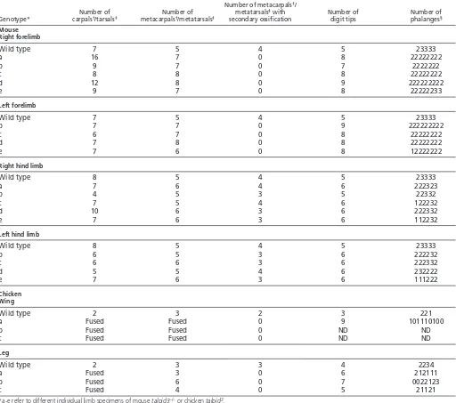
talpid3CKO mouse forelimbs were polydactylous, with up to nine digits (Fig. 5A⬘; compare Movies 1 and 2 in the supplementary material; see Fig. S4A in the supplementary material), which appeared, externally, to be fused (Fig. 5, compare insets in A⬘and A). Skeletal preparations revealed, however, only soft tissue syndactyly. Up to 16 carpals developed, compared with seven in wild-type mouse wrist, and were sometimes fused (see Fig. S4B,B⬘in the supplementary material; Table 2). Nearly all the digits appeared morphologically similar, usually consisting of two rather than three phalanges, including a terminal phalanx, and some digits were bifurcated (Fig. 5A⬘, arrow; Table 2). talpid3CKO mouse hind limbs were less affected than forelimbs, with usually just one extra digit, and digits also bifurcated (Fig. 5B⬘; compare
Movies 3 and 4 in the supplementary material; see Fig. S4C in the supplementary material; Table 2). As in mutant forelimbs, most hind limb digits only had two phalanges, although in several cases the penultimate posterior digit developed three phalanges (Fig. 5B⬘, asterisk; Table 2) and the most posterior metatarsal was of wild-type shape (Fig. 5B⬘, arrowhead).
[image:5.612.52.512.58.353.2]In addition to being polydactylous, talpid3CKO mouse limbs were short, with each skeletal element in both fore- and hind limbs being 40-70% shorter than wild-type counterparts (Fig. 6A), and the ossification of fore- and hind limb elements was abnormal and absent in digits at E17.5 (Fig. 5, compare Alizarin Red staining in A⬘,B⬘with that in A,B). Histological analysis of sections through growth plates confirmed that there was no subperiosteal bone deposited in the radius and ulna in mutant forelimbs; consequently, no bone collar formed, although calcified tissue stained with Alizarin Red formed internally (Fig. 6, compare wild type in B with talpid3 CKO in B⬘). Growth plates in long bones of talpid3 CKO forelimbs showed an increase in resting chondrocytes and a reduction in columnar chondrocytes and prehypertrophic chondrocytes, although chondrocytes in the long-bone centre still eventually underwent hypertrophy (Fig. 6, compare C with C⬘, proximal growth plate of ulna). Sections through digits at E17.5
Fig. 4. Loss of cilia from cells of talpid3CKO mouse limbs. (A,B)Wild-type mouse forelimb (A) and hind limb (B). Primary cilia axonemes (arrowed; stained for acetylated tubulin, green) project from centrosomes (stained for g-tubulin, red) on mesenchyme and ectoderm cells. (C)talpid3 CKO forelimb at E9.5, showing cilia (arrows). (D)talpid3CKO forelimb at E10.5. Primary cilia are absent from mesenchyme cells (arrowhead shows centrosome), but present on ectoderm cells (arrows). (E)talpid3CKO hind limb at E10.5; 2% of mesenchyme cells have primary cilium (arrow). Fl, forelimb; Hl, hind limb; m, mesenchyme and e, ectoderm, separated by dashed line. (F,F⬘) Phalloidin marks stress fibres (green) in wild-type mouse limb fibroblasts (F), which are reduced in talpid3–/–fibroblasts (F⬘); also note the increased number of filopodia around the cell circumference. (G,G⬘) Vinculin staining marks focal adhesions in wild-type mouse limb fibroblasts (G), which are absent in talpid3–/–fibroblasts (G⬘). (H-J⬘) Still images from film of scratch assay. Wild-type mouse limb fibroblasts migrate to close the scratch (delimited by red lines) within 10 hours (H-J), whereas talpid3–/–fibroblasts migrate more slowly (H⬘-J⬘). (K)The migration speed of wild-type mouse limb fibroblasts as compared with talpid3–/– fibroblasts. (L,L⬘) Plots tracing the movement of wild-type mouse limb fibroblasts (L) or talpid3–/–fibroblasts (L⬘) away from the scratch edge show that talpid3–/–fibroblasts lack directionality.
D
E
V
E
LO
P
M
E
N
confirmed the ossification failure (Fig. 6, compare wild type in D with talpid3CKO in D⬘, asterisk) and showed expansion of the joint-forming region (Fig. 6D⬘, bracket). Morphological changes in joints are presaged by alterations in Gdf5expression (Storm and Kingsley, 1999), which, in E13.5 wild-type forelimbs, precisely marked the joints (Fig. 6E), but in talpid3CKO forelimbs was more diffuse, encompassing a larger area (Fig. 6E⬘).
The general features of talpid3mouse CKO limbs resembled those of the talpid3 chicken mutant. talpid3 chicken mutant embryos surviving until day 10 have short wings and legs with an increased number of morphologically similar digits, although phalange number varies and ossification is impaired (Fig. 5C⬘,D⬘) (see also Ede and Kelly, 1964b; Macrae et al., 2010). In talpid3 CKO mouse forelimbs, digit cartilages were separate, with only soft tissue syndactyly, whereas in talpid3chicken mutant wings digit cartilages are fused (Fig. 5, compare C⬘with A⬘). In talpid3 chicken mutant legs, the extent of syndactyly is variable, with fusion of digit cartilages in some cases but only soft tissue syndactyly in others (Fig. 5D⬘) (see also Ede and Kelly, 1964b).
Patterning defects and skeletal defects in talpid3 CKO mouse limb buds, as in talpid3chicken mutant limb buds, can be ascribed to an inability to respond to Shh and Indian hedgehog (Ihh) signalling, respectively, as reflected in changes in the expression of Gli-regulated genes. At E10.5, Shhwas expressed in the polarising region of talpid3CKO mouse forelimb buds as in wild type (Fig. 7A,A⬘), but expression of the Gli activator targets Ptch1and Gli1 was lost (Fig. 7, compare B,C with B⬘,C⬘), whereas expression of Gli repressor targets, such as Hoxd13, was expanded anteriorly (Fig. 7D,D⬘, arrowheads) as was gremlin (Grem1 – Mouse Genome Informatics) (Fig. 7E,E⬘, arrowheads). Bmp4, which is normally expressed in the anterior limb bud, was reduced (Fig.
7F,F⬘). By contrast, the expression of genes upstream of Shh signalling, for example Hand2, was unaffected (Fig. 7G,G⬘), as was the expression of genes involved in proximodistal patterning, such as Hoxa11and Fgf8 (Fig. 7, compare H,I with H⬘,I⬘), although Hoxa13 expression was expanded anteriorly (Fig. 7J,J⬘). Sections of talpid3CKO humerus at E13.5 showed that Ihhis expressed in prehypertrophic chondrocytes as in wild type, but the absence of hypertrophic chondrocytes in the middle of the rudiment at this stage led to a single central zone of Ihh-expressing cells, whereas in wild type two zones were separated by hypertrophic chondrocytes no longer expressing Ihh (Fig. 6F,F⬘).At E13.5, Ptch1was expressed in the perichondrium surrounding developing digits in wild-type forelimbs, but no expression was detected in talpid3 CKO (Fig. 6G,G⬘).
Morphogenesis and digit spacing in talpid3 CKO
mouse limbs
[image:6.612.51.330.59.378.2]To compare limb shape, optical projection tomography (OPT) was used to generate 3D images of wild-type and talpid3 CKO forelimbs from E11.5-13.5 (Fig. 8A-C). By E11.5, hand plates of talpid3CKO forelimbs were already slightly broader than those of the wild type (Fig. 8Ab,D; see D⬘, which shows where width measurements of the hand plate were taken), and by E12.5 hand plates of talpid3 CKO forelimbs were clearly broader both anteriorly and posteriorly (Fig. 8Bb,D). talpid3CKO hand plates became broader still by E13.5, but distal outgrowth was reduced compared with wild type (Fig. 8Cb,D). Digital slices show that talpid3CKO hand plates were of comparable thickness to those of the wild type until E13.5, when the mutant expanded more ventrally than the wild type (Fig. 8Ac-Ce, arrows). The mean volume (±s.d.) of the hand plate at E11.5 was 395±76 mm3(n14) for wild type
Fig. 5. Skeletal patterns in wild-type and talpid3CKO mouse limbs. (A-B⬘) E17.5 wild-type (A,B) and talpid3 CKO (A⬘,B⬘) mouse forelimb (Fl) and hind limb (Hl) stained with Alcian Blue for cartilage and Alizarin Red for calcified tissue. The talpid3 CKO mouse forelimb and hind limb show polydactyly. Arrow (A⬘) indicates bifurcated digit. Asterisk (B⬘) indicates penultimate posterior digit with normal morphology. Arrowheads indicate normal posterior metatarsal (compare B with B⬘). Insets show wild-type (in A) and talpid3 CKO (in A⬘) forelimbs at E17.5; note the soft tissue syndactyly in the talpid3 CKO forelimb. (C-D⬘) Day 11 wild-type (C,D) and talpid3(C⬘,D⬘) chicken wing and leg stained with Alcian Blue. Note the cartilaginous syndactyly in the talpid3chicken wing and leg (dotted line marks edge of limb).
D
E
V
E
LO
P
M
E
N
and 547±179 mm3(n12) for talpid3 CKO (P0.0158, t-test). At E12.5, the mean volumes of the wild-type and talpid3 CKO hand plates were 804±215 mm3 (n6) and 1074±401 mm3 (n6), respectively (P0.2182, t-test). Assuming each digit occupies the same space, this would give 160.8 mm3per digit in E12.5 wild-type forelimbs with five digits, but only 134.25 mm3per digit in talpid3 CKO forelimbs with an average of eight digits.
A striking feature of the talpid3digit phenotype is syndactyly, involving only soft tissue in mouse but cartilaginous fusion in chicken talpid3 wing and sometimes leg. We investigated the formation of digital condensations and interdigital spaces in talpid3 CKO mutant mouse limbs. Wild-type and talpid3CKO littermates were collected between E11.5 and E13.5 (E11.5 n4 litters; E12.5 n3 litters; E13.5 n3 litters) and Sox9 expression in the left
forelimbs of each embryo and Raldh2(Aldh1a2– Mouse Genome Informatics) expression in right forelimbs was analysed. Sox9 is expressed in prechondrogenic cells and is required for cartilage differentiation (Akiyama et al., 2002), whereas Raldh2 is expressed in interdigital spaces between digit cartilage condensations (Niederreither et al., 1997; Schmidt et al., 2009; Kuss et al., 2009). Based on the patterns of expression obtained, the sequence of formation of digit condensations and interdigital spaces in wild-type and talpid3CKO limbs was deduced.
[image:7.612.51.563.72.525.2]In wild-type E11.5 mouse forelimbs, Sox9 was expressed throughout the digital plate. Then, first a Sox9-positive digit 4 condensation became discernible (Fig. 8E, asterisk), followed by rapid breakup of continuous Sox9 expression into digit 2 and 3 condensations, then digit 5, then digit 1 condensations (Fig. 8G);
Table 2. Analysis of limb development in E17.5 talpid3 CKO mouse and day 10/11 talpid3chicken embryos
Number of metacarpals†/
Number of Number of metatarsals‡with Number of Number of
Genotype* carpals†/tarsals‡ metacarpals†/metatarsals‡ secondary ossification digit tips phalanges§
Mouse Right forelimb
Wild type 7 5 4 5 23333
a 16 7 0 8 22222222
b 9 7 0 7 2222222
c 8 8 0 8 22222222
d 12 8 0 9 222222222
e 9 7 0 8 22222233
Left forelimb
Wild type 7 5 4 5 23333
b 7 7 0 9 222222222
c 6 7 0 8 22222222
d 7 8 0 8 22222222
e 7 6 0 8 12222222
Right hind limb
Wild type 8 5 4 5 23333
a 7 6 4 6 222323
b 4 5 3 5 22332
c 7 5 4 6 122232
d 10 6 3 6 222332
e 7 6 3 6 112232
Left hind limb
Wild type 8 5 4 5 23333
b 6 5 3 6 222232
c 6 6 3 6 222332
d 5 5 4 6 232222
e 7 6 3 6 111222
Chicken Wing
Wild type 2 3 2 3 221
a Fused Fused 0 9 101110100
b Fused Fused 0 ND ND
c Fused Fused 0 ND ND
Leg
Wild type 2 3 3 4 2234
a Fused 3 0 6 212111
b Fused 6 0 7 0022123
c Fused 4 0 5 21121
*a-e refer to different individual limb specimens of mouse talpid3–/–or chicken talpid3.
†Mouse forelimb or chicken wing. ‡Mouse hind limb or chicken leg.
§The number of phalanges including the terminal phalanx is given (0-4), in turn, for each digit tip. ND, not determined.
D
E
V
E
LO
P
M
E
N
Zhu et al. (Zhu et al., 2008) reported digit condensation in the order 4, 2, 5, 3, 1 using a Nog-lacZ knock-in allele to detect condensations rather than Sox9 expression. At the same time, Raldh2was expressed in the mid region of the bud tip (Fig. 8F), then separated into two blocks of expression representing the second and third interdigital spaces (Fig. 8H). At E12.5, all five digit condensations had formed in the forelimb (Fig. 8I,K) and the
[image:8.612.54.555.59.371.2]second, third and fourth interdigital regions were present, with the first interdigit following later (Fig. 8J). Raldh2expression was then detected anterior to digit 1 and posterior to digit 5 (Fig. 8L). By E13.5, the Sox9expression pattern mirrored digit shape (Fig. 8M); Raldh2expression became restricted to digit/interdigit boundaries (Fig. 8N). The spacing between digits was even in all wild-type forelimbs analysed.
Fig. 6. Skeletogenesis in wild-type and talpid3CKO mouse limbs. (A)The length of talpid3 CKO skeletal elements (red) as a percentage of wild-type elements (blue) normalised to one. (B,B⬘) Wild-type (B) and talpid3CKO (B⬘) mouse radius (r) and ulna (u), with cartilage stained with Alcian Blue at ends of elements, and bony collar (bc) stained with Alizarin Red. In talpid3CKO radius and ulna, cartilage is present at the ends of elements and also surrounds the Alizarin Red-stained (arrowheads) central region of calcified tissue. (C,C⬘) Longitudinal section through the proximal growth plate of wild-type (C) and talpid3 CKO (C⬘) ulna stained with Hematoxylin and Eosin. Brackets indicate different zones: dc, dividing chondrocytes; cc, columnar chondrocytes; phc, prehypertrophic chondrocytes; hc, hypertrophic chondrocytes; arrows indicate subperiosteal bone. Note only dividing and hypertrophic chondrocytes and no subperiosteal bone in talpid3 CKO ulna. (D,D⬘) Sections of middle digit stained with Hematoxylin and Eosin. Note defined joints (arrows), ossification (asterisk) and subperiosteal bone (arrowhead) in wild type (D), whereas the talpid3 CKO digit has an enlarged joint-forming region (bracket, D⬘). (E,E⬘) Gdf5 expression marks joints in wild-type (E) and talpid3CKO (E⬘) forelimbs at E13.5. (F,F⬘) Longitudinal section through humerus showing Ihh expression (dark blue) in two bands (arrows) of prehypertrophic chondrocytes in wild type (F) but in only a single band (arrow) of hypertrophic like-chondrocytes in talpid3CKO humerus (F⬘). (G,G⬘) Ptch1is expressed (arrows) in perichondrium surrounding digits in wild-type hand plate (G) but is absent in talpid3CKO hand plate (G⬘).
Fig. 7. Gene expression in wild-type and talpid3CKO mouse limbs. (A-J⬘) Whole-mount in situ hybridisation for the indicated genes in wild-type (A-J) and talpid3 CKO (A⬘-J⬘) mouse forelimb at E10.5 (A-B⬘) and E11.5 (C-J⬘). Limbs viewed from dorsal side, anterior up. Arrowheads indicate anterior extension of gene expression in talpid3–/–limb buds compared with wild type.
D
E
V
E
LO
P
M
E
N
[image:8.612.57.509.621.699.2]In talpid3CKO mouse limbs, digit condensation was delayed by 6-12 hours, interdigital spaces were not so precisely defined and digit spacing was not always so regular. Thus, whereas in E11.5 wild-type littermates digit 2, 3 and 4 condensations were apparent and discrete and the second and third interdigital spaces had formed, in talpid3 CKO limb buds Sox9 expression remained throughout the digital plate, although with weaker expression in the mid-posterior region at the limb tip (Fig. 8E⬘,G⬘, asterisk). Raldh2expression was increased, spreading diffusely throughout the mid-posterior limb region where Sox9 expression is reduced (Fig. 8F⬘,H⬘). Digit condensations expressing Sox9first appeared in the posterior of talpid3 CKO
forelimbs (Fig. 8G⬘, arrowhead), followed by rapid breakup of Sox9 expression as seen in wild-type forelimbs, resulting in many Sox9-expressing condensations throughout the broad digital plate (Fig. 8I⬘-M⬘). These condensations were not always evenly spaced; Raldh2 expression could be fragmented, not always extending from proximal to distal throughout the digital plate (Fig. 8J⬘-N⬘), consistent with the interdigital spaces not forming as regularly as in wild type.
[image:9.612.50.547.60.446.2]We also examined Raldh2 expression in talpid3chicken mutant wings and legs, which can show more severe syndactyly. In wild-type HH29 chicken wings and legs, Raldh2 was expressed in the interdigital spaces as in mouse, but also in a thin band just beneath
Fig. 8. Limb bud shape and digit spacing in wild-type and talpid3CKO mouse limbs. (A-Ce) Three-dimensional surfaces of right forelimbs from wild-type (A-C, red) and talpid3CKO (Aa-Ca, green) mice. A-Ae, E11.5; B-Be, E12.5; C-Ce, E13.5. Overlaid limbs (Ab-Cb) show the difference (arrows) in shape between wild type (red mesh) and talpid3CKO (transparent green) hand plates. Dashed line indicates the plane of sections in Ad-Cd. (Ac-Cc) Overlaid limbs viewed from a distal perspective. (Ad-Cd) Digital sections show that talpid3CKO limbs are thickened ventrally at E13.5 (arrows). (Ae-Ce) Overlaid limbs viewed from the anterior. (D,D⬘) Scatter plot (D) showing increasing hand plate width over limb length (measured from limb tip to spine, see D⬘) in talpid3CKO forelimbs. (E,G,I,K,M) Sox9expression in wild-type left forelimb from E11.5 to E13.5. Digit-forming condensations are marked with an asterisk (E,G) or numbered (G). (E⬘,G⬘,I⬘,K⬘,M⬘) Sox9expression in talpid3CKO left forelimb from E11.5 to E13.5. Region with reduced Sox9expression is marked with an asterisk (G⬘), and the first digit condensation is marked with an arrowhead (G⬘). (F,H,J,L,N) Raldh2expression in wild-type left forelimb from E11.5 to E13.5. Interdigits are numbered (H). (F⬘,H⬘,J⬘,L⬘,N⬘) Raldh2expression in talpid3CKO left forelimb from E11.5 to E13.5. (O,P)Raldh2expression in wild-type chicken wing (O) and leg (P). Expression is present in the interdigit and between the digit tip and apical ectodermal ridge (arrows). (O⬘,P⬘) Raldh2expression in talpid3chicken wing (O⬘) and leg (P⬘). Note Raldh2expression in a thin line between metacarpals/metatarsals and phalanges as well as beneath the apical ectodermal ridge (arrows), but no interdigital Raldh2expression. Limbs viewed from dorsal side, anterior up.
D
E
V
E
LO
P
M
E
N
the apical ectodermal ridge (Fig. 8O,P, arrows). In talpid3chicken mutant wings and legs at HH29, Raldh2 was expressed in a continuous band around the rim and in another continuous band between the fused phalanges and fused metacarpals/metatarsals (Fig. 8O⬘,P⬘, arrows). Thus, Raldh2 expression is continuous in talpid3chicken mutant wings and legs at a stage when interdigital spaces expressing Raldh2have formed in wild type.
DISCUSSION
Deletion of the highly conserved exons 11 and 12 of talpid3in the mouse abolishes its function and leads to a lack of primary cilia and hence abnormal Hh signalling. This extends our previous finding that these two exons are essential, but not sufficient, to rescue talpid3function in the chicken and is the first time that a null mouse has been generated based on work on a chicken mutant. Our ultrastructural observations – here on mouse and previously on chick (Yin et al., 2009) – show that, in mutant neural tube cells, the centrosome that will form the basal body of the cilium fails to dock with the apical cell membrane. A similar failure of centrosome positioning and/or docking is seen in cells lacking function in other genes encoding basal body proteins, including Ofd1 (oral-facial-digital syndrome 1) (Singla et al., 2010) and MKS1 (Meckel syndrome 1) (Dawe et al., 2007). Like cells of talpid3–/–mice, cells of Ofd1–/–(male mice) and Mks1–/–mouse mutants completely lack primary cilia. By contrast, loss of another centrosomal protein, RPGRIP1L (also known as FTM; which is responsible for Joubert syndrome type B and Meckel syndrome), results in fewer and/or malformed cilia (Vierkotten et al., 2007), and loss of BBS proteins (which are responsible for Bardet-Biedl syndrome) results in defects only in specialised cilia (for a review, see Goetz and Anderson, 2010).
Most features of the talpid3–/–mouse are similar to those of the talpid3mutant chicken and attributable to abnormal Hh signalling (Table 1). In talpid3–/–mouse mutants, the neural tube is dorsalised and midline facial structures are lost. Both defects reflect loss of Gli2 activator activity. In mouse neural tube, graded Shh signalling mediates dorsoventral patterning, with high Gli2 activity specifying ventral neural tube progenitors (Wijgerde et al., 2002), whereas in mouse face, Shh is required for the formation of midline structures. Patients with loss-of-function mutations in GLI2also have mid-facial hypoplasia (Roessler et al., 2003). By contrast, talpid2mutant chickens have an expanded facial midline, even though cells of this mutant have also been reported to lack cilia (Brugmann et al., 2010), resembling the hypertelorism seen in patients with GLI3 haploinsufficiency (Biesecker, 2008) and with Bardet-Biedl syndrome (Beales et al., 1999).
Embryonic lethality characterises both mouse and chicken talpid3 mutants but occurs earlier in mouse, although genetic background has an influence. The earlier demise of talpid3–/– mouse embryos might be due to vascularisation defects [as previously described in chicken talpid3 mutants (Davey et al., 2007)] impacting more severely on embryos with a placenta. talpid3–/–mouse embryos display randomised left/right asymmetry with respect to heart looping, consistent with the loss of nodal cilia (for a review, see Hirokawa et al., 2009). Preliminary observations suggest that talpid3chicken mutant embryos have normal left/right asymmetry. The reasons for this are not clear. Further detailed analysis is on-going and will be reported elsewhere. It has recently been suggested that cilia do not play a role in establishing left/right asymmetry in chicken embryos as they do in other vertebrates, but rather asymmetrical cell movements lead to preferential left-sided Shh expression (Gros et al., 2009).
Other mouse mutants with homozygous mutations in genes encoding IFT components (Kif3a and Ift88), Hh signalling components (Smoand Ptch1), as well as centrosomal proteins required for ciliogenesis (Ofd1, males), exhibit a similar gross morphological phenotype to the talpid3–/–mutant mouse. In all cases, embryos survive until E9.5-10.5, turning is compromised, heart looping is randomised and there are neural tube defects and loss of facial midline structures (Goodrich et al., 1997; Takeda et al., 1999; Murcia et al., 2000; Zhang et al., 2001; Ferrante et al., 2006). Mks1 mutant mice survive for longer (up until E18.5) even though the ciliogenesis defect is similar to that of talpid3–/–mice and they exhibit randomised heart looping and polydactyly (Weatherbee et al., 2009).
talpid3 CKO mouse limbs are remarkably similar to talpid3 chicken mutant limbs, i.e. they are polydactylous, short and syndactylous, even though effective loss of Talpid3 function, as witnessed by lack of cilia, is not abolished until E10.5, and then only in mesenchyme. This similarity suggests that Shh signalling is necessary only at later stages of limb development, fitting nicely with the phenotype of Shh–/– mouse embryonic limbs in which only structures distal to the elbow/knee are defective (Chiang et al., 2001). In talpid3 CKO mouse forelimbs, up to nine morphologically indistinguishable digits develop, whereas in hind limbs polydactyly is reduced, with just a single extra digit, and digits arising in the posterior are more patterned. Both the increase in digit number and the absence of pattern can be ascribed to abnormal Shh signalling resulting from a lack of cilia, and the expression of Gli-regulated genes in early limb buds is consistent with this. Digit morphology in talpid3 CKO limbs, particularly bifurcations, most closely resembles that in the limbs of mouse embryos that only express full-length Gli3 activator (Wang et al., 2007).
All forelimb digits and most digits in talpid3CKO mouse hind limbs lack one phalange. This brachydactyly, together with the shortening of long bones and accompanying growth plate changes and the failure to form subperiosteal bone, are seen in Ihh mouse mutants (St-Jacques et al., 1999). Calcified tissue is deposited in both talpid3CKO and Ihh mutant mouse long bones (St-Jacques et al., 1999); thus, maturation of chondrocytes occurs in talpid3 CKO mouse long bones despite the inability to respond to Ihh signalling as evidenced by the lack of Ptch1 expression. Calcified tissue has not been detected in talpid3mutant chicken long bones (Macrae et al., 2010).
Similar patterning and skeletal defects to those in talpid3CKO limbs have been seen in Prrx1conditional limb knockouts of Ift88, Kif3a and Ofd1, including polydactyly and abnormal ossification (Haycraft et al., 2007; Bimonte et al., 2010). Even though Ofd1 CKO mouse limbs end up with an almost identical skeletal phenotype to talpid3 CKO mouse limb buds, Ofd1 protein levels are not reduced at E10.5 (Bimonte et al., 2010), whereas in talpid3 CKO limb buds cilia are already absent by this stage. This suggests that timing cannot explain the difference in the extent of polydactyly between forelimb and hind limb. Limbs of patients with oral-facial-digital syndrome type 1, however, exhibit brachydactyly and syndactyly but not polydactyly [Online Mendelian Inheritance in Man (OMIM) ID #311200].
A striking difference between limbs of talpid3 CKO mice and talpid3chicken mutants is the extent of syndactyly. In the mouse, this only involves soft tissue, whereas in the chicken, especially wing, there is cartilaginous fusion. A possible reason for this difference is that talpid3 function is only knocked out in mesenchyme in the mouse, whereas in the chicken it is absent in both mesenchyme and ectoderm. Reciprocal recombination of
D
E
V
E
LO
P
M
E
N
ectoderm and mesenchyme from wild-type and talpid3chicken leg buds shows that the limb phenotype is determined by mesenchyme (Ede and Shamslahidjani, 1983). This finding suggests that the wild-type nature of the ectoderm in talpid3 CKO limb buds is not the reason for the differences in syndactyly. Formation of digit condensations and intervening interdigital spaces is delayed and irregular in talpid3CKO mouse limbs, whereas in talpid3chicken mutant limbs no interdigital spaces expressing Raldh2 are seen at a stage when this process is complete in wild type, correlating with extensive digital fusion. Interestingly, we showed sometime ago that implanting a bead soaked in retinoic acid or grafting a polarising region to wing buds of talpid3chicken mutant embryos could, to some extent, rescue syndactyly and result in digit separation (Francis-West et al., 1995).
Both mouse and chicken talpid3 mutant limb buds have a dramatically altered shape, being broader and shorter than wild-type limb buds. Donald Ede (Ede, 1971) suggested that the shape of the chicken mutant wing bud could be due to changes in cell proliferation or in the ability of cells to polarise. We found that talpid3–/–mouse cells take longer to migrate into a scratch and have less directionality than wild-type cells. This fits with recent work on cells from OPRK (Ift88Tg737Rpw) mice suggesting that primary cilia co-ordinate directional cell migration and chemotaxis (Schneider et al., 2010). The changed behaviour of talpid3–/– mutant cells is also intriguing in view of the suggestion that directional cell movement determines limb shape (Wyngaarden et al., 2010); thus, talpid3 CKO limbs could be a useful model to study morphogenesis.
Acknowledgements
We thank Alan Prescott, John James and Ursula Potter for electron microscopy; Louise Anderson and annex staff for their help; and Philip Beales, Malcolm Logan, Kate Nobes and Dominic Norris for discussions. We are grateful for support from the BBSRC (F.B., M.W., M.G.D.), MRC (F.B., N.A.) and The Royal Society (C.T.). Deposited in PMC for release after 6 months.
Competing interests statement
The authors declare no competing financial interests.
Supplementary material
Supplementary material for this article is available at
http://dev.biologists.org/lookup/suppl/doi:10.1242/dev.063602/-/DC1
References
Abbott, U. K., Taylor, L. W. and Abplanalp, H.(1959). A second talpid-like mutation in the fowl. Poult. Sci. 38, 1185.
Akiyama, H., Chaboisssier, M. C., Martin, J. F., Schedl, A. and de Crombrugghe, B.(2002). The transcription factor Sox9 has essential roles in successive steps of the chondrocyte differentiation pathway and is required for expression of Sox5 and Sox6. Genes Dev.16, 2813-2828.
Baker, K. and Beales, P. L.(2009). Making sense of cilia in disease: the human ciliopathies. Am. J. Med. Genet. C Semin. Med. Genet.151C, 281-295.
Beales, P. L., Elcioglu, N., Woolf, A. S., Parker, D. and Flinter, F. A.(1999). New criteria for improved diagnosis of Bardet-Biedl syndrome: results of a population survey. J. Med. Genet.36, 437-446.
Biesecker, L. G.(2008). The Greig cephalopolysyndactyly syndrome. Orphanet. J. Rare Dis.3, 10.
Bimonte, S., De Angelis, A., Quagliata, L., Giusti, F., Tammaro, R., Dallai, R., Ascenzi, M. G., Diez-Roux, G. and Franco, B.(2010). Ofd1 is required in limb
bud patterning and endochondral bone development. Dev. Biol.349, 179-191.
Brennan, J., Lu, C. C., Norris, D. P., Rodriguez, T. A., Beddington, R. S. and Robertson, E. J.(2001). Nodal signalling in the epiblast patterns the early
mouse embryo. Nature411, 965-969.
Brugmann, S. A., Allen, N. C., James, A. W., Mekonnen, Z., Madan, E. and Helms, J. A.(2010). A primary cilia-dependent etiology for midline facial disorders. Hum. Mol. Genet.19, 1577-1592.
Buxton, P., Davey, M. G., Paton, I. R., Morrice, D. R., Francis-West, P. H., Burt, D. W. and Tickle, C.(2004). Craniofacial development in the talpid3 chicken mutant. Differentiation72, 348-362.
Chiang, C., Littingtung, Y., Harris, M. P., Simandl, B. K., Li, Y., Beachy, P. A. and Fallon, J. F.(2001). Manifestation of the limb prepattern: limb development in absence of sonic hedgehog function. Dev. Biol. 236, 421-435.
Cole, R. K.(1942). The talpidlethal in the domestic foul. J. Hered.33, 82-86.
Davey, M. G., Paton, I. R., Yin, Y., Schmidt, M., Bangs, F. K., Morrice, D. R., Smith, T. G., Buxton, P., Stamataki, D., Tanaka, M. et al.(2006). The chicken talpid3 gene encodes a novel protein essential for Hedgehog signaling. Genes
Dev.20, 1365-1377.
Davey, M. G., James, J., Paton, I. R., Burt, D. W. and Tickle, C.(2007). Analysis of talpid3 and wild-type chicken embryos reveals roles for Hedgehog signalling in development of the limb bud vasculature. Dev. Biol. 301, 155-165.
Dawe, H. R., Smith, U. M., Cullinane, A. R., Gerrelli, D., Cox, P., Badano, J. L., Blair-Reid, S., Sriram, N., Katsanis, N., Attie-Bitach, T. et al.(2007). The Meckel-Gruber Syndrome proteins MKS1 and meckelin interact and are required for primary cilium formation. Hum. Mol. Genet.16, 173-186.
Ede, D. A.(1971). Control of form and pattern in the vertebrate limb. Symp. Soc. Exp. Biol. 25, 235-254.
Ede, D. A. and Kelly, W. A.(1964a). Developmental abnormalities in the head region of the talpid mutant of the fowl. J. Embryol. Exp. Morphol.12, 161-182.
Ede, D. A. and Kelly, W. A.(1964b). Developmental abnormalities in the trunk and limbs of the fowl. J. Embryol. Exp. Morphol.12, 339-356.
Ede, D. A. and Agerbak, G. S.(1968). Cell adhesion and movement in relation to the developing limb pattern in normal and talpid mutant chick embryos. J. Embryol. Exp. Morphol.20, 81-100.
Ede, D. A. and Shamslahidjani, M.(1983). Ectoderm/mesoderm recombination, dissociation and cell aggregation studies in normal and talpid mutant avian embryos. Prog. Clin. Biol. Res.110, 45-55.
Ferrante, M. I., Zullo, A., Barra, A., Bimonte, S., Messaddeq, N., Studer, M., Dollé, P. and Franco, B.(2006). Oral-facial-digital type I protein is required for primary cilia formation and left-right axis specification. Nat. Genet. 38, 112-117.
Fisher, M. E., Clelland, A. K., Bain, A., Baldock, R. A., Murphy, P., Downie, H., Tickle, C. R., Davidson, D. R. and Buckland, R. A.(2008). Integrating technologies for comparing 3D gene expression domains in the developing chick limb. Dev. Biol. 317, 13-23.
Francis-West, P. H., Robertson, K. E., Ede, D. A., Rodriguez, C., Izpisúa-Belmonte, J. C., Houston, B., Burt, D. W., Gribbin, C., Brickell, P. M. and Tickle, C.(1995). Expression of genes encoding bone morphogenetic proteins and sonic hedgehog in talpid (ta3) limb buds: their relationships in the signalling cascade involved in limb patterning. Dev. Dyn.203, 187-197.
Goetz, S. C. and Anderson, K. V.(2010). The primary cilium: a signalling centre during vertebrate development.Nat. Rev. Genet.11, 331-344.
Goodrich, L. V., Milenkovic´, L., Higgins, K. M. and Scott, M. P.(1997). Altered neural cell fates and medulloblastoma in mouse patched mutants. Science277, 1109-1113.
Gros, J., Feistel, K., Viebahn, C., Blum, M. and Tabin, C. J.(2009). Cell movements at Hensen’s node establish left/right asymmetric gene expression in the chick. Science324, 941-944.
Haycraft, C. J., Zhang, Q., Song, B., Jackson, W. S., Detloff, P. J., Serra, R. and Yoder, B. K.(2007). Intraflagellar transport is essential for endochondral bone
formation. Development134, 307-316.
Hirokawa, N., Tanaka, Y. and Okada, Y.(2009). Left-right determination: involvement of molecular motor KIF3, cilia, and nodal flow. Cold Spring Harb. Perspect. Biol. 1, a000802.
Hogan, B. and Lacy, E.(1994). Manipulating the Mouse Embryo: a Laboratory Manual. Plainview, NY: Cold Spring Harbor Laboratory Press.
Izpisúa-Belmonte, J. C., Ede, D. A., Tickle, C. and Duboule, D.(1992). The mis-expression of posterior Hox-4 genes in talpid (ta3) mutant wings correlates with the absence of anteroposterior polarity. Development114, 959-963.
Kuss, P., Villavicencio-Lorini, P., Witte, F., Klose, J., Albrecht, A. N., Seeman, P., Hecht, J. and Mundlos, S.(2009). Mutant Hoxd13 induces extra digits in a mouse model of synpolydactyly directly and by decreasing retinoic acid synthesis.
J. Clin. Invest.119, 146-156.
Lewis, K. E., Drossopoulou, G., Paton, I. R., Morrice, D. R., Robertson, K. E., Burt, D. W., Ingham, P. W. and Tickle, C.(1999). Expression of ptc and gli genes in talpid3 suggests bifurcation in Shh pathway. Development126, 2397-2407.
Logan, M., Martin, J. F., Nagy, A., Lobe, C., Olson, E. N. and Tabin, C. J.
(2002). Expression of Cre recombinase in the developing mouse limb bud driven by a Prxl enhancer. Genesis33, 77-80.
Macrae, V. E., Davey, M. G., McTeir, L., Narisawa, S., Yadav, M. C., Millan, J. L. and Farquharson, C.(2010). Inhibition of PHOSPHO1 activity results in impaired skeletal mineralization during limb development of the chick. Bone46, 1146-1155.
Matise, M. P., Epstein, D. J., Park, H. L., Platt, K. A. and Joyner, A. L.(1998). Gli2 is required for induction of floor plate and adjacent cells, but not most ventral neurons in the mouse central nervous system. Development125, 2759-2770.
Moorman, A. F., Houweling, A. C., de Boer, P. A. and Christoffels, V. M.
(2001). Sensitive nonradioactive detection of mRNA in tissue sections: novel
D
application of the whole-mount in situ hybridization protocol. J. Histochem. Cytochem.49, 1-8.
Murcia, N. S., Richards, W. G., Yoder, B. K., Mucenski, M. L., Dunlap, J. R. and Woychik, R. P.(2000). The Oak Ridge Polycystic Kidney (orpk) disease gene is required for left-right axis determination. Development127, 2347-2355.
Niedderreither, K., McCaffery, P., Dräger, U. C., Chambon, P. and Dollé, P.
(1997). Restricted expression and retinoic acid-induced downregulation of the retinaldehyde dehydrogenase type 2 (RALDH-2) gene during mouse development. Mech. Dev.62, 67-78.
Riddle, R. D., Johnson, R. L., Laufer, E. and Tabin, C.(1993). Sonic hedgehog mediates the polarizing activity of the ZPA. Cell75, 1401-1416.
Roessler, E., Du, Y. Z., Mullor, J. L., Casas, E., Allen, W. P., Gillessen-Kaesbach, G., Roeder, E. R., Ming, J. E., Ruiz i Altaba, A. and Muenke, M.(2003). Loss-of-function mutations in the human GLI2 gene are associated with pituitary anomalies and holoprosencephaly-like features. Proc. Natl. Acad. Sci. USA100, 13424-13429.
Saunders, J. W. and Gasseling, M. T.(1968). Ectodermal-mesenchymal interactions in the origin of limb symmetry. In Epithelial-mesenchymal Interactions(ed. R. Fleischmeyer and R. E. Billingham), pp. 78-97. Baltimore: Williams & Wilkins.
Schmidt, K., Hughes, C., Chudek, J. A., Goodyear, S. R., Aspden, R. M., Talbot, R., Gundersen, T. E., Blomhoff, R., Henderson, C., Wolf, C. R. et al.
(2009). Cholesterol metabolism: the main pathway acting downstream of cytochrome P450 oxidoreductase in skeletal development of the limb. Mol. Cell. Biol. 29, 2716-2729.
Schneider, L., Cammer, M., Lehman, J., Nielsen, S. K., Guerra, C. F., Veland, I. R., Stock, C., Hoffmann, E. K., Yoder, B. K., Schwab, A. et al.(2010). Directional cell migration and chemotaxis in wound healing response to PDGF-AA are coordinated by the primary cilium in fibroblasts. Cell. Physiol. Biochem. 25, 279-292.
Singla, V., Romaguera-Ros, M., Garcia-Verdugo, J. M. and Reiter, J. F.(2010). Ofd1, a human disease gene, regulates the length and distal structure of centrioles. Dev. Cell18, 410-424.
St-Jacques, B., Hammerschmidt, M. and McMahon, A. P.(1999). Indian hedgehog signaling regulates proliferation and differentiation of chondrocytes and is essential for bone formation. Genes Dev.13, 2072-2086.
Storm, E. E. and Kingsley, D. M.(1999). GDF5 coordinates bone and joint formation during digit development.Dev. Biol. 209,11-27.
Takeda, S., Yonekawa, Y., Tanaka, Y., Okada, Y., Nonaka, S. and Hirokawa, N.(1999). Left-right asymmetry and kinesin superfamily protein KIF3A: new insights in determination of laterality and mesoderm induction by kif3A–/– mice analysis.J. Cell Biol. 145, 825-836.
Tickle, C.(2006). Making digit patterns in the vertebrate limb.Nat. Rev. Mol. Cell Biol. 7, 45-53.
Tiecke, E., Turner, R., Sanz-Ezquerro, J. J., Warner, A. and Tickle, C.(2007). Manipulations of PKA in chick limb development reveal roles in digit patterning including a positive role in Sonic Hedgehog signaling. Dev. Biol. 305, 312-324.
Towers, M., Mahood, R., Yin, Y. and Tickle, C.(2008). Integration of growth and specification in chick wing digit-patterning. Nature452, 882-886.
Vierkotten, J., Dildrop, R., Peters, T., Wang, B. and Rüther, U.(2007). Ftm is a novel basal body protein of cilia involved in Shh signalling. Development134, 2569-2577.
Vortkamp, A., Franz, T., Gessler, M. and Grzeschik, K. H.(1992). Deletion of GLI3 supports the homology of the human Greig cephalopolysyndactyly
syndrome (GCPS) and the mouse mutant extra toes (Xt). Mamm. Genome3,
461-463.
Wang, C., Rüther, U. and Wang, B.(2007). The Shh-independent activator function of the full-length Gli3 protein and its role in vertebrate limb digit patterning. Dev. Biol. 305, 460-469.
Weatherbee, S. D., Niswander, L. A. and Anderson, K. V.(2009). A mouse model for Meckel syndrome reveals Mks1 is required for ciliogenesis and
Hedgehog signaling. Hum. Mol. Genet.18, 4565-4575.
Wijgerde, M., McMahon, J. A., Rule, M. and McMahon, A. P.(2002). A direct requirement for Hedgehog signaling for normal specification of all ventral progenitor domains in the presumptive mammalian spinal cord. Genes Dev.16, 2849-2864.
Wilkinson, D. G. and Nieto, M. A.(1993). Detection of messenger RNA by in situ hybridization to tissue sections and whole mounts. Methods Enzymol. 225, 361-373.
Wyngaarden, L. A., Vogeli, K. M., Ciruna, B. G., Wells, M., Hadjantonakis, A. K. and Hopyan, S.(2010). Oriented cell motility and division underlie early limb
bud morphogenesis. Development137, 2551-2558.
Yang, Y., Drossopoulou, G., Chuang, P. T., Duprez, D., Marti, E., Bumcrot, D., Vargesson, N., Clarke, J., Niswander, L., McMahon, A. et al.(1997). Relationship between dose, distance and time in Sonic Hedgehog-mediated regulation of anteroposterior polarity in the chick limb. Development124, 4393-4404.
Yin, Y., Bangs, F., Paton, I. R., Prescott, A., James, J., Davey, M. G., Whitley, P., Genikhovich, G., Technau, U., Burt, D. W. et al.(2009). The Talpid3 gene (KIAA0586) encodes a centrosomal protein that is essential for primary cilia
formation. Development136, 655-664.
Zhang, X. M., Ramalho-Santos, M. and McMahon, A. P.(2001). Smoothened mutants reveal redundant roles for Shh and Ihh signaling including regulation of L/R symmetry by the mouse node. Cell106, 781-792.
Zhu, J., Nakamura, E., Nguyen, M. T., Bao, X., Akiyama, H. and Mackem, S.
(2008). Uncoupling Sonic hedgehog control of pattern and expansion of the developing limb bud. Dev. Cell14, 624-632.