299
Introduction
Neural induction constitutes the first step in a complex sequence of events leading to the formation of the vertebrate nervous system. This process is believed to consist of a decision made by gastrula ectodermal cells between neural and epidermal fates (reviewed by Munoz-Sanjuan and Brivanlou, 2002; Wilson and Edlund, 2001). Early work in amphibians suggested that the decision to become neural is imposed by signals emanating from the dorsal mesoderm, or Spemann’s organiser (reviewed by Harland and Gerhart, 1997). More recently, numerous organiser-specific factors have been discovered, which include extracellular antagonists of bone morphogenetic proteins (BMPs) (reviewed by De Robertis et al., 2000).
In the ectoderm, the interplay between BMPs and their antagonists, such as Chordin (Chd), is currently considered as being central to neural induction (reviewed by Munoz-Sanjuan and Brivanlou, 2002). Xenopus ectodermal explants, which normally express Bmp4, develop as epidermis when cultured in the absence of organiser signals. When cell contacts and communication are disrupted in these explants, neural tissue
develops instead. Epidermis formation is, however, restored when dissociated explants are cultured in the presence of BMP4 protein. Conversely, intact ectodermal explants form neural tissue upon overexpression of BMP inhibitors, suggesting that these factors recapitulate neural induction by the organiser. This series of ex vivo experiments served as a basis for a model, known as the default model, which states that neural induction is a direct consequence of BMP inhibition in the ectoderm (Munoz-Sanjuan and Brivanlou, 2002). In this model, neural identity constitutes the ground state within the ectoderm and is revealed in the absence of instructive signals. A major difficulty with the default model is that it does not appear to account for neural induction in avian embryos. It was shown that organiser graft, but not BMP inhibition, leads to neural development in chick lateral epiblast (Streit et al., 1998). Only the cells located at the border between neural and non-neural territories can take on a non-neural identity upon BMP inhibition, suggesting that these cells are exposed to additional neuralising cues (Streit and Stern, 1999). Recent reports have indicated that FGF may constitute one of these signals (Streit et al., 2000; Wilson et al., 2000). Similar to organiser grafts,
Neural induction constitutes the first step in the generation of the vertebrate nervous system from embryonic ectoderm. Work with Xenopus ectodermal explants has suggested that epidermis is induced by BMP signals, whereas neural fates arise by default following BMP inhibition. In amniotes and ascidians, however, BMP inhibition does not appear to be sufficient for neural fate acquisition, which is initiated by FGF signalling. We decided to re-evaluate in the context of the whole embryo the roles of the BMP and FGF pathways during neural induction in Xenopus. We find that ectopic BMP activity converts the neural plate into epidermis, confirming that this pathway must be inhibited during neural induction in vivo. Conversely, inhibition of BMP, or of its intracellular effector SMAD1 in the non-neural ectoderm leads to epidermis suppression. In no instances, however, is BMP/SMAD1 inhibition sufficient to elicit neural induction in ventral ectoderm. By contrast, we find that neural specification occurs when weak eFGF or low ras signalling
are combined with BMP inhibition. Using all available antimorphic FGF receptors (FGFR), as well as the pharmacological FGFR inhibitor SU5402, we demonstrate that pre-gastrula FGF signalling is required in the ectoderm for the emergence of neural fates. Finally, we show that although the FGF pathway contributes to BMP inhibition, as in other model systems, it is also essential for neural induction in vivo and in animal caps in a manner that cannot be accounted for by simple BMP inhibition. Taken together, our results reveal that in contrast to predictions from the default model, BMP inhibition is required but not sufficient for neural induction in vivo. This work contributes to the emergence of a model whereby FGF functions as a conserved initiator of neural specification among chordates.
Key words: Xenopus, Vertebrates, Chordates, Neural induction, FGF, BMP, SMAD6, eFGF, Spemann’s organiser, Mesoderm, Ectoderm, SU5402
Summary
Neural induction in Xenopus requires early FGF signalling in
addition to BMP inhibition
Emilie Delaune, Patrick Lemaire and Laurent Kodjabachian*
Institut de Biologie du Développement de Marseille, Laboratoire de Génétique et Physiologie du Développement, CNRS-Université de la Méditerranée, Campus de Luminy, Case 907, 13288 Marseille Cedex 9, France
*Author for correspondence (e-mail: kodja@ibdm.univ-mrs.fr)
Accepted 11 November 2004
Development 132, 299-310
Published by The Company of Biologists 2005 doi:10.1242/dev.01582
Research article
De
velopment
De
soluble FGF8 can induce early neural marker gene expression in competent epiblast. The organiser does not, however, elicit neural induction if FGF receptor (FGFR) activity is blocked, in spite of the normal expression of chdin the graft (Streit et al., 2000). Interestingly, some authors suggested that perhaps FGF signalling in the chick is primarily involved in repressing the expression of Bmp genes (Munoz-Sanjuan and Brivanlou, 2002; Wilson and Edlund, 2001). A similar role has been proposed for early β-catenin activity in the Xenopusectoderm (Baker et al., 1999). According to this hypothesis, BMP inhibition in the chick, like in Xenopus, remains central to neural fate acquisition, and is achieved both via transcriptional repression and the activity of secreted antagonists. However, recent experiments on chick epiblast explants have suggested that FGF signalling also functions in a BMP-independent manner during neural induction (Wilson et al., 2001). Recent work in ascidians, which represent basal chordates, suggested that BMP inhibition is not involved in neural specification (Darras and Nishida, 2001), but that, here also, FGF plays an essential role as a direct inducer of early neural genes (Bertrand et al., 2003).
By contrast, FGFR signalling in Xenopus is currently considered to be essential for posterior neural development but not for neural induction (reviewed by Munoz-Sanjuan and Brivanlou, 2001; Munoz-Sanjuan and Brivanlou, 2002). This conclusion is primarily based on overexpression of a dominant-negative form of the FGFR1 receptor, which does not apparently suppress the formation of anterior neural features (Amaya et al., 1991; Holowacz and Sokol, 1999; Kroll and Amaya, 1996; Ribisi et al., 2000). However, several reports using antimorphic FGFR1 or FGFR4 receptors have suggested instead that FGF signalling plays a role during neural tissue development, leaving this controversial issue still unresolved (Hongo et al., 1999; Launay et al., 1996; Pera et al., 2003; Sasai et al., 1996).
We therefore made use of a broad panel of molecular and pharmacological tools to readdress the roles of the BMP and FGF signalling pathways during neural induction in Xenopus embryos. The in vivo results presented here are consistent with a model whereby pre-gastrula FGF signalling functions in addition to BMP inhibition to induce the nervous system, thus providing an evolutionary conserved alternative to the default model.
Materials and methods
Embryo manipulations and injections
Embryos were fertilised in vitro, dejellied, cultured and injected with synthetic capped RNAs (Ambion Megascript kit), as described (Carnac et al., 1996). Amounts of injected lacZRNA ranged from 250 to 500 pg. For all ventral ectoderm injection performed at the 16-cell stage, only embryos showing injected cells distant from the endogenous neural plate were considered for statistical analysis. We used amounts of RNAs encoding antimorphic FGF receptors similar to previous workers. The main difference with other studies lies in the fact that we poked twice (animally and marginally) a single dorsal blastomere at the four-cell stage, in order to obtain a more global RNA distribution in the progeny of the injected cell. Initial references for our expression constructs are: lacZ (Lemaire et al., 1995), XFD (Amaya et al., 1991), ∆FGFR4 (Hongo et al., 1999), wild-type FGFR4, tR4(Umbhauer et al., 2000), CABR(Hsu et al., 1998), tBR (Suzuki et al., 1994) and v-ras(Carballada et al., 2001). eFGFwas
cloned into pCS2+ (a gift from H. Isaacs). It was linearised with NotI and transcribed with Sp6 RNA polymerase. XSmad6was cloned into pBS-RN3 (Lemaire et al., 1995) (a gift from H. Yasuo). It was linearized with SfiI and transcribed with T3 RNA polymerase.
SU5402, and Noggin protein treatments
SU5402 (Calbiochem) was dissolved in DMSO (120 mM), and diluted in 0.1MBS for whole-embryo treatments, and in a low Ca2+, low Mg2+Ringer’s solution for animal explants treated with Noggin protein (LCMR, 43 mM NaCl, 0.85 mM KCl, 0.37 mM CaCl2, 5 mM HEPES, pH 7.2, 50 µg/ml gentamycin). We did not observe any effect of SU5402 below 5 µM. The severity of the phenotypes exhibited by SU5402-treated embryos increased in a dose-dependent manner. We arbitrarily defined five phenotypic classes at tadpole stage (Fig. 3A): class I embryos showed a slightly truncated tail; class II embryos lacked a tail and showed a slightly shorter trunk; class III embryos lacked all trunk and tail tissues; class IV embryos additionally showed a reduced head; class V embryos lacked all axial tissues, including head structures, but still formed a cement gland. As we noticed a variability between experiments in the amount of inhibitor necessary to yield class V embryos, we used in each experiment several concentrations ranging between 80 and 160 µM, and report data regarding the fifth class irrespective of the concentration. There was no obvious defects in cell cleavage, or in cell viability within the range of concentration used here. Gastrula stage SU5402-treated embryos were classified according to the phenotype of sibling embryos treated identically and grown up to tailbud or tadpole stage. Mouse recombinant Noggin/Fc chimeric protein (R&D systems) was resuspended in PBS, 0.1% BSA (50 µg/ml). Animal explants were treated with 2 µg/ml Noggin/Fc in LCMR supplemented with 0.5% BSA (47% of the fusion protein consists of the Noggin peptide), and stage 8 embryos were injected with 2 ng Noggin/Fc in the blastocoel.
In situ hybridisation, and immunostaining
Injected embryos were fixed for 30 minutes in MEMFA (100 mM MOPS, pH 7.4, 2 mM EGTA, 1 mM MgSO4, 3.7% formaldehyde), and analysed for lineage tracer distribution via β-galactosidase staining with X-gal or Salmon-gal substrate (Biosynth AG). Stained embryos were fixed in MEMFA for an additional hour, and processed for whole-mount in situ hybridisation with digoxygenin labelled probes (Roche) as described (Moos et al., 1995). Antisense riboprobes for noggin (Smith and Harland, 1992), chordin(Sasai et al., 1994), goosecoid (gsc) (Cho et al., 1991), cerberus (Bouwmeester et al., 1996), Sox2 (Mizuseki et al., 1998), opl (Kuo et al., 1998), dkk-1 (Glinka et al., 1998), hex(Newman et al., 1997), otx2(Pannese et al., 1995), Xcad3(Pownall et al., 1996), Xbra(Smith et al., 1991), Xnr2 (Jones et al., 1995), ADMP(Moos et al., 1995), Sox17α(Hudson et al., 1997), K81(Jonas et al., 1989), Slug(Mayor et al., 1995), Bmp4 (Hemmati-Brivanlou and Thomsen, 1995) were prepared as described in the respective references. After staining with BM Purple (Roche) and fixation in MEMFA, pigmented embryos were bleached (Mayor et al., 1995). Immunostaining with 12.101 (muscle) and 4d (anti-NCAM) antibodies was carried out as described (Carnac et al., 1996).
Results
BMP inhibition is required but not sufficient for neural induction in vivo
According to the default model, ectoderm cells form epidermis in response to BMP signals, and neural tissue in absence of BMP signalling (Munoz-Sanjuan and Brivanlou, 2002). We thus initiated our study of the molecular mechanisms of neural induction by addressing these predictions in whole Xenopus embryos.
We first analysed the fate of presumptive neural cells
De
velopment
De
subjected to ectopic BMP activation, via RNA microinjection of a constitutively active BMP receptor (CABR) in a single dorsal animal blastomere at the eight- or 16-cell stage. At the late gastrula stage, injected cells did not express the early neural markers Sox3(9/10 negative) and Sox2(45/47 negative), and instead expressed the epidermal marker cytokeratin 81 (K81) (35/35 positive; Fig. 1A, and not shown). Thus, consistent with ex vivo assays (Munoz-Sanjuan and Brivanlou, 2002), BMP activation in vivo is incompatible with neural induction, and elicits epidermis differentiation.
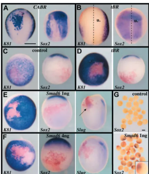
[image:3.612.46.290.198.482.2]Next, we wanted to address whether BMP inhibition in the presumptive non-neural ectoderm was sufficient for neural induction in vivo. As expected from previous works (Mizuseki et al., 1998; Munoz-Sanjuan and Brivanlou, 2002), BMP inhibition obtained by RNA injection of a dominant-negative BMP type I receptor (tBR) led to Sox2 activation (21/22 positive) and K81repression (16/16 negative) in animal caps (not shown). When tBR RNA was injected in the two left animal blastomeres (400 pg/blastomere) of the eight-cell embryo, we observed at the late gastrula stage a limited expansion of the neural plate (9/10 embryos showing expanded Sox2expression) at the expense of epidermis (10/10 embryos showing shrunk K81expression; Fig. 1B). We then restricted tBR misexpression to ectoderm distant from the endogenous neural plate, via injection at the 16-cell stage in a single ventral-most ectoderm cell (400 pg). This led to K81repression (48/53 negative), but not to Sox3(7/7 negative) or Sox2(45/46 negative) activation, suggesting that in these conditions BMP-deficient cells did not adopt a neural identity (Fig. 1D, and not shown). No transient Sox2activation was observed at the early gastrula stage either (not shown). Ventral animal injection of the maximal non toxic dose of tBR RNA (2 ng) still did not lead to Sox2activation (not shown). These data suggest that in vivo inhibition of the BMP pathway at the level of the receptor, although leading to epidermis suppression, is not sufficient to trigger neural induction in ectoderm distant from the endogenous neural plate. It has been proposed that additional inputs acting via the ERK pathway can antagonise the action of BMP at the level of SMAD1 (Pera et al., 2003). We thus reasoned that inhibition of SMAD1 by its specific intracellular inhibitor SMAD6 (Hata et al., 1998) may have a stronger effect than tBR misexpression. However, injection in a single ventral ectoderm cell of high amounts (1 or 4 ng) of Smad6RNA also led to epidermis repression (61/61 K81 negative with 1 ng; 26/26 K81negative with 4 ng) without neural induction (96/96 Sox2negative with 1 ng; 40/40 Sox2negative with 4 ng; Fig. 1E,F) in whole embryos. One caveat in these experiments lies in the fact that the degree of inhibition of the BMP pathway could be sufficient to suppress epidermis, but not to induce neural fates. We thus looked for additional indications that our conditions were indeed very effective at blocking BMP activities. The default model predicts that formation of tissues situated at the frontier between neural and epidermal territories requires intermediate levels of BMP inhibition (Marchant et al., 1998). Thus, it is expected that not only epidermis but fates emerging at the neural/non-neural border, including cement gland and neural crests, should not form under conditions of severe BMP inhibition. We found that ventral ectoderm cells injected with 1 ng Smad6RNA did not adopt a cement gland identity (not shown), but often expressed the neural crest marker Slug(11/17 positive), suggesting that in this condition BMP inhibition was partial (Fig. 1E). By contrast, Slug expression was not detected in ventral ectoderm cells injected with 4 ng Smad6RNA (17/17 negative; Fig. 1F). This suggests that BMP inhibition in this last assay was effective enough to prevent both epidermis and neural border fates from forming. Yet, no neural induction occurred as revealed by the lack of Sox2activation in cells injected with 4 ng Smad6RNA (Fig. 1F). It will be interesting, in future, to investigate the nature of those BMP-deficient cells that are no longer epidermal, but are neither cement gland, neural crest or neural cells.
Fig. 1. BMP inhibition in vivo is not sufficient for neural induction. In all panels, Sox2expression marks prospective neural tissue, K81 expression marks prospective epidermis, XBraexpression marks mesoderm, at the late gastrula stage 13, and Slugmarks neural crest progenitors at early neurula stage 15. Marker gene expression is revealed in purple, while β-galactosidase activity resulting from injection of lacZRNA as a lineage tracer is in red. (A) Sixteen-cell embryos injected with 100 pg CABRRNA in one dorsal-most animal blastomere. BMPR activation represses the neural marker Sox2and activates the epidermal marker K81. (B) Eight-cell embryos injected animally in the two left blastomeres with lacZ(250 pg/blastomere) and tBR(400 pg/blastomere) RNAs showing neural plate expansion and epidermis suppression (u, uninjected side). (C-G) Sixteen-cell embryos injected in one ventral-most animal blastomere with lacZ (C), lacZand tBR(400 pg, D), or lacZand Smad6(1 ng in E,G; 4 ng in F) RNAs. In whole embryos, BMP inhibition by Smad6represses epidermis, but does not induce neural tissue. Ectopic neural crest could form in embryos that received 1 ng (arrow), but not 4 ng, Smad6RNA. (G) Animal caps explanted at blastula stage 9 express Sox2in response to Smad6injection in ventral ectoderm cells. The inset in the lower panel illustrates that Sox2expression (purple) is restricted to the injected region (red). (A,B) Dorsal views, anterior towards the top. (C-F) Ventral views, anterior towards the top, except right panels in E,F, which are lateral views, anterior towards the top. Scale bars: 500 µm.
De
velopment
De
Another possibility to explain the lack of neural induction in vivo in our assays could be that ventral ectoderm cells are not able at all to respond to BMP inhibitors. We turned back to the animal cap assay to address this issue. We injected 1 ng Smad6RNA in a single ventral ectoderm cell at the 16-cell stage, explanted animal caps at late blastula stage, and analysed Sox2 expression in caps and in intact injected siblings. We found that neuralisation occurred in response to Smad6 expression in injected cells in explanted animal caps (15/24 Sox2positive; Fig. 1G), but not in intact embryos (11/11 Sox2 negative; Fig. 1E). These results indicate that our conditions of BMP inhibition are indeed sufficient to neuralise ventral ectoderm cells, which further supports the idea that the lack of neural induction by BMP inhibition in vivo is not merely quantitative. This also demonstrates that extirpation of the blastula ectoderm modifies the response to BMP inhibitors, which suggests that animal cap assays do not unambiguously reflect neural induction in the embryo. In summary, BMP/SMAD1 inhibition in vivo is sufficient to neuralise ectoderm cells in the vicinity but not away from the endogenous neural plate, suggesting that, as in the chick, additional inputs are required for neural induction.
Neural induction in vivo by conjugated FGF signalling and BMP inhibition
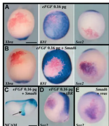
Because of works in amniotes (Streit et al., 2000; Wilson et al., 2000; Ying et al., 2003) and ascidians (Bertrand et al., 2003), we asked whether FGF signalling could play such a role by testing eFGF, a frog homolog of FGF4, in our ventral ectoderm assay. Consistent with previous work (Isaacs et al., 1994), injection of 3.2 pg eFGF RNA induced mesoderm as revealed by Xbra expression (23/23 positive), within and around injected cells (not shown). By contrast, injection of 0.16 pg eFGFRNA did not activate Xbraexpression (9/9 negative), nor did it significantly suppress K81 expression (4/10 no repression; 6/10 light reduction, example in Fig. 2A, centre), or activate Slug(12/12 negative, not shown) or Sox2expression (11/12 negative; Fig. 2A right). This suggests that at this dose eFGFdoes not efficiently antagonise the BMP pathway, which we showed in the previous section is a necessary condition for neural induction. We thus co-injected 0.16 pg eFGFand 1 ng Smad6RNAs, and found that this was now sufficient to repress K81 (12/12 negative) and activate Sox2 (21/21 positive), in absence of Xbraexpression (20/20 negative) at the late gastrula stage (Fig. 2B). Consistent with the activation of a stable neural programme, the injected territory was subsequently found to express the differentiation marker NCAM at tailbud stage (10/10 positive; Fig. 2C). Co-injection of 0.16 pg eFGFand 200 pg tBR RNAs gave similar results (11/11 K81 negative; 9/13 Sox2positive; 12/12 Xbranegative; Fig. 2D). FGF signals can activate multiple transduction pathways, including the ras/MAPK pathway (Powers et al., 2000). As this pathway is active in the early frog embryo (Schohl and Fagotto, 2002), we asked whether activated ras was able to induce ectopic neural tissue in BMP-deficient epidermis. We found that co-injection of 1 pg v-ras and 1 ng Smad6 RNAs led to Sox2 activation (12/12 positive), without Xbraexpression (11/11 negative; Fig. 2E and not shown). We conclude that in vivo and in the absence of detectable mesoderm induction, weak FGF or ras signalling combined with BMP inhibition is sufficient to induce neural tissue in ventral ectoderm.
FGF signalling is required for neural induction
[image:4.612.342.529.396.612.2]The results described above suggest that FGF signalling could participate to neural induction in vivo. We thus decided to evaluate neural development in embryos deficient for FGF function. To do this, we first blocked signalling at the level of the receptor via the use of the pharmacological FGFR inhibitor SU5402 (Mohammadi et al., 1997). This approach offers the possibility of efficiently and reversibly blocking FGF receptors in all cells of the embryo, at any given time, and has been shown to be effective in ascidians (Kim and Nishida, 2001), zebrafish (Shinya et al., 2001) and amniotes (Streit et al., 2000; Ying et al., 2003). We found that embryos cultured from the four-cell stage in the presence of SU5402 lost axial tissues from posterior to anterior, in a dose-dependent manner (Fig. 3A; see Materials and methods). In the most severely affected embryos (class V), mesoderm, neural tissue and neural crests were totally absent all along the body axis (Fig. 3B-H,J-M). Only epidermis, cement gland and endoderm formed normally in these embryos (Fig. 3I,O). Importantly, class V embryos completely lacked expression of the specific neural markers Sox2and opl, both at early (18/18 Sox2negative, and 5/5 opl negative at st10.5) and late gastrula stages (14/14 Sox2 negative, and 7/7 oplnegative at stage 13), suggesting that the neural programme was not initiated in these embryos (Fig. 3J-M). We then attempted to determine the fate, in class V embryos, of the cells normally destined to be part of the neural tissue. To achieve this, we injected RNA encoding the lineage tracer lacZat the 32-cell in presumptive anterior neural plate
Fig. 2. Neural induction in vivo by combined FGF signalling and BMP inhibition. (A-E) Sixteen-cell embryos were injected in one ventral-most animal blastomere with lacZand 0.16 pg eFGF(A); lacZ, 0.16 pg eFGFand 1 ng Smad6 (B,C); lacZ, 0.16 pg eFGFand 200 pg tBR(D); orlacZ, 1 ng Smad6and 1 pg v-rasRNAs (E). Low eFGF or activated ras signalling induces neural tissue, without Xbra-positive mesoderm, when combined with BMP inhibition in late gastrula stage 13 embryos. NCAM immunostaining at tailbud stage confirms the stable neural character of the induced tissue (arrow in C). (A,B,E) Ventral view, anterior towards the top; (C) lateral view, anterior towards the right; (D) lateral view, anterior towards the top. Scale bars: 500 µm.
De
velopment
De
cells and analysed the position of injected cells in tadpoles. Injected cells were found mostly in the eyes and the brain of control embryos. By contrast, in class V embryos these cells were found in the epidermis below the cement gland, and expressed K81(Fig. 3N,O). Thus, these cells appear to remain alive and to adopt a novel fate, ruling out the possibility that the lack of neural tissue produced by SU5402 is due to non-specific toxicity. Our data therefore support the idea that neural tissue is converted into epidermis in the absence of FGF activity.
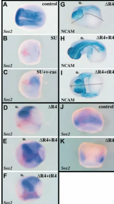
As ras functions downstream of FGF receptors, we asked whether it could bypass the action of SU5402, and directly rescue neural plate tissue. We found that animal injection of 5 to 20 pg v-ras RNA could restore Sox2 expression in late gastrula class V embryos (14/14 Sox2 negative in class V; 12/23 Sox2 positive upon v-ras injection; Fig. 4B,C). Consistently, v-ras also rescued NCAM expression in most SU5402-treated embryos at tailbud stage (not shown).
To further confirm that SU5402 specifically interferes with FGFR function, we analysed neural development in embryos misexpressing various antimorphic FGF receptors. Unilateral injection of 1 ng ∆R4 RNA, which encodes a
[image:5.612.182.562.71.445.2]dominant-negative membrane-bound form of FGFR4 (Hongo et al., 1999), in one dorsal blastomere at the four-cell stage led to a loss of Sox2 expression at the late gastrula stage (12/12 negative), and NCAM at tailbud stage (26/34 negative), without interfering with cement gland formation (Fig. 4D,G). Similarly, injection of XFD RNA (1-4 ng), a dominant-negative XenopusFGFR1 (Amaya et al., 1991), led to a loss of head features and of NCAM positive neural tissue (28/36 negative; not shown). Finally, misexpression of the secreted ectodomains of XenopusFGFR4 (a gift from H. Okamoto), or chick FGFR1 (Marics et al., 2002), which exhibit antimorphic effects by trapping FGF ligands outside the cells, also provoked the loss of the entire neural axis (not shown). To establish further the specificity of the phenotypes generated by these antimorphic receptors, we attempted to rescue neural development in ∆R4- and XFD-injected embryos. It is thought that truncated FGF receptors exert their negative effect via the formation of inactive heterodimeric complexes with normal receptors. Thus, increasing the amount of functional FGF receptors by RNA injection of wild-type counterparts must be sufficient to recover signalling. As shown in Fig. 4E,H, the presence of 1 ng FGFR4RNA rescued Sox2(10/11 positive) Fig. 3. FGFR signalling is required for
neural induction. (A) Four-cell embryos were treated until fixation with various concentrations of SU5402, as indicated. Phenotypic classes were defined according to the severity of axial deficiencies. Class V embryos lacked muscle, NCAM-positive neural tissue and neural crests [as revealed by 12.101 (B) and 4d (C) immunostaining at tailbud stage, or by SlugRNA hybridisation at neurula stage (D)]. Class V embryos at gastrula stages lacked prospective posterior and axial mesoderm (XBra, Xcad3, ADMP; E-G), prospective haematopoietic mesoderm (Xnr2, H), but contained prospective endodermal tissue (sox17α, I). (J-M) Sox2(J,K) and opl(L,M) expression are shown at early (J,L) or late (K,M) gastrula stages in control and class V embryos. No neural precursors are present in class V embryos. (N,O) Thirty-two-cell embryos were injected with 250 pg lacZRNA in one A1 blastomere (animal-most, dorsal-most). In untreated controls, injected cells populate mostly the eye and the brain and some head epidermis (N). In the presence of 120 µM SU5402, embryos were class V, and the injected cells were now found entirely in the epidermis below the cement gland (O). These cells expressed the marker K81, indicating that prospective neural cells were converted into epidermal progenitors
in absence of FGF activity. SU5402 is not toxic to ectoderm cells as they remain alive, and express β-galactosidase and K81. (A-C) Lateral view, anterior towards the left. (N) Lateral view, anterior towards the right. (D,G,J-M) Dorsal view, anterior towards the top. In M, the embryo is slightly tilted upwards. (E,F,H) Vegetal view, dorsal towards the top. (I) Hemisectioned embryo, dorsal towards the right. O, frontal view.
De
velopment
De
and NCAM (21/26 positive) expression in most embryos injected with 1 ng ∆R4 RNA. Rescue was effective in FGFR1/XFD co-injection experiments as well (not shown). It is probable that dominant-negative FGF receptors block most or all types of normal receptors through heterodimerisation of extracellular domains, which makes it difficult to ascribe a particular type to neural induction. In a first attempt to gain insight into this issue, we tried to rescue neural tissue in ∆ R4-injected embryos by co-expressing a ligand-independent
constitutively active form of FGFR4 (tR4) (Umbhauer et al., 2000). As tR4 lacks the extracellular domain normally found in FGFR4, it is unlikely to form heterodimers with wild-type or mutated receptors, and cannot correct the balance in this assay between normal and antimorphic FGFR4. Yet, both Sox2 (6/10 positive) and NCAM (8/12 positive) expression were recovered in embryos co-injected with 1 ng ∆R4and 4 ng tR4 RNAs (Fig. 4F,I). Thus, the indication is that downstream activation of the FGFR4 pathway is sufficient to correct the blockage of FGF signalling brought about by antimorphic receptors. In conclusion, all loss-of-function experiments performed here support the idea that FGFR activity is specifically required for the emergence of neural progenitors in Xenopus. We next analysed when and where FGFR signalling was necessary for neural induction.
Neural induction by FGF signalling is initiated prior to gastrulation
To assay the temporal requirements for FGFR activity upon neural development, we made use of the reversibility of the SU5402 action following washing, and varied the time of initiation and duration of treatment (Fig. 5). First, we found that treatment initiated at the two-cell stage and interrupted at the mid-blastula transition (MBT) yielded normal embryos. This confirms that SU5402 can be washed away, and indicates that FGFR activity is dispensable prior to MBT. Treatments initiated at the MBT induced phenotypes progressively more severe as their duration increased. Treatment starting at MBT and lasting 5.5 hours yielded class V embryos, whereas embryos developed anterior neural tissue (class IV) when the same 5.5 hours treatment started 2 or 3 hours past MBT. This early requirement for FGFR activity is consistent with the loss of Sox2 and opl expression in class V early gastrulae (Fig. 3J,L). Thus, as in the chick (Streit et al., 2000; Wilson et al., 2000), Xenopusneural induction in vivo requires pre-gastrula FGFR activity.
Neural induction requires intact FGF signalling in the ectoderm
[image:6.612.315.557.74.180.2]We showed that in the epidermis, weak FGF signalling combined with BMP inhibition is able to activate neural Fig. 4. Both pharmacological and antimorphic FGF inhibitors block
neural development in a specific manner. (B,C) Four-cell embryos were injected with lacZRNA (B), or lacZand 5 pg v-ras (C) RNAs, and embryos were treated with 120 µM SU5402. Neural plate tissue is rescued by v-ras injection in FGFR-deficient embryos. (D-I) Four-cell embryos were injected with lacZand 1 ng ∆R4 (dominant-negative FGFR4) (D,G), lacZ, 1 ng ∆R4and 1 ng FGFR4(E,H), or lacZ, 1 ng ∆R4and 4 ng tR4(activated FGFR4) RNAs (F,I). ∆R4 is not toxic to the cells as they retain β-galactosidase activity but no longer express Sox2. (G-I) β-Galactosidase reaction was omitted to optimise NCAM immunostaining at tailbud stage. Both FGFR4 and tR4 can rescue the loss of neural tissue due to ∆R4 misexpression. (J,K) Eight-cell embryos were injected in both animal blastomeres with lacZand ∆R4(200 pg/blastomere) RNAs in order to target presumptive neural tissue. ∆R4-injected cells do not express Sox2 at early gastrula stage 10.5. u, uninjected side. Broken lines in G-I indicate the midline. (A-I) Dorsal view, anterior towards the left. (J,K) Dorsal view, anterior towards the top.
F 2 6 8 9 10 10.5 12 35
blastula gastrula MBT
0 1.5 3 5 7 9 10.5 13.5 4 8
normal (20/20) cI (7/20) cII (13/20) 11
cIV (19/19) cV (18/18) cII (7/17) cIII (10/17) cIV (19/19) cIV (14/20) cV (6/20) cIV (16/16) 11 12.5
stage
[image:6.612.68.265.76.427.2]h.
Fig. 5. Time-window of requirement for FGFR activity in neural induction. Developmental stages are shown from fertilization (F) to stage 35, in register with time (in hours) elapsed at 23°C. Embryos were cultured in the presence of 80 µM SU5402 during the periods indicated with bars, and phenotypes were analysed at stage 35. Results are represented as the number of embryos of each class out of the total number of treated embryos. Starting 5.5 hours treatment at MBT, but not later, completely suppressed neural development.
De
velopment
De
markers in the absence of mesoderm (Fig. 2), which is consistent with a direct requirement of FGF activity in the presumptive neural
plate. We addressed this issue in whole embryos, and via in vitro neural induction assays.
First, we analysed the effect of restricting ∆R4expression to ectoderm cells by injection at the eight-cell stage in the two dorso-animal blastomeres (200 pg/blastomere). We found that at the early gastrula stage injected ectoderm cells did not express Sox2 (24/24 negative), thus supporting a direct requirement for FGF signalling at the time of neural induction in presumptive neural cells (Fig. 4J,K). We note that this is consistent with previous studies showing that animal expression of ∆R4 suppressed NCAM or N-tubulin expression, although these data could have been interpreted in terms of a requirement of FGF signalling for the maintenance, rather than induction of neural fates (Hongo et al., 1999; Pera et al., 2003).
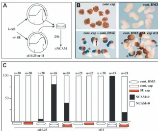
In a second series of experiments, we addressed whether ectoderm submitted to FGF inhibition could respond to the natural neural inducers produced by the organiser. Thus, organiser mesoderm was recombined with ectoderm from embryos treated or not with 80 µM SU5402 until the time of explantation, and washed before recombination (Fig. 6A). When compared with controls, pre-treatment with SU5402 caused a 50% reduction in the number of NCAM-positive conjugates made at the early gastrula stage (Fig. 6C). This reduction rose to 80% when recombination was performed at the mid-gastrula stage (Fig. 6C). Moreover, not only the number of NCAM-positive conjugates dropped dramatically upon pre-treatment with SU5402, but the amount of NCAM-positive cells within these conjugates, and the intensity of the staining in those cells were severely reduced (asterisks in Fig. 6B). We further noticed that in most cases, NCAM-negative conjugates still contained a cement gland (arrows in Fig. 6B), similar to what is observed in class V whole embryos (Fig. 3A). We conclude that FGFR activity is required to confer to the ectoderm its capacity to become neural in response to organiser signals.
Organiser gene expression in absence of FGF signalling
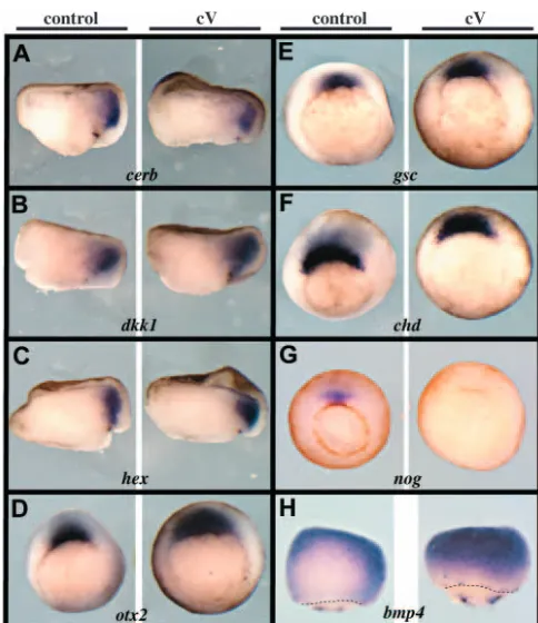
We noted in these recombination assays that it was more difficult to suppress neural induction in vitro than in vivo in siblings treated identically with the inhibitor. As the mesoderm does not form in absence of FGF signalling (see Fig. 3), it is possible that the complete lack of neural tissue in class V embryos is partly due to defective inductive properties of Spemann’s organiser mesoderm. To investigate the possible role of FGF signalling in the organiser, we analysed the expression of a large panel of genes known to be expressed in this territory and to play a role in neural induction. We found that most organiser genes were normally expressed in early gastrula class V embryos, at the time when the neural markers opl and Sox2 are lost in such embryos (Fig. 3J,L). Genes showing normal expression included goosecoid, otx2, Xlim1, hex, chordin, cerberus, dickkhopf1and crescent(Fig. 7A-F, and not shown). By contrast, the BMP antagonist nogginwas not expressed in class V embryos (Fig. 7G). Finally, we found that expression of the epidermis inducer Bmp4was upregulated in the dorsal ectoderm of early gastrula class V embryos (Fig. 7H). This is consistent with the conversion of neural precursors into epidermal cells in SU-treated embryos (Fig. 3).
Neural induction involves BMP/SMAD1-independent FGF signalling
[image:7.612.239.562.72.339.2]Both the loss of noggin expression, and the upregulation of Bmp4in class V embryos suggest that abnormal activation of the BMP pathway may cause or contribute to the loss of neural tissue in FGFR-deficient embryos. We thus attempted to rescue the lack of neural tissue in class V embryos via BMP inhibition. Single injection of tBR, Smad6or nogginRNAs in four-cell embryos did not rescue NCAM-expressing neural tissue in class V embryos (not shown). As these three factors exhibit different modes of BMP inhibition – extracellular Fig. 6. FGF activity is required in the ectoderm for
neural induction by Spemann’s organiser. (A) Ectoderm explants were taken at stage 10.25 or 11 from embryos that were treated (or not) with 80 µM SU5402. Animal caps were then washed and recombined with control organiser mesoderm explants prepared at the same stage. Conjugates were further cultured for 24 hours and analysed by immunostaining for NCAM expression.
(B) Typical results obtained in such assays. NCAM expression is in blue pointed by asterisks, arrows indicate cement glands. Conjugates made with stage 11 SU5402-treated animal caps show either no NCAM signal (counted as negative) or dramatically reduced NCAM signal (counted as positive). Furthermore, these conjugates retain cement gland tissue despite the lack of neural tissue. (C) Bar graph presenting the results as the percentage of NCAM-positive conjugates over the total number of conjugates in each condition. Pre-treatment with the FGFR inhibitor considerably reduces the number of conjugates showing neuralisation.
De
velopment
De
antagonism for Noggin, blockage of BMP signalling at the cell surface for tBR, and intracellular inhibition of SMAD1 action for SMAD6 – we reasoned that their concerted action should yield more complete BMP inhibition. Thus, all three RNAs, together with lacZ, were co-injected dorsally at the four-cell stage (4 ng Smad6+ 400 pg tBR+ 250 pg noggin) in SU5402-treated embryos. We found that Sox2 expression was not rescued in early gastrula class V embryos (13/13 negative), and that NCAM staining was not recovered either at tailbud stage (16/16 negative) (Fig. 8C,H). BMP inhibition was, however, visible in these embryos, as the epidermis was converted into cement gland (Fig. 8C). The same lack of rescue of NCAM expression resulted from injection of a high amount of Noggin protein (1 ng) in the blastocoele of class V embryos at late blastula stage (25/25 negative; Fig. 8D). Again, the epidermis was transformed into fully differentiated cement gland, indicating that BMP inhibition occurred in this assay. These experiments suggested that in the absence of FGF signals, the ectoderm cannot be neuralised by strong BMP inhibition.
[image:8.612.46.288.69.349.2]Further supporting this interpretation, we found that explanted animal caps could not be neuralised by tBR, nogginor Smad6 RNAs injection in the presence of SU5402 (not shown, Fig. 8H, Table 1), or when Noggin protein was added on SU5402 pre-treated ectoderm explants (Table 1). Further, untreated ectoderm explants did not undergo neuralisation upon co-incubation with Noggin protein and SU5402 (Table 1). These data demonstrate that neuralisation in whole embryos, as well as in BMP- or SMAD1-deficient animal caps requires intact FGFR signalling, both prior and during BMP inhibition. We conclude that FGFR signalling, although involved in the reduction of BMP activity in presumptive neural tissue, exerts some essential effects that are unlikely to result from the sole inhibition of BMP signalling
Fig. 7. Organiser gene expression in FGFR-deficient embryos. (A-H) Four-cell embryos were treated with 80 µM SU5402 until early gastrula stage 10.5 and fixed for in situ hybridisation. This treatment yielded class V embryos. In A-C, embryos were cut in two halves along the dorsoventral midline prior to hybridisation in order to improve probe penetration. The organiser genes cerberus (cerb; A), dickkopf1(dkk1, B), hex(C), otx2(D), goosecoid(gsc; E) and chordin(chd; F) are normally expressed in class V embryos, whereas noggin(nog; G) is not. Conversely, the epidermis inducer bmp4is ectopically expressed in the dorsal ectoderm of class V embryos (H). Thus, BMP signalling is probably upregulated in prospective neural tissue in absence of FGF activity. (A-C) Dorsal towards the right. (D-G) Vegetal view, dorsal towards the top. (H) Dorsal view, anterior towards the top, broken lines indicate the blastopore.
Fig. 8. Neural tissue development involves BMP/SMAD1-independent FGFR activity. (A-D) Stage 26 tailbud embryos immunostained for NCAM. (A) Untreated embryo; (B) class V embryo treated from stage 7 to 26 with 120 µM SU5402; (C) embryo treated similarly with SU5402 and which had received injection at the four-cell stage of lacZ, tBR(400 pg), noggin(250 pg) and Smad6(4 ng) RNAs; (D) embryo treated similarly with SU5402 and injected at blastula stage 9 with 1 ng Noggin protein. BMP inhibitors converted epidermis into cement gland (cement glands are outlined in B-D), but did not rescue neural tissue in class V embryos. (E-G) These embryos received the same treatment and injection as in A-C, but were harvested at the early gastrula stage 10.5, and analysed for Sox2expression. No rescue of Sox2 expression was obtained upon injection of the triple inhibitor combination in class V embryos. (H) Eight-cell embryos were injected in all four animal blastomeres with Smad6RNA (1 ng/blastomere) and cultured in absence or in presence of 120 µM SU5402 from stage 7, as indicated. Animal caps were excised at blastula stage 9, further cultured in the inhibitor until late gastrula stage 13, and harvested at tailbud stage. Siblings were class V in this experiment. Neural induction by Smad6is suppressed in absence of FGFR activity.
De
velopment
De
[image:8.612.315.554.70.337.2]Discussion
In this article, we have investigated, in vivo, the molecular mechanisms responsible for neural induction in Xenopus. We can draw three main conclusions that are of broad importance for the understanding of neural tissue specification during development and evolution, as well as for neural stem cell research. First, neural fates are unlikely to be specified by default as predicted by the prevailing model. Rather, and this is our second point, neural development appears to be initiated by weak pre-gastrula FGF signalling, acting in addition to BMP inhibition and directly in the ectoderm. Last, distinct levels of FGF signalling may help to position the prospective neurectoderm and mesoderm, suggesting that neural induction does not solely consist of a binary fate decision between epidermis and neural tissue. It may also involve a decision between neural and mesodermal identities. We now discuss these ideas in more details.
The default model of neural induction is based on assays where BMP signalling is manipulated in frog ectoderm explanted at blastula stages. In the most recent version of this model, ectoderm cells become epidermis when submitted to strong BMP signals, they adopt a border fate (including neural crest and cement gland) when submitted to weaker BMP signals, and they become neural in absence of BMP signalling (Munoz-Sanjuan and Brivanlou, 2002). Using assays in whole embryos, we first confirmed that BMP signalling promoted epidermis formation, and repressed neural development. Hence, epidermis development appears to be dominant over neural development. However, reducing BMP/SMAD1 signalling in the ventral ectoderm distant from the endogenous neural plate, to a level incompatible with epidermis, cement gland and neural crest development, did not elicit neural specification, strongly arguing against the default model. We demonstrate, however, that under these conditions, BMP inhibition is sufficient to neuralise ventral ectoderm cells upon explantation (Fig. 1G), making it unlikely that the lack of neural induction in vivo is only due to partial BMP inhibition. These results argue that neural induction in vivo does not consist of a simple epidermis-to-neural, or neural border-to-neural switch, which would be regulated by distinct levels of BMP signalling. Although it is necessary that BMP be downregulated for epidermis and neural border to be
suppressed, an additional signal(s) is required to initiate neural induction in the BMP-negative territory.
Several arguments allow us to propose that FGF signalling plays a positive role in this process. First, eFGF could transform BMP-deficient cells into neural progenitors in vivo, without inducing mesoderm. Second, using all available antimorphic FGF receptors and a pharmacological FGFR inhibitor, we found that the earliest steps of neural tissue development critically and specifically depends on FGF activity. These results confirm and extend previous studies. We note that in our assays, ∆R4 misexpression consistently led to more penetrant neural deficiencies than XFD, when equal amounts of RNAs were injected. This could suggest that FGFR4 is the main receptor involved in neural induction at blastula stages, in agreement with its higher level of expression (Hongo et al., 1999). Support for this idea also comes from the observation that tR4, a form of FGFR4 that is active in the absence of FGF ligands, can rescue neural phenotypes generated by ∆R4. In addition, our data indicate that the transduction pathway at work downstream of FGF receptors may involve ras, as it is sufficient to induce ectopic neural tissue in BMP-deficient ectoderm, and it can rescue neural plate tissue in SU5402-treated embryos. Our work thus provides initial hints as to the transduction cascade responsible for neural induction in frogs.
Interestingly, we have shown that neural induction in intact embryos requires FGF activity at pre-gastrula stages (Fig. 4J,K, Fig. 5). This early requirement for FGF signalling appears to be conserved between Xenopus (this work), zebrafish (Furthauer et al., 2004), chick (Streit et al., 2000; Wilson et al., 2000) and even ascidians (Bertrand et al., 2003). As this period of development is anterior to the formation of Spemann’s organiser, a possible interpretation of our results would be that neural induction is initiated by FGF signalling prior to gastrulation, and that this is maintained by organiser signals. However, it is equally possible that signals produced by the organiser, and in particular BMP antagonists, also act in the blastula at the time of FGF requirement (Kuroda et al., 2004; Wessely et al., 2001). Thus, further work is needed to compare
blastula
β-catenin BMP/Smad1
mesoderm mesoderm epidermis epidermis
neural neural
FGF high
low
bmp
chd CHD
fgf
Veg-T Xnrs
Maternal zygotic
XNRs
[image:9.612.320.557.75.169.2]nog NOG
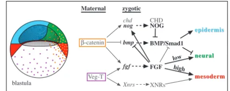
Fig. 9. A model for neural induction in Xenopus. See text for explanations. Coloured domains indicate prospective tissues. Orange and purple dots indicate β-catenin and VegT protein distribution in blastula nuclei, respectively. Arrows are not intended to represent direct regulation. The goal of this figure is to try and represent how the major molecular pathways known to impact on early embryonic patterning are integrated. It is not meant to give a complete view of this process. Regulatory arrows shown in black are inferred in part or totally from our work, while those shown in grey originate from other studies. Neural specification requires concomitant BMP inhibition, and low FGF signalling acting in a BMP-independent manner.
Table 1. Neural induction by BMP inhibition requires intact FGFR signalling in animal caps
Assay n % NCAM>0
Untreated cap + Noggin (stage 10.25)* 38 50
SU cap + Noggin (stage 10.25)* 36 0
Untreated cap + Noggin (stage 9)† 22 55
Untreated cap + Noggin + SU (stage 9)† 21 0
Untreated cap + Noggin (stage 10)† 40 60
Untreated cap + Noggin + SU (stage 10)† 38 3
Smad6 cap‡ 13 100
Smad6 cap‡+ SU (stage 13) 16 0
*1 µg/ml Noggin protein was added to animal caps pre-treated or not with 80 µM SU5402
†Untreated animal caps were cultured with 1 µg/ml Noggin protein in the
presence or not of 80 µM SU5402. ‡See legend to Fig. 7.
DMZ, dorsal marginal zone; cap, animal cap.
De
velopment
De
[image:9.612.45.294.95.187.2]the periods of requirement of FGF signalling and BMP inhibition for the normal activation of the neural programme.
We provide here multiple evidence that FGF signalling plays a direct role in the ectoderm during the process of neural induction. First, eFGF can cooperate with BMP inhibitors to neuralise ventral ectoderm cells in vivo, without mesoderm induction. Second, targeted ∆R4 injection in presumptive neural plate cells is sufficient to suppress early neural marker gene expression. Third, ectoderm explants submitted to the FGFR inhibitor SU5402 showed a severely reduced ability to respond to neural-inducing signals produced by the organiser, or to BMP inhibitors. In summary, both gain- and loss-of-function experiments suggest an essential role of FGF signalling directly in the ectoderm, consistent with ERK activation in prospective neural tissue at blastula/gastrula stages (Schohl and Fagotto, 2002; Uzgare et al., 1998).
In chick, the neural-inducing activity of FGF seems to be accounted for by both BMP-dependent and -independent functions. Our results point to a similar situation in Xenopus. In support of a BMP-dependent action of FGF, Bmp4 expression is upregulated in the dorsal ectoderm of FGFR-deficient embryos, which appears to be a conserved feature in vertebrates (Furthauer et al., 2004; Wilson et al., 2000). Moreover, nogginexpression is lost in class V embryos, which is further expected to upregulate BMP signalling. Finally, as it has been reported that activated ERK could inhibit SMAD1 (Pera et al., 2003), this mechanism could also attenuate BMP signalling in response to FGF. However, FGF does not appear to act solely via inhibition of BMP/SMAD1 signalling. First, the amount of eFGF that was found sufficient for neural specification in vivo, when combined to BMP inhibitors (tBR or SMAD6), does not efficiently repress epidermis formation on its own, nor does it activate a border fate (Fig. 2A). As both SMAD6 and FGF/ras are expected to block SMAD1 activity, the complementation reported here between these compounds is unlikely to be simply additive, and suggests that FGF/ras provide an independent information. Second, the loss of neural tissue because of FGFR inhibition in whole embryos cannot be rescued by increased BMP/SMAD1 inhibition (Fig. 8C,D,H). As this issue is crucial to propose a BMP/SMAD1-independent function of FGF, we attempted to block the BMP pathway simultaneously at multiple levels, outside the cell (Noggin), at the cell-surface (tBR) and inside the cell (SMAD6). Even in this case, no neural tissue was recovered in the complete absence of FGF activity. Although it is impossible to rule out that some very weak residual BMP signalling may persist in this assay and explain the lack of neural tissue, this possibility appears unlikely. Thus, we conclude that FGFR may function during neural induction via both dependent and BMP-independent routes (Fig. 9). The latter response is probably playing a crucial role directly on the future neurectoderm, as we show that FGFR-deficient animal caps cannot be neuralised by organiser tissue or BMP antagonists. Future work will aim to understand the nature of this response. Indeed, final proof for a BMP-independent action of FGF signalling requires the identification of a direct transcriptional target of the pathway in the neurectoderm, the activity of which would be required for neural specification, a situation that has so far been reached only in Ciona intestinalis(Bertrand et al., 2003).
A BMP-independent role of FGF in neural induction is incompatible with the default model, but not necessarily with
the data that led to its formulation (i.e. neural induction by BMP inhibition in animal caps). First, we found that following the same injection protocol in presumptive epidermal cells, BMP inhibition by SMAD6 could trigger neural induction in explants but not in whole embryos. Second, we show here that ectoderm explants pre-treated with the FGFR inhibitor are no longer able to become neural, supporting the idea that FGF activity is present and required for neuralisation in animal caps. One possibility to account for the difference between in vivo and ex vivo responses to BMP inhibition is that explanted caps are somehow subjected to an increased level of FGF signalling compared with the corresponding territory in intact embryos, as suggested by previous studies (Krain and Nordheim, 1999; LaBonne and Whitman, 1997). Dissociated frog blastula ectoderm constitutes another paradigm for neural induction. In theory, this treatment should prevent both BMP and FGF signalling, but leads nonetheless to neural differentiation. FGF signalling may, however, occur in an autocrine manner in dissociated cells such that it becomes sufficient to promote neural fates, when BMP is sufficiently suppressed by dispersion. Consistent with this idea, expression of dominant-negative FGFR4a significantly reduces neuralisation triggered by dissociation (Hongo et al., 1999). Interestingly, it has recently been reported that neural differentiation of mouse embryonic stem cells, cultured in isolation, requires autocrine FGF signalling (Ying et al., 2003). In this case, as in our study, BMP inhibition by Noggin could not compensate for the loss of FGFR activity in dispersed ES cells. Taken together, these data suggest that neural fate acquisition in Xenopus, chick and neural stem cells does not occur by default and may involve BMP-independent FGFR signalling, which may be autocrine. An important aspect of this work is that both gain- and loss-of-function experiments indicate the existence of a gradient of FGF activity that patterns the early embryo. The formation of prospective dorsal mesoderm expressing Xbra occurs at a higher level of FGF signalling than that of the presumptive neural tissue expressing Sox2 (Fig. 2). Likewise, posterior neural tissue requires higher levels of FGF signalling than anterior neural tissue (Fig. 3). Thus, incomplete inhibition of the pathway in the embryo may be sufficient to prevent mesoderm and posterior neural tissue formation, without interfering with anterior neural induction, providing a likely explanation for the maintenance in previous studies of head features in FGFR-compromised embryos (Amaya et al., 1991; Holowacz and Sokol, 1999; Kroll and Amaya, 1996; Ribisi et al., 2000). Our results indicate that weak FGF signalling is sufficient, in a BMP negative context, to promote neural identity, which makes complete inactivation particularly difficult to obtain and to ascertain.
We attempted to represent some of the most important genetic relationships relating to Xenopusneural induction in a schematic model (Fig. 9). In this model, presumptive mesoderm, neural ectoderm and epidermis are positioned at blastula stages in response to the maternal activities of VegT and β-catenin, relayed by the FGF and Nodal-related pathways.
β-catenin and FGF signalling repress Bmp4and activates BMP inhibitors (chordin, noggin), which generates a BMP-free region in the ectoderm, a condition necessary for neural tissue formation (Baker et al., 1999; Wessely et al., 2001). Consistently, β-catenin is broadly activated in dorsal mesoderm and ectoderm, where it activates FGF/ERK signalling (Schohl
De
velopment
De
and Fagotto, 2002; Schohl and Fagotto, 2003). The epidermis forms where BMP function is maintained in the ectoderm. While high-level FGF signalling combined with Nodal-related activity specify mesoderm in marginal cells, weak FGF signalling induces neural tissue in BMP-negative animal cells. This dose-dependent effect is consistent with the graded pattern of ERK activation seen in the embryo (Schohl and Fagotto, 2002). Further evidence for the ability of vertebrate embryonic cells to decide between a mesodermal and a neural fate include the conversion of mesoderm into neural cells in mouse and fish mutants of the FGF, Wnt and Nodal pathways, and in mutants of the T-box family (Chapman and Papaioannou, 1998; Ciruna et al., 1997; Feldman et al., 2000; Yoshikawa et al., 1997).
In conclusion, our work uncovers the crucial role played by pre-gastrula FGF signalling during Xenopusneural induction, in addition to the well-documented inhibition of BMP. The evidence presented here appears incompatible with the leading default model, and instead provides support for a shared use of FGF signalling in neural induction across the chordate phylum.
We acknowledge Drs R. Harland, H. Okamoto, H. Yasuo, C. Marcelle, P. Wilson, Y. Sasai, H. Isaacs and I. Dawid for plasmids. We are grateful to C. Stern and C. Linker for sharing data prior to publication and for insightful discussions. We thank members of our group for support, discussions, and comments on the manuscript. This work was supported by the CNRS.
References
Amaya, E., Musci, T. J. and Kirschner, M. W. (1991). Expression of a dominant negative mutant of the FGF receptor disrupts mesoderm formation in Xenopus embryos. Cell66, 257-270.
Baker, J. C., Beddington, R. S. and Harland, R. M. (1999). Wnt signaling in Xenopus embryos inhibits bmp4 expression and activates neural development. Genes Dev.13, 3149-3159.
Bertrand, V., Hudson, C., Caillol, D., Popovici, C. and Lemaire, P. (2003). Neural tissue in ascidian embryos is induced by FGF9/16/20, acting via a combination of maternal GATA and Ets transcription factors. Cell115, 615-627.
Bouwmeester, T., Kim, S., Sasai, Y., Lu, B. and de Robertis, E. M. (1996). Cerberus is a head-inducing secreted factor expressed in the anterior endoderm of Spemann’s organizer. Nature382, 595-601.
Carballada, R., Yasuo, H. and Lemaire, P. (2001). Phosphatidylinositol-3 kinase acts in parallel to the ERK MAP kinase in the FGF pathway during Xenopus mesoderm induction. Development128, 35-44.
Carnac, G., Kodjabachian, L., Gurdon, J. B. and Lemaire, P. (1996). The homeobox gene Siamois is a target of the Wnt dorsalisation pathway and triggers organiser activity in the absence of mesoderm. Development122, 3055-3065.
Chapman, D. L. and Papaioannou, V. E. (1998). Three neural tubes in mouse embryos with mutations in the T-box gene Tbx6. Nature391, 695-697.
Cho, K. W., Blumberg, B., Steinbeisser, H. and de Robertis, E. M. (1991). Molecular nature of Spemann’s organizer: the role of the Xenopus homeobox gene goosecoid. Cell67, 1111-1120.
Ciruna, B. G., Schwartz, L., Harpal, K., Yamaguchi, T. P. and Rossant, J.
(1997). Chimeric analysis of fibroblast growth factor receptor-1 (Fgfr1) function: a role for FGFR1 in morphogenetic movement through the primitive streak. Development124, 2829-2841.
Darras, S. and Nishida, H. (2001). The BMP/CHORDIN antagonism controls sensory pigment cell specification and differentiation in the ascidian embryo. Dev. Biol.236, 271-288.
De Robertis, E. M., Larrain, J., Oelgeschlager, M. and Wessely, O. (2000). The establishment of Spemann’s organizer and patterning of the vertebrate embryo. Nat. Rev. Genet.1, 171-181.
Feldman, B., Dougan, S. T., Schier, A. F. and Talbot, W. S. (2000). Nodal-related signals establish mesendodermal fate and trunk neural identity in zebrafish. Curr. Biol.10, 531-534.
Furthauer, M., van Celst, J., Thisse, C. and Thisse, B. (2004). Fgf signalling controls the dorsoventral patterning of the zebrafish embryo. Development 131, 2853-2864.
Glinka, A., Wu, W., Delius, H., Monaghan, A. P., Blumenstock, C. and Niehrs, C. (1998). Dickkopf-1 is a member of a new family of secreted proteins and functions in head induction. Nature391, 357-362.
Harland, R. and Gerhart, J. (1997). Formation and function of Spemann’s organizer. Annu. Rev. Cell Dev. Biol.13, 611-667.
Hata, A., Lagna, G., Massague, J. and Hemmati-Brivanlou, A. (1998). Smad6 inhibits BMP/Smad1 signaling by specifically competing with the Smad4 tumor suppressor. Genes Dev.12, 186-197.
Hemmati-Brivanlou, A. and Thomsen, G. H. (1995). Ventral mesodermal patterning in Xenopus embryos: expression patterns and activities of BMP-2 and BMP-4. Dev. Genet.17, 78-89.
Holowacz, T. and Sokol, S. (1999). FGF is required for posterior neural patterning but not for neural induction. Dev. Biol.205, 296-308.
Hongo, I., Kengaku, M. and Okamoto, H. (1999). FGF signaling and the anterior neural induction in Xenopus. Dev. Biol.216, 561-581.
Hsu, D. R., Economides, A. N., Wang, X., Eimon, P. M. and Harland, R. M. (1998). The Xenopus dorsalizing factor Gremlin identifies a novel family of secreted proteins that antagonize BMP activities. Mol. Cell1, 673-683.
Hudson, C., Clements, D., Friday, R. V., Stott, D. and Woodland, H. R.
(1997). Xsox17alpha and -beta mediate endoderm formation in Xenopus.
Cell91, 397-405.
Isaacs, H. V., Pownall, M. E. and Slack, J. M. (1994). eFGF regulates Xbra expression during Xenopus gastrulation. EMBO J.13, 4469-4481.
Jonas, E. A., Snape, A. M. and Sargent, T. D. (1989). Transcriptional regulation of a Xenopus embryonic epidermal keratin gene. Development 106, 399-405.
Jones, C. M., Kuehn, M. R., Hogan, B. L., Smith, J. C. and Wright, C. V.
(1995). Nodal-related signals induce axial mesoderm and dorsalize mesoderm during gastrulation. Development121, 3651-3662.
Kim, G. J. and Nishida, H. (2001). Role of the FGF and MEK signaling pathway in the ascidian embryo. Dev. Growth Differ.43, 521-533.
Krain, B. and Nordheim, A. (1999). Artefactual gene induction during preparation of Xenopus laevis animal cap explants. Int. J. Dev. Biol.43, 563-566.
Kroll, K. L. and Amaya, E. (1996). Transgenic Xenopus embryos from sperm nuclear transplantations reveal FGF signaling requirements during gastrulation. Development122, 3173-3183.
Kuo, J. S., Patel, M., Gamse, J., Merzdorf, C., Liu, X., Apekin, V. and Sive, H. (1998). Opl: a zinc finger protein that regulates neural determination and patterning in Xenopus. Development125, 2867-2882.
Kuroda, H., Wessely, O. and Robertis, E. M. (2004). Neural induction in Xenopus: requirement for ectodermal and endomesodermal signals via Chordin, Noggin, beta-Catenin, and Cerberus. PLoS Biol.2, E92.
LaBonne, C. and Whitman, M. (1997). Localization of MAP kinase activity in early Xenopus embryos: implications for endogenous FGF signaling. Dev. Biol.183, 9-20.
Launay, C., Fromentoux, V., Shi, D. L. and Boucaut, J. C. (1996). A truncated FGF receptor blocks neural induction by endogenous Xenopus inducers. Development122, 869-880.
Lemaire, P., Garrett, N. and Gurdon, J. B. (1995). Expression cloning of Siamois, a Xenopus homeobox gene expressed in dorsal-vegetal cells of blastulae and able to induce a complete secondary axis. Cell81, 85-94.
Marchant, L., Linker, C., Ruiz, P., Guerrero, N. and Mayor, R. (1998). The inductive properties of mesoderm suggest that the neural crest cells are specified by a BMP gradient. Dev. Biol.198, 319-329.
Marics, I., Padilla, F., Guillemot, J. F., Scaal, M. and Marcelle, C. (2002). FGFR4 signaling is a necessary step in limb muscle differentiation.
Development129, 4559-4569.
Mayor, R., Morgan, R. and Sargent, M. G. (1995). Induction of the prospective neural crest of Xenopus. Development121, 767-777.
Mizuseki, K., Kishi, M., Matsui, M., Nakanishi, S. and Sasai, Y. (1998). Xenopus Zic-related-1 and Sox-2, two factors induced by chordin, have distinct activities in the initiation of neural induction. Development 125, 579-587.
Mohammadi, M., McMahon, G., Sun, L., Tang, C., Hirth, P., Yeh, B. K., Hubbard, S. R. and Schlessinger, J. (1997). Structures of the tyrosine kinase domain of fibroblast growth factor receptor in complex with inhibitors. Science276, 955-960.
Moos, M., Jr, Wang, S. and Krinks, M. (1995). Anti-dorsalizing morphogenetic protein is a novel TGF-beta homolog expressed in the Spemann organizer. Development121, 4293-4301.
De
velopment
De
Munoz-Sanjuan, I. and Brivanlou, A. H. (2001). Early posterior/ventral fate specification in the vertebrate embryo. Dev. Biol.237, 1-17.
Munoz-Sanjuan, I. and Brivanlou, A. H. (2002). Neural induction, the default model and embryonic stem cells. Nat. Rev. Neurosci.3, 271-280.
Newman, C. S., Chia, F. and Krieg, P. A. (1997). The XHex homeobox gene is expressed during development of the vascular endothelium: overexpression leads to an increase in vascular endothelial cell number.
Mech. Dev.66, 83-93.
Pannese, M., Polo, C., Andreazzoli, M., Vignali, R., Kablar, B., Barsacchi, G. and Boncinelli, E. (1995). The Xenopus homologue of Otx2 is a maternal homeobox gene that demarcates and specifies anterior body regions. Development121, 707-720.
Pera, E. M., Ikeda, A., Eivers, E. and de Robertis, E. M. (2003). Integration of IGF, FGF, and anti-BMP signals via Smad1 phosphorylation in neural induction. Genes Dev.17, 3023-3028.
Powers, C. J., McLeskey, S. W. and Wellstein, A. (2000). Fibroblast growth factors, their receptors and signaling. Endocr. Relat. Cancer7, 165-197.
Pownall, M. E., Tucker, A. S., Slack, J. M. and Isaacs, H. V. (1996). eFGF, Xcad3 and Hox genes form a molecular pathway that establishes the anteroposterior axis in Xenopus. Development122, 3881-3892.
Ribisi, S., Jr, Mariani, F. V., Aamar, E., Lamb, T. M., Frank, D. and Harland, R. M. (2000). Ras-mediated FGF signaling is required for the formation of posterior but not anterior neural tissue in Xenopus laevis. Dev. Biol.227, 183-196.
Sasai, Y., Lu, B., Steinbeisser, H., Geissert, D., Gont, L. K. and de Robertis, E. M. (1994). Xenopus chordin: a novel dorsalizing factor activated by organizer- specific homeobox genes. Cell79, 779-790.
Sasai, Y., Lu, B., Piccolo, S. and de Robertis, E. M. (1996). Endoderm induction by the organizer-secreted factors chordin and noggin in Xenopus animal caps. EMBO J.15, 4547-4555.
Schohl, A. and Fagotto, F. (2002). Beta-catenin, MAPK and Smad signaling during early Xenopus development. Development129, 37-52.
Schohl, A. and Fagotto, F. (2003). A role for maternal beta-catenin in early mesoderm induction in Xenopus. EMBO J.22, 3303-3313.
Shinya, M., Koshida, S., Sawada, A., Kuroiwa, A. and Takeda, H. (2001). Fgf signalling through MAPK cascade is required for development of the subpallial telencephalon in zebrafish embryos. Development 128, 4153-4164.
Smith, J. C., Price, B. M., Green, J. B., Weigel, D. and Herrmann, B. G.
(1991). Expression of a Xenopus homolog of Brachyury (T) is an immediate-early response to mesoderm induction. Cell67, 79-87.
Smith, W. C. and Harland, R. M. (1992). Expression cloning of noggin, a new dorsalizing factor localized to the Spemann organizer in Xenopus embryos. Cell70, 829-840.
Streit, A. and Stern, C. D. (1999). Establishment and maintenance of the border of the neural plate in the chick: involvement of FGF and BMP activity. Mech. Dev.82, 51-66.
Streit, A., Lee, K. J., Woo, I., Roberts, C., Jessell, T. M. and Stern, C. D.
(1998). Chordin regulates primitive streak development and the stability of induced neural cells, but is not sufficient for neural induction in the chick embryo. Development125, 507-519.
Streit, A., Berliner, A. J., Papanayotou, C., Sirulnik, A. and Stern, C. D.
(2000). Initiation of neural induction by FGF signalling before gastrulation.
Nature406, 74-78.
Suzuki, A., Thies, R. S., Yamaji, N., Song, J. J., Wozney, J. M., Murakami, K. and Ueno, N. (1994). A truncated bone morphogenetic protein receptor affects dorsal-ventral patterning in the early Xenopus embryo. Proc. Natl. Acad. Sci. USA91, 10255-10259.
Umbhauer, M., Penzo-Mendez, A., Clavilier, L., Boucaut, J. and Riou, J.
(2000). Signaling specificities of fibroblast growth factor receptors in early Xenopus embryo. J. Cell Sci.113, 2865-2875.
Uzgare, A. R., Uzman, J. A., El-Hodiri, H. M. and Sater, A. K. (1998). Mitogen-activated protein kinase and neural specification in Xenopus. Proc. Natl. Acad. Sci. USA95, 14833-14838.
Wessely, O., Agius, E., Oelgeschlager, M., Pera, E. M. and de Robertis, E. M. (2001). Neural induction in the absence of mesoderm: beta-catenin-dependent expression of secreted BMP antagonists at the blastula stage in Xenopus. Dev. Biol.234, 161-173.
Wilson, S. I. and Edlund, T. (2001). Neural induction: toward a unifying mechanism. Nat. Neurosci.4, 1161-1168.
Wilson, S. I., Graziano, E., Harland, R., Jessell, T. M. and Edlund, T.
(2000). An early requirement for FGF signalling in the acquisition of neural cell fate in the chick embryo. Curr. Biol.10, 421-429.
Wilson, S. I., Rydstrom, A., Trimborn, T., Willert, K., Nusse, R., Jessell,
T. M. and Edlund, T. (2001). The status of Wnt signalling regulates neural and epidermal fates in the chick embryo. Nature411, 325-330.
Ying, Q. L., Stavridis, M., Griffiths, D., Li, M. and Smith, A. (2003). Conversion of embryonic stem cells into neuroectodermal precursors in adherent monoculture. Nat. Biotechnol.21, 183-186.
Yoshikawa, Y., Fujimori, T., McMahon, A. P. and Takada, S. (1997). Evidence that absence of Wnt-3a signaling promotes neuralization instead of paraxial mesoderm development in the mouse. Dev. Biol.183, 234-242.
