INTRODUCTION
In the developing vertebrate embryo, the order of Hox genes on the chromosome imposes their expression domains along the anteroposterior (A-P) axis (Krumlauf, 1994). Hoxa2 andHoxb2 exhibit the most-anterior expression domain, in the cranial neural crest that migrates to the second branchial arch (Prince and Lumsden, 1994; Nonchev et al., 1996; Mallo, 1997). In mice, Hoxa2 loss-of-function leads to a transformation of second branchial arch derivatives into the more anterior first branchial arch derivatives (Gendron-Maguire et al., 1993; Rijli et al., 1993; Barrow and Capecchi, 1999). In addition, ectopic expression of the homeobox gene Six2was also observed in these mutant embryos in territories normally controlled by Hoxa2 (Kutejova et al., 2005). This result, together with the demonstrated ability of Hoxa2 to bind to the Six2 promoter in vitro (Kutejova et al., 2005), suggest that repression of Six2 by Hoxa2 is a crucial step in the developmental pathway leading to second branchial arch formation.
Despite extensive genetic analysis, the molecular basis of Hox function is proving difficult to understand. Together with Six2, other likely Hox downstream targets have been identified in vertebrates (Pearson et al., 2005; Svingen and Tonissen, 2006), but it remains unclear for most of these genes whether they are regulated directly or indirectly by Hox proteins. With the exception of Hox genes themselves, it is also currently unknown how the activities of the few genes proven to be directly regulated by Hox proteins in vertebrate embryogenesis contribute to the function of Hox proteins (Serpente et al., 2005; Salsi and Zappavigna, 2006; Shaut et al., 2007). A conclusive characterization of the nature of direct downstream genes is essential to explain how Hox gene activities are converted into morphogenetic processes and to understand the transcriptional properties of Hox proteins as exerted on their target promoters. In addition, insight into the organization and the hierarchy of the pathways controlled by Hox proteins in vertebrates requires the analysis of the functional role of the direct downstream targets in the Hox pathway.
Here, we conclusively show that Hoxa2 directly controls Six2 transcription in the second branchial arch. Lack of control over Six2 transcription contributes to the generation of the Hoxa2mutant phenotype, with analysis of Six2; Hoxa2double mutants indicating that Hoxa2 controls additional downstream targets. We identify components of the IGF molecular pathway as targets of Hoxa2 regulation and correlate the changes in Six2expression with those in the expression of the gene encoding Igf-binding protein, Igfbp5, suggesting a role of Six2in mediating Hoxa2control over the IGF system.
MATERIALS AND METHODS
Production of anti-Hoxa2 antibodies
The C-terminal fragment of mouse Hoxa2 (amino acids 231 to 321) was expressed as a His-tag fusion in Escherichia coli. Four rabbits were immunized with the fusion protein by Biogenes (Berlin, Germany). Two specific anti-Hoxa2 antibodies, 43 and 44, were affinity purified from the sera showing the strongest response by coupling the immunogen to CNBr-activated Sepharose.
Chromatin immunoprecipitation
Chromatin immunoprecipitation was performed according to a standard protocol (Upstate Biotechnology, Lake Placid, NY) with the following modifications. Branchial arches and frontonasal mass of embryos were dissected in PBS. After fixing in 1% formaldehyde for 23 minutes at 4°C, embryonic tissues were desintegrated with a 25-gauge needle. The cross-linked material was sonicated to 200-1000 bp fragments (Vibracell sonicator; seven times for 10 seconds at 40% output) and the immunoprecipitations were performed starting with second branchial arches (50 pairs of second branchial arches from E10.5 embryos, or ten pairs of second branchial arches from E11.5 embryos) or five pairs of first branchial arches together with frontonasal mass and 3 μg of anti-Hoxa2 antibodies (43 or 44), 1 μg of anti-polymerase II antibody (Santa Cruz Biotechnology, Santa Cruz, CA), 1 μg of anti-Pbx1 antibody (Santa Cruz Biotechnology) or 3 μg of normal rabbit IgG. PCR amplifications were performed using the following primers: forward, 5⬘CTCGGGTTACCGGTGACTGAC -AGCGTCTCC-3⬘and reverse, 5⬘CTCTCCCTCCCGTCTA GCTC GCT -TGCAGCT-3⬘for the Six2 promoter; 5⬘GGCTGACTT TGGAGATGA -CTC-3⬘and reverse, 5⬘-GAATGCCTG CTCTA ACTGTTCAC-3⬘for the IP10(Cxcl10– Mouse Genome Informatics) promoter.
Mutant animals and phenotypic analyses
Hoxa2-null and Six2-null mutant mice have been described (Gendron-Maguire et al., 1993; Self et al., 2006). 900Six2-lacZtransgenic mice and the a2-Six2transgene are described by Kutejova et al. (Kutejova et al., 2005). The a2-Six2transgenic embryos were derived from a founder with no apparent phenotypic defects, which transmitted the transgene to the F2,
Six2 functions redundantly immediately downstream of
Hoxa2
Eva Kutejova1, Bettina Engist1, Michelle Self2, Guillermo Oliver2, Pavel Kirilenko3and Nicoletta Bobola1,3,*
Hox transcription factors control morphogenesis along the head-tail axis of bilaterians. Because their direct functional targets are still poorly understood in vertebrates, it remains unclear how the positional information encoded by Hox genes is translated into morphogenetic changes. Here, we conclusively demonstrate that Six2is a direct downstream target of Hoxa2 in vivo and show that the ectopic expression of Six2, observed in the absence of Hoxa2, contributes to the Hoxa2mouse mutant phenotype. We propose that Six2 acts to mediate Hoxa2 control over the insulin-like growth factor pathway during branchial arch development.
KEY WORDS: Six2, Hoxa2, Branchial arch, Mouse
Development 135, 1463-1470 (2008) doi:10.1242/dev.017624
1Department of Developmental Biology, Max-Planck Institute of Immunobiology,
Freiburg, Germany. 2Department of Genetics and Tumor Cell Biology, St Jude
Children’s Research Hospital, Memphis, TN, USA. 3Faculty of Human and Medical
Sciences, Stopford Building, The University of Manchester, Manchester M13 9PT, UK.
*Author for correspondence (e-mail: Nicoletta.Bobola@manchester.ac.uk)
Accepted 25 February 2008
D
E
V
E
LO
P
M
E
N
causing perinatal lethality and the skeletal defects expected by overexpression of Six2(Kutejova et al., 2005). Skeletal phenotypes were analyzed by Alcian Blue/Alizarin Red staining as described (Mallo and Brändlin, 1997). Whole-mount and tissue sections were analyzed by in situ hybridization as described (Kanzler et al., 1998), using Igfbp5 (Bobola and Engist, 2008) and Igf1(Weger and Schlake, 2005) probes. RT-PCR on second branchial arches of E10.5 embryos from a2-Six2transgenics was performed as described (Kutejova et al., 2005). Animals experiments were approved by the ethics committee of the Regierungspräsidium Freiburg.
Cell transfection, western blot and electrophoretic mobility shift assay
Mouse Six2, Hoxb2, Pbx1aand Pbx1bwere amplified from E11.5 second branchial arch cDNA using the following primers: 5⬘CAGCCGC -CACCATGTCCATGCTG-3⬘and 5⬘CTCTAGGAGCCCAGGTC CAC -AAGG-3⬘ for Six2; 5⬘AATGAATTCACCATGAATT TTGAATT -TGAGAGGGAG-3⬘ and 5⬘-AGGGAAACTGCAAGTCGATG-3⬘ for Hoxb2; 5⬘-AATAAGCTTACCATGGACGAGCAGCCGAGG-3⬘and 5⬘ -AATGGATCCTCAGTTGGAGGTATCAGAGTG-3⬘for Pbx1a; 5⬘AATA -AGCTTACCATGGACGAGCAGCCGAGG-3⬘and 5⬘AATGGATCCTC -ACTGTATCCTCCTGTCTG-3⬘ for Pbx1b; and cloned into pcDNA3 (Invitrogen). pcDNA3-Hoxb2-HA contains a HA tag inserted in-frame before the stop codon; the pcDNA3-Hoxa2-HA construct has been described (Kutejova et al., 2005).
HEK 293 cells were transfected using the calcium phosphate method, cultured for an additional 36 hours and lysed in buffer comprising 50 mM Tris-HCl pH 8.0, 250 mM NaCl, 1% NP40. Branchial arches and frontonasal mass of embryos were dissected in DMEM (Sigma) and total proteins were extracted using Trizol (Invitrogen) according to manufacturer’s instructions. The membranes were probed with anti-Pbx1 antibody (Santa Cruz Biotechnology) diluted 1:100.
Electrophoretic mobility shift assays were performed using T7-coupled TNT rabbit reticulocytes (Promega). The BstEII/SspI (from –181 to –48) fragment of the Six2promoter is described by Kutejova et al. (Kutejova et al., 2005). The oligonucleotide reproducing the sequence of the Six5 binding site in the Igfbp5promoter has been described (Sato et al., 2002); the sequence of the mutant oligonucleotide is 5⬘TGGGTGTTGG -GGAGCGCAAATTGCAGCTA-3⬘.
RESULTS
Hoxa2 binds to the Six2promoter in vivo
We used a chromatin immunoprecipitation assay (ChIP) to determine whether Hoxa2 directly regulates Six2activity in vivo. Briefly, two polyclonal antibodies against the non-conserved C-terminal portion of the Hoxa2 protein (amino acids 231 to 321) were raised in rabbits. The two antibodies specifically recognize Hoxa2 and do not cross-react with the Hoxa2 paralog Hoxb2 (Fig. 1A,B and data not shown). As an abundant source of Hoxa2 protein (Prince and Lumsden, 1994; Mallo, 1997; Nonchev et al., 1996), second branchial arches were isolated from E11.5 wild-type embryos. At this stage, Hoxa2 function is still required for second arch development (Santagati et al., 2005) and Six2is ectopically expressed in this territory in the absence of Hoxa2 (Kutejova et al., 2005). As negative controls, we used two embryonic regions colonized by Hox-negative cranial neural crest (Le Douarin and Kalcheim, 1999): first branchial arches and frontonasal mass (hereafter referred to as first arch, Fig. 1C). Cross-linked, sheared chromatin from second and first arches was immunoprecipitated using the two Hoxa2 polyclonal antibodies and analyzed by PCR for the presence of the highly conserved Six2 chromatin region recognized by Hoxa2 in vitro (Kutejova et al., 2005) (Fig. 1D). Second branchial arch immunoprecipitated chromatin showed a substantial enrichment for the most-proximal Six2 promoter region. No enrichment was detected with chromatin from first arches or from that immunoprecipitated in the presence of unrelated antibodies. Similar results were obtained with both of the polyclonal antibodies directed against Hoxa2 (Fig. 1E).
[image:2.612.137.555.57.262.2]The earliest stage at which we could detect Hoxa2 bound to the Six2 promoter was E10.5, corresponding to the appearance of ectopic Six2 expression in the mutant second branchial arch (Kutejova et al., 2005): second branchial arch chromatin immunoprecipitated in the presence of Hoxa2 polyclonal antibody showed a significant enrichment for the most-proximal Six2
Fig. 1. Hoxa2 binds to the Six2
promoter in vivo.(A,B) Western blot using anti-HA (A) and anti-Hoxa2 polyclonal antibody 43 (B) on whole extracts of human 293 cells transfected with empty vector (control), pCDNA3-Hoxa2-HA (Hoxa2-HA) or pCDNA3-Hoxb2-HA (Hoxb2-HA). Arrows indicate the expected position of Hoxb2-HA. (C) Side view of the facial region of an E11.0 mouse embryo, showing the areas isolated for ChIP (red, maxillary component of first arch and frontonasal mass; blue, second arch). (D) Schematic of the Six2
genomic locus around the
transcriptional start site (+1), with red boxes indicating the relative position of the two Hoxa2 binding sites identified in vitro (Kutejova et al., 2005) and gray arrows indicating the
position of the primers used for PCR amplification. (E) PCR amplification of the immunoprecipitated chromatin from E11.5 second branchial arch (2nd) or from frontonasal mass and first branchial arch (1st) using anti-Hoxa2 polyclonal antibodies 43 and 44 or normal rabbit IgG. (F) Same experiment as in E, using polyclonal anti-polymerase II antibodies to control for the quality of first arch chromatin: first arch chromatin is enriched for the Six2proximal promoter fragment, as expected for a gene actively transcribed in this area (Oliver et al., 1995). (G) PCR amplification with
Six2or IP10(Cxcl10; control) primers of E10.5 second branchial arch (2nd) chromatin, immunoprecipitated using anti-Hoxa2 antibody 43, or normal rabbit IgG. The number of PCR cycles is indicated in the bottom-right corner. EB, elution buffer. ChIP was performed on three independent pools of samples, and PCRs were performed in duplicate on each pool. Results shown are from a representative set.
D
E
V
E
LO
P
M
E
N
promoter region, whereas no enrichment was detected for an unrelated, control promoter (Fig. 1G). ChIP analysis of E9.5 embryos revealed no enrichment in Six2promoter in the presence of the specific antibody (not shown). These results demonstrate that at the stages (E10.5-11.5) when Hoxa2 actively represses Six2 transcription in the second branchial arch (Kutejova et al., 2005), Hoxa2 is bound to the Six2regulatory region in vivo.
Recruitment of Pbx1 to theSix2 promoter in vivo
is independent of Hox proteins
Members of the PBC family of proteins interact with Hox proteins and act as co-factors to modify their binding specificity in vitro (Moens and Selleri, 2006). In vertebrates, Hox proteins bind in a complex with Pbx1 on a Hox/Pbx bipartite binding site that is essential for the activity of the Hoxb1and Hoxb2enhancers (Jacobs et al., 1999; Ferretti et al., 2000).
The Six2promoter contains a Pbx/Meis binding site located a few nucleotides upstream of the binding sites recognized by Hoxa2 in vitro (Kutejova et al., 2005). ChIP assay using a Pbx1-specific antibody indicated that Pbx1 is bound to the Six2promoter in vivo (Fig. 2A). Moreover, Pbx1 was similarly detected on the Six2 promoter in immunoprecipitated chromatin extracted from embryonic areas where Hoxa2 is present (second branchial arches) and from areas where Hoxa2, or any other Hox proteins, are absent (first branchial arches and frontonasal mass) (Fig. 2A), indicating that the recruitment of Pbx1 to the Six2promoter does not depend on Hox proteins. The presence of the Pbx1 protein isoforms was confirmed in all embryonic areas examined (Fig. 2B-D).
Does Hoxa2 interfere with a Six2 autoregulatory mechanism?
Transcriptional repression of a target promoter can be achieved by a variety of mechanisms (Gaston and Jayaraman, 2003), some of which are difficult to investigate without identifying the proteins acting as activators of Six2.Six2is expressed in a large domain in
the first branchial arch and in a restricted one in the second branchial arch. Upon Hoxa2inactivation, an identical Six2expression pattern is observed in first and second arches (Kutejova et al., 2005), suggesting that the mechanism of activation is the same in both domains.
We and others previously showed that 1 kb of Six2promoter is sufficient to recapitulate Six2endogenous expression in various embryonic sites, including the branchial arches (Brodbeck et al., 2004; Kutejova et al., 2005). This promoter fragment is activated by Six2 and contains conserved Six-binding sites that are recognized by Six2 in vitro (Brodbeck et al., 2004) (N.B. and E.K., unpublished), suggesting that Six2activity in the branchial arches might rely on an autoregulatory loop of Six2 protein on its own promoter. To investigate whether Six2 controls its promoter in vivo, we introduced the 900Six2-lacZtransgene, containing the first 900 bp of the Six2promoter fused to a lacZreporter gene (Kutejova et al., 2005), into the Six2mutant background (Self et al., 2006). As shown in Fig. 3, lacZexpression was unchanged in the absence of Six2 in the branchial arches, maxilla and limbs at the stages examined. These results indicate that Six2 is not necessary to maintain the activity of its own promoter in these embryonic areas. The Six proteins share a conserved homeodomain, recognize similar binding sites and can substitute for each other in vivo and in vitro (Spitz et al., 1998; Ando et al., 2005; Grifone et al., 2005; Giordani et al., 2007; Kobayashi et al., 2007). Owing to the presence of other Six proteins in the branchial arches and their known capacity to compensate for each other, our experimental system cannot definitively rule out the possibility that a Six-dependent activation mechanism is nevertheless in place for the Six2promoter. Within the Six2 promoter, two Six2 binding sites overlap with or are in close proximity to Hoxa2 binding sites (Brodbeck et al., 2004) (Fig. 3E). In addition, Hoxa2 targets the 1 kb promoter fragment in vivo (Kutejova et al., 2005). However, an analysis of whether Hoxa2 might interfere with the binding of Six proteins to the Six2promoter revealed no change in the binding of Six2 to its promoter in the presence of increasing concentrations of Hoxa2 (Fig. 3F), despite the close arrangement of Six and Hoxa2 binding sites on the promoter.
Ectopic expression of Six2 contributes to the
Hoxa2mutant phenotype
[image:3.612.50.265.59.200.2]In the absence of Hoxa2, the skeletal derivatives of the second branchial arch do not form and are replaced by cartilage and bone that resemble, in shape and position, first arch skeletal derivatives (Gendron-Maguire et al., 1993; Rijli et al., 1993; Barrow and Capecchi, 1999). Since Hoxa2 in the second arch is found to be associated with the Six2 promoter and negatively regulates its transcription (Kutejova et al., 2005), we asked whether the upregulation of Six2observed in the absence of Hoxa2 is responsible for the Hoxa2 mutant phenotype. To test this possibility, we generated double Hoxa2; Six2-null mice. No obvious abnormalities are detected in first and second arch skeletal derivatives of Six2-null pups (Self et al., 2006) (data not shown). Double Hoxa2; Six2-null mutants were obtained by crossing compound heterozygotes, as Hoxa2and Six2single mutants die shortly after birth (Gendron-Maguire et al., 1993; Rijli et al., 1993; Barrow and Capecchi, 1999; Self et al., 2006). Analysis of middle-ear skeletal preparations from double-null newborns showed that the removal of Six2activity partially rescued the Hoxa2phenotype. As shown in Fig. 4, the gonial bone, which in the Hoxa2single mutant abnormally extends to connect the tympanic ring and its duplication (Gendron-Maguire et al., 1993; Rijli et al., 1993; Barrow and Capecchi, 1999), was Fig. 2. Pbx1 binds the Six2promoter in vivo.(A) PCR amplification
(39 cycles) of the immunoprecipitated chromatin from E11.5
frontonasal mass and first branchial arch (1st) or from second branchial arch (2nd) using anti-Pbx1 antibody or normal rabbit IgG. EB, elution buffer. ChIP was performed on two independent pools of samples, and PCRs were performed in duplicate on each pool. Results shown are from a representative set. (B,C) Western blot using anti-Pbx1 on whole extracts of human 293 cells transfected with pCDNA3-Pbx1a (Pbx1a) or pCDNA3- Pbx1b (Pbx1b), or whole extracts from frontonasal mass (FNM), first branchial arch (1st ba) and second branchial arch (2nd ba) of E11.5 embryos. (D) Ponceau staining of the membrane shown in C. Arrowhead, position of Pbx1a; arrow, position of Pbx1b.
D
E
V
E
LO
P
M
E
N
reduced to as much as normal size; the size of the duplicated mallei was also reduced in some of the double-mutant embryos. We observed incomplete penetrance in the extent of the rescue of the double-mutant phenotype as well as variability within the same embryo, ruling out background-dependent effects (Fig. 4D, Table 1). Complete rescue of the ectopic growth of the gonial bone could be observed already upon removal of a single Six2allele (Fig. 4E). Other aspects of the Hoxa2phenotype (Gendron-Maguire et al., 1993; Rijli et al., 1993; Barrow and Capecchi, 1999) remained unaffected. The partial rescue shows that one of the functional mechanisms by which Hoxa2 participates in the formation of the second branchial arch is via its repression of Six2expression in that territory.
Six2 partially mediates Hoxa2 control of the IGF pathway
The role of Six2 as a transcription factor does not in itself provide any explicit insight into its function downstream of Hoxa2. Furthermore, a straightforward analysis of the molecular function
[image:4.612.206.562.57.216.2]of Six2 in the Hoxa2 mutant is hindered by the incomplete penetrance of Hoxa2; Six2 mutant phenotypic rescue. A few downstream targets of Six transcription factors have been identified (Spitz et al., 1998; Li et al., 2002; Lagutin et al., 2003; Brodbeck et al., 2004; Ando et al., 2005; Chai et al., 2006; Giordani et al., 2007; Kobayashi et al., 2007), including Igf2and Igfbp5, genes encoding components of the IGF system (Sato et al., 2002). IGF positively regulates bone growth (Liu et al., 1993) and removal of Six2from the Hoxa2mutant mainly affects the growth of the ectopic gonial bone; moreover, IGF is required for the normal development of intramembranous bones that are part of, or develop in close proximity to, the middle-ear skeleton (Louvi et al., 1997). For these reasons, we analyzed the expression of various IGF components in wild-type and Hoxa2mutant embryos. Whereas Igf1 was upregulated in Hoxa2 mutant embryos, in which ectopic expression was detected below the developing otic vesicle at E11.5 (Fig. 5A,B), expression of Igfbp5, which appears in the second arch mesenchyme by E11.5, was strongly reduced in the absence of Hoxa2 (Fig. 5C,D). The other components of the Fig. 3. Regulation of the Six2promoter
by Six proteins.(A,C) E11.5 and E12.5 wild-type mouse embryos showSix2
promoter (–893 to +37)-driven lacZ
expression in the maxilla (m), first branchial arch (arrows), second branchial arch (arrowhead) and limbs (l), recapitulating Six2
endogenous expression. (B,D) E11.5 and E12.5 Six2mutant embryos show unchanged staining in these embryonic areas in the absence of Six2. (E) Nucleotide sequence of the proximal Six2 promoter, with Six-binding sites (highlighted in gray) and the sequence of Hoxa2 binding sites (red) indicated. The identification of binding sites is based for site 1 on the Six-binding consensus and footprinting analysis
[image:4.612.54.478.542.650.2](Spitz et al., 1998) (N.B. and E.K., unpublished) and for site 2 on the Six-binding consensus, in vitro binding and functional analysis (Spitz et al., 1998; Brodbeck et al., 2004) (N.B. and E.K., unpublished). Numbers indicate nucleotide positions relative to the transcriptional start site (+1). (F) Unchanged Six2 binding in the presence of Hoxa2. Six2 was translated in rabbit reticulocytes in vitro and incubated with a labeled Six2 promoter fragment (–181 to –48). Increasing amounts of pcDNA3-Hoxa2 programmed reticulocytes were added to the binding reaction mix, keeping the total amount of extract constant in each binding mix by adding unprogrammed reticulocytes. Arrow, the Six2-probe complex; arrowhead, the Hoxa2-probe complex. No ternary complex was observed; however, addition of Hoxa2 did not perturb Six2 binding to the probe.
Fig. 4. Middle-ear skeletal phenotype of Hoxa2–/–; Six2–/–and Hoxa2–/–; Six2+/– mutant mice.(A-E) Dissected middle-ear skeleton from
wild-type (A), Hoxa2–/–(B), Hoxa2–/–; Six2–/–(C,D) and Hoxa2–/–; Six2+/–(E) E18.5 embryos. (A) Malleus (m), incus (i), gonial bone (g) and tympanic ring (t) are derived from first arch; stapes (s) is second arch-derived. (B-E) In the absence of Hoxa2, the stapes disappears and duplicated first arch elements form in the second arch (asterisks). The gonial bone abnormally extends to connect the wild-type tympanic ring with its duplicated counterpart. (C) Removal of Six2rescues the ectopic growth of the gonial bone. The malleal duplication is also reduced (compare double-ended arrows in B and C). (D) The extent of the rescue of the ectopic growth of the gonial bone in Hoxa2–/–; Six2–/–was variable, but the ectopic gonial bone, when present, never extended to reach the duplicated tympanic ring (arrowhead), as invariably observed in Hoxa2–/–embryos (arrowhead in B).
(E) Complete reversal to a wild-type gonial bone was observed also in Hoxa2–/–; Six2+/–embryos.
D
E
V
E
LO
P
M
E
N
IGF system examined (Igf2, Igfbp2,Igfbp3,Igfbp4) showed no change at this stage (data not shown). These results show that Hoxa2 acts upstream of the IGF system in the second branchial arch where it controls, directly or indirectly, the expression of Igf1 and of the gene encoding the Igf-binding protein, Igfbp5.
Since bothSix2and Igfbp5are differentially expressed in the Hoxa2mutant and as Six5, a Six2 homolog, regulates Igfbp5by binding to and activating its promoter (Sato et al., 2002), we tested whether Six2 might act upstream of Igfbp5 and account for the Igfbp5downregulation observed in Hoxa2mutant embryos. For these experiments, we analyzed Igfbp5 expression in embryos transgenically expressing Six2in the second branchial arch, rather than using the double mutants whose phenotypic variability precludes rigorous analysis of Six2 molecular function. In the transgenic embryos, Six2 overexpression results in phenotypic characteristics of the Hoxa2mutant (Kutejova et al., 2005). Igfbp5 expression was reduced in the second branchial arch of E11.5 embryos overexpressing Six2(n=3, Fig. 5E,F). Igfbp5expression was also noticeably reduced in the anterior forelimb, an area where the expression of the transgene generates a strong phenotype (not shown). The embryos analyzed were derived from the same founder, allowing a direct correlation between Six2expression levels (Fig. 5G), the observed phenotypic effects (Kutejova et al., 2005) and the effects on Igfbp5mRNA levels.
These results indicate that Hoxa2 regulates the IGF pathway in the second branchial arch and suggest at least a partial role for Six2 in mediating this Hoxa2 function.
Six2 interacts directly with the Igfbp5 promoter in vitro
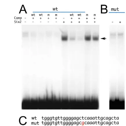
Regulation of Igfbp5by Six5, a Six2 homolog, is mediated by direct binding to a short, perfectly conserved sequence in the Igfbp5 proximal promoter (Sato et al., 2002). To test whether Six2 directly repressesIgfbp5via binding to the Igfbp5promoter, we performed an electrophoretic mobility shift assay (EMSA) using the Six5 binding site identified in theIgfbp5promoter as a probe. Whereas no binding was detected when the probe was incubated with unprogrammed reticulocytes, incubation of the probe with in vitro translated Six2 resulted in the formation of a retarded complex (Fig. 6A). This complex represents the specific interaction of Six2 with the probe because its formation was competed by an excess of cold wild-type oligonucleotide at different molar concentrations. The same molar excess of a mutant oligonucleotide, containing one
nucleotide substitution in the Six5 binding site (Fig. 6C), left the complex unaffected (Fig. 6A). Finally, no binding was detected when Six2 was incubated in the presence of the mutant oligonucleotide used as a probe (Fig. 6B).
These data show that Six2 recognizes the Six5 binding site in the Igfbp5promoter and that this interaction is sequence-specific, and suggest that Six2 could directly repress Igfbp5transcription.
DISCUSSION
[image:5.612.328.537.61.380.2]Transcriptional control of Six2expression in vivo Our demonstration that Hoxa2 is associated in vivo with the Six2 promoter fragment shown to be involved in Six2repression firmly identifies Six2as a gene directly regulated by Hoxa2 in vivo. In the absence of its repressor Hoxa2, expression of Six2in the second arch is first detected at E10.5. Although we detected Hoxa2 bound to the Six2 promoter at this stage, we were unable to detect Hoxa2 associated with the Six2promoter at E9.5, when Hoxa2 is already present in the second branchial arch. It is possible that Hoxa2 Table 1. Incomplete penetrance in the middle-ear skeletal
phenotype of Hoxa2–/–; Six2–/–and Hoxa2–/–; Six2+/–mutants
Genotype (n) Wild-type Reduced Mutant
Hoxa2–/–; Six2–/–(7) 3 11 0
Hoxa2–/–; Six2+/–(6) 2 4 6
Hoxa2–/–; Six2+/+(6) 0 0 12
Wild-type, Reduced and Mutant refer to the appearance of the gonial bone (and the extent of the rescue, evaluated as complete, partial or absent, respectively). Since left and right middle-ear skeleton appeared different within each embryo, numbers in the columns refer to the middle-ear skeletons analyzed (two per embryo). Complete reversal to the wild-type situation was observed in three middle-ear
skeletons of seven Hoxa2–/–; Six2–/–embryos (3/14 sides showed complete rescue)
and two middle-ear skeletons of six Hoxa2–/–; Six+/–embryos (2/12 sides showed
complete rescue). The ectopic gonial bone was reduced in 11 of 14 sides of
Hoxa2–/–; Six2–/–embryos and in four of 12 sides of Hoxa2–/–; Six+/–embryos. The
frequency of a Hoxa2mutant-looking gonial bone was 0/14 sides in Hoxa2–/–; Six2–/–
and 6/12 in Hoxa2–/–; Six2+/–embryos. No correlation between the extent of the
rescue and the left or right side of the embryos was observed. In double mutants, the presence of a wild-type-looking gonial bone was associated with a noticeable reduction in the width of the duplicated mallei.
[image:5.612.49.299.81.126.2]n, Number of embryos analyzed for each genotype.
Fig. 5. Expression of Igf1and Igfbp5in wild-type and mutant mouse embryos.(A-F) In situ hybridization on whole-mount E11.5 wild-type (A,C,E), Hoxa2mutant (B,D) and a2-Six2transgenic embryos (F) using Igf1(A,B) and Igfbp5(C-F) probes. The arrow in A and B indicates the embryonic area where Igf1is upregulated in the mutant. The arrow in C and D indicates the domain where Igfbp5expression in the Hoxa2 mutant is lost. In E and F, arrows and arrowheads indicate areas of Igfbp5expression in the second branchial arch; note the reduced Igfbp5expression in the second branchial arch of embryos overexpressing Six2. I, first branchial arch; II, second branchial arch. (G) Semi-quantitative RT-PCR on RNA extracted from E10.5 second branchial arches of wild-type (wt) and a2-Six2(tg) embryos using primers specific for Six2and Gapdh.
D
E
V
E
LO
P
M
E
N
recruitment to the Six2promoter coincides with gene activation, i.e. as a result of changes in chromatin accessibility or of interaction with the activator(s).
The same Six2promoter fragment that bound to Hoxa2 was also associated in vivo with Pbx1, a member of a family of proteins that act as Hox co-factors in both Drosophila and vertebrate development. Binding of Pbx1 to the Six2promoter in vivo occurred independently of Hoxa2 or other Hox proteins. This in vivo result, together with our previous in vitro data (Kutejova et al., 2005), indicate that Pbx1 and Hoxa2 bind independently of each other on the Six2 promoter, where they recognize two separate sites. Although this finding is in apparent contrast with previous studies in vertebrates, in which Pbx and Hox proteins bind in a complex on specific Hox/Pbx bipartite sites to regulate the Hoxb1and the Hoxb2 enhancers, it should be noted that: (1) the number of Hox targets analyzed in vertebrates to date is too small to extract general rules about the transcriptional properties of Pbx and Hox proteins on their target promoters; and (2)Hoxb1and Hoxb2are activated by Hox proteins and repression might require a different binding architecture of Pbx and Hox proteins on the promoter.
Pbx1mutant mice display defects in the neural crest-derived skeletal elements of the second branchial arch. One of these defects, the elongation of the lesser horn of the hyoid bone, is phenocopied by transgenic embryos overexpressing Six2in the second arch (Selleri et al., 2001; Kutejova et al., 2005). This is suggestive of a role for Pbx1 in controlling Six2levels in the second branchial arch. Insight into the functional relevance of
Six2promoter occupancy by Pbx1 in the absence of Hox proteins, and of the simultaneous presence on Six2promoter of Pbx1 and Hoxa2 for Six2regulation, awaits analyses in single Pbx1and combined Hoxa2/Pbx1mutants.
The molecular mechanism responsible for Six2activation in the first branchial arch (and most likely in the second branchial arch of the Hoxa2 mutant) is unknown. Although activation of Six2 expression through the cooperation of Eya1, Pax2 and Hoxa11 has recently been described in kidney development (Gong et al., 2007), the situation is rather different in the branchial arches, where Hoxa2 acts as a repressor. In addition, Eya1 expression is mainly restricted to the epithelia in this embryonic area, whereas Six2is expressed in the mesenchyme (Kutejova et al., 2005) (data not shown). The capacity of Six2 to activate the promoter fragment that recapitulates Six2 expression in the branchial arches instead suggests the involvement of Six proteins in Six2 promoter activation in vivo (Brodbeck et al., 2004; Kutejova et al., 2005). We have ruled out any Six2 requirement for Six2transcription, but we cannot exclude the in vivo relevance of Six-mediated activation owing to the likely compensatory effect between other Six proteins present in the branchial arches (Grifone et al., 2005). However, even if such a mechanism does activate the Six2promoter, Hoxa2 repression of Six2 transcription does not appear to be the result of mutually exclusive binding of Hoxa2 and Six proteins to their closely spaced binding sites on the Six2promoter. The discovery and analysis of additional Hoxa2 targets will undoubtedly shed light on the transcriptional properties of Hoxa2 and the requirements to switch from a repressor to an activator role.
Molecular function of Six2 in the second branchial arch
[image:6.612.52.270.54.273.2]Ectopic expression of Six2in the second branchial arch reproduces a molecular aspect of the Hoxa2 mutant phenotype, i.e. downregulation of the gene encoding the Igf-binding protein, Igfbp5. A possible direct regulation of Igfbp5by Six2 is consistent with the ability of Six5 and Six1 to activate the Igfbp5promoter (Sato et al., 2002), and is supported by the ability of Six2 to recognize the Six5 binding site in theIgfbp5promoter. The opposite effects of Six5 and Six2 on Igfbp5expression are in line with the documented ability of Six proteins to function both as transcriptional activators and repressors (Li et al., 2003; Brugmann et al., 2005). Together with Igfbp5, Igf1is also affected in the Hoxa2mutant. The IGF system positively controls bone development and growth, with cranial and facial bones displaying the most dramatic defects in the absence of IGF signaling (Liu et al., 1993; Louvi et al., 1997). The activity of the IGF system is regulated by six insulin-like growth factor-binding proteins able to bind Igf1 and Igf2 directly and to control the pool of free IGF proteins available for interaction with the cognate receptors to transduce the signal in target cells. In most cases, this interaction leads to down-modulation of IGF signaling (Clemmons, 1998; Collett-Solberg and Cohen, 2000). We indeed found that expression of Igfbp5negatively affects bone development and growth in the craniofacial area (Bobola and Engist, 2008), where it partially reproduces the effects of Hoxa2 overexpression (Kanzler et al., 1998). In addition, these effects are IGF-dependent (Bobola and Engist, 2008). Hoxa2 control of second arch skeletal development could be exerted, at least in part, via a decrease in IGF signaling, resulting from downregulation of Igf1and upregulation of its potential negative regulator Igfbp5. An increase in IGF signaling is expected to result in increased bone formation, which is indeed a phenotypic characteristic of the Hoxa2mutant (Kanzler et al., 1998).
Fig. 6. Six2 binds the Six5 binding site identified in the mouse
Igfbp5promoter.(A) Incubation of the wild-type oligonucleotide probe in the presence of unprogrammed reticulocytes does not result in the formation of a specific retarded complex. Incubation of the probe with Six2-programmed reticulocytes gives rise to a retarded complex (arrow), which is competed by the addition of cold double-stranded wild-type oligonucleotide (wt), but not by oligonucleotide with a nucleotide substitution in the Six5 binding site (m). Cold oligonucleotides were added at 100- and 500-fold molar excess. (B) Incubation of the labeled mutant oligonucleotide does not result in the formation of a specific complex in the presence of
Six2-programmed reticulocytes. (C) Sequence of wild-type and mutant oligonucleotides. The nucleotide changed in the mutant oligonucleotide is highlighted in red.
D
E
V
E
LO
P
M
E
N
Alternative functional organization downstream of Hoxa2
Removal of ectopic Six2 expression from the Hoxa2 mutant indicates that Six2is only one of the genes controlled by Hoxa2 and that Hoxa2 regulates second arch development by activation and/or repression of additional targets. In addition, the variability observed in the rescue of the gonial bone phenotype in Hoxa2; Six2-null mutants indicates a high degree of redundancy, with other genes able to substitute for Six2function.
The double-mutant phenotype can be explained by either of two models. The simplest interpretation of the rescue observed in the double mutant is that the ectopic expression of Six2specifically promotes the growth of the gonial bone. The rescue of the double mutant is limited because Six2function is restricted to the control of a specific process. Hoxa2 also regulates other as yet unknown genes, the loss- or gain-of-function of which in the absence of Hoxa2 would promote some of the various specific phenotypes observed in Hoxa2 mutant mice. However, because the ectopic gonial bone is also the phenotypic component most sensitive to Hoxa2 dosage (Santagati et al., 2005), a rescue in this aspect of the phenotype might simply reflect the immediate readout of the phenotype to any change introduced into the Hoxa2mutant (in this specific case, the loss of Six2ectopic activity). In this alternative model, Six2 has broader effects on the development of the second branchial arch and the limited rescue does not rest in the control by Six2of a restricted aspect of the phenotype, but rather in Six2acting redundantly with other genes that can partially compensate for modifications in its activity. Additional observations are in support of a broader function for Six2in the generation of the Hoxa2phenotype, beyond that in gonial bone growth. Contrary to the predominant rescue of intramembranous bone formation in the double mutant lacking Six2, Six2ectopic expression in the second branchial arch (Kutejova et al., 2005) expands the chondrogenic domains and affects the size and shape of second arch cartilages. The main effect of Six2on cartilage when ectopically expressed can be explained by the transient expression of Six2in the second arch of these wild-type embryos (supplied with a functional Hoxa2 protein), in which expression declines before it affects later developmental processes, such as intramembranous bone formation. The analysis of Six2function in different experimental systems indicates, therefore, that Six2can control both chondrogenesis and intramembranous bone formation in the second arch, i.e. the different processes that contribute to the Hoxa2mutant phenotype. Indeed, removal of Six2 from the Hoxa2 mutant partially rescues the duplication of the malleus, albeit at a lower frequency than for the rescue in the gonial bone phenotype. A broader function of Six2in the generation of the Hoxa2phenotype is also supported by Six2spatiotemporal expression in the mutant second arch (Kutejova et al., 2005).
The proposed molecular function of Six2, i.e. to control Igfbp5, is compatible with both models. IGF signaling could have global effects on the development of the second arch, exerted through a direct effect on bone formation, or mediated through cell proliferation, with a final impact on both chondrogenesis and intramembranous bone formation. In that case, any change in IGF signaling would most likely be perceived first by the aspect of the phenotype most sensitive to changes, i.e. the ectopic gonial bone. It seems reasonable to assume that control over a broad mechanism such as IGF signaling is diverse and likely to involve several genes. Six2could be one of those genes, acting to decrease the levels of the Igf-binding protein Igfbp5. Removal of the ectopic Six2would only partially affect the state of IGF signaling, owing to the presence of the additional regulators that compensate for the loss of Six2,
explaining the variability of the phenotype. Alternatively, Igfbp5 function in the second arch could be restricted to the growth of the gonial bone by local inhibition of IGF signaling, in which case repression of Igfbp5by Six2 would directly lead to increased growth of the gonial bone. Overall, the wide-ranging effects of Six2on second arch skeletogenesis, the broad expression of this gene in the mutant second arch, and the apparent redundancy of its function favor the hypothesis that Six2is part of a network of genes with overlapping functions exerted downstream of Hoxa2. The conclusive identification of Six2as a direct target of Hoxa2, together with the only partial rescue observed in the double mutant, points to the requirement for Hoxa2-mediated activation and/or repression of other target genes in addition to Six2. A redundant organization downstream of Hoxa2 would preclude the identification of its target genes on the basis of analyses of their separate functions and would require a more complicated experimental approach, i.e. the generation of triple and quadruple mutants. In the light of these perspectives, the lack of rescue of the skeletal phenotype in double mutants of Hoxa2and in two additional previously identified targets (Bobola et al., 2003) (M. Mallo, personal communication) might warrant a revisit.
We thank D. Solter for support; M. Mallo, M. Hoffmann, S. Saccani and D. van Essen for critical reading of the manuscript; Thomas Schlake for the Igf1probe; and L. Elsby for assistance. This work was supported in part by National Institutes of Health grant R21DK068560, Cancer Center Support grant CA-21765, and the American Lebanese Syrian Associated Charities (ALSAC) to G.O.
References
Ando, Z., Sato, S., Ikeda, K. and Kawakami, K.(2005). Slc12a2 is a direct target of two closely related homeobox proteins, Six1 and Six4.FEBS J.272, 3026-3041.
Barrow, J. R. and Capecchi, M. R.(1999). Compensatory defects associated with mutations in Hoxa1 restore normal palatogenesis to Hoxa2 mutants.
Development126, 5011-5026.
Bobola, N. and Engist, B. (2008). IGFBP5 is a potential regulator of craniofacial skeletogenesis. Genesis46, 52-59.
Bobola, N., Carapuco, M., Ohnemus, S., Kanzler, B., Leibbrandt, A., Neubuser, A., Drouin, J. and Mallo, M.(2003). Mesenchymal patterning by Hoxa2 requires blocking Fgf-dependent activation of Ptx1. Development 130, 3403-3414.
Brodbeck, S., Besesnbeck, B. and Englert, C.(2004). The transcription factor Six2 activates expression of the Gdnf gene as well as its own promoter. Mech. Dev.121, 1211-1222.
Brugmann, S. A., Pandur, P. D., Kenyon, K., Pignoni, F. and Moody, S.(2005). Six1 promote a placodal fate within the lateral neurogenic ectoderm by functioning both as a transcriptional activator and repressor. Development131, 5871-5881.
Chai, L., Yang, J., Di, C., Cui, W., Kawakami, K., Lai, R. and Ma, Y.(2006). Transcriptional activation of the SALL1 by the human SIX1 homeodomain during kidney development. J. Biol. Chem.281, 18918-18926.
Clemmons, D.(1998). Role of insulin-like growth factor binding proteins in controlling IGF action. Mol. Cell. Endocrinol.140, 19-24.
Collett-Solberg, P. F. and Cohen, P.(2000). Genetics, chemistry and function of the IGF/IGFBP system. Endocrine12, 121-136.
Ferretti, E., Marshall, H., Popperl, H., Machonochie, W., Krumlauf, R. and Blasi, F.(2000). Segmental expression of Hoxb2 in r4 requires two separate sites that integrate cooperative interactions between Prep1, Pbx and Hox proteins.
Development 127, 155-166.
Gaston, K. and Jayaraman, P. S.(2003). Transcriptional repression in eukaryotes: repressor and repression mechanism. Cell. Mol. Life Sci.60, 721-741. Gendron-Maguire, M., Mallo, M., Zhang, M. and Gridley, T.(1993). Hoxa-2
mutant mice exhibit homeotic transformation of skeletal elements derived from cranial neural crest. Cell75, 1317-1331.
Giordani, J., Bajard, L., Demignon, J., Daubas, P., Buckingham, M. and Maire, P.(2007). Six proteins regulate the activation of Myf5 expression in embryonic mouse limbs. Proc. Natl. Acad. Sci. USA104, 11310-11315. Gong, K.-Q., Yallowitz, A. R., Sun, H., Dressler, G. R. and Wellik, D. M.
(2007). A Hox-Eya-Pax complex regulates early kidney developmental gene expression. Mol. Cell. Biol.27, 7661-7668.
Grifone, R., Demignon, J., Houbron, C., Souil, E., Niro, C., Seller, M. J., Hamard, G. and Maire, P.(2005). Six1 and Six4 homeoproteins are required for Pax3 and Mrf expression during myogenesis in the mouse embryo. Development
132, 2235-2249.
D
E
V
E
LO
P
M
E
N
Jacobs, Y., Schnabel, C. A. and Cleary, M.(1999). Trimeric association of Hox and TALE homeodomain proteins mediates Hoxb2 hindbrain enhancer activity.
Mol. Cell. Biol.19, 5134-5142.
Kanzler, B., Kuschert, S. J., Liu, Y.-H. and Mallo, M.(1998). Hoxa2restricts the chondrogenic domain and inhibits bone formation during development of the branchial area. Development125, 2587-2597.
Kobayashi, H., Kawakami, K., Asashima, M. and Nishinakamura, R.(2007). Six1 and Six4 are essential for Gdnf expression in the metanephric mesenchyme and ureteric bud formation, while Six1 deficiency alone causes mesonephric-tubule defects. Mech. Dev.124, 290-303.
Krumlauf, R.(1994). Hox genes in vertebrate development. Cell78, 191-201. Kutejova, E., Engist, B., Mallo, M., Kanzler, B. and Bobola, N. (2005). Hoxa2
downregulates Six2 in the neural crest derived mesenchyme. Development132, 469-478.
Lagutin, O. V., Zhu, C. C., Kobayashi, D., Topczewski, J., Shimamura, K., Puelles, L., Russell, H. R., McKinnon, P. J., Solnica-Krezel, L. and Oliver, G. (2003). Six3 repression of Wnt signaling in the anterior neuroectoderm is essential for vertebrate forebrain development. Genes Dev.17, 368-379. Le Douarin, N. M. and Kalcheim, C.(1999). The Neural Crest. Cambridge, UK:
Cambridge University Press.
Li, X., Perissi, V., Liu, F., Rose, D. W. and Rosenfeld, M. G.(2002). Tissue-specific regulation of retinal and pituitary precursor cell proliferation. Science
297, 1180-1183.
Li, X., Oghi, K. A., Zhang, J., Krones, A., Bush, K. T., Glass, C. K., Nigam, S. K., Aggarwal, A. K., Maas, R., Rose, D. W. and Rosenfeld, M. G.(2003). Eya protein phosphatase activity regulates Six1-Dach-Eya transcriptional effects in mammalian organogenesis. Nature426, 247-254.
Liu, J.-P., Baker, J., Perkins, A. S., Robertson, E. J. and Efstratiadis, A.(1993). Mice carrying null mutations of the genes encoding insulin-like growth factor I (Igf-1) and type 1 IGF receptor (Igf1r). Cell75, 59-72.
Louvi, A., Accili, D. and Efstratiadis, A.(1997). Growth-promoting interaction of IGF-II with the insulin receptor during mouse embryonic development. Dev. Biol.189, 33-48.
Mallo, M.(1997). Retinoic acid disturbs mouse middle ear development in a stage-specific fashion. Dev. Biol. 184, 175-186.
Mallo, M. and Brändlin, I.(1997). Segmental identity can change independently in the hindbrain and rhombencephalic neural crest. Dev. Dyn.210, 146-156. Moens, C. and Selleri, L.(2006). Hox cofactors in vertebrate development. Dev.
Biol.291, 193-206.
Nonchev, S., Vesque, C., Maconochie, M., Seitanidou, T.,
Ariza-McNaughton, L., Frain, M., Marshall, H., Sham, M. H., Krumlauf, R. and Charnay, P.(1996). Segmental expression of Hoxa-2in the hindbrain is directly regulated by Krox-20. Development122, 543-554.
Oliver, G., Wehr, R., Jenkins, N. A., Copeland, N. G., Cheyette, B. N., Hartenstein, V., Zipursky, S. L. and Gruss, P.(1995). Homeobox genes and connective tissue patterning. Development121, 693-705.
Pearson, J. C., Lemons, D. and McGinnis, W.(2005). Modulating Hox gene functions during animal body patterning. Nat. Rev. Genet.6, 893-904. Prince, V. and Lumsden, A.(1994). Hoxa-2expression in normal and transposed
rhombomeres: independent regulation in the neural tube and neural crest.
Development120, 911-923.
Rijli, F. M., Mark, M., Lakkaraju, S., Dierich, A., Dolle, P. and Chambon, P. (1993). A homeotic transformation is generated in the rostral branchial region of the head by disruption of Hoxa-2, which acts as a selector gene. Cell75, 1333-1349.
Salsi, V. and Zappavigna, V.(2006). Hoxd13 and Hoxa13 directly control the expression of the EphA7 Ephrin tyrosine kinase receptor in developing limbs. J. Biol. Chem.281, 1992-1999.
Santagati, F., Minoux, M., Ren, S. Y. and Rijli, F. M.(2005). Temporal requirement of Hoxa2 in cranial neural crest skeletal morphogenesis.
Development132, 4927-4936.
Sato, S., Nakamura, M., Cho, D. H., Tapscott, S. J., Ozaki, H. and Kawakami, K.(2002). Identification of transcriptional targets for Six5: implication for the pathogenesis of myotonic dystrophy type 1. Hum. Mol. Gen.11, 1045-1058. Self, M., Lagutin, O. V., Bowling, B., Hendrix, J., Cai, Y., Dressler, G. R. and
Oliver, G.(2006). Six2 is required for suppression of nephrogenesis and progenitor renewal in the developing kidney. EMBO J.25, 5214-5228. Selleri, L., Depew, M. J., Jacobs, Y., Chanda, S. K., Tsang, K. Y., Cheah, K. S.,
Rubenstein, J. L., O’Gorman, S. and Cleary, M. L. (2001). Requirement for Pbx1 in skeletal patterning and programming chondrocyte proliferation and differentiation. Development128, 3543-3557.
Serpente, P., Tumpel, S., Ghyselinck, N. B., Niederreither, K., Wiedemann, L. M., Dolle, P., Chambon, P., Krumlauf, R. and Gould, A. P. (2005). Direct crossregulation between retinoic acid receptor βand Hox genes during hindbrain segmentation. Development132, 503-513.
Shaut, C. A., Saneyoshi, C., Morgan, E. A., Knosp, W. M., Sexton, D. R. and Stadler, H. S.(2007). HOXA13 directly regulates EphA6 and EphA7 expression in the genital tubercle vascular endothelia. Dev. Dyn.236, 951-960. Spitz, F., Demignon, J., Porteu, A., Kahn, A., Concordet, J. P., Daegelen, D.
and Maire, P.(1998). Expression of myogenin during embryogenesis is controlled by Six/sine oculis homeoproteins through a conserved MEF3 binding site. Proc. Natl. Acad. Sci. USA95,14220-14225.
Svingen, T. and Tonissen, K. F.(2006). Hox transcription factors and their elusive mammalian gene targets. Heredity 97, 88-96.
Weger, N. and Schlake, T.(2005). Igf-I signalling controls the hair growth cycle and the differentiation of hair shafts. J. Invest. Dermatol.125, 873-882.