Nanoparticles as a Platform for Antimicrobial
Drug Delivery
Satish Patel
1, Deependra Singh
1, Shikha Srivastava
1, Manju Singh
1, Kamal Shah
2,
Durgesh Nandini Chauahn
3, Nagendra S Chauhan
4,*1University Institute of Pharmacy, Pt. Ravishankar Shukla University, India 2Institute of Pharmaceutical Research, GLA University, Mathura, India
3Columbia Institute of Pharmacy, Raipur (C.G.), India 4Drug Testing Laboratory and Research Center (AYUSH Lab), India
Copyright©2017 by authors, all rights reserved. Authors agree that this article remains permanently open access under the terms of the Creative Commons Attribution License 4.0 International License
Abstract
The hasty developments in nanotechnology will have intense impact on various areas of therapeutic applications like drugs and bioactive delivery, tissue engineering, and other application. This review mainly emphasized on the nanoparticle systems for delivery of antimicrobial drug. Various antimicrobial agents have been used to kill or inhibit the growth of microbes such as bacteria, fungi and viruses. As the pharmacological actions of these drugs have been well established, feeble delivery and degradation could lead to insufficient therapeutic index and cause side effects. Nanoparticles have many advantages like small and controllable size, large surface area to mass ratio, and functionalizable structure. These properties make the nanoparticles, a suitable delivery system of antimicrobials. Along with this, it also overcomes the limitations intra-additional antimicrobial therapeutics. In last few years, development and fabrication of antimicrobial agent in nanoparticle systems has evolved as an innovative and promising alternative that increases therapeutic effectiveness and overcoming the undesirable side effects. In this chapter, we focused on the current progress and challenges in producing nanoparticle system for antimicrobial drugs delivery for the treatment of microbial diseases.Keywords
Nanoparticles, Nanotechnology, Antimicrobial Drug Delivery1. Introduction
Microbial infections remain foremost causes of morbidity and mortality in hospitals around the world. The innovation of miracle drugs called antibiotics in the 20th century leading a histrionic reduction in death and illness from these infectious diseases. Plentiful antimicrobial agents have been
used to eradicate the microbes. Despite the fact the therapeutic efficiency of available drugs and treatment has been well proven, incompetent and deprived delivery of antimicrobial agents could consequence in insufficient therapeutic index and causing adverse effects like nausea, vomiting, irritation, scaling and gut microbial flora reduction. Along with developing multidrug resistant microbial strains, however, variations in the society, environment, technology and developing microorganisms are contributing to the emergence of new diseases and development of antimicrobial resistance (1). The continuing presence of antibiotic resistance in pathogenic and opportunistic microorganisms forces the pharmaceutical companies and the researcher community to continuously develop novel antibacterial drug, drug targets and delivery system to improve the activity against multidrug-resistant bacteria. In the current scenario, the progress of molecular nanostructures through precise particle size and shape is of prominent interest in biomedical applications like delivery of antimicrobial agents. Nanoscale materials/polymers have emerged up as novel antimicrobial agents and delivery system because of high surface area to volume ratio and distinctive properties (2; 3). Due to various metallic and polymeric nanoparticles (4) have confirmed antimicrobial activities, the development of novel applications in this field makes them an attractive alternative to antibiotics.
diseases (7).
In earlier decades, the application of nanotechnology, particularly the use of nanoparticles for antibiotic/drug delivery, has produced noteworthy impact in management of infectious diseases. Various systems for delivery of nanoparticles like gels, liposome, polymeric nanoparticles, dendrimers, and inorganic nanoparticles, have acknowledged momentous attention. Antimicrobial molecules loaded into nano-carriers by different methods like chemical synthesis, cross-linking, physical encapsulation, adsorption, to show an improved pharmacokinetic profile and therapeutic index when related to their free form (8). Innumerable nanoparticle-based drug delivery systems have been approved for clinical use to treat a variety of infectious diseases, and a number of formulations are now under various stages of pre-clinical and clinical tests (9). Due to advancement in fabrication, green synthesis multifunctional nanoparticles, various inventive approaches have emerged, more work on improving on nanoparticle therapeutic efficacy against microbial infection. In this chapter, we discuss about nanoparticle approaches having significant potential to improve upon current treatments. Researches and investigations were made in this arena provide wonderful prospects for alternative and more effective antimicrobial approaches that amend the pharmacokinetics properties of antibiotics and produce novel antibiotics with novel inhibition mechanisms.
2. Nanoparticles
The focus of pharmaceutical research is being steadily shifted from the development of new chemical entities to the development of novel drug delivery system of existing drug molecule to maximize their effect in term of therapeutic and patient protection (10). Various novel drug delivery system like transdermal patches, Microspheres, Nanoparticles, Resealed erythrocytes, Ocusert, Depots, Liposomes, Niosomes, Buccal films etc., offer several advantages over the conventional drug delivery systems (11). Moreover, the development of novel drug delivery systems is going to be the outmost need of pharmaceutical industry especially after enforcement of product patent. Nanoparticles attract an increasing interest in contemporary drug research because they could be used as a very efficient drug delivery system. Nanotechnology is evolving as a rapidly growing field with its application in various fields of science and research for the purpose of producing new substances and molecules at the nanoscale level (12). The word “nano” is used to show one billionth of a meter or 10-9. The term “Nanotechnology” was first used by a Japanese scientist Professor Norio Taniguchi in 1974 to define precision manufacturing of materials at the level of nanometer. Nanotechnology deals with novel technology that provides a platform for development and exploration of biological systems, and offers various stimulus models for bio-assembled
components. Nano-structured materials whichever inorganic or organic exhibited numerous features like optical and catalytic. These properties mainly depend on the size and shape of prepared nanoparticles.
Nanoparticles are defined as particulate dispersions or solid particles or groups of atoms in the size range of 1-100nm. “Nano” is a Greek word identical to dwarf means extremely small. The use of nanoparticles is getting impulse in the present century as they having defined chemical, optical and mechanical properties.
Nanoparticles, first developed around 1970, they were initially devised as carriers for vaccines and anticancer drugs. The drugs may be enclosed inside the sphere of the nanoparticle or linked to the surface. Once they are at the target site, the drug payload may be released from the nanoparticle by diffusion, swelling, erosion or degradation. Active systems are also possible, e.g. drug release in response to the input of external energy such as targeted ultrasound, light or magnetic field. Nanoparticles have been successfully used in nano-chemistry to enhance the immobilization and activity of catalysts, (13) in medical and pharmaceutical nano-engineering for delivery of therapeutic agents, (14) in chronic disease diagnostics, and in sensors (15). Nanoparticles have been also used in clothing and in the food industry to limit bacterial growth (16; 17).
2.1. Properties of Nanoparticles
Bulk material should have constant physical properties regardless of its size, but at the nanoscale size-dependent properties are often observed. Thus, the properties of materials change as their size approaches the nanoscale and as the percentage of atoms at the surface of a material becomes significant.
For bulk materials larger than one micrometer (or micron), the percentage of atoms at the surface is insignificant in relation to the number of atoms in the bulk of the material. The interesting and sometimes unexpected properties of nanoparticles are therefore largely due to the large surface area of the material, which dominates the contributions made by the small bulk of the material.
Nanoparticles of usually yellow gold and gray silicon are red in color; gold nanoparticles melt at much lower temperatures (nearly 300oC for 2.5 nm size) than the gold slabs (1064oC).
Suspensions of nanoparticles are possible since the interaction of the particle surface with the solvent is strong enough to overcome density differences, which otherwise usually result in a material either sinking or coating in a liquid.
gold nanoparticles appear deep red to black in solution.
The high surface area to volume ratio of nanoparticles provides a tremendous driving for drug diffusion.
Moreover, nanoparticles have been found to impart some extra properties to various day to day products. For example the presence of titanium dioxide nanoparticles imparts what we call the self-cleaning effect, and the size being nano-range, the particles cannot be observed. Zinc oxide particles have been found to have superior UV blocking properties compared to its bulk substitute. This is one of the reasons why it is often used in the preparation of sunscreen lotions, and is completely photostable.
2.2. Advantages of Nanoparticles
Nanoparticles can offer significant advantages over the traditional delivery mechanisms in terms of various aspects (18). Nanoparticles have capability of both passive and active drug targeting due to its particle size and surface characteristics. With the use of nanoparticles, there is increase in drug therapeutic efficacy and reduction in side effects due to altering organ distribution of the drug and subsequent clearance of the drug. Nanoparticles also control and sustain the release of the drug. On the basis of choice of
matrix constituents, nanoparticles release and degradation properties can be easily modulated. Nanoparticles also have an advantage of Site-specific targeting. It can be achieved by using targeting ligands to surface of particles. Nanoparticles used for various routes of administration including oral, nasal, parenteral, intraocular etc. Along with these nanoparticles have high stability, capability to transport both hydrophilic and hydrophobic molecules, high ratio of surface area to mass, high reactivity, prolonged systemic circulation, precise drug targeting and concurrent delivery of multiple drugs (18).
2.3. Limitations of Nanoparticles
In spite of these advantages, nanoparticles do have limitations.
For example, their small size and large surface area can lead to particle-particle aggregation, making physical handling of nanoparticles difficult in liquid and dry forms.
In addition, small particles size and large surface area readily result in limited drug loading and burst release.
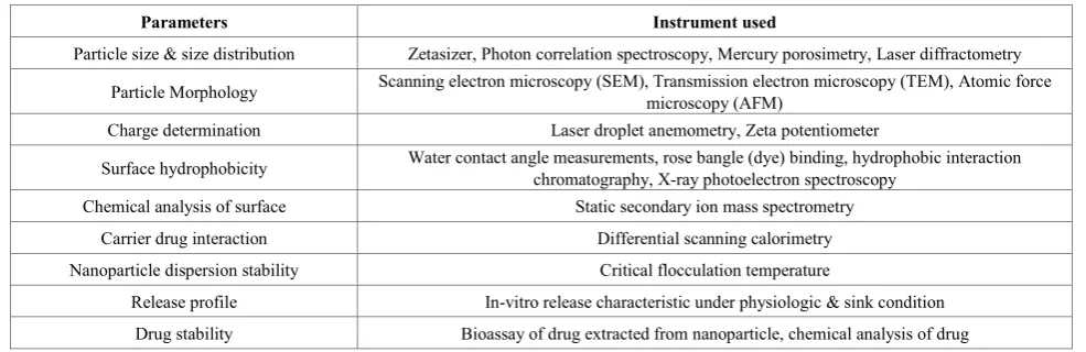
Table 1 state about the different parameters for characterization nanoparticles and instruments used for these characterizations.
Table 1. Different Parameters for characterization of Nanoparticles
Parameters Instrument used
Particle size & size distribution Zetasizer, Photon correlation spectroscopy, Mercury porosimetry, Laser diffractometry Particle Morphology Scanning electron microscopy (SEM), Transmission electron microscopy (TEM), Atomic force microscopy (AFM) Charge determination Laser droplet anemometry, Zeta potentiometer
Surface hydrophobicity Water contact angle measurements, rose bangle (dye) binding, hydrophobic interaction chromatography, X-ray photoelectron spectroscopy Chemical analysis of surface Static secondary ion mass spectrometry
Carrier drug interaction Differential scanning calorimetry Nanoparticle dispersion stability Critical flocculation temperature
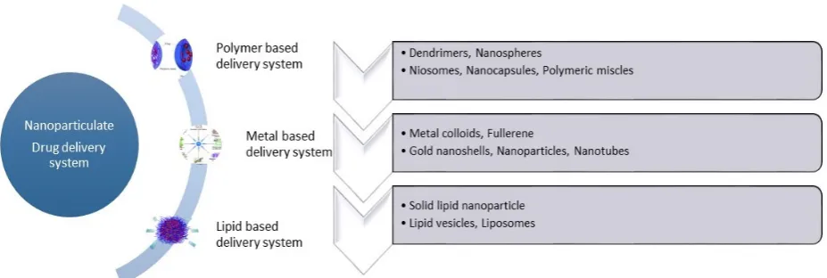
[image:3.595.59.552.442.602.2]Figure 1. Types of nano-particulate delivery systems
3. Nanoparticles as Platform for
Antimicrobials Delivery
These characteristic physicochemical properties have advantages in delivering and targeting to the target site, consequently minimizing adverse effect. Additionally, these could move in cells through endocytosis and consequently release the antibacterial agent to eliminate infections. In fact, a number of researches on nanoparticles based antibiotics have been developed and reviewed.
3.1. Metallic Nanoparticles
The continuous growing resistance of microbes to antibiotics causes severe health complications in current years. To confrontation with these problems, modern researcher identified the novel application by combining nanotechnology with intrinsic antimicrobial activity of the metals. Metallic nanoparticles are being utilized in many field of science with engineering comprising medical fields also and are still attract the scientists to invent new dimensions for their respective worth which is generally credited to their corresponding small sizes. In present scenario, the research in nanoparticle field is mainly focused on its synthesis or green synthesis not much on its applications. Research mainly focused on green synthesis
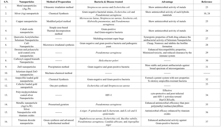
Table 2. List of metallic nanoparticles
S.N. Nanoparticles Method of Preparation Bacteria & Disease treated Advantage Reference
1. Metal nanoparticles (Ag, Pt) Ultrasonic irradiation method Streptococcus aureus and Escherichia coli Show antimicrobial activity of metals 23
2. Silver (Ag) nanoparticle Chemical Synthesis Gram negative bacterial strains, Pseudomonas aeruginosaEscherichia coli and Show antimicrobial activity of such hybrids materials 10, 24
3. Copper nanoparticles Modified polyol method Micrococcus luteusKlebsiella pneumoniae, Streptococcus aureus, Eescheria coli, , and Pseudomonas
aeruginosa Show antimicrobial activity of metals 25
4. nanoparticles Cobalt oxide Thermal decomposition Simple urea-based method
Gram-positive
And Gram-negative bacteria Show antimicrobial activity of metals 26
5. Quercetin-Acetylcholine- Selenium Nanoparticles --- Multidrug-resistant super bugs antibacterial activity of Selenium Nanoparticles Synergistic properties of both drug enhance the 27
6. Nanoparticles CaO Microwave irradiated synthesis Gram negative and gram positive bacteria and pathogenic yeast Cheap, Nontoxic and inhibits the biofilm formation 28
7. Dextran-and polyacrylic acid-coated ceria
nanoparticles --- Pseudomonas aeruginosa
Enhanced biocompatibility properties, Minimized toxicity, and reduced clearance by the
immune system 29
8. Carboxyl-capped bismuth Nanoparticles --- Helicobacter pylori --- 30
9. ZnO nanoparticles Precipitation method Gram negative and gram positive bacteria More stable and potent antibacterials against broad spectrum of microorganisms 31
10. Selenium doped ZnO nanoparticles Mechano-chemical method --- --- ---
11. Ampicillin loaded gold nanoparticles Chemical Synthesis Gram-negative and Gram-positive bacteria Formed a potent system with new properties To destroy ampicillin-resistant bacteria 32
12. Cefaclor loaded gold nanoparticle One pot synthesis Escherichia coli and Streptococcus aureus --- 33
13. Polyvinylpyrrolidone coated silver
Nanoparticles --- HIV-I
Effective
cyto-protective and post-infected anti-HIV-1 activities toward
Hut/CCR5cells.
34
14. Metallic nanoparticle (Ag,Cu,W) Pressurised gyration Pseudomonas aeruginosa Enhanced antimicrobial efficiency than pure poly(methyl methacrylate)fibres 35
15. Nanoparticles with Silver
titanium oxides ---
A.niger, P.spinulosum and S.chartarum, and E.coli and S.
epidermidis Antimicrobial efficacy enhanced by titanium oxides 36
16. Titanium dioxide nanoparticles Green synthesis and advanced hydrothermal method Pseudomonas aeruginosa, Candida albicans, Staphylococcus aureus, Escherichia coli, Bacillus subtilis, and Aspergillus niger
Enhanced antibacterial activity against
[image:5.808.49.761.95.504.2]Table 3. List of Metal/Metal oxide nanoparticle with their mechanism of action
S. No. Metal/ Metal oxide nanoparticle Mechanism of action Reference
1. Silver
Create holes in the cell wall of microbes by increasing the cell membrane permeability and in capacitating the respiratory chain. Inhibit and interrupt protein structure by binding to thiol and amino groups.
Photo-catalytic and induce reactive oxygen species.
Poisoning respiratory enzymes and components of the microbial electron transport system Binds to surface, altering the membrane function, and
Bind to DNA bases and inhibiting replication
38, 39, 40, 41, 42
2. Titanium dioxide Stimulates reactive oxygen species burst. Photo-catalytic
Reactive oxygen species destruct the cell membrane, DNA, and macromolecules and various functions of the bacterial cell
43, 44
3. Gold Generate holes in the cell wall resulting in the leakage of cell contents and cell death Destroy bacteria via photo-thermal heating Bind to the DNA of bacteria and inhibit the uncoiling and transcription of DNA
33, 45
4. Iron oxide Break DNA strands, can initiate lipid per oxidation or inactivate the enzymes Depolymerize the polysaccharides, 46
5. Magnesium oxides Inhibit essential enzymes of the bacteria Inducing reactive oxygen species 44
6. Zinc Oxide
Cell wall damage due to ZnO-localized interaction Enhanced membrane permeability
Internalization of nanoparticles due to loss of proton motive force and uptake of toxic dissolved zinc ions led to mitochondria weakness, intracellular outflow, and releasing expression of oxidative stress caused eventual cell growth inhibition and cell death
Reactive oxygen generation
47, 48, 49
7. Nitric Oxide Affecting reactive nitrogen species than reactive oxygen species 50
8. Copper oxide Interact with DNA molecules and intercalate with nucleic acid strands. Membrane disruption and Reactive oxygen species production
Disrupt biochemical processes. 43
[image:6.808.53.785.87.391.2]Table 4. Limitation and toxicity of Metallic nanoparticle
S. No. Metallic nanoparticle Limitation/Toxicity Reference
1. Silver Aggregation of particles into large particles due to high surface energy leads to loss of antibacterial activity. Oral toxicity, eye irritation, corrosion, and dermal toxicity 52 2 Silicon oxide 10 nm particle having strong impact on cell viability and genotoxic effects 52 3. Gold nanoparticles (4-5 nm) Inflammation, accretion of gold in lungs, and an increased number of macrophages 53
3.2. Polymeric Nanoparticles
Polymeric nanoparticles with biodegradable and biocompatible polymers are good candidates as particulate carrier for antimicrobial agent and drug delivery and there has been considerable interest in the use of nanoparticles as potential delivery systems. Numerous investigations have shown that nanoparticles can not only improve the stability of therapeutic agents against enzymatic degradation, but by modulating polymer characteristics, they can also achieve desired therapeutic levels in target tissues for the required duration for optimal therapeutic efficacy (54, 1). Depending on their composition and intended use, they can be administered orally, parenterally, or locally (54). Different nanoparticles manufacturing methods were described allowing modification of physicochemical characteristics such as size, structure, morphology, surface texture and composition to meet different requirements.
Nanoparticles act on microbes by releasing antibiotics, antimicrobial peptides, and antimicrobial agents or by contact-killing like quaternary ammonium compounds etc. Cationic groups (with long hydrophobic chains) are capable to lysis the membrane to penetrate and burst the bacterial membrane and lead to death (52, 55).
Lots of the nanoparticle therapeutics in research are polymeric nanoparticles, which have been comprehensively explored as platform for drug delivery. Polymeric nanoparticles are fabricated from biocompatible and biodegradable polymers. Polymeric nanoparticles are formulated by a self-assembly process using block-copolymers comprising of two or more polymer chains with different hydrophilicity. Polymeric nanoparticles have been framed to encapsulate either hydrophilic or hydrophobic drug molecules, as well as macromolecules
such as proteins, peptides and nucleic acids (56).
The main issues in this field are the loading efficiency, stability of bioactive agent during preparation and release, release profiles and surface modification. Particles size and surface property (surface charge and hydrophobic or hydrophilic property) are primary factors for the in-vivo fate of nanoparticles. Surface modification of nanoparticles has been achieved mainly by two methods:
Surface coating with hydrophilic
polymers/surfactants
Development of biodegradable copolymers with charged functional group or hydrophilic segments. 3.2.1. Methods of Preparation of Polymeric Nanoparticles
Method of preparation are mainly depends upon the physico-chemical characteristic of polymers and the drugs. They are classified as-
Amphiphilic macromolecular cross linking I Heat cross-linking
II Chemical cross-linking
Polymerization methods I Emulsion polymerization II Dispersion polymerization III Interfacial complexation
IV Interfacial condensation polymerization
Polymer precipitation methods
I Solvent extraction/ evaporation methods. II Solvent displacement methods
III Salting out.
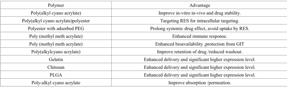
Table 5. Polymer used in nanoparticle preparation with advantages
Polymer Advantage
Poly(alkyl cyano acrylate) Improve in-vitro in-vivo and drug stability. Poly(alkyl cyano acrylate)polyester Targeting RES for intracellular targeting.
Polyester with adsorbed PEG Prolong systemic drug effect, avoid uptake by RES. Poly (methyl meth acrylate) Enhanced immune response.
Poly (methyl meth acrylate) Enhanced bioavailability ,protection from GIT Poly(alkylcyano acrylate) Improve retention of drug /reduced washout.
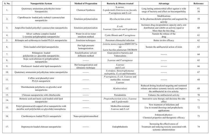
[image:7.595.63.544.601.747.2]Table 6. List of polymeric nanoparticles
S. No. Nanoparticles System Method of Preparation Bacteria & Disease treated Advantage Reference
1. Quaternary ammonium polyethylen imine Nanoparticles Chemical Synthesis S.epidermidis,Enterococcus faecalis, S.aureus, P.aeruginosa and E.coli
Long lasting antimicrobial effect against a wide
range of bacteria and effect on biocompatibility 57, 58
2. Ciprofloxacin- loaded poly-isobutyl cyanoacrylate nanoparticles Emulsion polymerization Mycobacterium avium In the pharmacokinetic properties and augment the Modifications
activity 59
3. Ampicillin-loaded polyethyl cyanoacrylate nanoparticles Emulsion polymerization S.aureus, S.faecalis and S.epidermisE.coli, Increases drug encapsulation capacity and a size distribution and exhibit an activity equal or
More than the free drug. 60 4. L-tyrosine polyphosphate nanoparticles Silver–carbene complex-loaded Water-in-oil-in-water emulsion method Cystic fibrosis and P.aeruginosa Sustain the release of the drug 61 5. Rifampin and azithromycin loaded PLGA nanoparticles Emulsion techniques Persistent chlamydial infection Better invitro efficacy 62
6. Nisin-loaded solid lipid nanoparticles Hot high pressure homogenization Listeria monocytogenesnd DMST2871a
Lacto bacillus plantarum TISTR850 Sustain the antibacterial action of nisin 63
7. polylactide-co- glycolide nanoparticles Rifampicin- loaded Emulsification/ solvent diffusion method Gram-positive bacteria and Tuberculosis --- 64
8. Kojic acid-chitosan-tri-polyphosphate nanoparticles --- S.aureus and P.aeruginosa --- 65
9. Florfenicol- loaded solid lipid nanoparticle Hot homogenisation and ultrasonic technique and S.aureus E.coli --- 66
10. Quaternary ammonium polyethylene imine nanoparticles --- maltophilia, E.coli S.viridans, Stenotrophomonas and Peritonitis --- 67
11. Caffeic acid phenethyl ester PLGA nanoparticle --- P.aeroginosamethicillin- resistant , E.coli, S.aureus and S.aureus --- 68
12. Thioridazinein poly(lactic-co-glycolic) acid nanoparticles --- M.tuberculosis Reduced dosing localized targeting and sustained release and reduce systemic toxicity and improve
the antibacterial in-vivo acticity 69 13. Chitosan nanoparticles with chlorhexidin --- Periodontitis Enhance the antibacterial activity 70
14. Retinoic acid and lauric acid loaded solid lipid nanoparticles --- Propionibacterium acnesand S.epidermidis, S.aureus Innovative acne therapy and decrease the side effect 71
15. Poly(l-glutamicacid)-capped silver nanoparticles with ascorbic acid poly(lactide-co-glycolide) nanospheres --- Methicillin-resistant S.aureus and E.coli Use in wound dressings and prophylactic New treatment of infections and
applications 72
16. Clarithromycin loaded PLGA nanoparticle Nano-precipitationmethod Chemical properties and therapeutic efficacy Enhanced physico- 73
17. Daptomycin-loaded chitosan nanoparticles --- Endophthalmitis Treatment and reducing toxicity associated with Increasing the effectiveness of
[image:8.808.42.775.89.550.2]18. Clindamycin loaded PLGA hydroxyapatite nanoparticle
Sono chemical homogeneous precipitation and ultrasonic de-agglomeration
Wound healing Enhance drug absorption and delivery 75
19. Ciprofloxacin loaded Glycosylated polyacrylate nanoparticles Bacillus anthracisS. aureus, Enhanced bioavailability, Increased therapeutic
Efficacy 76
20. Azithromycin loaded PLGA nanoparticle Nano-precipitationmethod Chemical properties and therapeutic efficacy Better physico- 77
21. Rifampicin, isoniazid,
pyrazinamide loaded Alginate nanoparticles
M. tuberculosis
Increase drug load, Pharmacokinetic activity and
Therapeutic efficacy 78 22. Rifampicin loaded PLGA nanoparticle Emulsification/ solvent diffusion method Gram-positive and Gram-negative bacteria Enabling drug penetration to parasites present in macrophages 64
23. Halofantrine loaded PLA nanoparticle Malaria Improved stability, lower toxicity than free form in solution and rapid uptake by tissues 79
24. Saquinavir loaded Polyethylene oxide Modified poly(epsilon carprolactone)
Nanoparticles HIV
Shield Saquinavir from enzyme cytochrome C
Metabolism and avoid P-efflux pump 80
25. PLGA nanoparticle entrapped eugenol and trans-cinnamaldehyde Emulsion evaporation method Salmonell aand Listeria Mask the hydrophobic property 81
Table 7. List of Polymers for nanoparticle fabrication
S. No. Polymer Composition Action through Reference
1. Poly-𝜀𝜀-lysine cationic homopeptide of L-lysine Disrupting the cell membrane of bacteria 83
2. ammonium Quaternary compounds.
Benzalkonium chloride, stearalkonium
chloride, and cetrimonium chloride Interact with surface of bacteria and denature structural proteins and enzymes 52
3. Chitosan N-acetylglucosamine polymer chitin Interrupted membrane functions like destabilization of membrane protein and membrane lysis, affect
respiration and cell division, drug efflux system 84
3.3. Techniques to Determine Antibacterial Activity of Nanoparticles
Various experimental methods and assays are available for measuring the antibacterial efficacy of nanoparticles. Each methods are unique and having their advantage and disadvantage. These methods/assays are (85):
Optical density measurement
Spread-plate(colony counts on agar)
Cell counting devices
Live/dead fluorescent stain MTS/MTT/XTT assays
Crystal violet staining
4. Conclusions
In summary, it can be concluded that nanotechnology developed as a new approach in pharmaceutical sciences and research grasps great promise for overcoming complications related to delivery of antimicrobial and infection curing approaches. Various antimicrobial substances are unusable due to their instability, physicochemical limitations, enzyme degradation and cytotoxicity. Along with these problems, many microbes and their strains were getting resistant to the available antibiotics become as problem that surges the necessity to develop novel antibacterial substances. These issues were reasons for emergence of nanotechnology that provide many option for antimicrobials. The small size of the nanoparticles is very suitable for delivery of antibacterial substances.
REFERENCES
[1] Lam S, Wong EHH, Boyer C, Qiaoa GG. Antimicrobial Polymeric Nanoparticles. Progress in Polymer Science, 2017. https://doi.org/10.1016/j.progpolymsci.2017.07.007
[2] Morones JR, Elechiguerra JL, Camacho A, Ramirez JT. The bactericidal effect of silver nanoparticles. Nanotechnology 2005; 16: 2346–53.
[3] Kim JS, Kuk E, Yu KN, Kim JH, Park SJ, Lee HJ, et al. Antimicrobial effects of silver nanoparticles. Nanomed Nanotechnol Biol Med 2007; 3: 95-101.
[4] Zazo H, Millan, Colino CI, Lanao JM. Applications of Metallic Nanoparticles in Antimicrobial Therapy.
Antimicrobial Nanoarchitectonics,2017;411-17
[5] Gajjar P, Pettee B, Britt DW, Huang W, Johnson WP, Anderson J. Antimicrobial activities of commercial nanoparticles against an environmental soil microbe, Pseudomonas putida KT2440. Journal of Biological Engineering. 2009; 3: 9-22.
[6] Nakazato G, Kobayashi RKT, Seabra AB, Duran N. Use of nanoparticles as a potential antimicrobial for food packaging. Food Preservation, 2017; 413-447.
[7] Ferrari M. Cancer nanotechnology: opportunities and challenges. Nat Rev Cancer, 2005, 5, 3, 161-71.
[8] Gao W, Thamphiwatana S, Angsantikul P, Zhang L. Nanoparticle Approaches against Bacterial Infections. Wiley Interdiscip Rev Nanomed Nanobiotechnol. 2014; 6(6): 532– 547.
[9] Zhang L, Pornpattananangkul D, Hu CMJ, Huang CM. Development of nanoparticles for antimicrobial drug delivery. Curr Med Chem. 2010; 17:585–594.
[10]Das MR, Sarma RK, Saikia R, Kale VS, Shelke MV, Sengupta P. Synthesis of silver nanoparticles in an aqueous suspension of graphene oxide sheets and its antimicrobial activity. Colloids and Surfaces B: Biointerfaces 83 (2011) 16–22.
[11]Parmar B, Mandal S, Petkar C, Patel LD, Sawant KK. Valsatran loaded solid lipid nanoparticle: development, characterization and in-vitro and ex-vivo evaluation. Int j Pharma Sci Nanotech, 2011, 4, 3, 1483-90.
[12]Albrecht MA, Evan CW, Raston CL. Green chemistry and the health implications of nanoparticles. Green Chem 2006; 8: 417–32.
[13]Wang P. Nanoscale biocatalyst systems. Curr Opinion Biotechnol 2006; 17: 574-9.
[14]Zhang L, Gu FX, Chan JM, Wang AZ, Langer RS, Farokhzad OC. Nanoparticles in medicine: therapeutic applications and developments. Clin Pharmacol Ther 2008; 83: 761-9. [15]Hong B, Kai J, Ren Y, Han J, Zou Z, Ahn CH, et al. Highly
sensitive rapid, reliable, and automatic cardiovascular disease diagnosis with nanoparticle fluorescence enhancer and MEMS. Adv Exp Med Biol 2008; 614: 265-73.
[16]Vigneshwaran N, Kathe AA, Varadarajan PV, Nachane RP, Balasubramanya RH. Functional finishing of cotton fabrics using silver nanoparticles. J Nanosci. Nanotechnol. 2007; 7: 1893-7.
[image:10.595.62.547.91.185.2]markers and a potential source of new therapeutic agents. Biochim Biophys Acta 2004; 1696: 1-14.
[18]Kim D, Park S, Lee JH, Jeong YY, Jon S. Anti-biofouling polymer coated gold nanoparticle as a contrast agent for in vivo x ray commuted tomography imaging. J Am Chem Sci, 129, 7661-7665.
[19]Gopinath V, Ali DM, Priyadarshini S, Priyadarshini NM, Thajuddin N, Velusamy P. Biosynthesis of silver nanoparticles from Tribulus terrestri sand its antimicrobial activity: A novel biological approach. Colloids and Surfaces B: Biointerfaces 96 (2012) 69-74.
[20]Gong P, Li H, He X, Wang K, Hu J, Tan W, et al. Preparation and antibacterial activity of Fe3O4@Ag nanoparticles. Nanotechnology 2007; 18: 604–11.
[21]Gu H, Ho PL, Tong E, Wang L, Xu B. Presenting vancomycin on nanoparticles to enhance antimicrobial activities. Nano Lett 2003; 3(9):1261–3.
[22]Ahmed S, Ahmad M, Swami BL, Ikram S. A review on plants extract mediated synthesis of silver nanoparticles for antimicrobial applications: A green expertise. Journal of Advanced Research (2015)
http://dx.doi.org/10.1016/j.jare.2015.02.007.
[23]Cho KH, Park JE, Osaka T, Park SG. The study of antimicrobial activity and preservative effects of nanosilver ingredient. Electro chimica Acta 51 (2005) 956–960.
[24]Shaker MA, Shaaban MI. Synthesis of silver nanoparticles with antimicrobial and anti-adherence activities against multidrug-resistant isolates from Acinetobacter baumannii. Journal of Taibah University Medical Sciences, 12, 4, 2017, 291-297.
[25]Ramyadevi J, Jeyasubramanian K, Marikani A, Rajakumar G, Rahuman AA. Synthesis and antimicrobial activity of copper nanoparticles. Materials Letters 71 (2012) 114–116
[26]Khan S, Ansari AA, Khan AA, Ahmad R, Al-Obaid O, Al-Kattan W. In vitro evaluation of anticancer and antibacterial activities of cobalt oxide nanoparticles. J BiolInorg Chem. 2015 Dec; 20(8):1319-26.
[27]Huang X, Chen X, Chen Q, Yu Q, Sun D, Liu J. Investigation of functional selenium nanoparticles as potent antimicrobial agents against superbugs. Acta Biomater. 2015 S1742-7061(15)30170-7. doi: 10.1016/j.actbio.2015.10.041. [28]Roy A, Gauri SS, Bhattacharya M, Bhattacharya J.
Antimicrobial activity of caO nanoparticles. J Biomed Nanotechnol. 2013;9(9):1570-8
[29]Wang Q, Perez JM, Webster TJ. Inhibited growth of Pseudomonas aeruginosa by dextran- and
polyacrylicacid-coated ceria nanoparticles. Int J Nanomedicine. 2013; 8:3395-9.
[30]Nazari P, Dowlatabadi-Bazaz R, Mofid MR, Pourmand MR, et al. The antimicrobial effects and metabolomic footprinting of carboxyl-capped bismuth nanoparticles against Helicobacter pylori. Appl Biochem Biotechnol. 2014, 172(2):570-9.
[31]Siddique S, Shah ZH, Shahid S, Yasmin F. Preparation, characterization and antibacterial activity of ZnO nanoparticles on broad spectrum of microorganisms. Acta Chim Slov. 2013; 60(3):660-5.
[32]Brown AN, Smith K, Samuels TA, Lu J, Obare SO, Scott ME. Nanoparticles Functionalized with Ampicillin Destroy Multiple Antibiotic-Resistant Isolates of Pseudomonas
aeruginosa and Enterobacter aerogenes and
Methicillin-Resistant Staphylococcus aureus. Applied and Environmental Microbiology, 2012, p. 2768–2774.
[33]Rai A, Prabhune A, Perry CC. Antibiotic mediated synthesis of gold nanoparticles with potent antimicrobial activity and their application in antimicrobial coatings. J. Mater. Chem. 2010; 20:6789-6798.
[34]Sun RW, Chen R, Chung NP, Ho CM, Lin CL, Che CM. Silver nanoparticles fabricated in Hepes buffer exhibit cytoprotective activities toward HIV-1 infected cells. Chem Commun (Camb). 2005; 40:5059–61.
[35]Illangakoona UE, Mahalingama S, Wanga K, Cheong YK, Canales E, Ren GG et al., Gyrospun antimicrobial nanoparticle loaded fibrous polymeric filters. Materials Science and Engineering: C 74, 2017, 315-324.
[36]Chi Chen Y, Yu KP. Enhanced antimicrobial efficacy of thermal-reduced silver nanoparticles supported by titanium dioxide. Colloids and Surfaces B: Biointerfaces, 154, 2017, 195-202.
[37]Sundrarajan M, Bama K, Bhavani m, Jegatheeswaran S, Ambika S, Sangili A, Nithya P, Sumathi R. Obtaining titanium dioxide nanoparticles with spherical shape and antimicrobial properties using M. citrifolia leaves extract by hydrothermal method. Journal of Photochemistry and Photobiology B: Biology, 171, 2017, 117-124.
[38]Beyth N, Yudovin-Farber I, Perez-Davidi M, Domb AJ, and Weiss EI, “Polyethylenimine nanoparticles incorporated into resin composite cause cell death and trigger biofilm stress in-vivo,” Proceedings of the National Academy of Sciences of the United States of America, 107, 51, 22038–22043, 2010.
[39]Sondi I and Salopek-Sondi B, “Silver nanoparticles as antimicrobial agent: a case study on E. coli as a model for Gramnegative bacteria,” Journal of Colloid and Interface Science, 275, 1, 177–182, 2004.
[40]Choi O, Deng KK, Kim NJ, Ross Jr. L, Surampalli RY,Hu Z, “The inhibitory effects of silver nanoparticles, silver ions, and silver chloride colloids on microbial growth,” Water Research, 42, 12, 3066–3074, 2008.
[41]Percival, S.L., Bowler, P.G., Russell, D., 2005. Bacterial resistance to silver in wound care. J. Hosp. Infect. 60, 1–7. [42]Wright, J.B., Lam, K., Burrell, R.E., 1994. Wound
management in an era of increasing bacterial antibiotic resistance: a role for topical silver treatment. Am. J. Inf. Control 26, 572–577.
[43]Pelgrift RY, Friedman AJ, “Nanotechnology as a therapeutic tool to combat microbial resistance,” Advanced Drug Delivery Reviews, 65, 13-14, 1803–1815,2013.
[44]Blecher K, Nasir A, Friedman A, “The growing role of nanotechnology in combating infectious disease,” Virulence, 2, 5,395–401, 2011.
[46]Weinberg ED. Iron loading and disease surveillance. Emerg Infect Dis. 1999; 5(3): 346-352.
[47]Sirelkhatim A, Mahmud S, Seeni A, Kaus NHM, Ann LC, Bakhori SKM, Hasan H, Mohd SK. Review on Zinc Oxide Nanoparticles: Antibacterial Activity and Toxicity Mechanism. Nano-Micro Lett. (2015) 7(3):219–242
[48]Reddy KM, Feris K, Bell J, Wingett DG, Hanley C, Punnoose A. Selective toxicity of zinc oxide nanoparticles to prokaryotic and eukaryotic systems. Appl Phys Lett. 2007; 90(213902):2139021–2139023.
[49]Padmavathy N, Vijayaraghavan R. Enhanced bioactivity of ZnO nanoparticles – an antimicrobial study. Sci Technol Adv Mat. 2008; 9(3):35004–35010.
[50]Han G, Martinez LR, Mihu MR, Friedman AJ, Fried-man JM, Nosanchuk JD. “Nitric oxide releasing nanoparticles are therapeutic for Staphylococcus aureus abscesses in a murine model of infection,” PLoS ONE, 2009, 4, 11.
[51]Ansari MA, Khan HM, Khan AA, Cameotra SS, Saquib Q, Musarrat J, “Interaction of Al2O3nanoparticleswith Escherichia coli and their cell envelope biomolecules,” Journal of Applied Microbiology, 116,772–783,2014.
[52]Beyth N, Houri-Haddad Y, Domb A, Hazan R. Alternative Antimicrobial Approach: Nano-Antimicrobial Materials. Evidence-Based Complementary and Alternative Medicine, 2015, 246012, 16. http://dx.doi.org/10.1155/2015/246012 [53]Maneewattanapinyo P, Banlunara W, Thammacharoen C,
Ekgasit S, Kaewamatawong T, “An evaluation of acute toxicity of colloidal silver nanoparticles,” The Journal of Veterinary Medical Science, 73,11,1417–1423,2011.
[54]Campos AD, Diebold Y, Carvalho ELS, Sanchez A, Alonso MJ. Chitosan nanoparticles as new ocular drug delivery systems: in vitro stability, in-vivo fate, and cellular toxicity. Phafrma Res, 2004, 21, 5, 803-810
[55]Lichter JA, Rubner MF. “Polyelectrolyte multilayers with intrinsic antimicrobial functionality: the importance of mobile polycations,” Langmuir, 25, 13, 7686–7694, 2009.
[56]Wang AZ, Farokhzad OC, Langer R, Nanoparticle Delivery of Cancer Drugs. Annu. Rev. Med. 2012. 63:185–98
[57]Beyth N, Houri-Haddad Y, Baraness-Hadar L, Yudovin-Farber I, Domb AJ, Weiss EI. Surface antimicrobial activity and biocompatibility of incorporated polyethylenimine nanoparticles. Biomaterials 29 (2008) 4157–4163.
[58]Jiaoa Y, Niua L, Maa S, Lic J, Tayd FR, Chena JH. Quaternary ammonium-based biomedical materials: State-of-the-art, toxicological aspects and antimicrobial resistance. Progress in Polymer Science, 71, 2017, 53-90. [59]Fawaz F, Bonini F, Maugein j, Lagueny AM.
Ciprofloxacin-loaded poly-iso-butylcyano acrylate nanoparticles: pharmacokinetics and in vitro antimicrobial activity. International Journal of Pharmaceutics 168 (1998) 255-259.
[60]Fontana G, Pitarresi G, Tomarchio V, Carlis B, Biagio PLS. Preparation, characterization and in vitro antimicrobial activity of ampicillin-loaded polyethylcyanoacrylate nanoparticles. Biomaterials 19 (1998) 1009-1017.
[61]Hindi KM, Ditto AJ, Panzner MJ, Medvetz DA, Hovis CE, Hilliard JK et al. The antimicrobial efficacy of sustained release silver–carbene complex-loaded L-tyrosine polyphosphate nanoparticles: Characterization, in vitro and in vivo studies. Biomaterials 30 (2009) 3771–3779.
[62]Toti US, Guru BR, mCpHARLIN cm, Wykes SM, Panyam J, Hudson JA. Targeted delivery of antibiotics to intracellular chlamydial infections using PLGA nanoparticles. Biomat, 2011, 32, 27, 6606-13
[63]Prombutara P, Kulwatthanasal Y, Supaka N, Sramala I, Chareonpornwattana S, Production of nisin-loaded solid lipid nanoparticles for sustained antimicrobial activity. Food Control 24 (2012) 184-190.
[64]Esmaeili F, Hosseini-Nasr M, Rad-Malekshahi M, Samadi N, Atyabi F, Dinarvand R. Preparation and antibacterial activity evaluation of rifampicin-loaded poly lactide-co-glycolide nanoparticles. Nanomedicine.2007; 3(2):161-167.
[65]Chaudhary J, Lakhawat S, Pathak AN.Elucidation on enhanced application of synthesized kojic acid immobilized magnetic and chitosan tri-polyphosphate nanoparticles as antibacterial agents. IET Nanobiotechnol. 2015 Dec; 9(6):375-80. doi: 10.1049/iet-nbt.2014.0077.
[66]Wang T, Chen X, Lu M, Li X, Zhou W. Preparation, characterisation and antibacterial activity of a florfenicol-loaded solid lipid nanoparticle suspension. IET Nanobiotechnol. 2015 Dec; 9(6): 355-61.
[67]Ortega A, Farah S, Tranque P, Ocaña AV, Nam-Cha SH, Beyth N, et al. Antimicrobial evaluation of quaternary ammonium polyethyleneimine nanoparticles against clinical isolates of pathogenic bacteria. IET Nanobiotechnol. 2015, 9(6):342-8.
[68]Arasoglu T, Derman S, Mansuroglu B. Comparative evaluation of antibacterial activity of caffeic acid phenethyl ester and PLGA nanoparticle formulation by different methods. Nanotechnology. 2015 Dec 2; 27(2):025103. [69]Vibe CB, Fenaroli F, Pires D, Wilson SR, Bogoeva V,
Kalluru R, et al.. Thioridazine in PLGA nanoparticles reduces toxicity and improves rifampicin therapy against mycobacterial infection in zebrafish. Nanotoxicology. 2015 Nov 17:1-9.
[70]Barreras US, Méndez FT, Martínez RE, Valencia CS, Rodríguez PR, Rodríguez JP. Chitosan nanoparticles enhance the antibacterial activity of chlorhexidine in collagen membranes used for periapical guided tissue regeneration. MaterSciEng C Mater Biol Appl. 2016 Jan 1; 58:1182-7. [71]Silva EL, Carneiro G, De Araújo LA, Trindade Mde J,
Yoshida MI, et al. Solid Lipid Nanoparticles Loaded with Retinoic Acid and Lauric Acid as an Alternative for Topical Treatment of Acne Vulgaris. J Nanosci Nanotechnol. 2015; 15(1): 792-9.
[72]Stevanovic M, Uskokovic V, Filipovic M, Škapin SD, Uskokovic D. Composite
PLGA/AgNpPGA/AscHnanospheres with combined osteoinductive, antioxidative, and antimicrobial activities. ACSAppl Mater Interfaces. 2013, 25; 5(18):9034-42.
drug delivery system. Colloid Surf B.2011;88 (1):39-44. [74]Silva NC, Silva S, Sarmento B, Pintado M. Chitosan
nanoparticles for daptomycin delivery in ocular treatment of bacterial endophthalmitis. Drug Deliv. 2015 Oct; 22(7):885-93.
[75]Vukomanovic M, Zavasnik-Bergant T, Bracko I, et al. Poly(D,L-lactide-co-glycolide)/hydroxyapatite
coreshellnanospheres. Part 3: properties of hydroxyapatite nano-rods and investigation of a distribution of the drug within the composite. Colloid Surf B. 2011; 87(2):226-35. [76]Turos E, Shim JY, Wang Y, Greenhalgh K, Reddy GS,
Dickey S, Lim DV. Antibiotic-conjugated polyacrylate nanoparticles: new opportunities for development of anti-MRSA agents. Bioorg Med Chem Lett. 2007; 17: 53-56. [77]Mohammadi G, Valizadeh H, Barzegar-Jalali M, et al.
Development of azithromycin-PLGA nanoparticles: physicochemical characterization and antibacterial effect against Salmonella typhi. Colloid Surf B. 2010; 80(1):34-39. [78]Ahmad Z, Pandey R, Sharma S, Khuller GK. Alginate
nanoparticles as anti-tuberculosis drug carriers: formulation development, pharmacokinetics and therapeutic potential. Ind J Chest Dis Allied Sci 2005; 48:171–6.
[79]Mosqueira VC, Loiseau PM, Bories C, Legrand P, Devissaguet JP, Barratt G. Efficacy and pharmacokinetics of
intravenous nanocapsule formulations of halofantrine in
Plasmodium berghei-infected mice. Antimicrob Agents
Chemother. 2004; 48(4):1222-1228.
[80]Shah LK, Amiji MM. Intracellular delivery of saquinavir in biodegradable polymeric nanoparticles for HIV/AIDS. Pharm Res. 2006; 23: 2638-2645.
[81]Gomes C, Moreira RG, Castell-Perez E. Poly (DL-lactide-co-glycolide) (PLGA) nanoparticles with entrapped trans-cinnamaldehyde and eugenol for antimicrobial delivery applications. J Food Sci. 2011; 76(2): N16-24.
[82]SinCheow W, Wook Chang M, Hadinoto K. Antibacterial Efficacy of Inhalable Levofloxacin-Loaded Polymeric Nanoparticles Against E. coli Biofilm Cells: The Effect of Antibiotic Release Profile. Pharm Res. 2010; 27:1597–1609. [83]Hiraki J, “Basic and applied studies on 𝜀𝜀-polylysine,” Journal
of Antibacterial and Antifungal Agents, vol.23, pp. 349–354, 1995.
[84]Ibrahim M, Tao Z, Hussain A et al., “Deciphering the role of Burk holder iacenocepacia membrane proteins in antimicrobial properties of chitosan,” Archives of Microbiology, 196 ,1,9-16, 2014.