Microstructural Studies of Milled and Annealed ZnFe
2
O
4
Nanostructures Using X-Ray Diffraction and Mössbauer
Spectroscopy
O. Ould Fella
1,*, M. Tamine
1, N. Randrianantoandro
2, J. M. Grenèche
21Laboratoire de Physique et Chimie Quantique, Département de Physique, Faculté des Sciences, Université de Tizi-Ouzou BP 17RP,
15000 Tizi-Ouzou, Algérie
2Institut des Molécules et Matériaux du Mans (IMMM)-UMR CNRS 6283, Université du Maine, Le Mans Cedex 72085, France
*Corresponding Author: [email protected]
Copyright © 2013 Horizon Research Publishing All rights reserved.
Abstract
Nanostructured zinc ferrite ZnFe2O4 wasmilled at high energy during 12 hours and then the portions of the obtained powders were calcined at 400°C and 600°C during 24 hours. X-ray diffractogramms recorded for various samples show that compared to the grain size value of as-prepared sample the average grain size obtaining from the milling process decreases after 12 hours of milling duration and increases with increasing annealing temperature. Then, the inversion and lattice parameters increase with milling process and decrease with increasing annealing temperature. Magnetic behaviours analyzed by means of 57Fe Mössbauer
spectra recorded at 300K and 77K indicate that only milled sample without annealing is magnetically ordered at room temperature. At 77K, spectrum of milled zinc ferrite displays a magnetic state confirming the existence of cationic distribution after milling duration whereas the spectra of annealed samples exhibit the presence of superparamagnetic state.
Keywords
Ferrite. Zinc Ferrite. X-Ray Diffraction. Mössbauer Spectroscopy. High Energy Ball-Milling. Annealing Process
1.Introduction
Spinel ferrites with formula MFe2O4 (M= Zn, Ni, Fe, Co,
Cu…) have many interests as storage information, catalysis [1]. It contains metal transitions which are interesting from the magnetic properties point of view because they show various magnetic behaviours such as paramagnetism, antiferromagnetism, ferrimagnetism and spin glass or spin cluster behaviour [2,3]. In the spinel structure, there are twice as many octahedral (B) cationic sites as tetrahedral cationic (A) sites. If M2+ occupies only tetrahedral
(octahedral) sites, the spinel is direct (inverse).
Bulk ZnFe2O4 depicts a long range order below 10K as
well as short range at higher temperature [5] and exhibits a normal spinel structure when Zn2+ and Fe3+ ions occupy
tetrahedral (A) and octahedral (B) spinel sites, respectively [4]. Indeed, if the tetrahedral sites are exclusively occupied by non-magnetic ions (Zn), magnetic interaction occurs only between Fe3+ ions in the octahedral sites and the weak
negative B-B interaction leads to an antiferromagnetic long-range order. On the other hand, ZnFe2O4 exhibits
ferrimagnetic order as a result of competition among A-A, A-B and B-B exchange interactions. Since the superexchange interactions is stronger for more closely situated cations and for bonds angle closer to 180°, the superexchange interaction of A-O-B is strongest, that B-O-B is weaker, and that A-O-A the weakest.
Many experimental techniques have been used to explore inverted spinel structure in zinc ferrite in which Fe3+
occupies simultaneously octahedral and tetrahedral sites. Thus, this cationic inversion was observed by means of X-ray diffraction [6,7,8], EXAFS studies [9,10,11], neutron diffraction [12,13] and Mössbauer spectrometry [14,15,16]. It is well established from the literature that the values of the inversion parameter (δ) ranging from 0.03 to 0.6 are strongly dependent on the preparation procedure [13,17,18]. The increasing cationic inversion with the diminution of grains sizes induces drastic changes in magnetic properties, magnetization and ordering temperature. In addition, the reduction of grain size leads to novel properties as compared to the bulk material, due to small volume (superparamagnetism [19]) or high surface/volume ratio (spin canting [20]).
unusual physical behaviors which are subject of an increasing interest [23]. In order to provide some additional lightings on the analysis and the comprehension of these properties, we explore the role of milling as well as annealing process on the microstructural and magnetic properties of zinc ferrite.
In our experiments studies, nanostructured powders of zinc ferrites have been milled during 12 hours in a planetary ball milled (fritsh pulverisette P7) with high energy (intensity I=8, 1000tr/mn). Portion of the 12 hours milled sample were annealed during 24 hours under temperature of 400°C and 600°C. The Maud procedure is used to treat and analyze patterns of X-rays diffraction. Mössbauer spectra have been realized at room temperature and 77K in order to study the magnetic properties of different samples. In order to situate the context of this work; it is important to mention that although the work reported in reference [32] and that exposed here have some similarity, nevertheless an important difference should be noted. To argue this, it is important to emphasize that the microstructure analysis of zinc ferrite nanoparticles obtained from thermal treatment of their mixed hydroxides at 400° C-600° C has been recently investigated by means of X-rays diffraction and Mössbauer spectroscopy which reveals the asymmetric broadening and the large line widths of spectral lines confirming the presence of superparamagnetic behaviour [32]. Here, a chemical method is used to synthesize the zinc ferrite nanoparticles since the mixed hydroxides are prepared by co-precipitation from their nitrate solutions. In contrast, in our work the used procedure to elaborate the zinc ferrite nanoparticles is based on the ball-milling technique with a certain duration followed by the annealed treatment at 400°C and 600°C during 24 hours. Thus, firstly, the obtained zinc ferrite nanoparticles are not related with any synthesized methods. Secondly, the study of the structural and magnetic properties of the milled powders of zinc ferrite treated at different temperatures was performed. Unlike many studies described in the literature, our investigations focuses on the analysis of the microstructure and magnetic properties of these powders as a result of a high-energy mechanical milling followed by heat treatment. The annealing was performed on a milled zinc ferrite unlike previous work where the annealing is performed directly on the nanoparticle [32]. From these considerations, the main objective of the investigations carried out in this work is to clarify both the role and influence of the mechanical grinding as well as thermal treatment on microstructure parameters and magnetic properties of zinc ferrite nanostructures.
2. Experiments
The milling of commercial coarse-grained powders of ZnFe2O4 (Alfa Cesar) was carried out in argon atmosphere
with a planetary ball milled (Fritsch pulverisette, P7) using tungsten carbide balls with a ball to weight powder ratio of 5:1. Small amounts of powder were extracted from the vial after 12 hours of milling and annealed at 400°C and 600°C during 24hours. For structural analysis, X-ray diffraction
(XRD) patterns were obtained employing a Phillips X’pert diffractometer with tension 40kV and intensity 30 mA. A wavelength λ = 1.5405Å was used to obtain data in the range of 10° <2θ< 80°, (with step of 0.02°).
Mössbauer spectra were recorded in a transmission geometry using a 57CO/Rh ray source. Spectra obtained in
zero magnetic field (B=0T) for temperatures T= 300K, T=77K are adjusted using Mosfit program [24]. The isomer shift was referred to that of α-Fe at 300K.
3. X-Ray Diffraction
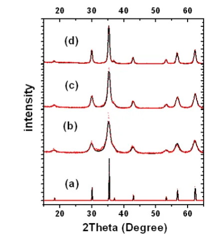
[image:2.595.318.525.263.498.2]X-ray diffraction recorded for as-prepared (0hours), 12 hours milled as well as 12 hours milled and annealed at 400°C, 600°C during 24 hours samples are shown in figure 1.
Figure 1. X-ray diffractograms (CuK) of ZnFe2O4.(a): as-prepared.(b):
12 hours milled sample.(c): 12 hours milled sample annealed at 400°C during 24h.(d): 12 hours milled sample annealed at 600°C during 24h.
The X-ray diffraction patterns for all samples display the fundamental peaks of cubic spinels, essentially matching that of ZnFe2O4 (Franklinite JCPDS No, 22-1012). After milling,
the diffractions peaks become broadened with small intensities indicating a decrease of cristallinity and crystallite size. It is notable that the line broadening gradually decreased with increasing annealing temperature which probably due to the growth of grain size at higher temperature and presumably also to reduction in strain and improved particle crystallinity. An identical result was previously observed on ZnFe2O4 nanoparticles synthesized
Table 1. XRD lattice parameter (a), inversion parameter (δ) and particle size (d) values.
Samples a (nm) δ d (nm)
As-prepared 8.441 250
Milled during 12hours 8.458 0.45 4
Milled during 12 hours and annealed at 400°C for
24hours 8.450 0.26 12
Milled during 12 hours and annealed at 600°C for
24hours 8.447 0.16 20
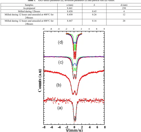
Figure 2. Mössbauer spectra for T=300K.(a): as-prepared(b): 12 hours milled sample.(c): 12 hours milled sample annealed at 400°C during 24h.(d): 12
hours milled sample annealed at 600°C during 24h
Due to the small value of the lattice parameter for as-prepared sample in comparison with those corresponding to other (milled during 12 hours, milled during 12 hours and annealed during 24hours at 400°C and 600°C), one concludes the existence of a mixture of normal and inverse structure in the milled as well as in the milled and annealed samples. The lattice parameter as well as the inversion parameter increase initially after 12 hours of milling process then decrease with increasing of annealing temperature. The milling process favours a redistribution of Fe3+ and Zn2+ in
interstitial sites. The substituting of some Zn2+cations with
larger ionic radius instead of Fe3+ cations in octahedral sites
outcomes of the increasing in the lattice parameter value as reported in the literature [26, 27] and the decreasing in the inversion parameter. Calcining at higher temperature causes grain growth and also provides sufficient energy to allow reordering Zn2+ ions into the tetrahedral sites. Consequently,
the lattice parameter (inversion parameter) decreases (increases). To close this section, it is important to emphasize that in contrast to that of nanostructured fluoride powders where a second component assigned the disordered grain boundaries should be introduced [28], here only single component allows us to describe X-ray pattern for the ball-milled samples as shown in figure1. In addition, no significant contribution of grain boundaries (<15at. %) is depicted from X-ray diffractograms.
4. Mössbauer Studies
Room temperature 57Fe Mössbauer spectra recorded for
various samples are illustrated in figure 2.
from iron cation occupying the octahedral sites. After milling process, the ball milled samples showed spontaneous magnetization. This anomalous behaviour has been already cited in some works: A higher magnetization obtained at room temperature in nanosize ZnFe2O4 as reported by
Kamiyama et al. [12]. Moreover, Upadhyay et al. [29] showed the existence of anomalous magnetization behaviour for nanocrystalline zinc ferrite with average grain size ≈ 4 nm measured using Squid magnetometer in the temperature range 5-300K. This anomalous behaviour can be explained by the reduction of grains size which favours the configuration where some cations of Zn2+ prefer to occupy B
sites with a simultaneous occupancy of A- sites by Fe3+
cations making the system partially inverted [17]. With annealing process, the spontaneous magnetization disappears and the corresponding spectra obtained for milled and calcined samples at 400°C and 600°C show quadrupoles doubletsin agreement with the results described by Aljuraide et al. for ZnFe2O4 nanoparticles with grains size of 12-37 nm
[32]. In addition, the vanishing spontaneous magnetization may be caused by the grains size magnitude estimated around 12-20nm for our samples. Let us note that for smaller grains size (6-7nm) the existence of spontaneous magnetization is not detected at room temperature and the inversion degree measured by means of Fe-extended X-ray absorption fine structure (EXAFS) studies is estimated around 80% (40%) for grains size of 4 nm (6 nm). It is well known that for grains size equal to 7 nm and more, the inversion degree is negligible thus showing the existence of normal spinel structure [29]. This is the situation obtained for our samples where the grains size value are given to be superior to 7 nm thus leading to the low value of inversion degree (see table 1) and reflecting the disappearance of the magnetization after annealing process. This means that annealing process is opposite with the milling process where Zn2+ cations prefer to return in tetrahedral site and
simultaneously Fe3+ cations migrate to octahedral sites. The
isomer shift and quadrupole splitting values are characteristic of Fe3+ charge state. There is no evidence for
the presence of Fe2+ charge state due to oxygen vacancies
[image:4.595.60.297.622.736.2]produced by milling process as previously described in the literature [30]. The hyperfine parameters values are listed in table 2.
Table 2. Isomer shift (IS), quadrupole splitting (∆) and hyperfine magnetic
field <Bhyp.> values for spectra recorded at 300K.
Samples (mm /s) IS (mm /s) ∆ <BHyp>
(T)
As-prepared 0.33 0.34
Milled during 12hours 0.31 12.0
Milled during 12 hours and annealed at 400°C
for 24hours
0.33
0.32 0.49 0.47 Milled during 12 hours
and annealed at 600°C for 24hours
0.31
0.32 0.38 0.39
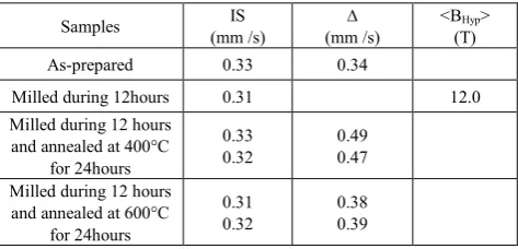
For 77K the obtained spectra are shown in figure 3.
Figure3. 77K Mössbauer spectra (a): as-prepared (b): 12 hours milled
sample. (c): 12 hours milled sample annealed at 400°C during 24h. (d): 12 hours milled sample annealed at 600°C during 24h
The spectrum of as-prepared sample shows a quadrupole doublet which can be refined using ∆ = 0.35 mm/s and IS = 0.42 mm/s. The 12 hours milled sample spectrum which is adjusted using hyperfine field distribution shows a magnetic sextet typical of the hyperfine splitting for iron. This result confirms the existence of cationic inversion produced by the milling process. Nevertheless, the sample calcined at 400°C consists of a magnetically ordered component and a quadupole doublet which is significant of the presence of superparamagnetic behaviour of small particles. The measured average time relaxation values are estimated around 1,3.10-9 s (2.10-9 s) for the annealed samples 400°C
(600°C). The average relaxation time is calculated using Neel-Brown formula concerning superparamagnetism relaxation time (ζ) and may be expressed as in reference [33]:
)
/
exp(
0
KV
kT
τ
τ
=
,where T characterizes the temperature; k: denotes the Boltzmann constant; K is the magnetic anisotropy constant; V measures the volume of single domain and
τ
0 is run over the order of 10-12 10-9 s. Defining the blocking temperature(TB) as a transition temperature between superparamagnetic
slow relaxation) it is then possible to consider two magnetic behaviour regions. Below TB, the relaxation time is greater
than measurement time
τ
m (2. 10-8s). The magnetization ofthe grains is considered as "blocked" in the time scale of measurements thus leading to the observation of magnetic sextet. Above TB, the grains magnetization will return
numerous times during the measurements so that the measured zero magnetization appear. As a result, the collapse followed by the complete vanishing of the magnetic sextet and then the apparition of a quadrupole splitting doublet are depicted.
The magnetic component disappears with the increasing annealing temperature and the quadrupole doublet remains the only dominant component. Here the corresponding refinement is realized using two quadrupole doublets: the first is attributed to Fe3+ cations occupying octahedral sites
and the additional doublet may be assigned to Fe3+ ions from
[image:5.595.62.298.419.519.2]the A-sites. The obtained relative intensity of the magnetic component is found to be 0%, 100 %, 68 % and 0 % for the as–prepared sample, milled during 12 hours, milled during 12 hours and annealed at 400°C and 600°C, respectively. This is significant with the increase (decrease) of inversion parameter with milling (annealing) process. The corresponding numerical values of Mössbauer hyperfine parameters obtained from the adjustment model are resumed in table 3.
Table 3. Hyperfine parameters and various relative proportions (%) values
for spectra recorded at 77K.
Samples (mm /s) IS (mm /s) ∆ <BHyp>
(T) Prop. (%)
As-prepared 0.42 0.35 0 100
Milled during 12hours 0.42 41.5 100
Milled during 12 hours and
annealed at 400°C for 24hours 0.41 0.42 0.37 26.8 32 68 Milled during 12 hours and
annealed at 600°C for 24hours 0.43 0.42 0.36 0.41 72 28
5. Conclusion
Nanostructured zinc ferrite was milled during 12 hours at high energy. Portion of the powder were annealed during 24 hours at 400°C and 600°C. XRD analysis showed that milled as well as milled and annealed samples present a cubic spinel structure. Based on the grain size value of the as-prepared sample the average grain size for the milled sample is found to be decreases after 12 hours of milling and increases with increasing annealing temperature. The milling process favors disorder whereas the annealing process causes grains growth and improves crystallinity of samples. The increasing inversion parameter with the decreasing grains size is depicted. Magnetic properties studies are carried out using 57Fe Mössbauer spectrometry. At room temperature,
12 hours milled sample without annealing displays a spontaneous magnetization while the annealed samples show a nonmagnetic state. This anomalous behaviour may-be
explained by the presence of cationic redistribution induced by the milling effect. A careful inspection of the Mössbauer spectra recorded for 77K confirms the existence of this cationic distribution which occurs after milling and the presence of a superparamagnetic state configuration for the annealed samples is revealed. Furthermore, the investigations carried out in this work clarify both the role and influence of mechanical grinding as well as thermal treatment on microstructure and magnetic properties of ferrites zinc nanostructures. Thus, it was established the mechanical milling and annealing react opposite leading to new magnetic and structural properties.
Further studies are in progress to analyze the magnetic behaviours of milled as well as milled and annealed zinc ferrite nanostructures by focusing on the realized zero-field and In-field Mössbauer spectra in order to confirm the ferrimagnetic character of the samples, to determine the relative proportions of different components and also to explore the orientation of the magnetic moments in the two sites which can generate the presence of spin canting effect.
Acknowledgements
The first author (O. Ould Fella) would like to thank the staff of the IMMM (UMR CNRS 6283 – Université du Maine (France)) particularly Dr. J.M. Greneche and Nirina Randrianantoandro during their stay at the university and for encouragements and stimulating discussions.
REFERENCES
[1] Sun, S., Murray, C. B., (1999) ‘Synthesis of monodisperse cobalt nanocrystals and their assembly into magnetic superlattices ’,J. Appl. Phys., Vol. 85, pp. 4325-4331. [2] Villain, J, (1979) ‘Insulating Spin Glasses ’,Z. Phys. B., Vol.
33, pp. 31-42.
[3] Pavljukhin, Yu. T, Medikov, Ya. Ya, Boldyrev, V. V., (1983) ‘Magnetic and chemical properties of mechanically activated zinc and nickel ferrites’, Mater. Res. Bull., Vol. 18, pp. 1317-1327.
[4] Smith, S. and Wijn, H P. (1961) Ferrites, ed. Philips Library, Amsterdam.
[5] Schiessel, W., Potzel, W., Karzel, H., Steiner, M., Kalvius, G. M., Martin, A., Krause, M. K., Halevy, I., Gal, J., Shäfer, W., Will, G., Hillberg, M. and Wäppling, R. (1996) ‘ Magnetic properties of the ZnFe2O4 spinel’,Phys. Rev. B., Vol. 53, pp.
9143-9152.
[6] Sepelak, V., Tkacova, K., Rycov, A I., (1993) ‘Rietveld analysis of mechanically activated powdered zinc ferrite’, Crystal. Res .Technol. Issue1, Vol. 28, pp. 53-56.
[7] Chinnasamy, C. N., Narayanasamy, A., Ponpandian, N., Chattopadhyay, K., (2001) ‘The influence of Fe3+ ions at
[8] Stewart, S. J., Al-Omari, I, A, Sives, F. R., Widatallah, H M., (2010) ‘Non-equilibrium cation influence on the Néel temperature in ZnFe2O4’, Journal of Alloys and Compounds,
Vol. 495, pp. 506-508.
[9] Jeyadevan, B., Tohji, K. and Nakatsuka, K. (1994) ‘Structure analysis of coprecipitated ZnFe2O4 by extended X-ray
absorption fine structure ’, J. Appl. Phys., Vol. 76, pp. 6325-6327.
[10] Ammar, S., Jouini, N. Fiévet, F., Stephan, O., Marhic, C., Richard. M., Villain. F., Cartier, Ch., Brice. S., Sainctavit. Ph., (2004) ‘Influence of the synthesis parameters on the cationic distribution of ZnFe2O4 nanoparticles obtained by forced
hydrolysis in a polyol medium ’,Journal of Non-Crystalline Solids, Vol. 345-346, pp. 658-662.
[11] Stewart, S. J., Figueroa, S. J. A., Ramallo Lopez, J. M., Marchetti, S. G., Bengoa, J. F., Prado, R. J., Requejo, F. G., (2007) ‘ Cationic exchange in nanosized ZnFe2O4 spinel
revealed by experimental and simulated near-edge absorption structure’,Phys. Rev. B., Vol. 75, pp. 073408-073411. [12] Kamiyama, T., Haneda, K., Sato, T., Ikeda, S. Asano, H.,
(1992) ‘Cation distribution in ZnFe2O4 fine studied by
neutron powder diffraction ’,Solid. State. Commun. 7, Vol. 81, pp. 563-566.
[13] Goya, G. F., Rechenberg, H.R., Chen, M. and Yelon, W. B., (2000) ‘Magnetic irreversibility in ultrafine ZnFe2O4
particles ’,J. Appl. Phys., Vol. 87, pp. 8005-8007.
[14] Chinnasamy, C. N., Narayanasamy, A., Ponpondian, N., Chattopadhyay, K., Guerault, H. and Grenèche, J. M. (2000) ‘Magnetic properties of nanostructured ferrimagnetic zinc ferrite’, J. Phys: Condens. Matter, Vol. 12, pp. 7795-7805. [15] Ammar, S., Jouini, N. Fiévet, F., Beji, Z., Smiri, L., Moliné,
P., Danot, Michel. and Grenèche, J. M. (2006) ‘Magnetic properties of zinc ferrite nanoparticles synthesized by hydrolysis in a polyol medium’, J. Phys.: Condens. Matter,
Vol. 18, pp. 9055-9069.
[16] Goya, G. F. and Leite, E. R. (2003) ‘Ferrimagnetism and spin canting of Zn57Fe
2O4 nanoparticles embedded in ZnO
matrix ’, J. Phys: Condens. Matter, Vol. 15, pp. 641-645. [17] Hamdeh, H. H., Ho, J. C., Oliver, S. A., Willey, R. J., Oliveri,
G. and Busca. (1997) ‘Magnetic properties of partially-inverted zinc ferrite aerogel powders’,J. Appl. Phys., Vol. 81, pp. 1851-1857.
[18] Oliver, S. A., Harris, V., Hamdeh, H. H.and Ho, J. C. (2000) ‘Large zinc cation occupancy of octahedral sites in mechanically activated zinc ferrite powder’, J. Appl. Phys. Lett., Vol. 76, pp. 2761-2763.
[19] Bean, C. P., Livingston, J. D., (1959). ‘Superparamagnetism ’, J. Appl. Phys., Vol. 30, 120S-129S.
[20] Martinez, B., Obrados, X., Balcells, Li., Rouanet, A., Moty, C., (1998) ‘ Low temperature spin-glass transition in γ-Fe2O3
nanoparticles ’,Phys. Rev. Lett., Vol. 80, pp. 181-184. [21] Sepelak, V., Steinike, U., Uecker, D., Wiβmann, S., Becker,
K., (1998)’ Structural disorder in mecanosynthesised zinc ferrite’, J. Solid State Chem., Vol. 135, pp. 52-58.
[22] Nachbaur, V., Tauvel, G., Verdier, T,. Jean, M., Juraszek, J., Houvet, D., (2009)’ Mecanosynthesis of partially inverted zinc ferrite’, Journal of Alloys and Compounds, Vol. 473, pp.
303-307.
[23] Ikenaga, T., Ohgaito, Y., Matsushima, H., Suzuki, T., (2004)’ Preparation of zinc ferrite in the presence of carbon material and its application to hot-gas cleaning’ Fuel, Vol.83, pp. 661-669.
[24] Grenèche, J. M., ‘‘Programme Mossfit“ Université Du Maine (Le Mans-France).
[25] Lutterotti, L. and Scardi, P. (1990) ‘Simultaneous structure and size-strain refinement by the Rietveld method ’,J. Appl. Cryst., Vol. 23, pp. 246-252.
[26] Raeisi Shahraki, R., Ebrahimi, M., Seyyed Ebrahimi, S.A., Masouspanah, S. M., (2012) ’ Structural characterization and magnetic properties of superparamagnetic zinc ferrite nanoparticles synthesized by the coprecipitation method’, J. Magn. Magn. Mater., Vol. 324, pp.3762-3765.
[27] Anantharaman, A., Jaghatersan. S., Malini. K, Sibdhu. S., Narayanasamy, A., Chinnasamy, C., Jacobs. J., Reijne. S., Seshan. K., Smits. R., (1998)’ On the magnetic properties of ultra-fine zinc ferrites’ J. Magn. Magn. Mater., Vol.189, pp.83-88.
[28] Guerault, H., Grenèche, J.M., (2000) ‘Microstructural modeling of nanostructured fluoride powders prepared by mechanical milling ’, J. Phys.: Condes. Matter, Vol. 12, pp. 4791-4798.
[29] Upadhyay, C., Verma, H. C., Sathe, V., Pinpale, A. V., (2007)’ effect of size and synthesis route on the magnetic properties of chemically prepared nanosized ZnFe2O4’ J. Magn. Magn.
Mater., Vol. 312, pp.271-88.
[30] Goya, G. F. and Rechenberg, H. R. (1999) ‘Ionic disorder and Néel temperature in Zn Fe2O4 nanoparticles’, J. Magn.
Magn.Mater., Vol. 196-197, pp. 191-192.
[31] Wang, L., Zhou, Q. and Li, F. (2004) ‘Ionic disorder and Yaffet-Kittel angle in nanoparticles of ZnFe2O4 prepared by
Sol-gel method’, Phys. Stat. Sol., Vol. (b) 241, pp. 377-341. [32] Aljuraide, N. I., Mousa, M. A. A., Mostafa, N. Y.,
El-Shobaky, G. A., Hamdeh, H. H. and Ahmed. M. A. (2012) ‘Microstructure analysis of zinc ferrite nanoparticles by means of X-ray powder diffraction and Mössbauer spectroscopy’, Int. J. Nanoparticles, Vol. 5, No. 1, pp. 56-63. [33] Neel, L. (1949) ‘Theory of magnetic after effect of