Neuroscience Institute Dissertations Neuroscience Institute
4-19-2010
Functional Substrates of Social Odor Processing within the
Functional Substrates of Social Odor Processing within the
Corticomedial Amygdala: Implications for Reproductive Behavior
Corticomedial Amygdala: Implications for Reproductive Behavior
in Male Syrian Hamsters
in Male Syrian Hamsters
Pamela Mary Maras
Georgia State University
Follow this and additional works at: https://scholarworks.gsu.edu/neurosci_diss
Part of the Neuroscience and Neurobiology Commons
Recommended Citation Recommended Citation
Maras, Pamela Mary, "Functional Substrates of Social Odor Processing within the Corticomedial
Amygdala: Implications for Reproductive Behavior in Male Syrian Hamsters." Dissertation, Georgia State University, 2010.
https://scholarworks.gsu.edu/neurosci_diss/1
This Dissertation is brought to you for free and open access by the Neuroscience Institute at ScholarWorks @ Georgia State University. It has been accepted for inclusion in Neuroscience Institute Dissertations by an authorized administrator of ScholarWorks @ Georgia State University. For more information, please contact
FUNCTIONAL SUBSTRATES OF SOCIAL ODOR PROCESSING WITHIN THE
CORTICOMEDIAL AMYGDALA: IMPLICATIONS FOR REPRODUCTIVE BEHAVIOR IN
MALE SYRIAN HAMSTERS
by
PAMELA M. MARAS
Under the direction of Aras Petrulis
ABSTRACT
Adaptive reproductive behavior requires the ability to recognize and approach possible
mating partners in the environment. Syrian hamsters (Mesocricetus auratus) provide a useful
animal model by which to study the neural processing of sexual signals, as mate recognition in
this species relies almost exclusively on the perception of social odors. In the laboratory, male
hamsters prefer to investigate female odors compared to male odors, and this opposite-sex odor
preference provides a sensitive measure of the underlying neural processing of sexual stimuli. In
addition to chemosensory cues, reproductive behavior in hamsters also requires sufficient levels
chemosensory and hormone signals are processed within an interconnected network of ventral
forebrain nuclei, and within this network, the posteromedial cortical amygdala (PMCo) and
medial amygdala (MA) are the only nuclei that both receive substantial chemosensory input and
are also highly sensitive to steroid hormones. Although a large body of evidence suggests that
the MA is critical for generating attraction to sexual odors, the specific role of the PMCo in
regulating odor-guided aspects of male reproductive behavior has never been directly tested.
Furthermore, detailed analyses of the MA suggest that separate, but interconnected sub-regions
within this nucleus process odors differently. Specifically, the anterior MA (MeA) receives the
majority of chemosensory input and responds to a variety of social odors, whereas the
posterodorsal MA (MePD) receives less chemosensory input but contains the vast majority of
steroid receptors. In order to further elucidate how the PMCo and/or MA process sexual odors,
this dissertation addressed the following research questions: (1) Is the PMCo required for the
expression of either opposite-sex odor preferences or male copulatory behavior? (2) Are
functional interactions between MeA and MePD required for the expression of opposite-sex odor
preferences? (3) How do MeA and MePD regulate odor responses within the MePD and MeA,
respectively? (4) Are odor and/or hormone cues conveyed directly between MeA and MePD?
Together, these experiments provide a comprehensive analysis of the functional and
neuroanatomical substrates by which the brain processes sexual odors and generates appropriate
behavioral responses to these stimuli.
FUNCTIONAL SUBSTRATES OF SOCIAL ODOR PROCESSING WITHIN THE
CORTICOMEDIAL AMYGDALA: IMPLICATIONS FOR REPRODUCTIVE BEHAVIOR IN
MALE SYRIAN HAMSTERS
by
PAMELA M. MARAS
A Dissertation Submitted in Partial Fulfillment of the Requirements for the Degree of
Doctor of Philosophy
in the College of Arts and Sciences
Georgia State University
Copyright by
Pamela M. Maras
FUNCTIONAL SUBSTRATES OF SOCIAL ODOR PROCESSING WITHIN THE
CORTICOMEDIAL AMYGDALA: IMPLICATIONS FOR REPRODUCTIVE BEHAVIOR IN
MALE SYRIAN HAMSTERS
by
PAMELA M. MARAS
Committee Chair: Aras Petrulis
Committee: Timothy Bartness
Anne Murphy
Walter Wilczynski
Electronic Version Approved:
Office of Graduate Studies
College of Arts and Sciences
Georgia State University
DEDICATION
I would like to dedicate this Dissertation to Ashley. I could never have done this without you.
Thank you for taking the stress out of my life and making me so happy every day. You know I
ACKNOWLEDGEMENTS
I would like to thank the many people who have provided guidance and support throughout my
graduate education.
First and foremost is my mentor, Aras Petrulis, who has provided an extremely supportive
environment for me to develop as a scientist, and from whom I have learned so much about
creating a successful and happy lab. I have thoroughly enjoyed working, thinking, and writing
with Aras, and I am especially proud to be his first PhD student.
I would also like to acknowledge the other fantastic members of my dissertation committee, Tim
Bartness, Anne Murphy, and Walt Wilczynski. Deepest thanks to Tim Bartness for his
overwhelming support. His faith in my abilities continues to motivate me. Anne Murphy has
continually gone out of her way not only to help me with certain techniques, but also to provide
career advice and support. She provides an extra mentor to so many students, and we are all very
lucky to have her on our side. I would like to thank Walt Wilczynski for his service during both
my qualifying exam and dissertation process. He has provided a unique point of view to my
work and has helped me to conceptualize social communication in a broader context.
I would also like to thank my undergraduate faculty mentors from Florida State University, Dr.
James Smith and Dr. Frank Johnson, who are responsible for getting me excited about
neuroscience. I am very grateful to them.
I would like to thank the members of the Petrulis lab: Laura Been, Marc Badura, Dr. Christine
Lewis, Luis Martinez, and Fidel. Thank you all for the friendship and support, and for putting up
with my incessant need to organize. We have the best darn lab environment in the history of the
also wish to acknowledge the wonderful undergraduate students that I have had the pleasure of
mentoring, in particular: Don Bearden, Larry Burrell, Laurie Hale, Nina King, Anaam
Mohammed, and Victoria Smith. I hope you learned something.
I would not have made it through this crazy process without the support of some great friends,
including: Marc Badura, Laura Been, Kelli Duncan, Desiree Krebs-Kraft, Stacie Lin-Taylor,
Varenka Lorenzi, Joe Normandin, and Victoria Smith. Laura, you are the best mini-me ever and
so much more. I promise to keep in touch.
Finally, I would like to acknowledge the love and support provided by my parents and family.
Mom and Dad, thank you for always cheering me on and being so proud, even though none of
your friends understand what it is I actually do. Barb and Fred, thank you for welcoming me into
your family and giving me so much encouragement. Jerry, Matt, Sarah, and Beckie, thank you
TABLE OF CONTENTS
ACKNOWLEDGEMENTS V.
LIST OF TABLES X.
LIST OF FIGURES XI.
CHAPTER 1: Introduction 1
Overview 2
Chemosensory regulation of rodent reproductive behavior 3
Chemosensory-hormone interactions 5
Candidate sites for chemosensory-hormone integration 6
Functionally distinct sub-regions within MA 9
Goals of Dissertation 12
Chapter 1 Figures 13
CHAPTER 2: The Posteromedial Cortical Amygdala Regulates Copulatory
Behavior, but not Sexual Odor Preference, in Male Syrian Hamsters (Mesocricetus auratus)
16
Abstract 17
Introduction 18
Materials and Methods 20
Results 28
Discussion 32
Acknowledgements 39
Chapter 2 Tables 40
CHAPTER 3: Lesions that Functionally Disconnect the Anterior and Posterodorsal
Sub-regions of the Medial Amygdala Eliminate Opposite-sex Odor Preference in
Male Syrian Hamsters (Mesocricetus auratus)
49
Abstract 50
Introduction 51
Materials and Methods 54
Results 61
Discussion 64
Acknowledgements 72
Chapter 3 Tables 73
Chapter 4 Figures 75
CHAPTER 4: The Anterior Medial Amygdala Transmits Social Odor Information to
the Posterior Medial Amygdala and Related Forebrain Nuclei
78
Abstract 79
Introduction 80
Materials and Methods 81
Results: Experiment 1 90
Results: Experiment 2 93
Discussion 95
Acknowledgements 103
Chapter 4 Tables 104
Chapter 4 Figures 106
Amygdala: Integration of Odor and Hormone Signals
Abstract 118
Introduction 119
Materials and Methods 121
Results 127
Discussion 131
Acknowledgements 138
Chapter 5 Tables 139
Chapter 5 Figures 142
CHAPTER 6: General Discussion 148
Overview 149
Differential roles for PMCo and MA 150
Integration of chemosensory and hormone cues within MA 152
An extended circuit for generating attraction to sexual odors 156
The role of the corticomedial amygdala in regulating other social behaviors 158
Structure and function of the amygdala in other species 165
Summary 170
Chapter 6 Figures 172
REFERENCES 175
LIST OF TABLES
Table 2.1 Surgical coordinates for electrolytic lesions of the PMCo 40
Table 2.2 Summary of behavioral measures from Clean Y-maze tests 41
Table 2.3 Summary of derived behavioral measures from male copulatory tests 42
Table 2.4 Comparison of copulatory behavior between SHAM males and males with
damage primarily outside the PMCo (Non-PMCoX)
43
Table 3.1 Coordinates for electrolytic lesions of MeA or MePD 73
Table 3.2 Summary of behavioral measures from Clean tests in the 3-choice
apparatus
74
Table 4.1 Coordinates for lesion surgery in Experiments 1 and 2 104
Table 4.2 Patterns of Fos expression within ventral forebrain nuclei in response to
social odors
105
Table 5.1 Total numbers of CTB+, Fos+, and Fos/CTB+ cells (per mm2) within MeA
and MePD
139
Table 5.2 Total numbers of Fos+ cells (per mm2) within MeA and MePD observed
ipsilateral or contralateral to CTB injection.
140
Table 5.3 Total numbers of CTB+, AR+, and AR/CTB+ cells (per mm2) within MeA
and MePD
LIST OF FIGURES
Figure 1.1 Flow of chemosensory information through the corticomedial amygdala 13
Figure 1.2 MeA and MePD regulate distinct aspects of odor preference 14
Figure 1.3 Proposed model of MeA-MePD interaction during social odor processing 15
Figure 2.1 Timeline of surgeries and behavioral testing 44
Figure 2.2 Lesion reconstruction 45
Figure 2.3 Behavioral results from sexual odor preference and discrimination tests 46
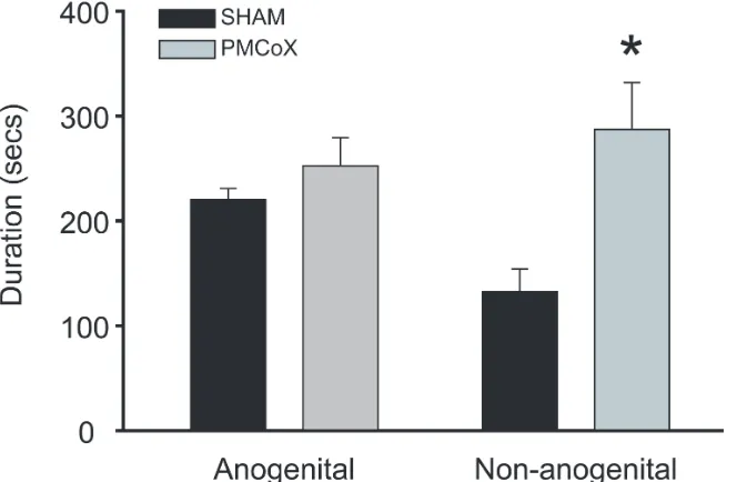
Figure 2.4 Total durations of investigation behavior during the male copulatory
behavior test
47
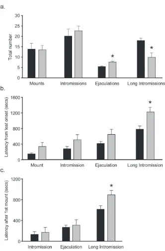
Figure 2.5 Mating events during the male copulatory behavior test 48
Figure 3.1 Lesion reconstruction 75
Figure 3.2 Summary of results from Odor Preference tests 76
Figure 3.3 Summary of results from Odor Discrimination tests 77
Figure 4.1 Counting domains for analysis of Fos-immunoreactivity 106
Figure 4.2 Lesion reconstruction for Experiment 1 107
Figure 4.3 Summary of effects of MeA or MePD lesions on Fos-ir throughout the
MA
108
Figure 4.4 Representative photomicrographs of Fos-ir within MeP 109
Figure 4.5 Representative photomicrographs of Fos-ir within MeA 110
Figure 4.6 Summary of effects of MeA or MePD lesions on Fos-ir within ventral
forebrain nuclei
111
Figure 4.7 Lesion reconstruction for Experiment 2 113
Figure 4.9 Representative photomicrographs of Fos-ir within MeP 115
Figure 4.10 Summary of effects of excitotoxic MeA lesions on NeuN-ir 116
Figure 5.1 Counting domains for analysis of CTB, Fos, and AR 142
Figure 5.2 Verification of CTB injections 143
Figure 5.3 Co-localization of CTB and Fos 144
Figure 5.4 Comparison of Fos/CTB double-labeling between males exposed to female
odors or male odors
145
Figure 5.5 Co-localization of CTB and AR 146
Figure 6.1 Updated model of MeA-MePD interaction during social odor processing 172
Figure 6.2 Proposed model of the neural circuit regulating behavioral responses to
different categories of social odors
173
Figure 6.3 Summary of the major afferent and efferent connections of the medial
amygdala
Overview
Adaptive reproductive behavior requires integrating external signals about available
mates in the environment with internal signals about an individual’s own reproductive state
(Wingfield et al., 1997). Indeed, reproductive fitness is optimal when the expression of
reproductive behaviors is restricted toward appropriate mating partners (ie. conspecific,
opposite-sex) and also coincides with the maximum fecundity of the individual (Wingfield et al., 1997;
Gowaty & Hubbell, 2009). The attempt to mate when these conditions are not met would not
only be a costly waste of energetic resources, but may even have severe consequences for an
individual’s immediate survival (Groning & Hochkirch, 2008; Gowaty & Hubbell, 2009). In
rodents (Brennan, 2004; Keverne, 2004), as well as many other species (Johnston, 1983;
Rodriguez, 2004), mate recognition relies heavily on the perception of chemical signals released
by conspecifics, whereas reproductive state is tightly linked to circulating levels of gonadal
steroid hormones (Gomes & VanDemark, 1974). The mechanisms by which the brain integrates
these external and internal signals remain poorly understood, although the expression of
reproductive behavior in rodents involves an extended network of ventral forebrain nuclei that
process chemosensory and/or steroid hormone cues (Wood, 1997; 1998). Within this circuit, the
posteromedial cortical amygdala (PMCo) and medial amygdala (MA) are the only nuclei that
both receive substantial direct chemosensory input and are also highly sensitive to steroid
hormones (Wood, 1997; 1998), suggesting a unique role for these nuclei in the integration of
chemosensory and hormone signals. The current review will therefore focus on the roles of the
PMCo and MA in processing sexual odors and generating reproductive behavior in rodents, with
Chemosensory regulation of rodent reproductive behavior
As in many mammalian species (Johnston, 1983; Rodriguez, 2004), social
communication in rodents involves the active release and detection of chemical signals
(Johnston, 1990; Brennan, 2004; Keverne, 2004). Social odors convey a wide range of
information about the sender, including species, sex, kinship, and even social or reproductive
status (Johnston, 1983; Rich & Hurst, 1999; Hurst & Beynon, 2004; Brennan & Kendrick, 2006;
Arakawa et al., 2008). In the contexts of reproduction, females of many rodent species advertise
their sexual receptivity through active scent-marking behaviors, and these sexual odors are
highly attractive to conspecific males (Johnston, 1983; Coquelin, 1992; Matochik et al., 1992).
For example, female Syrian hamsters display a highly stereotyped vaginal marking behavior,
during which the female lowers and thrusts her pelvis, depositing vaginal secretion onto the
substrate (Johnston, 1979; Been & Petrulis, 2007). These vaginal secretions serve as a potent
chemical attractor for male hamsters and are sufficient to stimulate the expression of male
copulatory behaviors (Johnston, 1974; 1975; Johnston & Kwan, 1984; Petrulis & Johnston,
1995). Male hamsters are attracted to other components of female odors as well (Johnston, 1986)
and display robust preferences to approach and investigate female odors compared to male odors,
referred to as an opposite-sex odor preference (Johnston, 1981; Steel, 1982; Maras & Petrulis,
2006; Ballard & Wood, 2007).
In rodents, social odors are processed by two, anatomically distinct chemosensory
systems, the main and accessory olfactory systems (Meredith, 1991; Restrepo et al., 2004).
Sensory receptors of the main olfactory system, located in the main olfactory epithelium,
respond best to low molecular weight, volatile components of social odors (Meredith, 1991;
system, located in the vomeronasal organ, are thought to process high molecular weight,
non-volatile components of social odors (Meredith, 1991; Keverne, 1999; Halpern &
Martinez-Marcos, 2003; Rodriguez, 2004). Together, these chemosensory systems regulate the expression
of most rodent social behaviors, including male reproductive behaviors (Hull et al., 2002;
Keverne, 2004; Restrepo et al., 2004; Keller et al., 2009).
In particular, the expression of reproductive behavior in male Syrian hamsters relies
heavily on chemosensory processing. Either olfactory bulbectomy (Murphy & Schneider, 1970)
or simultaneous deafferentation of the main and accessory olfactory systems (Powers & Winans,
1975) completely eliminates the expression of copulatory behavior in male hamsters. In contrast
to rats (Larsson, 1975), previous sexual experience does not mitigate these deficits in hamsters
(Murphy & Schneider, 1970). Chemosensory processing is also required for a male hamster’s
attraction to approach and investigate female odors (Powers et al., 1979), as well as the
pre-copulatory anogenital investigation of a receptive female (Murphy & Schneider, 1970; Devor &
Murphy, 1973; Powers & Winans, 1975). Thus, both the appetitive and consummatory aspects of
male reproductive behavior in hamsters require on the chemosensory detection of sexual odors.
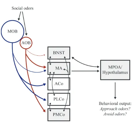
Social odors are processed initially within the main and accessory olfactory bulbs (MOB
and AOB, respectively), which send projections to the ventral forebrain primarily via the lateral
olfactory tract (Figure 1.1) (Kratskin & Belluzzi, 2003; Lin et al., 2005). MOB efferents travel to
a broad network of secondary olfactory nuclei, including the olfactory tubercle, piriform and
entorhinal cortices, anterior and posterolateral cortical amygdala, and MA (Scalia & Winans,
1975; Cleland & Linster, 2003). In contrast to these wide projections, the AOB sends limited
projections specifically to MA and PMCo, as well as some modest projections to the bed nucleus
information reaches downstream forebrain nuclei, such as the medial preoptic area (MPOA),
either directly from MA or indirectly through the BNST (Wood, 1997). Disruption of the flow of
chemosensory information at any point in this circuit disrupts the expression of male
reproductive behavior in many rodent species, including hamsters (Murphy & Schneider, 1970;
Devor, 1973; Macrides et al., 1976; Lehman et al., 1980; Lehman & Winans, 1982; Lehman et
al., 1983; Powers et al., 1987).
Chemosensory-hormone interactions
In addition to chemosensory cues, male reproductive behavior is also regulated by
internal signals of reproductive state via changes in circulating levels of gonadal steroid
hormones (Beyer et al., 1976; Morin & Zucker, 1978; Hull et al., 2002). In hamsters,
testosterone and its primary metabolites, estradiol and dihydrotestosterone, are critical not only
for the expression of male copulatory behavior (Morin & Zucker, 1978; Powers et al., 1985), but
also for male’s attraction to investigate female odors (Steel, 1982; Powers & Bergondy, 1983;
Powers et al., 1985; Petrulis & Johnston, 1995). Given the importance of both chemosensory and
hormone cues for the expression of reproductive behavior in rodents, it is perhaps not surprising
that these signals are processed by largely overlapping neural circuits. In fact, dense populations
of steroid receptor-containing neurons are found within many of the ventral forebrain nuclei that
receive either direct or indirect chemosensory input (Wood, 1997; Figure 1.1). Specifically,
androgen receptor (AR) and estrogen receptor (ER) containing neurons are found within the MA,
PMCo, BNST, and MPOA (Doherty & Sheridan, 1981; Simerly et al., 1990; Wood et al., 1992;
Li et al., 1993). This overlap of chemosensory and hormone-sensitive nuclei highlights the
In addition to these anatomical data, there is substantial evidence for functional
interactions between chemosensory and steroid hormones systems in male hamsters. Exposure to
female odors rapidly increases circulating levels of testosterone in male hamsters (Macrides et
al., 1974; Pfeiffer & Johnston, 1992). This increase in testosterone relies on chemosensory
processing via the vomeronasal organ (Pfeiffer & Johnston, 1994), and likely involves the
activation of GnRH-expressing cells within the MPOA (Meredith & Fernandez-Fewell, 1994).
Furthermore, gonadal hormones are critical for male hamster’s attraction to female odors;
castrated male hamsters display low levels of investigation of female odors, and testosterone
treatment fully restores this behavior (Steel, 1982; Powers & Bergondy, 1983; Powers et al.,
1985; Petrulis & Johnston, 1995). These data suggest that chemosensory processing can
modulate gonadal physiology, and vice versa. Within the brain, hormones appear to act within
specific forebrain nuclei to generate attraction to sexual odors, as unilateral testosterone implants
into either the MPOA/BNST or MA increase anogenital investigation in castrated male hamsters
(Wada et al., 1990; Wood & Newman, 1995c; Wood, 1996; Wood & Williams, 2001). Unilateral
olfactory bulbectomy ipsilateral to the steroid implant, however, prevents this increase (Wood &
Newman, 1995b; Wood & Coolen, 1997), suggesting that attraction to sexual odors requires the
neural integration of chemosensory and hormone cues. Although the mechanisms of
chemosensory-hormone integration remain unclear, they likely involve brain areas that process
both types of information (Wood & Coolen, 1997).
Candidate sites for chemosensory-hormone integration
Within the ventral forebrain, the PMCo and MA are the only nuclei that both receive
substantial chemosensory input and are also highly sensitive to gonadal steroid hormones
constitute the main components of the “vomeronasal amygdala” (Scalia & Winans, 1975;
Kevetter & Winans, 1981a; Pro-Sistiaga et al., 2007). The MA receives direct projections from
the MOB (Scalia & Winans, 1975; Pro-Sistiaga et al., 2007), and both nuclei share extensive
indirect connections with secondary nuclei of the main olfactory system, including the anterior
cortical and posterolateral cortical nuclei of the amygdala (Kevetter & Winans, 1981b). In male
hamsters, neurons within PMCo and MA display increases in Fos expression following exposure
to female odors (Fiber et al., 1993; Kollack-Walker & Newman, 1997; Fewell & Meredith,
2002), indicating that these areas are activated during processing of sexually relevant odors.
Finally, both PMCo and MA contain dense populations of steroid receptor-containing neurons
(Doherty & Sheridan, 1981; Simerly et al., 1990; Wood et al., 1992), and many of these
steroid-sensitive neurons are activated during mating (Wood & Newman, 1993). These data indicate that
the PMCo and/or MA mediate odor-guided aspects of male reproductive behavior.
PMCo. Despite the anatomical evidence detailed above, few studies have addressed the
role of the PMCo in regulating reproductive behavior. In hamsters, males with large lesions of
the corticomedial amygdala display deficits in male copulatory behavior (Lehman et al., 1983).
As these lesions damaged several nuclei, the specific role of the PMCo in regulating copulatory
behavior cannot be determined from this study. Using more discrete lesions, Romero and
colleagues demonstrated that the PMCo is required for the preference to investigate intact males
compared to castrated males in female rats (Romero et al., 1990), indicating that the PMCo is
critical for generating attraction to sexually relevant odors. Finally, in male hamsters, neurons
within PMCo display elevated levels of Fos expression following either copulatory behavior or
exposure to female odors (Kollack & Newman, 1992; Kollack-Walker & Newman, 1995;
that the PMCo is involved in processing sexual odors and regulating male reproductive behavior,
this hypothesis has never been directly tested. Consequently, the goal of Chapter 2 will be to
assess the effects of specific lesions of PMCo on opposite-sex odor preferences, as well as the
expression of copulatory behavior, in male hamsters.
MA. Relative to PMCo, much more is known about the role of the MA in processing
social odors and regulating various aspects of social behavior. Increases in immediate early gene
expression within the MA have been reported following exposure to many different types of
social odors (Fiber et al., 1993; Coolen et al., 1997; Dielenberg et al., 2001; Day et al., 2004;
Meredith & Westberry, 2004; Choi et al., 2005; Kiyokawa et al., 2005), suggesting that MA
plays a significant role in social odor processing. In fact, the MA is critical for the expression of
several odor-guided social behaviors, including maternal (Numan et al., 1993; Keller et al.,
2004), aggressive (Koolhaas et al., 1980; Luiten et al., 1985; Potegal et al., 1996a), and
defensive behaviors (Dielenberg & McGregor, 2001; Li et al., 2004). Regarding reproductive
behavior, lesions of MA reduce or eliminate the expression of male copulatory behaviors in
many rodent species (Lehman et al., 1980; Lehman & Winans, 1982; Kondo, 1992; Stark et al.,
1998; Heeb & Yahr, 2000; Kondo & Sachs, 2002). In male hamsters, MA lesions completely
eliminate male copulatory behavior and dramatically decrease the pre-copulatory investigation of
the receptive female (Lehman et al., 1980; Lehman & Winans, 1982). MA also mediates
behavioral responses to sexual odors outside of the copulatory sequence, as MA lesions eliminate
the preference to investigate opposite-sex odors in male (Maras & Petrulis, 2006) and female
(Petrulis & Johnston, 1999) hamsters. Similarly, MA lesions in male rats eliminate the
preference to investigate odors from estrus females compared to ovariectomized females (Kondo
al., 1998). Taken together, these data highlight a critical role for the MA in processing social
odors and generating appropriate behavioral responses to these stimuli in a variety of behavioral
contexts, including reproduction.
Functionally distinct sub-regions within MA
Although MA receives both chemosensory and hormonal information, several lines of
evidence suggest that these signals are processed by separate sub-regions of the MA (Wood,
1997). Specifically, the anterior MA (MeA) receives substantial chemosensory input from both
main and accessory olfactory systems, whereas the posterodorsal MA (MePD) receives only
limited chemosensory input, primarily from the accessory olfactory system (Scalia & Winans,
1975; Kevetter & Winans, 1981a; b; Lehman & Winans, 1982; Coolen & Wood, 1998). Data
from several immediate early gene studies indicate that these differences in chemosensory input
correspond to functional differences in how MeA and MePD process social odors; whereas the
MeA is activated by a wide variety of social odors, including conspecific and heterospecific
odors, the MePD is activated only in response to conspecific odors (Day et al., 2004; Meredith &
Westberry, 2004; Kiyokawa et al., 2005; delBarco-Trillo et al., 2009; Samuelsen & Meredith,
2009). Furthermore, the MeA responds equally to presentations of opposite- and same-sex odors,
whereas the MePD preferentially responds to opposite-sex odors (Samuelsen & Meredith, 2009).
These data suggest that MeA is involved in processing many categories of social odors, whereas
the MePD is limited to processing sexually relevant (opposite-sex conspecific) odors.
In contrast to the processing of chemosensory information, the processing of hormonal
cues within the MA occurs primarily within the MePD. Indeed, the vast majority of AR- and
1990; Wood et al., 1992). Moreover, testosterone implants into MePD, but not MeA, facilitate
male copulatory behavior in castrated male hamsters (Wood & Newman, 1995c), suggesting that
MePD is in fact more responsive to steroid hormones than MeA. Interestingly, the anatomical
separation of chemosensory and hormone processing that is observed within MA is also
observed within sub-regions of the BNST, MPOA and hypothalamus (Wood, 1997), and this
parallel processing of chemosensory and hormone signals may therefore represent a fundamental
principle of the ventral forebrain network.
Results from several lesion studies highlight important functional differences between the
MeA and MePD in regulating the output of male reproductive behavior. Whereas MeA lesions
completely eliminate male hamster copulatory behavior (Lehman et al., 1980), similar to deficits
observed following bilateral destruction of the olfactory bulbs (Murphy & Schneider, 1970),
males with MePD lesions still mate, but display alterations in the temporal pattern of the
copulatory sequence (Lehman et al., 1983). More recently, our laboratory has shown that MeA
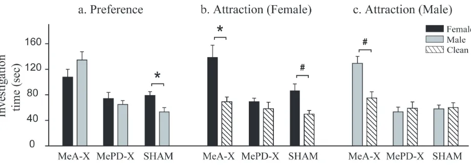
and MePD mediate distinct aspects of opposite-sex odor preference (Maras & Petrulis, 2006).
Specifically, we tested males with lesions of either MeA or MePD for their (1) preference to
investigate female odors or male odors when presented simultaneously and (2) attraction to
investigate each sexual odor when presented opposite clean (neutral) odors. Although lesions of
either MeA or MePD eliminate the preference to investigate opposite-sex odors, these lesions are
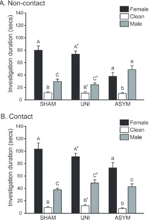
associated with qualitatively different patterns of odor investigation (Figure 1.2). Males with
MePD lesions fail to investigate female odors longer than clean odors, indicating a decreased
attraction to investigate opposite-sex odors. In contrast, males with MeA lesions remain highly
attracted to female odors, but are also highly (and inappropriately) attracted to male odors. This
opposite-sex odors, whereas MeA evaluates the sexual relevance of odor stimuli and directs
investigation specifically toward opposite-sex odors.
Taken together, these studies indicate that MeA and MePD differentially process
chemosensory and hormone signals and consequently, regulate distinct aspects of reproductive
behavior. Given the substantial reciprocal connections between MeA and MePD (Gomez &
Newman, 1992; Coolen & Wood, 1998), we hypothesize that these sub-regions interact during
the processing of social odors (Figure 1.3). We predict that MeA functions as a chemosensory
filter to identify the sexual relevance of odor stimuli and regulate odor responses within MePD.
Specifically, MeA may enhance responses to opposite-sex odors, but suppressresponses to
same-sex odors, within MePD. Furthermore, we hypothesize that MePD provides positive feedback
onto MeA during the processing of sexually relevant odors, such that MePD normally enhances
responses to opposite-sex odors within MeA. Together, these sub-regions can modulate
behavioral responses to different categories of social odors through projections to downstream
nuclei, such as the BNST and MPOA (Kevetter & Winans, 1981a; Gomez & Newman, 1992;
Coolen & Wood, 1998). Importantly, if this model is correct, then we expect that (a) disrupting
the interactions between MeA and MePD will eliminate the preference to investigate
opposite-sex odors (Chapter 3) and (b) odor responses within MeA and MePD will be altered in the
absence of MePD or MeA, respectively (Chapter 4). Finally, if the connections between MeA
and MePD provide a mechanism for chemosensory-hormone integration, then we expect that
Goals of Dissertation
The fundamental goal of this research is to identify the functional substrates by which the
brain recognizes sexual signals in the environment and generates appropriate reproductive
responses to these stimuli. Using Syrian hamsters as a model species, this dissertation combines
the use of behavioral, lesion, and neuroanatomical techniques to provide a comprehensive
analysis of how the corticomedial amygdala processes sexual odors and regulates odor-guided
aspects of reproductive behavior. Specifically, we addressed the following research questions:
(1) Is the PMCo required for the expression of either opposite-sex odor preferences or copulatory
behavior? (2) Are functional interactions between MeA and MePD required for the expression of
opposite-sex odor preferences? (3) How do MeA or MePD regulate odor responses within the
MePD or MeA, respectively? (4) Are odor and/or hormone cues conveyed directly between MeA
and MePD? Together, these results fill a critical gap in our knowledge regarding fundamental
mechanisms by which the brain processes sexually relevant stimuli and identify possible
substrates for the integration of chemosensory and hormone signals that is required for male
reproductive behavior in rodents. Although these mechanisms are critical for odor processing
specifically related to reproductive behaviors, they are also likely important for appropriate
Chapter 1 Figures
CHAPTER 2: The Posteromedial Cortical Amygdala Regulates Copulatory Behavior, but
not Sexual Odor Preference, in the Male Syrian Hamster (Mesocricetus auratus)
Pamela M. Maras and Aras Petrulis
Neuroscience Institute
Georgia State University, Atlanta, Georgia, USA 30302
Abstract
In rodent species, the expression of reproductive behavior relies heavily on the perception
of social odors, as well as the presence of circulating steroid hormones. In the Syrian hamster,
chemosensory and hormonal cues are processed within an interconnected network of ventral
forebrain nuclei that regulates many aspects of social behavior. Within this network, the
posteromedial cortical amygdala (PMCo) receives direct projections from the accessory olfactory
bulbs and contains a dense population of steroid receptor-containing neurons. Consequently, the
PMCo may be important for generating odor-guided aspects of reproductive behavior, yet little is
known regarding the role of this nucleus in regulating these behaviors. Thus, the present study
tested male hamsters with site-specific electrolytic lesions of the PMCo for their (a) sexual odor
preference in a Y-maze apparatus (b) sexual odor discrimination in a habituation-dishabituation
task and (c) copulatory behavior when paired with a sexually receptive female. PMCo-lesioned
males preferred to investigate female odors over male odors and were able to discriminate
between these odor sources. However, PMCo lesions were associated with several alterations in
the male copulatory pattern. First, PMCo-lesioned males displayed increased investigation of the
female’s non-anogenital region, suggesting that the PMCo may be involved in directing
appropriate chemosensory investigation during mating. Second, PMCo lesions altered the
temporal pattern of the mating sequence, as PMCo-lesioned males took longer than
Sham-lesioned males to reach sexual satiety, as indicated by the delayed expression of long
intromissions. This delayed onset of satiety was associated with an increased number of
ejaculations compared to Sham-lesioned males. Importantly, these data provide the first direct
Introduction
In many rodent species, including the Syrian hamster, male reproductive behavior relies
heavily on the perception of sexual odors (Hull et al., 2002). Male hamsters are highly attracted
to female odors, and these chemosignals stimulate the expression of copulatory behaviors
(Johnston, 1974; 1975; 1986) via both the main and accessory olfactory systems (Murphy &
Schneider, 1970; Winans & Powers, 1977; Meredith, 1991; Restrepo et al., 2004). In addition to
chemosensory processing, male hamster sexual behavior also requires the presence of circulating
gonadal steroid hormones (Morin & Zucker, 1978; Powers & Bergondy, 1983; Powers et al.,
1985; Petrulis & Johnston, 1995). Consequently, the expression of male reproductive behavior in
the hamster involves the integration of chemosensory and hormonal cues (Wood, 1998), and this
integration likely occurs within the extended network of ventral forebrain nuclei known to
regulate mating behavior (Wood & Newman, 1995b; Wood & Coolen, 1997).
Previous research has identified several critical brain areas within this network, including
the medial preoptic areas (MPOA), bed nucleus of the stria terminalis (BNST) and medial
amygdala (MA), which regulate various aspects of male reproductive behavior (Lehman et al.,
1980; Lehman et al., 1983; Powers et al., 1987; Maras & Petrulis, 2006). Although the
posteromedial cortical nucleus of the amygdala (PMCo) is also part of the ventral forebrain
circuit (Wood, 1997), the function of this nucleus in guiding male sexual behavior remains
largely unknown. However, several lines of evidence suggest that it may be involved in
reproductive behavior. First, the PMCo has reciprocal connections with BNST and MA (Kevetter
& Winans, 1981a; Gomez & Newman, 1992; Coolen & Wood, 1998; Wood & Swann, 2005), as
well as strong connections with chemosensory circuitry. Specifically, the PMCo receives direct
from the main olfactory bulbs via the anterior and posterolateral cortical nuclei of the amygdala
(Kevetter & Winans, 1981b). Second, the PMCo is sensitive to gonadal steroids, as it contains
dense populations of steroid receptor-containing neurons (Simerly et al., 1990; Wood et al.,
1992; Wood & Newman, 1993; Shughrue et al., 1997). In rats, this nucleus is sexually dimorphic
Caerols et al., 1998) and is masculinzed by perinatal estradiol treatment
(Vinader-Caerols et al., 2000). Finally, in male hamsters, neurons within the PMCo display elevated levels
of c-fos expression, an indirect measure of neuronal activity, following either copulatory
behavior or exposure to female odors (Kollack & Newman, 1992; Wood & Newman, 1993;
Kollack-Walker & Newman, 1995; Fernandez-Fewell & Meredith, 1998; Fewell & Meredith,
2002).
Together, these data indicate that the PMCo may function to regulate odor-guided aspects
of male reproductive behavior, yet to our knowledge, this hypothesis has never been directly
tested. Consequently, we assessed the effects of electrolytic lesions of the PMCo on both
appetitive and consummatory aspects of reproductive behavior in male Syrian hamsters. Males
were tested first for their preference to investigate female odors over male odors in a Y-maze
apparatus, as well as for their ability to discriminate between these odors in a
habituation-dishabituation task. Males were then tested for their copulatory behavior to satiety when paired
with a receptive female. Due to the PMCo’s substantial projections from the accessory olfactory
bulbs (Scalia & Winans, 1975), we hypothesized that this nucleus would regulate male
reproductive behavior primarily through its processing of vomeronasal information. In male
hamsters, the role of the vomeronasal organ in regulating sexual behavior changes with sexual
experience. Specifically, although the vomeronasal organ is critical for the expression of sexual
processing in sexually experienced males (Meredith, 1986). As the current study is an initial
attempt to identify a functional role for the PMCo in regulating these behaviors, we wanted
experimental conditions that specifically rely on accessory olfactory processing and therefore
used sexually naïve male subjects. Our results show that the PMCo regulates distinct aspects of
male hamster copulatory behavior, although this nucleus is not critical for the expression of
sexual odor preferences.
Materials and Methods
Animals
All animals in this study were Syrian hamsters (Mesocricetus auratus) purchased from
Charles River Laboratory at three weeks of age and singly-housed until the age of behavioral
testing (3-6 months). Subjects were sexually naïve males that had been gonadectomized and
implanted subcutaneously with testosterone Silastic capsules prior to lesion surgery (see below).
Ovariectomized, hormone-primed female hamsters served as stimulus animals for the copulatory
behavior tests (see below). A separate group of gonadally-intact male and female hamsters were
used to provide social odor stimuli. Subjects were unrelated to, and had no previous contact with,
stimulus females or odor donors. All animals were housed in solid-bottom Plexiglas cages (36
cm X 30 cm X 16 cm) and maintained on a reversed 14-h light/ 10-h dark photoperiod (lights
off/on at 9 am/7 pm). Food and water were available ad libitum. All animal experiments were
carried out in accordance with the National Institutes of Health Guide for the Care and Use of
Laboratory Animals (NIH Publications No. 80-23; revised 1996) and were approved by the
Georgia State University Institutional Animal Care and Use Committee. All efforts were made to
Surgical procedures
Gonadectomy and testosterone implantsin male subjects. In male hamsters, exposure to
female odors causes a rapid increase in serum testosterone levels (Macrides et al., 1974; Pfeiffer
& Johnston, 1992), and it is possible that lesions of the PMCo may alter this testosterone surge.
Thus, in order to equalize steroid hormone levels between experimental groups, all subjects were
gonadectomized and provided with physiological levels of exogenous testosterone (Maras &
Petrulis, 2006). Males were anesthetized with 1-2% isoflurane (mixed with 100% oxygen).
Following a midline abdominal incision, testicles were bilaterally removed via cauterization of
the ductus deferens and blood vessels. Immediately following gonadectomy, males were given
chronic testosterone (Sigma Chemical Co., St. Louis, MO, USA) replacement via a 20 mm
Silastic capsule (i.d. 1.57 mm, o.d. 2.41 mm, Dow Corning, Midland, MI) that was implanted
subcutaneously between the scapulae.
Ovariectomy and hormone priming of stimulus females. Stimulus females for copulatory
behavior tests were anesthetized with 1-2% isoflurane (mixed with 100% oxygen) and
ovariectomized via bilateral flank incisions. Immediately following ovariectomy, stimulus
females were given chronic estradiol (Sigma Chemical Co., St. Louis, MO, USA) treatment via a
5 mm Silastic capsule (i.d. 1.57 mm, o.d. 2.41 mm, Dow Corning, Midland, MI, USA) that was
implanted subcutaneously between the scapulae. Females were allowed at least two weeks for
recovery prior to being used as stimulus animals in copulatory behavior tests. To induce
behavioral receptivity, females were given a subcutaneous injection of 0.25 mg of progesterone
(dissolved in sesame oil, 2.5 mg/ml) (Sigma Chemical Co., St. Louis, MO, USA) four hours
Electrolytic lesions. One to two weeks following gonadectomy, male subjects were
randomly assigned to either PMCo lesion (PMCoX, n = 22) or sham lesion (SHAM, n = 7)
group. Subjects were anesthetized with 1-2% isoflurane (mixed with 7:3 oxygen: nitrous oxide)
and positioned in a stereotaxic apparatus so that the skull was flat. The temporal muscles were
retracted from the skull and small holes were drilled to expose the dura. Bilateral electrolytic
lesions were made using a platinum/iridium electrode (0.25 mm diameter, 0.45 mm uninsulated
tip, Frederick Haer & Co., Bowdoinham, ME, USA) and by passing 1 mA of anodal current from
a lesion-making device (Ugo Basile, Comerio, VA, Italy). As the PMCo extends over 2 mm in
the rostral-caudal direction, and the size, shape and location of the nucleus varies along its
length, we used a combination of multiple small lesions in order to generate maximal damage of
the PMCo while limiting collateral damage to nearby nuclei. Therefore, each PMCoX male
received a total of five bilateral penetrations, and the current duration varied across penetrations
(Table 2.1). Sham surgeries were identical to lesion surgeries except that the electrode was
lowered 1.5 mm above the target coordinate and no current was passed. Gel foam (Pharmacia &
Upjohn Co., Kalamazoo, MI, USA) was used to pack the holes, and the incision was closed with
wound clips.
Behavioral testing
To determine the role of the PMCo in generating responses to sexual odors, as well as
male copulatory behavior, subjects were given a series of behavioral tests beginning 2 to 3 weeks
after lesion surgery (Figure 2.1). First, subjects were tested for their preference to investigate
female odors over male odors in a Y-maze apparatus (Sexual odor preference). Subjects were
then tested for their ability to discriminate between these odor sources in
copulatory behavior when paired with a sexually receptive female (Male copulatory behavior).
All testing was done during the first four hours of the dark phase of the photoperiod and under
dim light.
Odor Stimuli. Male and female odor stimuli used for sexual odor preference and sexual
odor discrimination tests were collected from cages that had housed a single odor donor and had
not been changed for 10-13 days. Odor stimuli consisted of 12 g of soiled cotton bedding (4
Nestlets, ANCARE, Bellmore, NY); 50 ml of soiled corncob litter; one damp cotton gauze pad
that was used to wipe along the inner walls of the odor donor cage; and an additional damp gauze
pad that was used to wipe the odor donor’s bilateral flank and anogenital regions. For female
odor stimuli, vaginal secretion was collected onto an additional gauze pad by inducing an estrous
donor female into lordosis and gently palpating the vaginal area with a disposable probe. Clean
odor stimuli consisted of unsoiled components identical to those of the social odor stimuli. All
odor stimuli were stored in plastic bags at 4°C until 30 minutes prior to use. Odor samples older
than one month were discarded, and care was taken to ensure that subjects were not tested with
the same individual’s odor more than once. Clean latex gloves were worn while collecting odor
samples to prevent contamination of odor cues. To conserve stimulus odors, each odor was used
for two consecutive sexual odor preference or discrimination tests.
Sexual odor preference. Subjects were tested for their preference to investigate female
over male odor stimuli when presented in a maze apparatus (Maras & Petrulis, 2006). The
Y-maze consisted of a stem arm (61 cm long) and two side arms (68 cm long). All arms of the
maze were 10 cm wide, with walls 10 cm high. Each side arm had a stimulus chamber (20 cm
long) at its distal end, in which odor stimuli (see above) were placed. Stimulus chambers had
were exposed to only the volatile components of the odor stimuli. A start chamber (20 cm long),
with a removable, perforated door, was located at the distal end of the stem arm. An electric fan
located behind the start chamber pulled air from the stimulus chambers through the entire length
of the Y-maze (airflow rate of 2.0 km/hr, measured at the start box). The top of the Y-maze was
secured with a clear Plexiglas top to allow for overhead video recording of the subject’s
behavior.
Subjects were tested in a sequence of two Y-maze tests, separated by 24 hours. First, to
habituate the subjects to the Y-maze and obtain baseline behavioral data, subjects were tested
with clean odor stimuli in both stimulus chambers of the Y-maze (Clean). Subjects were then
tested for their sexual odor preference by placing male and female odors in opposite stimulus
chambers (Preference). For all tests, subjects were placed in the start chamber for one minute,
after which, the door was removed and subjects were allowed nine minutes to explore the
Y-maze. All surfaces of the Y-maze were thoroughly cleaned with 50% alcohol and allowed to dry
between subjects.
Video recordings of Y-maze tests were digitized onto a computer and scored using the
Observer for Windows, version 5.0 (Noldus Information Technology B.V., Wageningen, The
Netherlands). All observers were blind to the condition of the subject, and different observers
reached at least an 85% inter-observer reliability score prior to coding behavior. Both the time
spent investigating the stimulus chambers and the numbers of entries into each arm of the
Y-maze were scored. Investigation of the stimulus chamber was coded when the subject made
contact with, or directed its nose within 1 cm of, the stimulus chamber door. Arm entry was
Sexual odor discrimination. A habituation-dishabituation model was used to test
discrimination between male and female odors. This approach involves repeated presentations of
the same odor source followed by a test presentation of a novel odor source. A decrease in
investigation during the repeated presentations indicates a perception of the odors as being the
same or familiar. An increase in investigation of the novel odor compared to the last presentation
of the habituated odor indicates an ability to discriminate between the two odors (Johnston,
1993; Baum & Keverne, 2002).
The testing sequence consisted of four, 3-minute presentations of repeated odors
(habituation) followed by a fifth, 3-minute presentation of a novel odor (test). Five-minute
inter-trial intervals separated each odor presentation. As we have previously shown that male hamsters
do not consistently habituate to repeated presentations of female odors (Maras & Petrulis, 2006),
all subjects were tested using male odors as the habituation stimuli and female odors as the test
stimuli. Subjects were presented with a different male’s odor on each of the habituation trials so
that subjects were habituated to the sexual identity of the repeated odor, rather than to the
individual identity of an odor donor.
Odor stimuli were presented in modified 50 ml polypropylene collection tubes, with ½
cm holes drilled 1 cm apart along the surface of the tube. Wire mesh lined the inner surface of
the odor container to prevent contact with the odor stimulus. Thus, subjects were exposed to only
the volatile components of the odor stimuli during these tests. Odor containers were placed in the
center of the subject’s home cage and investigation was scored when the subject’s nose
contacted, or came within 1cm of, the odor container. Total investigation times were measured
using a stopwatch. Odor containers were cleaned with 50% alcohol and allowed to air dry for 24
Male copulatory behavior. Subjects were tested for their copulatory behavior when
paired with a receptive stimulus female hamster in a clear, Plexiglas arena. An angled mirror was
placed under the testing arena to provide a view of the ventral surface of the animals (in addition
to the side view). Males were placed into the empty testing arena for five minutes prior to the
addition of the stimulus female. Copulatory tests lasted 30 minutes, at which time the stimulus
female was removed.
Copulatory behavior tests were video-recorded and the male’s behavior was later scored
using the Observer for Windows, version 5.0 (Noldus Information Technology B.V.,
Wageningen, The Netherlands). The total number and latencies (from test onset) of several
behavioral measures were scored: mounts without intromissions (mounts), mounts with
intromissions (intromissions), ejaculations, and long intromissions. Long intromissions are
distinguished from regular intromissions in that the male does not quickly dismount the female
following vaginal penetration, but instead displays a repetitive thrusting pattern (Bunnell et al.,
1977; Parfitt & Newman, 1998). Importantly, the expression of long intromissions is associated
with the onset of sexual satiety in this species (Bunnell et al., 1977; Parfitt & Newman, 1998). In
addition, the total durations of time the male spent investigating the female’s anogenital region,
investigating the female’s head or body region (non-anogenital) and self-grooming were also
scored. Finally, several derived measures of copulatory behavior were also analyzed:
Post-ejaculatory interval (latency to display a mount or intromission after each ejaculation), the
number of intromissions to reach each ejaculation, and mounting efficiency (the total number of
Histology and lesion verification
Following the last behavioral test, subjects were injected with an overdose of sodium
pentobarbital (Nembutal, 100 mg/kg) and transcardially perfused with 200 ml of 0.1M
phosphate-buffered saline (PBS, pH 7.4) followed by 200 ml of phosphate-buffered formalin
(10%). Brains were post-fixed in phosphate-buffered formalin (10%) overnight and then
cryoprotected for 48-72 hours in 30% sucrose in PBS solution. Coronal sections (40-µm) of
brain tissue were sectioned on a cryostat (-20°C) and stored in PBS until mounting. Every third
section was mounted onto glass slides using a 1% gelatin mounting solution and stained with
cresyl violet.
Sections were examined under a light microscope for the location and extent of lesion
damage as compared with published hamster neuroanatomical plates (Morin & Wood, 2001).
Brain sections from subjects with minimum- and maximum-sized lesions were captured at 5X
magnification by a Zeiss Axiocam using Axiovision 4.0 software (2002). These lesions were
traced onto anatomical plates using Adobe Illustrator CS 11.0 software (2003).
Blood collection and radioimmunoassay
Blood samples were collected from the inferior vena cava immediately prior to perfusion
and stored in vacutainer collection tubes (VWR, West Chester, PA., 4 ml draw, red/gray) on ice
until centrifuging. Samples were centrifuged at 3200 rpms, at 4°C for 20 minutes and serum was
stored in 200µl aliquots at -20°C until assay. Testosterone levels (ng/ml) were measured by
radioimmunoassasy kits from Diagnostics System Laboratories (DSL 4000 Testosterone), with a
sensitivity range of 0.05-22.92 ng/ml and an inter-assay variance of 6%, previously validated for
Data analysis
All data were analyzed using SPSS 11.0 (SPSS Inc., Chicago, IL, USA) for Windows and
are reported as mean ± SEM. To establish investigatory preferences in each Y-maze test (Clean,
Preference), 2 (Lesion group: PMCoX, SHAM) X 2 (Stimulus; Clean test: left, right; Preference:
female, male odor) ANOVAs were performed. In addition, independent t-tests were used to
detect group differences in the total number of arm entries made during each Y-maze test.
For the sexual odor discrimination tests, data were analyzed using a 2 (Lesion group) X 5
(Odor presentation: Male 1-4, Female) ANOVA. For post-hoc analysis, pairwise comparisons
with Bonferroni corrections were used to compare investigation times between Male 1 vs. Male
4 and Male 4 vs. Female presentations.
Group differences in all copulatory measures were detected using independent t-tests.
Furthermore, to detect changes in post-ejaculatory intervals or the number of intromissions to
reach each ejaculation across the duration of the copulatory test, separate 2 (Lesion group) X 2
(First, Last ejaculatory series) ANOVAs were performed.
Results
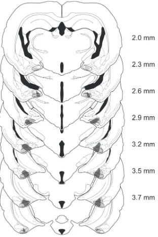
Lesion verification
Males were included in the PMCoX lesion group (n = 11) only if they had extensive
bilateral damage of the PMCo. Specifically, all males in the PMCoX group had at least 50%
bilateral damage that included the middle three sections of the PMCo (Figure 2.2). In four of
these males, damage extended into the rostral sections of the PMCo, whereas in seven subjects,
or copulatory behavior between males with rostral or caudal spread of lesion damage (all p >
.05). Males were excluded from the PMCoX group if there was any damage to the posterior
medial amygdala (n = 2) or if there was substantial sparing of the PMCo (n = 9).
In addition to damage of the PMCo, a subset of PMCoX males also had minimal damage
(< 10% in any section) to adjacent nuclei, including the posterior basomedial (BMP, n = 5),
posterior basolateral (n = 6) and posterolateral cortical amygdala (n = 6), and the
amygdalopiriform transition area (n = 3). Importantly, damage to these regions was mostly
unilateral and never complete. All PMCoX males also had some lesion damage to the
amygdalohippocampal area (AHi). In four males, this damage was minimal (< 10%), whereas in
seven males, damage to the AHi was moderate (≤ 50%). There were no differences in either the
preference or copulatory behavior between males with minimal or moderate damage to the AHi
(all p > .05).
Behavioral measures
Sexual odor preference. In the Clean test, there were no significant differences between
investigation levels of the two sides of the Y-maze, F(1, 16) = 1.139, p > .05, or between
experimental groups, F(1, 16) = .228, p > .05; there was also no significant interaction between
these factors, F(1, 45) = .001, p > .05 (Table 2.2). Furthermore, when the investigation times
were summed for the left and right arms, PMCoX and SHAM males did not differ in their total
duration of investigation of the clean stimulus chambers, t(16) = .478, p > .05. Levels of activity,
as measured by the total number of arm entries, were also not different between PMCoX and
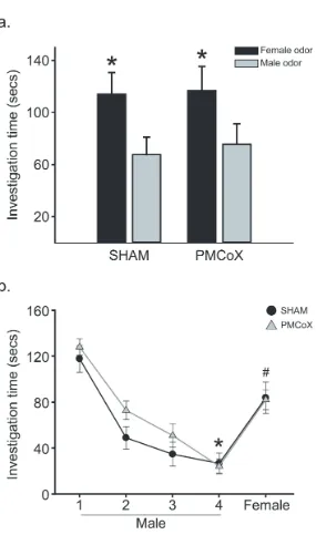
In the Preference test, subjects investigated female odors longer than male odors, F(1, 16)
= 7.760, p < .05, with no difference in investigation between experimental groups, F(1, 16) =
.029, p > .05, or significant interaction between odor stimulus and experimental group, F(1, 16)
= .019, p > .05 (Figure 2.3a). In addition, PMCoX and SHAM males spent similar amounts of
time investigating female odors, t(16) = -.054, p > .05, and male odors, t(16) = -.248, p > .05.
Sexual odor discrimination. There was a significant difference in investigation times
across stimulus presentations, F(4, 16) = 32.359, p < .05 (Figure 2.3b). There was no significant
difference between lesion groups, F(1, 16) = 2.908, p > .05, nor was there a significant
interaction between stimulus presentation and lesion group, F(4, 16) = 2.294, p > .05. Post-hoc
pairwise comparisons detected significant differences in investigation times between the first and
fourth presentations of male odors, t(16) = 10.120, p < .05 , as well as between the fourth
presentation of the male odor and the test presentation of the female odor, t(16) = -11.151, p <
.05.
Male copulatory behavior. When tested with a sexually receptive female, all male
subjects ejaculated and all, except one PMCoX male, reached sexual satiety, as indicated by the
expression of long intromissions. However, PMCoX and SHAM males did differ in the
expression of several aspects of the male copulatory sequence.
PMCoX males investigated the female’s non-anogenital region significantly longer than
SHAM males, t(16) = -2.309, p < .05, although groups did not differ in their duration of
anogenital investigation, t(16) = -1.218, p > .05 (Figure 2.4). PMCoX males also displayed less
self-grooming than SHAM males, t(16) = 2.953, p < .05 (PMCoX = 454 ± 29.0 seconds; SHAM
Although PMCoX and SHAM males displayed equal numbers of mounts, t(16) = .093, p
> .05, and intromissions, t(16) = -.626, p > .05, PMCoX males displayed more ejaculations, t(16)
= -3.320, p < .05, and less long intromissions, t(16) = 2.831 p < .05, compared to SHAM males
(Figure 2.5a). PMCoX males also took slightly longer than SHAM males to express mounts,
intromissions and ejaculations from test onset, although these differences were not statistically
significant (all p > .05; Figure 2.5b). PMCoX males, however, took significantly longer than
SHAM males to express long intromissions, t(16) = -2.634, p < .05 (Figure 2.5b).
Because the latencies to initiate mating (display the first mount) were slightly different
between experimental groups, and this difference could affect the latencies to display subsequent
mating behaviors, we also analyzed the latencies to display intromissions, ejaculations, and long
intromissions after correcting for the latency to first mount (Figure 2.5c). After mating began,
PMCoX and SHAM males had comparable latencies to display intromissions, t(16) = -.329, p >
.05, and ejaculations, t(16) = -.314, p > .05. PMCoX males, however, still took longer than
SHAM males to display long intromissions, t(16) = -2.314, p < .05.
PMCoX and SHAM males did not differ in the duration of their first post-ejaculatory
interval, t(16) = .075, p > .05. In both PMCoX and SHAM males, post-ejaculatory interval
durations increased across the ejaculatory series, F(1,16) = 26.155, p < .05, and there was no
difference between groups in the pattern of this increase, F(1,16) = 2.789, p > .05. PMCoX and
SHAM males also did not differ in the number of intromissions to reach the first ejaculation,
t(16) = .358, p > .05. In both groups, fewer intromissions were required to reach the last
ejaculation compared to the first ejaculation, F(1,16) = 26.807, p < .05, and there was no
and SHAM males were comparable in their mounting efficiency, t(16) = -.407, p > .05. Table 2.3
summarizes these derived measures of male copulatory behavior.
Males with damage primarily outside the PMCo. For an additional comparison, we also
analyzed the copulatory behavior of a subset of males that were excluded from the PMCoX
group (Non-PMCoX, n = 6). In this subset, males had less than 20% damage of the PMCo and
moderate to substantial damage of the AHi and/or BMP. Importantly, males with damage
primarily outside the PMCo did not differ from SHAM males in any of the copulatory behavior
measures analyzed (all p > .05, Table 2.4).
Testosterone assay
There was no difference in testosterone levels (ng/ml) between PMCoX and SHAM
males, F(1,16) = .006, p > .05 (PMCoX = 5.972 ± 0.699; SHAM = 5.880 ± 1.043). The ranges
of testosterone levels in both groups (PMCoX = 2.82 − 8.05 ng/ml; SHAM = 2.66 − 7.70 ng/ml)
were within the physiological range reported for this species (Moore et al., 2004).
Discussion
The present results demonstrate that the PMCo regulates two distinct aspects of the
mating sequence in male Syrian hamsters. First, the PMCo may be involved in directing
appropriate chemosensory investigation during mating, as males with lesions of the PMCo
displayed increased investigation of the female’s non-anogenital region compared to SHAM
males. Second, the PMCo may regulate sexual satiety, as PMCo-lesioned males took longer than
SHAM males to display long intromissions, an indication of the onset of sexual satiety in this
species (Bunnell et al., 1977; Parfitt & Newman, 1998). This delayed onset of satiety was
ejaculations, compared to SHAM males. In contrast to these effects on copulatory behavior,
males with lesions of the PMCo preferred to investigate female odors over male odors, as did
SHAM males, and were able to discriminate between male and female odors in a
habituation-dishabituation task.
Electrolytic lesion technique
This study used multiple, small electrolytic lesions to generate discrete damage targeted
at the PMCo. One limitation of this technique is that damage is not restricted to neuronal cell
bodies but also includes fibers of passage. Consequently, it is possible that PMCo lesions
disrupted anatomical connections of nearby brain areas. However, the primary fiber tracts
associated with the PMCo are the accessory olfactory bulb efferents traveling along the ventral
surface of the brain to the PMCo itself (Kevetter & Winans, 1981a; Kemppainen et al., 2002). As
the PMCo is the most caudal target of these fibers (Scalia & Winans, 1975), damage to the
ventral surface does not disconnect the accessory olfactory bulb from other brain areas.
Furthermore, males with lesion damage primarily to nuclei outside the PMCo, including the AHi
and/or BMP, displayed copulatory behavior similar to that of SHAM males, suggesting that the
behavioral deficits observed in PMCo-lesioned males do not simply reflect a disconnection of
nearby brain areas. The use of excitotoxins for making lesions would reduce many of these
concerns, as they spare fibers of passage, but we have found that they do not produce reliable,
controllable lesion damage in this nucleus (unpublished observations).
The role of the PMCo in male copulatory behavior
Unlike lesions of the MPOA or MA, which eliminate male copulatory behavior in many