R E S E A R C H A R T I C L E
Open Access
Effect of oral administration of ethanolic extract
of
Vitex negundo
on thioacetamide-induced
nephrotoxicity in rats
Farkaad A Kadir
1†, Normadiah M Kassim
1*, Mahmood A Abdulla
2†and Wageeh A Yehye
3†Abstract
Background:Oxidative stress due to abnormal induction of reactive oxygen species (ROS) molecules is believed to be involved in the etiology of many diseases. Evidences suggest that ROS is involved in nephrotoxicity through frequent exposure to industrial toxic agents such as thioacetamide (TAA). The current investigation was designed to explore the possible protective effects of the leaves ofVitex negundo(VN) extract against TAA-induced
nephrotoxicity in rats.
Methods:Twenty fourSprague Dawleyrats were divided into four groups: (A) Normal control, (B) TAA (0.03% w/v in drinking water), (C) VN100 (VN 100 mg/kg + TAA) and (D) VN300 (VN 300 mg/kg + TAA). Blood urea and serum creatinine levels were measured,supraoxide dismutase (SOD), catalase (CAT) and malondialdehyde (MDA) levels of renal tissue were assayed. Histopathological analysis together with the oxidative stress nicotinamide adenine dinucleotide phosphate (NADPH) oxidase p22phox in kidney sections were examined in all experimental groups. Results:Blood urea and serum creatinine levels were increased in TAA group as a result of the nephrotoxicity compared to the VN100 and VN300 groups where, the levels were significantly decreased (p< 0.05). Renal MDA level was significantly decreased (p< 0.05) in the VN-treated groups with increased CAT and SOD activities compared to the TAA group. Light microscopic examination of renal tissues stained by H&E stain and Masson’s Trichrome for TAA-treated groups revealed severe histopathological changes, whereas specimens obtained from VN-treated groups showed only mild changes. These findings were supported by immunohistochemical results. Conclusions:VN extract acts as a natural potent antioxidant to prevent ongoing TAA-induced nephrotoxicity in rats, both biochemically and morphologically.
Keywords:Vitex negundo, Thioacetamide, Urea, Creatinine, Histopathology, Immunohistochemistry, Nephrotoxicity
Background
Kidneys are highly vulnerable to damage caused by re-active oxygen species (ROSs), likely due to oxidative stress by polyunsaturated fatty acids in the composition of renal lipids [1]. This damage can also be caused by a high volume of blood flowing through it, and filtering large amounts of toxins, which can concentrate in kid-ney lobules [2]. The kidkid-ney’s response to toxicants var-ies by multiple morphological patterns beginning with tubular or interstitial changes to nephropathy [3]. It has
been strongly implicated that (NADPH) oxidase, as a major source of ROSs production in the kidney [4] could have a role the in development of renal oxidative damage.
Nephrotoxicity is a poisonous effect due to drugs and its overdose on the kidneys. Thioacetamide (TAA) is an or-ganic compound with the formula CH3CSNH2. It is
ori-ginally used as a fungicide, and is a potent hepatotoxin [5]. It can serve as a source of sulphur in the synthesis of or-ganic compounds such as rubber chemicals, curing agents, cross linking agents, metallurgy, pesticides, and pharma-ceuticals [5]. TAA is the most potent nephrotoxic sub-stance because of its rapid elimination and cumulative injury when it is given intermittently,presumably by free
* Correspondence:normadiah_mk@um.edu.my
†Equal contributors
1
Department of Anatomy, Faculty of Medicine, University of Malaya, Kuala Lumpur 50603, Malaysia
Full list of author information is available at the end of the article
radical-mediated lipid [2,6]. Metabolic studiesof TAA-induced tissue damage suggest that TAA is metabolized by the mixed function oxidase system to its toxic metabo-lites sulfine (sulfoxide) and sulfene (sulfone)which are then distributed among several organs,including plasma, liver, kidney, bone marrow, adrenals and other tissues [7]. Later, TAA undergoes an extensive metabolism to acetate and it is excreted through the urine within 24 hours [5,8].
Vitex negundo (VN) is commonly called five leaved chaste tree. It is a large aromatic shrub or small and slen-der with quadrangular branchlets, about 2 to 5 m in height, distributed mainly in tropical to temperate regions, especially in Malaysia, India at the warmest zones and Western Himalayas [9].
The leaves have a typical five foliolate pattern in pal-mate arrangement measuring4-10 cm long and bluish purple in colour which become dark when ripened. The whole parts of the plant have shown to be a potent source of natural antioxidants [10], 1, 2 di-substituted idopyranose, the isolated compound from VN extract has shown protection of hepatocytes, nephrocytes and pancreatic β-cells in streptozotocin-induced diabetes, probably by its action against NF-kB and induced-nitric oxide synthase iNOS mediated inflammation [11]. A preliminary acute toxicity study of ethanolic leaf extract of VN in albino rats by oral rout carried out by Tandon, 2005 [12] found it to be practically nontoxic, as its LD50 dose was recorded as 7.58 g/Kg body weight with no histomorphological changes in liver, kidney, stom-ach, heart and lung at any dose of the extract studied. VN has various traditional uses in treating stomach-ache, eye disease, inflammation, enlargement of spleen, bronchitis, asthma and painful teething in children. It is also used as an antihelmintic, promoting hair growth [13], and the juice of the leaves used in treating ulcers and swelling of joints [14]. Literature review reveals that the plant of VN possesses analgesic and antinociceptive activity [15], hepatoprotective activity against TAA [16], antituberculardrugs [12], CCl4 [17] and Ibuprofen via
inhibition of lipid peroxidation [18].
In this study, VN extract was utilized in a rat model to evaluate its possible protective effects on TAA-induced nephrotoxicity. Data were collected on serum creatinine level, blood urea level, kidney and body weight ratio, glutathione content, and lipid peroxidation in the kidney tissues, as well as determination of p22phox expression and histopathological changes in the kidneys after ad-ministering VN extract in the adult male SD rats.
Methods
Collection and preparation of plant extract
Fresh leaves of VN plant were obtained from Kampung Baru, Sungai Ara, Penang, Malaysia. The botanical identity was determined and authenticated in the Department of
Pharmacy, Faculty of Medicine, University Malaya, Kuala Lumpur, Malaysia with voucher specimen number (KLU 34968). The plant was dried and grounded to a fine pow-der. Next, the powder was homogenized in 95% ethanol at a ratio of 1:10 of plant to ethanol, and left to soak for four days at 25°C with occasional shaking and stirring. Later, the mixture was filtered through filter paper, and the resulting liquid was concentrated at a reduced pressure at 45°C to obtain a dark gummy–green extract. The percent-age yield of VN crude extract was 18%. The extract was then dissolved in Tween 20 (10% w/v) and administered orally to rats in concentrations of 100 and 300 mg/kg body weight.
Preparation of TAA
TAA (from Sigma-Aldrich, Switzerland) and all other chemicals used were of analytical grade and purchased mostly from Sigma-Aldrich and Fisher. TAA stock solu-tion was prepared by dissolving30 mg pure TAA which is in crystal form in 100 ml distilled water (0.03% w/v) until all the crystals were dissolved. The solution was given to the rats as their daily drinking water [19].
Experimental animals
A healthy adult maleSprague Dawleyrats weighing 180– 200 gm were obtained from the Animal House Unit, Fac-ulty of Medicine, University of Malaya, Malaysia. They were kept in wire-bottomed cages at 25±3°C, at 50–60% humidity, and a 12 h light-dark cycle for at least a week be-fore the experiment. They were maintained under standard housing conditions with free access to a standard diet and water ad libitum during the experiment. The experimental protocol was approved by the Institutional Animal Care and Use Committee, University of Malaya (UM IACUC) with an ethical no. ANA/18/05/2012/FAAK. Throughout the experiment, all criteria for taking care of animals pre-pared by the National Academy of Sciences and outlined in the“Guide for the Care and Use of laboratory Animals” were compiled [20].
The animals were randomly divided into four experimen-tal groups, with each group consisting of six rats, and given the following treatments: Group A: Normal control group, received per oral treatment of 10% Tween 20 (5 ml/kg) daily for 12 weeks; Group B: TAA group, received TAA 0.03% w/v in drinking water daily for 12 weeks; Group C: VN100 group, received TAA 0.03% w/v in drinking water and 100 mg/kg body weight of VN extract daily for 12 weeks; Group D: VN300 group,received TAA 0.03% w/v in drinking water and 300 mg/kg body weight of VN ex-tract daily for 12 weeks.
fasting. The rats were anaesthetized by intramuscular in-jection of 50 mg/kg ketamine mixed with xylazine 5 mg/ kg. Blood samples were collected and serum was obtained for estimation of creatinine and blood urea levels. Kidney samples were dissected, trimmed of connective tissues, washed using normal saline to eliminate blood contamin-ation, dried by blotting with filter paper and weighed. The kidneys were excised into two halves. One-half was kept in isotonic formalin for histopathological assessment and the other half was kept in the freezer under -80°C for prepar-ing kidney homogenate for the malondialdehyde (MDA), catalase (CAT) and supraoxide dismutase(SOD) assays.
Assessment of renal function
A blood sample was withdrawn through the jugular vein and collected into a plain tube with activated gel for detec-tion of urea and creatinine levels. The samples were allowed to clot, centrifuged and the serum samples were sent for analysis using a standard automated technique in the Central Diagnostic Laboratory (CDL), University of Malaya Medical Centre, according to the procedures de-scribed by the manufacturers.
Preparation of kidney homogenates
Kidney homogenates (10% w/v) were prepared by hom-ogenizing kidney tissue in cold 50 mM potassium phos-phate buffer saline (pH 7.4) using a tissue homogenizer (DAIHAN Sci., Seoul, Korea). The cell debris was removed by centrifugation at 4500 rpm for 15 minutes at 4°C using a refrigerated centrifuge Rotofix 32 (Hettich Zentrifugen, Germany). The supernatant was used for estimating the followingin vivoantioxidant using commercially available kits (Cayman Chemical Company, USA): (MDA) or thio-barbituric acid reactive substance (TBARS) (Item No. 10009055), (CAT) (Item No. 707002) and (SOD) (Item No. 706002). All assays were performed according to the instruction manual of the manufacturers.
Histopathological assessment
The lower half of the right kidney was examined for histo-pathological changes. All kidneys specimens were examined under a light microscope. The kidneys were fixed in 10% formalin and then embedded in paraffin wax before sec-tioning at 5μm thickness and stained with haematoxylin-eosin and Masson’s trichrome stains. Kidney sections from the six rats in each group were examined by two dependent observers. Morphological analysis there were carried out according to Houghton et al., [21] and were graded as fol-lows: Grade 0 (normal), Grade 1 (when changes were lim-ited to the tubulointerstitial areas of focal granulovascular epithelial cell degeneration and granular debris in the tubu-lar lumina with or without evidence of desquamation in small foci (<1% of total tubule population involved by des-quamation), Grade 2 (when tubular epithelial necrosis and
desquamation were easily seen but involved less than half of the cortical tubules), Grade 3 (when more than half of the proximal tubules showing necrosis and des-quamation, but with intact tubules easily identified, and Grade 4 (when there was complete or almost complete proximal tubular necrosis).
Immunohistochemical study
For the quantitative determination of p22 phox anti-body, immunohistochemistry kit for p22 phox (CS9): sc-130551 (Santa Cruz Biotechnology, INC.) was used. Briefly, sections were deparaffinized in xylene and hydrated in a series of graded alcohol. Deparaffinized sections were treated in a microwave oven in a citrate buffer at 95°C for 15 minutes, and immersed in 3% hydrogen peroxide in methanol for 10 min to abolish endogeneous peroxidase ac-tivities. The sections were immersed in normal goat serum for 15 min, incubated withmouse monoclonal antibody as a marker for NADPH oxidase subunits (diluted to 1:50) at 37°C for 60 min, and then incubated with goat anti-mouse IgG conjugated with horseradish\ peroxidise (HRP) (diluted to 1:200). The reaction products were visualized using 3-30-deiaminobenizidine tetrahydrochloride and hydrogen peroxide. A cytoplasmic brown granule was marked as positive expression of p22 phox.
Statistical analysis
[image:3.595.305.540.100.170.2]All data were expressed as mean ± standard error of the mean (SEM) and statistical analysis was performed using SPSS for Windows version 17.0 (SPSS Inc. Chicago, IL, USA). One-way analysis of variance (ANOVA) followed
Table 1 Effect of VN on body and kidney weights
Group Body weight (g) Kidney weight (g)
Normal control 219.8 ± 29.88** 0.95 ± 0.23
TAA 177.5 ± 7.1* 0.93 ± 0.119
VN100 + TAA 199.3 ± 15.98 0.94 ± 0.22
VN300 + TAA 204.3 ± 10.70 0.99 ± 0.29
Data were stated as mean±SEM. Means with different superscripts are significantly different.*
P <0.05 versus normal control group and**
P <0.05 versus TAA group.
Table 2 Effect of VN on blood urea and serum creatininelevels
Group Urea (mmol/L) Creatinine (mol/L)
Normal control 4.46 ± 0.45** 23 ± 1.7**
TAA 8.73 ± 1.31* 52.3 ± 7.5*
VN100 + TAA 6.28 ± 0.59*,** 35.66 ± 7.3*,**
VN300 + TAA 5.73 ± 0.973** 29.33 ± 3.4**
Data were stated as mean±SEM. Means with different superscripts are significantly different.*
P <0.05 versus normal control group and**
[image:3.595.305.539.638.708.2]by Bonferroni post hoc test was applied to test for statis-tically significant differences between groups atp< 0.05.
Results
Gross pathology
During the course of this study, continuous daily adminis-tration of 0.03% w/v TAA in drinking water was not associ-ated with animal mortality. The external appearance of the
kidneys from animals treated with TAA in drinking water as well as VN-treated groups revealed a smooth, glistening capsule with no petechial hemorrhage was noted.
Body and kidney weight
[image:4.595.57.539.101.183.2]In the present study, the body weight of rats adminis-tered with TAA were reduced significantly (p <0.05) in comparison to the normal control group. Although there was increased in body weight of VN300 + TAA
Table 3 Effects of VN on somein vivoantioxidant parameters in all experimental groups
Group CAT SOD MDA
(nmol/mg protein) (U/mg protein ) (nmol/mg protien)
Normal control 27.24 ± 34.32 32.23 ± 7.14 38.74 ± 2.61
TAA 21.57 ± 24.87* 22.3 ± 8.58* 107.14 ± 3.71*
VN100 + TAA 24.75 ± 105.09 26.73 ± 0.93** 78.85 ± 2.26**
VN300 + TAA 25.26 ± 56.01 27.47 ± 3.44** 54.35 ± 1.73**
Data were stated as mean±SEM. Means with different superscripts are significantlydifferent.*
P <0.05 versus normal control group and**
P <0.05 versus TAA control group.
B
C
D
GL BM
Ai
Aii
[image:4.595.58.539.342.675.2]and VN100 + TAA groups, the increased in body weight was not significant (p <0.05) compared to TAA group. Concurrently, no noticeable concurrent significant dif-ference in the weights of the kidneys for all groups was shown (Table 1).
Biochemical determination
Rats from the TAA-treated group exhibited significantly increased (p <0.05) levels of blood urea and serum cre-atinine compared to the normal control group. On the other hand increases in these parameters were prevented by concurrent treatment of animals with VN 100 mg/ kg, and more effectively, with VN 300 mg/kg which re-sulted in nearly normalized levels of these parameters (Table 2).
CAT and SOD are some of the enzymes of the intrinsic antioxidant defense system [22] which are responsible for the dissemination of free radicals such as superoxide radi-cals. Table 3 shows that SOD was significantly (p< 0.05) increased in high and low dose VN-treated groups but de-creased in the TAA group due to long term excretion of free radicals. Generally, rats treated only with TAA, had significantly higher levels of MDA (p< 0.05) than normal rats and the other experimental treated groups. Notably, experimental rats treated with low dose and high dose VN extract had significantly lower levels (p< 0.05) of renal MDA compared to TAA group. These results suggest that treatment with VN extract may protect renal tissue from further damage.
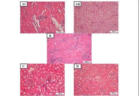
Histopathological findings
Histopathological examination of sections from rat kidneys treated with TAA showed impaired renal morphology throughout with severe and generalized tubular epithelial cell necrosis associated with diffuse tubular swelling, glomerular congestion and inflammatory cell infiltration. Kidneys from animals concurrently treated with VN 100 mg/kg body weight + TAA 0.03% in their drinking water showed mild to moderate inflammatory cell infiltration and tubular epithelial cell necrosis (Figure 1C), while concur-rent treatment with VN 300 mg/kg body weight provided the best morphological protection against TAA-induced renal damage (Figure 1D and Figure 2D).
Discussion
Various useful drugs like acetaminophen, gentamicin and some environmental and industrial toxicants can cause severe renal damage through activation of these drugs to highly reactive free radicals [23]. One of the most extensively studied chemicals and industrial toxicants is TAA. TAA is known to induce centrilobular hepatic ne-crosis, liver cirrhosis, hepatocellular carcinoma and bile duct proliferation [5,8,16,24,25] with injury to the terminal portion of the proximal renal tubule [7]. After administra-tion of TAA, this chemical undergoes an extensive metab-olism forming sulfine (sulfoxide) and sulfene (sulfone). Both are circulated through several vital organs in body before finally being transformed into acetate and excreted into urine within 24 hours [8].
B
C D
A
BM
[image:5.595.58.540.451.685.2]GL
TAA administration (0.03% w/v) in drinking water for 12 weeks found to cause renal toxicity as assessed by bio-chemical analysis of urea and creatinine, MDA and anti-oxidant status of SOD and CAT. As a measure of renal function status, blood urea and creatinine are often regarded as reliable markers [26]. The increased urea and creatinine levels resulting from TAA administration in this study (Table 2), are in agreement with a previous study done by Begum et al., [2]. High values of blood urea and serum creatinine indicate renal damage [27,28] and this may be correlated with the significant and progressive weight loss in the TAA group (Table 1). As for the VN100 and VN300 groups, although there was increased body weight in both groups, the increase was not significantly different compared in the TAA group (Table 1).
From this study, pretreatment with VN 100 mg/kg ex-tract could partly prevent TAA-induced kidney damage as shown by the decreasing levels of urea and creatinine.
These parameters were almost significantly normalized by the administration of VN 300 mg/kg extract (Table 2). This result is consistent with many previous studies done using other traditional plants [29], and is strongly attributed to the scavenging free radicals and reduced lipid peroxidation mechanisms.
Free radicals are believed to play a major role in the de-velopment of TAA-induced toxicity [30], hence, when oxi-dative stress is overwhelming, the various inherent defense mechanisms such as the antioxidant defense mechanisms, intercellular concentration of CAT and SOD activities be-come significantly impaired and insufficient [31]. This is because during oxidative stress, the body uses its defense mechanism to minimize the process of lipid peroxidation using these antioxidant enzymes, thus, the activity of these enzymes becomes higher in early stages of TAA induction. When the oxidative insult was continued for a long period, the enzymes become depleted and unable to fight against the free radicals, suggesting advance stages of TAA-induced peroxidation. This was evident in the present study with the significant increase (p <0.05) in MDA level, renal damage in TAA group compared to the normal con-trol group.
[image:6.595.58.538.89.313.2]Several phytochemical studies have revealed the pres-ence of volatile oils, lignans, flavonoid like flavones, luteolin-7-glucoside and glycosides in VN [9]. In addition, VN ethanolic extract possesses radical scavenging activity probably due to higher concentration of flavonoids and al-kaloids [32]. This was evident in the increased (CAT and SOD) levels in this study as well as the decreased in MDA
Table 4 Histopathological grading of the kidney sections for all experimental groups
Pathological grading
Group N 0 I II III IV Average of grades
Normal 6 6 0 0 0 0 0.0**
TAA 6 0 0 0 2 4 3.33 ± 0.51*
VN 100 + TAA 6 0 1 1 4 0 2.00 ± 0.63**
VN 300 + TAA 6 1 3 2 0 0 1.16 ± 1.34**
Data were stated as mean±SEM. Means with different superscripts are significantly different.*
P <0.05 versus normal control group and**
p <0.05 versus TAA control group.
A
B
C
D
[image:6.595.58.292.625.707.2]GL BM
Figure 3Photomicrographs of renal sections for immunohistochemical evaluation of 22phox protein expression in all experimental groups. A: Normal control, showing normal glomerulus (GL) in the cortical area, with intact Bowman’s capsule (BM) and normal
level in the VN-treated groupsas shown in Table 3. Pre-sumably, this is due to the antioxidant properties of the plant extract, which may account for the nephroprotective action of VN against TAA-induced nephrotoxicity experi-mental models.
TAA is toxic to selected populations of cells (hepato-cytes, proximal convoluted tubular cells in kidney and cor-tical thymocytes) [7], and several animals studies have shown that toxin-induced renal tubular damage plays a crucial role in the reduction of glomerular filtration rate ei-ther through obstruction and back leak of renal filtrate or secondary to ROS [33]. The p22phox is one of the subunits of the NADPH oxidase which generates ROS that plays an important role in the development of many kidney diseases [4,34]. Thus, VN is important in protecting the kidney from TAA-induced injury, through the improvement in renal function and antioxidant status. Furthermore, these findings were corroborated by the renal histological and immunohistochemical results, which revealed more exten-sive and marked tubular and glomerular damage with ap-parent expression of p22phox protein in the TAA-treated group. Similar changes were also reported by Edward et al., [7] demonstrating structural changes in renal tissue. How-ever, in the VN-treated groups, renal microscopic changes were alleviated and the renal histological archi-tecture was almost normalised, especially, at the maximum dose of 300 mg/kg as shown in Figures 1, 2, 3 and Table 4. Although the exact mechanism of TAA-induced neph-rotoxicity is not well understood, but based on several previous studies [27,28,35,36] we can postulate that the protective effect of VN extract is via its antioxidant and/or free radical scavenging activities due to its high concentration of flavonoids and alkaloids content [9].
Conclusions
The overall results from this study demonstrated TAA-induced renal injury by biochemical analysis, histopatho-logical features and immunohistochemistry analysis. The concurrent treatment with VN extract clearly provided a considerable degree of protection in a dose-dependent manner against the deleterious renal side effects of TAA. In conclusion, VN could act on the kidney as a potent nat-ural antioxidant to prevent ongoing TAA-induced nephro-toxicity, thus, these results constitute a lead towards discovering a novel herb in traditional and complementary medicine.
Competing interests
The authors declare that they have no competing interest.
Authors’contributions
Conceived and designed the experiments: FK, NMK, MAA and WAY. Performed the experiments FAK, WAY. Analyzed the data: FK and WAY. Contributed reagents, materials and analysis tools: FK, NMK, MAA and WAY. Wrote the paper: FK and WAY. Editing the paper: FK, NMK, MAA and WAY. All authors read and approved the final manuscript.
Acknowledgments
The authors express their gratitude to the staffs of the Faculty of Medicine, Animal House for the care and supply of rats. Financial support from (PG087-2012B) and (UM/MoHE/HIR Grant E000045-20001) from University of Malaya, Malaysia, are gratefully acknowledged.
Author details 1
Department of Anatomy, Faculty of Medicine, University of Malaya, Kuala Lumpur 50603, Malaysia.2Department of Biomedical Science, Faculty of
Medicine, University of Malaya, Kuala Lumpur 50603, Malaysia.
3Nanotechnology & Catalysis Research Centre, (NANOCAT), University of
Malaya, Block 3A, Institute of Postgraduate Studies Building, Kuala Lumpur 50603, Malaysia.
Received: 14 April 2013 Accepted: 24 October 2013 Published: 30 October 2013
References
1. Ozbek E:Induction of Oxidative Stress in Kidney.Int J Nephrol2012,
2012:1–9.
2. Begum Q, Noori S, Mahboob T:Antioxidant effect of sodium selenite on thioacetamide-induced renal toxicity.Pakistan Journal of Biochemistry and Molecular Biology2011,44(1):21–26.
3. Silva FG:Chemical-induced nephropathy: a review of the renal tubulointerstitial lesions in humans.Toxicol Pathol2004,32(2):71–84. 4. Etoh T, Inoguchi T, Kakimoto M, Sonoda N, Kobayashi K, Kuroda J,
Sumimoto H, Nawata H:Increased expression of NAD (P) H oxidase subunits, NOX4 and p22phox, in the kidney of streptozotocin-induced diabetic rats and its reversibity by interventive insulin treatment.
Diabetologia2003,46(10):1428–1437.
5. Kadir FA, Othman F, Abdulla MA, Hussan F, Hassandarvish P:Effect of Tinospora crispa on thioacetamide-induced liver cirrhosis in rats.Indian J Pharmacol2011,43(1):64–68.
6. Dashti HM, Mathew TC, Jadaon MM, Ashkanani E:Zinc and liver cirrhosis: biochemical and histopathologic assessment.Nutrition1997,13(3):vi–212. 7. Edward A, Baker EAS:Nonhepatic Thioacetamide Injury. The morphologic Features of proximal renal tubular injury.Am J Pathol1974,74(3):576–590. 8. Spira B, Raw I:The effect of thioacetamide on the activity and expression
of cytosolic rat liver glutathione-S-transferase.Mol Cell Biochem2000,
211(1):103–110.
9. Gautam L, Shrestha S, Wagle P, Tamrakar B:Chemical constituents from Vitex negundo (Linn.) of nepalese origin.Scientific world2010,6(6):27–32. 10. Raghavendra H, Lakshmanashetty VBN, Madhumathi G, Hiremath aVK:In
vitro Antioxidant Activity of Vitex negundo L. Leaf Extracts.Chiang Mai Journal of Science2010,37(3):489–497.
11. Manikandan R, Thiagarajan R, Beulaja S, Sivakumar MR, Meiyalagan V, Sundaram R, Arumugam M:1,2 di-substituted idopyranose from Vitex negundo L. protects against streptozotocin-induced diabetes by inhibiting nuclear factor-kappa B and inducible nitric oxide synthase expression.Microsc Res Tech2011,74(4):301–307.
12. Tandon VR:Medicinal uses and biological activities of Vitex negundo.
Natural product radiance2005,4(3):162–165.
13. Singh P, Mishra G, Srivastava S, Sangeeta K, Khosa R:Phytopharmacological review of vitex negundo (Sambhalu).Pharmacologyonline2011,2:1355–1385. 14. Nadkarni KM:Dr. K. M. Nadkarni&s Indian materia medica. 1.1994.
Popular Prakashan.
15. Zheng C-J, Huang B-K, Han T, Zhang Q-Y, Zhang H, Rahman K, Qin L-P:
Antinociceptive activities of the liposoluble fraction from Vitex negundo seeds.Pharm Biol2010,48(6):651–658.
16. Kadir FA, Kassim NM, Abdulla MA, Yehye WA:Hepatoprotective Role of Ethanolic Extract of Vitex negundo in Thioacetamide-Induced Liver Fibrosis in Male Rats.Evidence-Based Complementary and Alternative Medicine2013,2013.
17. Avadhoot Y, Rana A:Hepatoprotective effect of Vitex negundo against carbon tetrachloride-induced liver damage.Arch Pharm Res1991,
14(1):96–98.
18. Mahalakshmi R, Rajesh P, Balasubramanian V, Rajesh Kanan V:
Hepatoprotective activity on Vitex negundo Linn.(Verbanaceae) by using Wistar albino rats in Ibuprofen induced model.Int J Pharmacol2010,
19. Müller A, Machnik F, Zimmermann T, Schubert H:Thioacetamide-induced cirrhosis-like liver lesions in rats usefulness and reliability of this animal model.Exp Pathol1988,34(4):229–236.
20. National research council. Guide for the care and use of laboratory animals.8th edition Washington, DC: The National Academies Press. 21. Houghton DC, Plamp CE 3rd, DeFehr JM, Bennett WM, Porter G, Gilbert D:
Gentamicin and tobramycin nephrotoxicity. a morphologic and functional comparison in the rat.Am J Pathol1978,93(1):137–152. 22. Ercal N, Gurer-Orhan H, Aykin-Burns N:Toxic metals and oxidative stress
part I: mechanisms involved in metal-induced oxidative damage.
Curr Top Med Chem2001,1(6):529–539.
23. Olagunju J, Adeneye A, Fagbohunka B, Bisuga N, Ketiku A, Benebo A, Olufowobi O, Adeoye A, Alimi M, Adeleke A:Nephroprotective activities of the aqueous seed extract of Carica papaya Linn. in carbon tetrachloride induced renal injured Wistar rats: a dose-and time-dependent study.
Biol Med2009,1(1):11–19.
24. Stankova P, Kucera O, Lotkova H, Rousar T, Endlicher R, Cervinkova Z:The toxic effect of thioacetamide on rat liver in vitro.Toxicol In Vitro2010,
24(8):2097–2103.
25. Madani H, Talebolhosseini M, Asgary S, Naderi G:Hepatoprotective activity of Silybum marianum and Cichorium intybus against thioacetamide in rat.Pakistan J Nutr2008,7(1):172–176.
26. Gowda S, Desai PB, Kulkarni SS, Hull VV, Math AAK, Vernekar SN:Markers of renal function tests.North Am J Med Sci2010,2(4):170.
27. Adeneye AA, Benebo AS:Protective effect of the aqueous leaf and seed extract of Phyllanthus amarus on gentamicin and acetaminophen-induced nephrotoxic rats.J Ethnopharmacol2008,118(2):318–323. 28. Khalid MS:Nephroprotective effect of the ethanolic extract of Lantana camara
Linn flower on acute dose of Cisplatin induced renal injured rats; 2012. 29. El-Adawi H, El-Azhary D, Abd El-Wahab A, El-Shafeey M, Abdel-Mohsen M:
Protective effect of milk thistle and grape seed extracts on fumonisin B1 induced Hepato-and nephro-toxicity in rats.J Med Plant Res2011,
5(27):6316–6327.
30. Mohammed AA, Mahmood Ameen A, Salmah I, Zahra AA:
Hepatoprotective effects of Orthosiphon stamineus extract on thioacetamide-induced liver cirrhosis in rats.Evidence-Based Complementary and Alternative Medicine2011,2011.
31. Parlakpinar H, Tasdemir S, Polat A, Bay-Karabulut A, Vardi N, Ucar M, Acet A:
Protective role of caffeic acid phenethyl ester (cape) on gentamicin-induced acute renal toxicity in rats.Toxicology2005,207(2):169–177. 32. Tiwari OP, Tripathi YB:Antioxidant properties of different fractions of
Vitex negundo Linn.Food Chemistry2007,100(3):1170–1176.
33. Leena P, Balaraman R:Effect of green tea extract on cisplatin induced oxidative damage on kidney and testes of rats.Ars Pharmaceutica
2005,46:5–18.
34. Hu Z, Ren L, Wang C, Liu B, Song G:Effect of chenodeoxycholic acid on fibrosis, inflammation and oxidative stress in kidney in high-fructose-fed wistar rats.Kidney Blood Press Res2012,36(2):85–97.
35. Sarumathya K, Vijay T, Jayakanthia J, Dhana M:A protective effect of Caesalpinia sappan (CS) on acetaminophen induced nephrotoxicity and oxidative stress in male albino rats.J Pharmacol Toxicol2011,2:11–21. 36. Khan SA, Priyamvada S, Khan W, Khan S, Farooq N, Yusufi ANK:Studies on
the protective effect of green tea against cisplatin induced nephrotoxicity.Pharmacol Res2009,60(5):382–391.
doi:10.1186/1472-6882-13-294
Cite this article as:Kadiret al.:Effect of oral administration of ethanolic extract ofVitex negundoon thioacetamide-induced nephrotoxicity in
rats.BMC Complementary and Alternative Medicine201313:294. Submit your next manuscript to BioMed Central
and take full advantage of:
• Convenient online submission
• Thorough peer review
• No space constraints or color figure charges
• Immediate publication on acceptance
• Inclusion in PubMed, CAS, Scopus and Google Scholar
• Research which is freely available for redistribution