Georgia State University Georgia State University
ScholarWorks @ Georgia State University
ScholarWorks @ Georgia State University
Biology Theses Department of Biology
Spring 4-4-2012
Actions of Selective Estrogenic Drugs Implanted Into the Medial
Actions of Selective Estrogenic Drugs Implanted Into the Medial
Amygdala on Male Rat Mating Behavior
Amygdala on Male Rat Mating Behavior
Anna I. Dunigan
Follow this and additional works at: https://scholarworks.gsu.edu/biology_theses
Recommended Citation Recommended Citation
Dunigan, Anna I., "Actions of Selective Estrogenic Drugs Implanted Into the Medial Amygdala on Male Rat Mating Behavior." Thesis, Georgia State University, 2012.
ACTIONS OF SELECTIVE ESTROGENIC DRUGS IMPLANTED INTO THE MEDIAL AMYGDALA ON MALE RAT
MATING BEHAVIOR
by
ANNA DUNIGAN
Under the Direction of Andrew N. Clancy
ABSTRACT
Estrogen stimulation of the medial amygdala (MEA) of the brain promotes male rat mating behavior.
However, selective stimulation of either of the estrogen receptor subtypes found in the MEA (ERα or
ERβ) does not support mating behavior. We tested the hypothesis that dual stimulation of ERα and ERβ
is required to activate estrogen-dependant neural circuits in the MEA responsible for mating by local
treatment of MEA with a combination of selective estrogenic agonists: propyl pyrazole triol (PPT, an
ERα agonist ) and diarylpropionitrile (DPN, an ERβ agonist) administered to castrated, DHT maintained
male rats. Estradiol (E2) or cholesterol (Chol) MEA implants served as positive and negative controls
respectively. The animals receiving a mixture of PPT and DPN into the MEA displayed higher levels of
mating behavior than the Chol treated animals but lower levels of mating behavior than the E2 treated
animals.
ACTIONS OF SELECTIVE ESTROGENIC DRUGS IMPLANTED INTO THE MEDIAL AMYGDALA ON MALE RAT
MATING BEHAVIOR
by
ANNA DUNIGAN
A Thesis Submitted in Partial Fulfillment of the Requirements for the Degree of
Master of Science
in the College of Arts and Sciences
Georgia State University
Copyright by Anna Dunigan
ACTIONS OF SELECTIVE ESTROGENIC DRUGS IMPLANTED INTO THE MEDIAL AMYGDALA ON MALE RAT
MATING BEHAVIOR.
by
ANNA DUNIGAN
Committee Chair: Andrew N. Clancy
Committee: Therese Poole
Laura Carruth
Electronic Version Approved:
Office of Graduate Studies
College of Arts and Sciences
Georgia State University
ACKNOWLEDGMENTS
This work was made possible by the collaboration of many people.
To all of you I am greatly thankful.
I would like to express my gratitude to Dr. Andrew Clancy, whose lectures evoked a love for the
endocrine system within me. I thank him for his patience, support, and invaluable guidance throughout
my education. I also thank my committee members, Dr. Laura Carruth and Dr. Therese Poole, for their
advice and words of encouragement. Special thanks to my lab mates Nancy (Drew) Russell, Ejiro Ogaga,
Candace Style, and Henrietta Mitchell for helping me complete this work.
To Dr. Kyla Turpin Ross, Kathy Rockwell, Dr. Peggy Ogden, and Dr. Carmen Eilertson, thank you
for giving me the opportunity to fund my education, to further my knowledge and understanding of
anatomy and physiology, to attain public speaking skills, and to discover my love for teaching. I also
express my appreciation to LaTesha Morrison Warren for taking care of administrative issues
throughout my graduate education.
I would like to thank my parents, Igor and Irina Semenenko, for supporting me now and always,
for allowing me to experiment and to find myself; and my husband, Patrick Dunigan, who helped me
TABLE OF CONTENTS
ACKNOWLEDGEMENTS iv
LIST OF FIGURES vi
1. INTRODUCTION 1
2. MATERIALS AND METHODS 4
2.1. Animals 4
2.2. Female rat surgical procedures 4
2.3. Sexual behavior screening 4
2.4.Cannulae Implantation, Castration and Postoperative Screening 5
2.5. Histological Analysis 6
2.6. Statistical Analysis 6
3. RESULTS 7
4. DISCUSSION 19
LIST OF FIGURES
FIGURE 1: MALE SEXUAL BEHAVIORAL FREQUENCIES. 12
FIGURE 2: MALE SEXUAL BEHAVIORAL LATENCIES. 14
FIGURE 3: MALE SEXUAL BEHAVIOR. 16
1. INTRODUCTION
Testosterone (T) is required for male rat mating behavior to occur. Castrated males show a decline in
mating behavior [Davidson, 1966a, 1966b] but mating can be restored with administration of exogenous
T [Christensen and Clemens, 1974; Davidson, 1966a, 1966b]. At least two biologically active T derivatives
are formed in the male rat brain, the estrogen estradiol (E2) and the androgen dihydrotestosterone
(DHT). E2 is produced from T by aromatase [Naftolin et al., 1975], whereas DHT is produced by the
conversion of T to DHT by 5α reductase [Martini, 1982; Massa et al., 1972]. Alone, neither E2 [Baum and
Vreeburg, 1973; Davidson, 1969] nor DHT [Davidson, 1966a; Feder, 1971; McGinnis and Dreifuss, 1989]
are as effective as T in their ability to fully maintain mating. Nevertheless, combined treatment with
physiological doses of E2 and DHT restores mating behavior to levels comparable to T-treated animals
[Baum and Vreeburg, 1973], suggesting that both androgenic and estrogenic derivatives of T jointly play
a role in expression of copulatory behavior by male rats.
Fadrozole (Fad), a non-steroidal aromatase inhibitor, blocks the conversion of T into E2 [Bonsall
et al., 1992; Lipton et al., 1990]. Systemic administration of Fad and T to castrated male rats significantly
reduces mating behavior compared to rats treated with water and T [Bonsall et al., 1992]; reinforcing
the important role that E2 plays in mating behavior. When gonadally intact male rats are treated with
Fad infusions directly into the MEA, the animals displayed significantly fewer intromissions and
ejaculations with significant increases in mount and intromission latencies and the post ejaculatory
interval in comparison to controls that receive saline infusions into the MEA [Huddleston et al., 2006],
suggesting that estrogen is required in MEA to support mating. Mounts and intromission, but not
ejaculations, were maintained at levels not significantly different from preoperative values in gonadally
intact male rats treated with Fad systemically and bilateral E2 implants in the MEA. In contrast, mounts,
intromissions, and ejaculations declined significantly in gonadally intact rats receiving systemic Fad and
aromatization of T into E2 in the MEA is critical for expression of mounting and intromission and that E2
contributes to activation of neural circuits in MEA presumed to be involved in the motivation of mating
behavior.
For estrogen to have a physiological effect, it must bind to estrogen receptors (ER) in its target
cells. Two types of ERs are well known and studied: estrogen receptor alpha (ERα) and estrogen
receptor beta (ERβ) [Koike et al., 1987; Kuiper et al., 1996; Mosselman et al., 1996]. The two receptors
are coded by different genes and exhibit a high degree of similarity in their ligand (E, 58%) and DNA
binding domains (C, 96%) whereas other domains, A, B, D, and F, are poorly conserved [Mosselman et
al., 1996]. Both receptors bind with high affinity to steroidal estrogens but vary in their ability to bind to
nonsteroidal ligands [Charn et al., 2010; Lindberg et al., 2003; Paulmurugan et al., 2011]. A smaller ERβ
ligand binding cavity [Hillisch, 2004] may account for this difference and prove to be useful
experimentally when trying to selectively activate one of the receptors while being assured that the
other subtype remains inactive. It is known that estrogen receptors undergo dimerization and may form
homodimers or heterodimers [Cowley et al., 1997]. The similarity in the C domain, a domain responsible
for DNA binding and dimerization of receptors, may account for the ability of ERα and ERβ to form
heterodimers. Heterodimer formation is possible in cells where both ER subtypes colocalize. Findings
reported by several laboratories indicate that ERα and ERβ are co-expressed in MEA [Greco et al., 2001;
Greco et al., 2003; Mitra et al., 2003; Shughrue et al., 1997; Shughrue et al., 1998]. Furthermore, in
female rat’s dorsal portion of posteriodorsal MEA (MEApd) both ERα and ERβ containing cells are
activated by mating and express mating-induced Fos-ir, a marker of neuron activity, whereas, in the
ventral portion of MEApd, only ERβ cells are Fos expressive [Greco et al., 2003]. This highlights the
importance of both ER receptor subtypes in the MEA in the expression of mating behavior.
When an ERα antisense oligodeoxynucleotide (AS-ODN) complementary to the mRNA of ERα
reduce mating behavior in gonadally intact male rats [Paisley et al., 2012]. Conversely, AS-ODN infusion
of the medial preoptic area (MPO) significantly reduced copulation, which suggests that the sexual
response of the MPO to estrogen is mediated by ERα to a greater degree than the response to estrogen
of the MEA [Paisley et al., 2012]. Moreover, neither MEA implants of propyl pyrazole triol (PPT [Stauffer
et al., 2000]), an ERα agonist, nor diarylpropionitrile (DPN [Meyers et al., 2001]), an ERβ agonist,
maintained mating in castrated male rats administered DHT s.c. whereas E2 MEA implants were effective
[Russell et al., 2012]. Taken together, it appears that estrogen may not act through a single ER pathway
in MEA because ERα alone is not necessary and ERβ alone is not sufficient to promote the sexual
response of MEA to estrogen. However, because estrogen is required in MEA, it is possible that ERα and
ERβ normally work together to mediate the sexual response of MEA to estrogen. Therefore, we
hypothesized that, in MEA, both ERα and ERβ must be simultaneously activated in order to promote
expression of mating behavior.
To test this hypothesis we examined the effects of combined implantation of the selective
non-steroidal estrogenic ERα and ERβ agonistic drugs, PPT and DPN respectively, locally into the MEA on
mating behavior of castrated DHT s.c. maintained male rats. As a positive control, E2 was implanted into
MEA, and as a negative control, cholesterol (Chol) was implanted into MEA. We expected that animals
receiving a mixture of ERα and ERβ agonists would show high levels of mating behavior comparable to
2. MATERIALS AND METHODS
2.1. Animals
Male and female Sprague Dawley rats obtained from Charles River Laboratories were housed in
polycarbonate cages, 22 x 44 x 18 cm, with free access to food and water in the Georgia State University
vivarium. Male rats were housed two per cage until surgery; post-operatively, male rats were housed in
individual cages. Female rats were housed two per cage throughout the experiment except during
post-operative recovery. The animals were kept in a climate controlled rat colony on a reversed 14:10 hour
reverse light: dark cycle (lights off at 0930 hours EST). Animal care and surgical procedures were in
accordance with the Georgia State University IACUC and the NIH Guide for the Care and Use of
Laboratory Animals (NIH Publ. No. 85-23, revised 1985).
2.2. Female rat surgical procedures
Female rats were anesthetized with isoflurane (5% gas at 1.0 LPM oxygen exchange rate to
induce and 2-3% gas at 0.3 LPM oxygen to maintain), ovariectomized through a midline abdominal
incision and implanted s.c. in the scapular region with a 6 mm Silastic crystalline E2-filled capsules.
Postoperatively, the female rats received intramuscular injections of Penicillin (0.1 ml in benzathine
penicillin G+ procaine penicillin G) and carprofen (5 mg/kg) to reduce the possibility of infection and to
alleviate pain during recovery. The females were given at least 7 days to recover before the initiation of
weekly sexual behavioral tests. On behavioral test days, female rats received s.c. an injection of
progesterone (1 mg progesterone in 0.2 ml of sesame oil) at least 4-6 hours prior to being paired with
males to render them sexually receptive.
2.3 Sexual behavior screening
Male rats were screened for the display of sexual behavior in weekly 30 minute mating tests
over a 4 week period. The tests were performed under a red light illumination during the dark phase of
50 cm polycarbonate cage for a 30 minute period. The recorded data included: mount frequency (MF)-
number of mounts without penetration; intromission frequency (IF) – number of mounts with
penetration; ejaculation frequency (EF) – number of ejaculations; mount latency (MLAT)- time passed
from the start of the test until the first mount or intromission; ejaculation latency (ELAT)- time passed
from the first mount or intromission until the first ejaculation; post-ejaculatory interval (PEI) – time from
the first ejaculation to the subsequent mount or intromission. The data were recorded by testers blind
to the experimental status of the animals in order to minimize experimenter bias. Based on the obtained
ejaculatory frequencies the animals were assigned into 3 matched groups, those receiving: 1) estradiol
to the MEA (E2 group), the positive control, 2) cholesterol to the MEA (Chol group), the negative control
and 3) a 50%/50% wt/wt (50/50) mixture of PPT and DPN to the MEA (PD group).
2.4. Cannulae Implantation, Castration and Postoperative Screening
The male rats were anesthetized with isoflurane as described above and the testes were
removed through an abdominal incision. The animals received a 10 mm Silastic capsule filled with DHT
s.c. in the scapular region. DHT capsules of this size have been reported to produce physiological levels
of circulating DHT [Ando et al., 1998; Lugg et al., 1995; Parte and Juneja, 1992]. While still deeply
anesthetized, the male rats were then placed into a stereotaxic instrument and implanted intracranially
with bilateral 22-gauge stainless steel, ethylene oxide sterilized guide cannulae aimed at the MEA (level
skull coordinates: AP=-3.2 mm, ML=3.5 mm, DV=-8.2 mm [Swanson, 1998]. 28-gauge stainless steel,
ethylene oxide sterilized inner cannulae filled with the appropriate drug, either a 50/50 mixture of PPT
and DPN, or E2, or Chol, were inserted extending 1 mm below the guide cannulae. Postoperatively, the
male rats received intramuscular injections of Penicillin (0.1 ml in benzathine penicillin G+ procaine
penicillin G) and carprofen (5 mg/kg) to reduce the possibility of infection and to alleviate pain during
recovery. The males were given at least 7 days to recover prior to the onset of post-operative
cannulae two days prior and one day following a behavior test to ensure that the MEA was chronically
exposed to the appropriate drug. Microscopic inspection of the cannulae tips removed from the
experimental animals showed that the drug was present at the cannulae tips at least 98% of the time.
The animals were behaviorally tested for 30 minutes each week as described above for 7 consecutive
weeks.
2.5. Histological Analysis
After the testing period was terminated, the male rats were euthanized with an overdose of
sodium pentobarbital (100 mg/kg i.p.) and perfused transcardially for 5min with physiological saline
followed by a minimum of 300 ml of 4% paraformaldehyde in 0.1 M phosphate buffer (PB) solution (pH
7.4). The skulls were partially opened and stored in the perfusion fixative overnight. The following day,
the brains were dissected and stored in a solution of 30% sucrose in 0.1 M PB for at least 48 hrs. Frozen
coronal brain sections (40 µm) through the amygdala were collected in 0.1 M PB, mounted on
gel-albumin coated slides, and stained with toluene blue to microscopically verify cannulae placement.
2.6. Statistical Analysis
Kruskal–Wallis one-way analysis of variance was used to determine if there was a significant
in-between group difference during the terminal pre-operative, first operative, and terminal
post-operative tests [Siegel, 1956]. If the results showed a significant group difference, pairwise Mann–
Whitney U tests were used to determine the origin of the group difference by comparing E2 to Chol, E2
to PD, and PD to Chol groups. Wilcoxon signed-rank test was used to determine within group differences
between the data values obtained during terminal pre-operative and terminal post-operative tests
[Siegel, 1956]. Fisher’s test was used to compare the proportions of animals that mated in each group
3. RESULTS
The three groups showed robust mating during the operative period and on the third
pre-operative test (terminal pre-pre-operative test) there were no significant group differences in any of the
behavioral indices. Mating behavior diverged significantly between groups during the post-operative
period. Specifically, castrated, DHT maintained male rats receiving Chol implants to the MEA showed a
significant, progressive decline in all indices between the terminal pre-operative test and seventh
post-operative test (terminal post-post-operative test). Conversely, except for an initial significant decline in all
mating indices on the first post-operative test, E2- MEA implanted rats continued mating such that, on
most behavioral indices by the terminal post-operative test, these animals mated at levels not
significantly different from those during the terminal pre-operative test. All behavioral indices of the PD
group declined significantly between the last pre-operative test and 1st post-operative test but were
stably maintained at this lower level throughout the post-operative period and were intermediate
between the Chol and E2 groups. The proportion of male rats in the PD group that mounted or
intromitted during the terminal post-operative tests did not differ significantly from the proportion of
male rats in the E2 group that mounted or intromitted during the terminal post-operative test.
Conversely, the proportion of animals receiving Chol-MEA implants mounted or intermitted at rates that
were significantly lower than those in both the PD and E2 groups.
Although the three groups did not differ significantly from each other in MF during the terminal
pre-operative trial, all 3 groups showed a significant decline in MF on the first post-operative trial (Chol
p<0.025; E2 p<0.029; PD p<0.001) but did not differ significantly from each other. By the terminal
post-operative test, however, the MF displayed by the E2 group returned to levels that were not significantly
different from terminal pre-operative values. Conversely, the Chol group showed a steady decline in MF
and by the terminal post-operative test, the frequency of mounts performed by the animals in this
first post-operative trials (p<0.003 and p<0.005 respectively). In the PD group, after an initial significant
drop in MF between the terminal pre-operative and the first post-operative tests, MF was maintained at
a steady rate during the post-operative trials such that the MF was not significantly different between
the first and the terminal post-operative tests. However, MF was significantly lower (p<0.005) on the
terminal post-operative test than on the terminal pre-operative test. Between-group pairwise
comparison conducted on the terminal post-operative tests showed that rats in E2 group mounted at
significantly higher frequencies of than rats in both PD and Chol groups (p< 0.040; p<0.003 respectively),
although MF measures for the PD group were numerically, but not significantly, higher than those of the
Chol group. A similar pattern was seen for MIF (mount and intromission frequency) in all of the groups.
Between-group pairwise comparison of MIF performed by male rats during the terminal post-operative
trial showed that MIF rates were significantly higher in E2 group than in Chol group (p<0.003) but MIF
was not significantly different in the PD group compared to the E2 group, nor was the PD group
significantly different compared to the Chol group (Figure 1A, 1C).
The three groups were not significantly different in the number of Intromissions during the
terminal pre-operative trial. Immediately after the surgery, on the first post-operative test, IF in each of
the groups decreased significantly (Chol p<0.003; E2 p<0.009; PD p<0.001) compared to the terminal
pre-operative trial. Thereafter, however, the IF was maintained at a steady level in the E2 and PD groups
so that by terminal post-operative test the IF was not significantly different from the values obtained
during the first post-operative test for each group. However, in both the E2 and PD groups these values
were significantly lower than those on the terminal pre-operative trial (E2 p<0.009; PD p<0.001). The
Chol group showed a progressive decline in IF throughout the post-operative trials such that the values
obtained during the terminal post-operative trial were significantly lower compared to both terminal
comparison showed that three groups did not differ from each other in IF during the terminal
post-operative trial (Figure 1B).
There were no significant between-group differences in ejaculation frequency (EF) on the
terminal pre-operative trial. During the first post-operative trial the values for EF declined in all of the
groups; although, only the Chol and PD groups show a significant reduction in EF compared to the
terminal pre-operative trial (p<0.044; p<0.007 respectively). EF continued to decline in each of the three
groups throughout the post-operative trials and were significantly lower by the terminal post-operative
test compared to terminal pre-operative test (Chol p<0.005; E2 p<0.012; PD p<0.002). However, on the
terminal post-operative trial EF displayed by male rats in the E2 and PD groups did not differ significantly
in comparison to EF seen during the first post-operative trial. On the other hand, Chol group ejaculated
significantly fewer times on the terminal postoperative test compared to first post-operative test
(p<0.018). Pairwise between- group comparison of EF observed during the terminal post-operative trial
showed no significant differences in ejaculatory rates between the three groups (Figure 1D).
During the terminal pre-operative trial, there were no significant differences between the three
groups in any of the latency measures, MLAT, ELAT, and PEI. In-between and within-group differences
developed after the male rats were castrated and received a DHT s.c. capsule and an implant of Chol, E2,
or PPT and DPN into the MEA. Overall, Chol group displayed highest latency measures while E2 group
had the shortest latency measures and the PD group values were intermediate between the two other
groups. Post-operatively, MLAT increased numerically in all three groups, but the elevation between the
last pre-operative and the first post-operative tests was only significant in the PD group (p<0.008). MLAT
of the male rats in the Chol group continued to rise throughout the post-operative testing period and by
the terminal post-operative trial was significantly higher than during the terminal pre-operative and the
first post-operative trials (p<0.003; p<0.005 respectively). MLAT of the E2 group remained slightly
trial, but was not significant. The male rats that received PD-MEA implants displayed significantly longer
MLAT during the terminal post-operative test when compared to the terminal pre-operative values
(p<0.006), but these values were not significantly different from those on the first post-operative test.
Between-group pairwise comparison of MLAT observed during the terminal post-operative trial revealed
that in the Chol group MLAT was significantly higher than that of E2 group (p<0.003) but no other
significant differences in MLAT occurred between the Chol and PD or E2 and PD groups (Figure 2A).
Post-operatively, a similar trend was observed in ELAT as with MLAT (Figure 2B). During the first
post-operative test, all of the groups showed a significant elevation of PEI compared to the terminal
pre-operative trial (Chol p<0.026; E2 p<0.006; PD p<0.001). During the terminal post-pre-operative trial, the PEI
measure in the Chol group was significantly longer from the measures obtained during the terminal
pre-operative and first post-pre-operative tests (p< 0.005; p<0.017 respectively). The lowest PEI measure
occurred in the E2 group and the PEI of PD group was intermediate compared to E2 and Chol groups.
Specifically, in both, the E2 and the PD, groups the PEI was significantly longer during the terminal
post-operative test when compared to the terminal pre-post-operative test (p<0.004; p<0.001 respectively) but
were not significantly different from the values obtained during the first post-operative test.
Between-group pairwise comparison of PEI observed during the terminal post-operative trial showed no
significant differences between the three groups (Figure 1C).
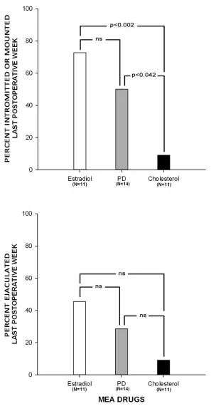
The proportions of animals that mounted or intromitted during the terminal post-operative test in the E2
(9 of 11) or PD (7 of 14) groups were significantly greater than the proportions of animals that that
mounted or intromitted in the Chol group (1 of 11) (p<0.002, p<0.042 respectively). Moreover, the
proportions of the animals in the E2 group that mounted or intromitted on the terminal post-operative
trial did not differ significantly from that in the PD group (Figure 3A). The proportion of animals that
ejaculated during the terminal post-operative test was highest in the E2 group (5 of 11) and lowest in the
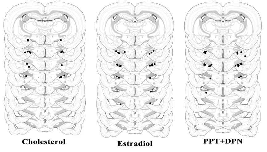
These values did not differ significantly between the three groups (Figure 3B). The histological analysis
Time course of E2 (n=11), PPT and DPN (n=14) , and Chol (n=11) effects on mating frequencies of castrated, DHT maintained male rats. The three groups were not significantly different during the pre-operative trials. The differences in behavioral indices arose post-pre-operatively. A: Mount Frequency. Post-operatively, all 3 groups showed a significant decline in MF between the last pre-operative and the first post-operative tests. MF returned to values not different from those obtained during the pre-operative trials in the E2-MEA group. Animals in the Chol-MEA group showed a steady decline in MF. Male rats that received PD-MEA implants were intemediate. B: Intromission Frequency. Post-operatively, all 3 groups showed a significant decline in IF between the last pre-operative and the first post-operative tests. IF did not return to pre-operative lvels in any of the groups. C: Mount and Intromission
Frequency. MIF follows the same pattern as MF. D: Ejaculation Frequency. Post-operatively, all 3 groups showed a significant decline in EF compared to pre-operative levels. EF continued to decrese throughout the post-operative trials so that by the terminal post-operative test all the groups showed siglificantly lower EF compared to terminal pre-operative test and the groups did not differ significantly from each other.
* Within-group difference during the terminal pre-oprerative test and first post-operative test, p<0.05. + Within-group difference during the terminal pre-oprerative test and terminal post-operative test, p<0.05.
# Within-group difference during the first post-operative test and terminal post-operative test, p<0.05. E/C- Group difference between Estradiol and Cholesterol, p<0.05.
Time course of E2 (n=11), PPT and DPN (n=14) , and Chol (n=11) effects on mating latencies of castrated, DHT maintained male rats. The three groups were not significantly different during the pre-operative trials. The differences in behavioral indices arose post-operatively.A: Mount Latency. During the first post-operative test, MLAT increased for all of the groups, however, only in the PD group was this significant. Throughut the post-operative trials MLAT continued to rise for all of the groups numerically. During the terminal post-operative trial E2-MEA displayed the lowest MLAT values that were
numerically but not significantly higher compared to the pre-operative values. PD-MEA and Chol-MEA groups showed significantly higher MLAT than on the terminal post-operative test compared to the terminal pre-operative values. These two groups were not significantly different from each other, yet numerically Chol-MEA group displayed longer MLAT than the PD-MEA group. B: Ejaculation Latency.
ELAT MIF followed the same trend as MLAT. C: Post Ejaculatory Interval. Each group showed a significant elevation of PEI on the first post-operative trial. The values for PEI continued to rise throughout the post-operativetests and were significantly higer for all of the groups on the terminal post-operative test.
* Within-group difference during the terminal pre-oprerative test and first post-operative test, p<0.05. + Within-group difference during the terminal pre-oprerative test and terminal post-operative test, p<0.05.
# Within-group difference during the first post-operative test and terminal post-operative test, p<0.05. E/C- Group difference between Estradiol and Cholesterol, p<0.05.
Terminal post-operative mating behaviors (percentages) for the E2, PD, and Chol groups. A: Percent Intromitted or Mounted. A large proportion (9 of 11, 81.8%) of castrated DHT maintained male rats given E2-MEA implaints mounted and intromitted and the proportion of rats receiving PD-MEA implants
that mounted and intromitted (7 of 14, 50.0%) did not differ significantly from the E2-MEA group. Only 1
FIGURE 4: CANNULAE PLACEMENT TO THE MEA.
4. DISCUSSION
Because, in the MPO, mating behavior is supported by activation of a single receptor (ERα), [Russell et
al., 2012] it was expected that the estrogen-sensitive neural circuits of MEA might work similarly.
However, this is not the case. Stimulation of MEA ERα alone by PPT (ERα agonist) did not mimic the
actions of E2 and did not support mating behavior [Russell et al., 2012]. Moreover, stimulation of MEA
ERβ by DPN (ERβ agonist) alone also does not promote mating [Russell et al., 2012]. Therefore, because
in the MEA the effect of either PPT or DPN is more limited and selective than that of E2, the objective of
this experiment was to determine if simultaneous stimulation of both ER subtypes is required for
activation of the estrogen-sensitive neural circuits within MEA that mediate mating behavior.
The hypothesis was confirmed by examining the mating response of male rats implanted with a
combination of selective estrogenic agonists of ERα and ERβ (PPT and DPN respectively) into the MEA
and by comparison of this group to controls receiving E2 or Chol MEA implants. Because only MEA and
not other brain areas important for mechanical execution of copulatory behavior, such as MPO, were
stimulated, we expected post-operative behavioral indices to be lower for all of the groups; we also
expected that only mounts and intromissions, but not ejaculations, would be maintained [Huggleston
et.al., 2003]. Therefore, ejaculation frequency, ejaculation latency, and post ejaculatory interval did not
figure prominently in the evaluation of the data although these data are included in the results.
During the pre-operative trials animals in all groups (PD, E2, and Chol) mated vigorously and
were not significantly different from each other on any behavioral indices. The number of mounts and
intromissions declined significantly in all groups post-operatively as expected. Mounts and intromissions
displayed by the E2-MEA group returned to the pre-operative levels by the terminal post-operative trial,
while the mounts and intromissions displayed by animals receiving Chol-MEA implants declined
progressively throughout the postoperative period and were significantly lower on the terminal
PD-MEA implants failed to maintain mounts and intromissions at the pre-operative levels they did maintain
these mating behavior indices at the levels not significantly different from those seen during the first
pre-operative trial. Between-group statistical comparison of the mounts and intromissions exhibited by
the rats in the three groups on the terminal post-operative week showed that PD group was not
significantly different from the E2 group, nor was it significantly different from the Chol group.
Furthermore, on the terminal post-operative trial the proportions of animals that displayed mounts or
intromissions in the PD-MEA and E2-MEA groups were significantly higher than the proportion of animals
displaying mounts or intromissions in the Chol-MEA group and the PD-MEA group did not differ
significantly from the E2-MEA group. These findings suggest that MEA responded to activation by
combined administration of ERα and ERβ selective estrogenic agonists, however, the response did not
precisely mimic that of E2. Nevertheless, we conclude that ERα and ERβ play a role in activation of
estrogen sensitive circuits in MEA responsible for mediation of mating behavior because combined
application of PPT and DPN produced results that were numerically and in some instances significantly
higher than those observed in Chol treated animals while singular application of either of the drugs did
not produce these effects [Russell et al., 2012].
Although activation of MEA through simultaneous administration of PPT and DPN was
successful, dual administration of these ER agonists did not mimic the action of E2 in the MEA. The
plausible explanations for this may lie in the dosage of drugs administered. The animals in the PD group
were given a 50/50 mixture of PPT and DPN because an appropriate ratio of PPT/DPN required to
activate E2-dependent circuits in the MEA is not known. Perhaps, if the ratio of the drugs had been
altered, we would have observed the rats displaying mating behavior at the levels not statistically
different from rats receiving E2-MEA treatment. When ERα expression was knocked out or knocked
down in MEA, the mating was not affected [Paisley et al., 2012]. This previous finding, together with the
required in lower concentrations than ERβ. Thus, a 50/50 mixture of PPT and DPN might not have
provided enough of ERβ agonist to full activate the circuits in MEA.
Although our findings provide some information about the role of E2 in MEA to promote mating
behavior, the mechanisms of E2 action is MEA are still not well understood. Several studies showed that
ERα and ERβ are co-expressed in cells of MEA and that in female rats the cells that co-express both
receptor subtypes also exhibit FOS-ir in response to mating stimuli [Greco et al., 2003; Shughrue et al.,
1997; Shughrue et al., 1998]. It is also known that ERα and ERβ undergo dimerization to form
homodimers and heterodimers during the reaction cascade that results in gene activation leading to
expression of mating behavior [Cowley et al., 1997]. ERs are able to form heterodimers if both receptor
subtypes are colocalized within a cell and it has been proposed that different functions are rendered
based on the type of dimer that could be formed in an area [Shughrue et al., 1997; Shughrue et al.,
1998]. However, a new finding showed that heterodimers may be formed when only one subtype is
bound by a ligand [Paulmurugan et al., 2011]. Therefore, when PPT alone was given, it is possible that
ERα/ERβ heterodimer was formed along with an ERα homodimer and when DPN was given alone, it is
possible that ERα/ERβ heterodimer was formed along with an ERβ homodimer. Neither of these pairs of
dimers were sufficient to activate E2-dependent neuronal circuits in MEA [Russell, et al., 2012].
However, when a combination of PPT and DPN was administered to MEA, it is possible that ERα/ERβ
heterodimer, an ERα/ERα and an ERβ/ERβ homodimers were also formed and that all three dimers are
required to facilitate mating behavior.
The results of this experiment might appear to contradict previous findings because it was
shown that, in the MEA, ERα is not necessary for male rats to display mating behavior [Paisley et al.,
2012]. However, the current findings suggest that ERα nevertheless participates in the signaling pathway
which leads to the display of mating behavior by male rats. There are multiple plausible explanations for
mating behavior on its own and instead must be coupled to another receptor in order to generate the
response. Although ERβ might normally be coupled to ERα, there might be other possibilities. Because
mating behavior persists when ERα mRNA expression is inhibited [Paisley et al., 2012], it is possible that
ERβ may couple to another non-ERα receptor (not yet identified). Another possibility could involve two
different pathways, one of which engages dimers of ERα and ERβ, while the other proceeds through a
novel type of E2 receptor not linked to ERα or ERβ. Yet another interpretation may emerge from this
experiment. It is reasonable to assume that when expression of ERα mRNA was inhibited in MEA by
AS-ODN infusion [Paisley, et al., 2012], it did not completely knock out ERα, but rather only knocked down
ERα expression. The reduced level of ERα in MPO may have been too low to support the mating
behavior if ERα is primarily responsible for E2 actions in MPO [Paisley et al., 2012; Russell et al., 2012].
However, low residual levels of ERα in MEA might still be enough to sustain the response of the MEA to
E2 because they may aid ERβ even if ERα is not the primary E2 receptor of MEA. To further investigate
the actions of E2 in MEA an anatomical study of male MEA to test if the cells co-expressing ERα and ERβ
also would exhibit Fos-ir upon mating stimulation might be valuable. Nevertheless, it now appears that
E2 dependent circuits in the MEA responsible for activation of mating behavior in male rats proceed
through simultaneous activation of both ERα or ERβ and that both receptor subtypes are important in
REFERENCES
Ando S., Aquila S., Beraldi, E., Canonaco, M., Panno, M.L., Valenti, A., Dessi-Fulgheri, F., 1988.
Physiological changes in androgen plasma levels with elapsing of time from Castration in adult
male rats. Horm. Metab. Res. 20, 96–99.
Baum, M.J., Vreeburg, J.T.M., 1973. Copulation in castrated male rats following combined treatment
with estradiol and dihydrotestosterone. Science 182(109), 283-285.
Bonsall, R.W., Clancy, A,N., Michael, R.P., 1992. Effects of the nonsteroidal aromatase inhibitor,
fadrozole, on sexual behavior in male rats. Horm. Beh. 20, 240-254.
Charn, T. H., Liu, E. T., Chang, E.C., Lee, Y.K., Katzenellenbogen, J.A., Katzenellenbogen, B.S., 2010.
Genome-Wide Dynamics of Chromatin Binding of Estrogen Receptors α and β: Mutual
Restriction and Competitive Site Selection. Mol. Endocrinol. 24 (1), 47-59.
Christensen L.W., Clemens L.G., 1974. Intrahypothalamic implants of testosterone or estradiol and
resumption of masculine sexual behavior in long-term castrated male rats. Endocrinology
95(4), 984-990.
Cowley, S.M., Hoare, S., Mosselman, S., Parker, M.G., 1997. Estrogen receptors α and β form
heterodimers on DNA. J. Biol. Chem. 272, 19858-19862
Davidson, J.M., 1966a. Activation of the male rat’s sexual behavior by intracerebral implantation
Davidson, J.M., 1966b. Characteristics of sexual behavior in male rats following castration. Anim.
Behav. 14, 266-272.
Davidson, J.M., 1969. Effects of estrogen on the sexual behavior of male rats. Endocrinology
84(6), 1365-1372.
Feder, H.H., 1971. The comparative actions of testosterone propionate and 5α-androstan-17β-ol-
3-one propionate on the reproductive behavior, physiology and morphology of male rats. J
Endocrinol. 51(2), 241-252.
Feder, H.H., Naftolin, F., Ryan, K.J., 1974. Male and female sexual responses in male rats given
estradiol benzoate and 5 alpha-androstan-17 beta-ol-3-one propionate. Endocrinology
94, 136-141.
Greco, B., Allegretto, E.A., Tetel, M.J., Blaustein, J.D., 2001. Coexpression of ER beta with ER alpha and
progestin receptor proteins in the female rat forebrain: Effects of estradiol treatment,
Endocrinology 142 (12), 5172-5181.
Greco, B., Blasberg M.E., Kosinski E.C. , Blaustein, J.D., 2003. Response of ERalpha-IR and ERbeta-IR cells
in the forebrain of female rats to mating stimuli. Horm. Behav. 43, 444–453.
Hillisch, A., Peters, O., Kosemund, D., Müller, G., Walter, A., Schneider, B., Reddersen, G., Elger, W.,
Fritzemeier, K., 2004. Dissecting Physiological Roles of Estrogen Receptor α and β with Potent
Huddleston, G.G., Michael, R.P., Zumpe, D., Clancy, A.N., 2003. Estradiol in the male rat
amygdala facilitates mounting but not ejaculation. Physiol. Behav. 79, 239-246.
Huddleston, G.G., Paisley, J.C., Clancy, A.N., 2006. Effects of estrogen in the male rat medial
amygdala: infusion of an aromatase inhibitor lowers mating and bovine serum albumin
conjugated estradiol implants do not promote mating. Neuroendocrinology 83, 106-116.
Koike, S., Sakai, M., Muramatsu, M., 1987. Molecular cloning and characterization of rat estrogen
receptor cDNA. Nucleic Acids Res. 15, 2499–2513.
Kuiper, G.G.J.M., Enmark, E., Pelto-Huikko, M., Nilsson, S. , Gustafsson, J.A., 1996. Cloning of a novel
estrogen receptor expressed in rat prostate and ovary. Proc. Natl. Acad. Sci. 93, 5925–5930.
Lindberg, M.K., Moverare, S., Skrtic, S., Gao, H., Dahlman-Wright, K., Gustafsson, J., Ohlsson, C., 2003.
Estrogen Receptor (ER)-β Reduces ERα-Regulated Gene Transcription, Supporting a “Ying Yang”
Relationship between ERα and ERβ in Mice. Mol. Endocrinol. 17(2), 203-208.
Lipton, A., Harvey, H.A., Demers, L.M., Hanagan, J.R., Mulagha, M.T., Kochak, G.M., Fitzsimmons, S.,
Sander, S.I., Santen R.J., 1990. A phase I trial of CGS 16949A: A new aromatase inhibitor. Cancer
65, 1279-1285.
Lugg, J.A., Rajfer, J., González-Cadavid, N.F., 1995. Dihydrotestosterone is the active androgen in the
Martini, L., 1982. The 5-alpha reduction of testosterone in the neuroendocrine structures: biochemical
and physiological implications. Endocr. Rev. 3, 1-25.
Massa, R., Stupnicka, E., Kniewald, Z., Martini, L., 1972.The transformation of testosterone into
dihydrotestosterone by the brain and the anterior pituitary. J Steroid Biochem. 3, 385-399.
McGinnis, M.Y., Dreifuss, R.M., 1989. Evidence for a role of testosterone androgen receptor interactions
in mediating masculine sexual behavior in male rats. Endocrinology 124, 618-626.
Meyers, M.J., Sun J., Carlson, K.E., Marriner, G.A., Katzenellenbogen, B.S., Katzenellenbogen,
J.A., 2001. Estrogen receptor-potency-selective ligands: structure-activity relationship studies of
diarylpropionitriles and their acetylene and polar analogues. J. Med. Chem. 44, 4230–4251
Mitra, S.W., Hoskin, E., Yudkovitz, J., Pear, L., Wilkinson, H.A., Hayashi, S., Pfaff, D.W., Ogawa, S., Rohrer,
S.P., Schaeffer, J.M., McEwen, B.S., Alves, S.E., 2003. Immunolocalization of estrogen receptor
beta in the mouse brain: Comparison with estrogen receptor alpha. Endocrinology 144 (5),
2055-2067.
Mosselman, S., Polman, J., Dijkema, R., 1996. ERβ beta: identification and characterization of a novel
human estrogen receptor. FEBS Lett. 392, 49–53.
Naftolin, F., Ryan, K.J., Davies, I.J., Reddy, V., Flores, F., Petro, Z., Kuhn, M., White, R.J., Takaoka, Y.,
Wolin L., 1975. The formation of estrogens by central neuroendocrine tissues. Recent Prog.
Paisley, J.C., Huddleston, G.G., Denman, H.N., Carruth, L.L., Petrulis, A., Grober, M.S., Clancy,
A.N., 2012. Sexual Responses of the Male Rat Medial Preoptic Area and Medial Amygdala to
Estrogen. I: Site Specific Suppression of Estrogen Receptor Alpha. Horm. Behav. in print.
Parte, P., Juneja, H.S., 1992. Temporal changes in the serum levels of gonadotropins and Testosterone
in male rats bearing subcutaneous implants of 5 alpha-dihydrotestosterone. Int. J. Androl., 15,
355–364.
Paulmurugan, R., Tamrazi, A., Massoud, T. F., Katzenellenbogen, J.A., Gambhir, S.S., 2011. In Vitro and in
Vivo Molecular Imaging of Estrogen Receptor α and β Homo- and Heterodimerization:
Exploration of New Modes of Receptor Regulation. Mol. Endocrinol. 25 (12), 2029-2040.
Russell N.V., Ogaga-Mgbonyebi, E.V., Habteab,B., Dunigan, A.I., Tesfay, M., Clancy, A.N., 2012. Sexual
Responses of the Male Rat Medial Preoptic Area and Medial Amygdala to Estrogen. II: Site
Specific Effects of Selective Estrogenic Drugs. Horm. Behav. in print.
Shughrue, P.J., Lane, M.V., Merchenthaler, I., 1997. Comparative distribution of estrogen receptor-α and
-β mRNA in the rat central nervous system. J. Comp. Neurol. 388 (4), 1096-9861.
Shughrue, P.J., Scrimo, P.J., Merchenthaler, I., 1998. Evidence for the colocalization of estrogen receptor
beta mRNA and estrogen receptor-alpha immunoreactivity in neurons of the rat forebrain.
Siegel, S., 1956. Nonparametric Statistics for the Behavioral Sciences. McGraw-Hill, New York.
Stauffer, S.R., Coletta, C.J., Tedesco, R., Nishiguchi, G., Carlson, K., Sun, J., Katzenellenbogen, B.S.,
Katzenellenbogen, J.A., 2000. Pyrazole ligands: structure-affinity/activity relationships and
estrogen receptor-alpha-selective agonists. J Med Chem. 43(26), 4934-4947.
Södersten, P., 1973. Estrogen-activated sexual behavior in male rats. Horm. Behav. 4, 247–256.
Swanson, L.W., 1998. Brain maps: Structure of the Rat Brain, ed 2, revised. Elsevier, Amsterdam.
Takaoka, Y., Wolin L., 1975. The formation of estrogens by central neuroendocrine tissues. Recent Prog.