www.impactjournals.com/oncotarget/
Oncotarget, Vol. 5, No. 5
Heat shock proteins in multiple myeloma
Lei Zhang
1, Jacqueline H.L. Fok
1and Faith E. Davies
11 Haemato-Oncology Research Unit, Division of Molecular Pathology, Cancer Therapeutics and Clinical Studies, The Institute
of Cancer Research, London, UK
Correspondence to: Faith E. Davies, email: faith.davies@icr.ac.uk
Keywords: heat shock proteins, multiple myeloma, protein folding, ER stress, haematology
Received: November 7, 2013 Accepted: January 17, 2014 Published: January 19, 2014
This is an open-access article distributed under the terms of the Creative Commons Attribution License, which permits unrestricted use, distribution, and reproduction in any medium, provided the original author and source are credited.
ABSTRACT:
Heat shock proteins are molecular chaperones with a central role in protein folding and cellular protein homeostasis. They also play major roles in the development of cancer and in recent years have emerged as promising therapeutic targets. In this review, we discuss the known molecular mechanisms of various heat shock protein families and their involvement in cancer and in particular, multiple myeloma. In addition, we address the current progress and challenges in pharmacologically targeting these proteins as anti-cancer therapeutic strategies
INTRODUCTION
Heat shock proteins are a group of highly conserved
proteins found in both eukaryotic and prokaryotic cells.
They are involved in a wide range of cellular processes
such as assisting protein folding and degradation of
misfolded proteins, intracellular trafficking, modulating
signalling pathways and regulating immune responses
[1-5]. The multi-functional nature of heat shock proteins
enables them to play critical roles in the regulation of
protein homeostasis and cell survival. Although the
proteins are frequently associated with the cellular stress
response, they also play an important role in supporting
normal cellular processes such as development and
differentiation [6-8].
They were accidentally discovered in 1962, as a
set of genes whose expression is elevated by heat shock
in
Drosophila melanogaster
[9]. It is now known that
heat shock proteins function as molecular chaperones
and can play many roles in the cell in addition to
modulating the heat shock response. In mammalian cells,
they are classified into five families according to their
molecular weight: Hsp100, Hsp90, Hsp70, Hsp60 and
small heat shock proteins including Hsp27. Members
of each family can be either constitutively expressed or
cell event induced, and can be found in defined cellular
compartments carrying out specific functions.
There are numerous lines of evidence which link
heat shock proteins to the pathogenesis of cancer. They
are found to be overexpressed in a wide range of cancers
and are implicated in cell survival, apoptosis, invasion,
metastasis and escape of immune surveillance. As a
tumour progresses, it becomes increasingly dependent
on these proteins to adapt to its microenvironment and to
stabilise the large amount of oncogenic proteins produced
which support growth and survival. The different heat
shock protein families are being studied extensively as
potential anti-cancer targets for two main reasons: (1).
heat shock proteins interact with multiple cancer related
client proteins/pathways and targeting them may lead to
the inhibition of multiple cancer causing pathways; (2).
some cancers rely on heat shock proteins to survive the
proteotoxic stress induced by the production of excessive
proteins/oncogene products.
Multiple myeloma is a cancer resulting from the
malignant proliferation of plasma cells in the bone marrow
and one important feature of myeloma plasma cells is the
secretion of excessive monoclonal paraproteins [10].
Despite recent advances in treatment and the use of high
dose chemotherapy, the majority of patients relapse even
after successful initial treatment. To date, the disease
remains incurable with a median survival of 4 years.
There is therefore an urgent need for better treatments
and new drugs. In recent years, heat shock proteins have
become attractive potential therapeutic targets in multiple
myeloma, as the ability to deal with proteotoxic stress as
a result of paraprotein production is critical for myeloma
cell survival [10, 11]. Importantly, several inhibitors of
Hsp90 have demonstrated activity against myeloma cells
shock protein families and their implication in cancer
development and progression concentrating particularly,
on multiple myeloma. We also discuss the role of heat
shock proteins as potential therapeutic targets in multiple
myeloma and discuss the supporting pre-clinical and
clinical data.
The mechanisms and functions of heat shock
family proteins
Hsp90
Multiple Hsp90 family proteins exist in different
subcellular locations. These include the cytoplasmic
Hsp90α (inducible) and Hsp90β (constitutive),
mitochondrial TNF receptor-associated protein 1 (TRAP1)
and endoplasmic glucose regulated protein 94 (Grp94). All
of the proteins are highly abundant and the cytoplasmic
isoform is essential for cell survival [16], and studies
in yeast demonstrate that Hsp90 may interact with more
than 10% of the yeast proteome [17]. Unlike other heat
shock proteins involved in general protein folding
tasks, Hsp90 is found to interact only with a group of
selective client proteins, many in a more mature folding
conformation compared to Hsp70 substrates. Rather than
protein folding, Hsp90 is more commonly associated with
client protein maturation or functions to maintain a client
protein in a specific folding conformation required for
its activity, for example, to respond to activation signals
such as phosphorylation. The client proteins identified
to date consist mainly of protein kinases, receptors and
transcription factors, and many of these are involved in
cell cycle control and signalling pathways [18-24]. The
list of client proteins identified for Hsp90 is increasing, yet
the molecular basis for substrate selectivity is still largely
unknown as client proteins have no obvious common
sequence motifs. A current list of Hsp90-interacting
proteins has been maintained by Didier Picard and can
be found at (http://www.picard.ch/downloads/downloads.
htm), which includes client proteins as well as
co-chaperones.
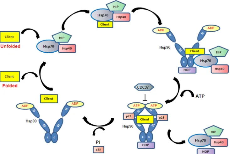
Hsp90 proteins exist as homodimers of subunits
consisting of an N-terminal ATPase domain, a C-terminal
dimerisation / protein interaction domain, and a middle
domain associated with client protein binding [25]. ATPase
activity is essential for the chaperoning activity of Hsp90
[26, 27]. Following the addition of ATP, Hsp90 undergoes
a conformational change, which induces an open to shut
conformation shift [28], with transient dimerisation of
the N-terminal domains and N-M domain association
(Figure 1) [29, 30]. The middle segment of Hsp90 has
been identified as the binding site for protein kinase
[image:2.612.122.488.449.695.2]PKB/Akt and is implicated as the main site for client
protein interactions [31]. This segment can also interact
with cochaperones and is required for N-terminal ATPase
activity. The C-terminal domain is involved in dimerisation
and contains a highly conserved EEVD sequence which
is required for the binding of tetratricopeptide repeat
(TPR) containing family of cofactors, such as HOP [32,
33]. The C-terminal domain also contains an alternative
ATP-binding site [34, 35], but how this contributes to the
overall function of Hsp90 remains unclear.
The Hsp90 homodimer functions like a molecular
clamp, using ATP binding and hydrolysis to drive the
confirmation change cycle of Hsp90 which is required
to facilitate the binding and release of client proteins.
It works in a multi-chaperone complex and the current
proposed mechanism is that client proteins first bind to
Hsp70. ATP hydrolysis of Hsp70 by Hsp40 stabilises the
initial client/Hsp70/40/HIP complex, which then interacts
with Hsp90 in ADP-bound open conformation via Hop,
presenting the client protein to Hsp90 [36]. When Hsp90
exchanges ADP to ATP, its open to shut conformational
change leads to the dissociation of Hsp70/40 and HOP
and the association of another set of co-chaperones
such as CDC37 and p23 to form the mature complex
[37, 38]. In this mature state the client protein becomes
activated (Figure 1). Studies of glucocorticoid receptor
(GR) activation demonstrate that Hsp90 and Hsp70 are
absolutely required for GR activation (i.e. opening of the
steroid binding cleft in GR), whereas cochaperones such
as Hop, Hsp40 and p23 help to facilitate the chaperoning
activity by client protein recruitment (HOP), ATP
hydrolysis of Hsp70 (Hsp40) and stabilisation of
Hsp90-ATP conformation (p23) [39-41]. To date more than 20
Hsp90 co-chaperones have been identified and all are
involved in the recruitment of client proteins, control
of client protein maturation and modulation of ATPase
activity. It is thought that the binding and release of
specific co-chaperones in an orderly way may control the
activity / selectivity of Hsp90, and that different client
proteins require a different set of co-chaperones. Given
the complexity of the Hsp90 chaperoning system, a full
understanding of the molecular mechanism is still lacking.
In addition to the cytoplasmic Hsp90s, protein
quality control requires the functions of compartment
specific Hsp90s located within various cellular organelles.
TRAP1 is an Hsp90 located in the mitochondria involved
in mitochondrial protein folding, cytoprotection and
mitochondrial integrity [42, 43]; whereas Grp94 is the only
Hsp90 residing in the endoplasmic reticulum (ER), where
it plays a critical role in regulating ER protein homeostasis
by chaperoning highly selective client proteins such as
immunoglobulins [44], targeting misfolded client proteins
for ER-associated degradation (ERAD) [45] and storing
Ca
2+to regulate ER calcium flux [46]. Both are implicated
in promoting tumour progression [43, 46].
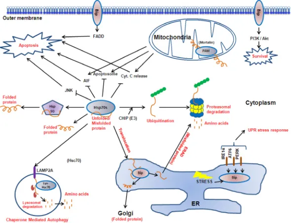
Hsp70
Eukaryotic cells also express a range of Hsp70
proteins in various subcellular localisations. Family
members include the constitutively expressed Hsc70
and stress induced Hsp72 in the cytoplasm, Bip (Grp78)
localised in the endoplasmic reticulum and mortalin/Grp75
in the mitochondria. Similar to Hsp90, Hsp70 protein
consists of a N-terminal ATPase domain where ATP
exchange acts as the driving force of the conformational
change required for target protein binding and release; a
substrate binding domain with affinity for hydrophobic
amino acid residues; and a C-terminal domain containing
an EEVD motif for co-chaperone binding and functioning
as a ‘lid’ which controls the availability of the substrate
binding domain to target proteins [47]. Hsp70 forms a
complex with its cochaperone Hsp40 and a nucleotide
exchange factor such as Bag-1 and HspBP1. Hsp40
stimulates Hsp70 assisted protein folding by interacting
with Hsp70 and promoting ATP hydrolysis, resulting in
a closed conformation and tight binding of substrate,
whereas a nucleotide exchange factor stimulates the
release of ADP and binding of ATP, thereby opening the
binding pocket for substrate release [48].
During protein synthesis, partially synthesized
and incompletely folded polypeptide chains expose
hydrophobic regions that need to be protected from
misfolding and aggregation. Hsp70 assists the
de novo
folding of 15-20% of all bacterial proteins, and this figure
is thought to be even higher in eukaryotes [3]. It interacts
with a wide spectrum of nascent polypeptide chains
co- and posttranslationally, with preference for chains
between 30-75kDa [49-51]. It utilises ATP driven cycles
of substrate binding and release to carry out chaperoning
functions, preventing aggregation by maintaining a low
free substrate concentration, while enabling free substrate
to fold to its native state [52-56]. On the other hand,
the binding and release cycles may also induce specific
unfolding of a misfolded polypeptide or pull apart
aggregated proteins for them to be refolded to their correct
state [56].
The family members found at different cellular
localisations fulfill specific roles. Collectively, they
form a key part in the cellular mechanism maintaining
protein homeostasis and cell survival (Figure 2). They
play central housekeeping functions in the cell as part
of a complex network working with co-chaperones and
downstream chaperoning systems such as Hsp90. In
addition to assisting the folding of newly synthesized
and refolding of misfolded proteins discussed above,
they translocate target proteins across membranes [57],
as well as directing protein degradation by the
ubiquitin-proteasome pathway [58] or autophagy [59]. An increasing
number of signal transduction proteins and transcription
factors are known to transiently interact with the Hsp70
complex [60], and together with the Hsp90 complex, the
Hsp70 system is linked to cell cycle regulation, apoptosis
and differentiation.
signal transductions [64-66]. Mitochondria matrix
localised Hsp70 (mortalin) forms part of the presequence
translocase-associated motor (PAM) complex which acts
as the driving motor of protein translocation from the
cytoplasm into the mitochondria [67]. Whereas cytosolic
Hsp70 is required for the post-translational translocation
of secretory proteins destined to the ER, by holding the
fully transcribed polypeptide in an incompletely folded
state for translocation [68, 69]
.
In addition the cytoplasmic
inducible Hsp72 and its cognate protein Hsc70 are
responsible for the folding of proteins in the cytoplasm
as well as the recruitment of E3 ubiquitin ligases such as
CHIP to tag target proteins for proteasomal degradation
[70]. Hsc70 also participates in chaperone-mediated
autophagy, a type of lysosomal degradation which
selectively targets specific proteins. Cytosolic Hsc70
binds to a target protein and presents it to the lysosome
receptor LAMP-2A. At this site the substrate protein is
subsequently unfolded and translocated into lysosome,
a process which is assisted by the lysosomal resident
Hsc70 [71, 72]. In addition to their roles in maintaining
the cellular protein program, cytoplasmic Hsp70 inhibits
both the caspase dependent and independent apoptosis
pathways at multiple levels [73, 74].
Small heat shock proteins - Hsp27
In contrast to Hsp90 and Hsp70, small heat shock
proteins are a family of ATP-independent chaperones.
With a small size between 15-30kDa, they oligomerise
to form homo or hetero-oligomers with up to 50 subunits
[75], which determines their chaperoning activity. In
addition to their phosphorylation status, cell-cell signalling
and various protein modifications also modulate their
oligomerisation [76].
Hsp27 belongs to this family and functions to
prevent protein aggregation by directly binding misfolded
substrates, and promoting protein refolding by interaction
with the Hsp70 chaperone complex. In addition, Hsp27
can directly prevent cell death by interfering with key
components of the apoptosis pathway, such as blocking
the formation of the apoptosome by binding to cytochrome
c released from the mitochondria [77], and by interacting
with Daxx, a mediator of Fas-induced apoptosis [78].
Under stress conditions, Hsp27 is also directly
involved in the ubiquitin-proteasome pathway by binding
to the 26S proteasome and multi-ubiquitin chains, to
facilitate the degradation of a selective range of target
proteins [79]. By doing so, Hsp27 can mediate its
cytoprotective effect at multiple levels by facilitating the
degradation of various apoptotic and cell cycle proteins.
For example, Hsp27 can enhance the anti-apoptotic
activtity of the transcription factor NF-κB, as the presence
of Hsp27 in the proteasome-protein substrate complex
is required for the degradation of I-κBα, the inhibitor of
NF-κB
[79]. Hsp27 can also promote
the degradation of
the cell cycle inhibitor p27
Kip1, thereby avoiding cell cycle
arrest during stress [80].
Hsp60/Hsp10
[image:4.612.156.451.460.686.2]Hsp60 and Hsp10 form the mitochondrial
chaperonin complex, which is involved in mitochondrial
protein folding. The understanding of the structure and
Figure 2: The Hsp70 family proteins.
Hsp70 protein isoforms (Bip, cytoplasmic Hsp70s, lys-Hsc70 and mortalin) reside at varioussubcellular localisations to perform specific roles in protein folding, translocation, degradation and signal transduction, thereby mediating
function of chaperonin has mainly come from studies
performed on the bacterial chaperonin, GroEL and GroES.
GroEL (Hsp60) is an oligomer formed by monomers
arranged into two stacked heptameric rings [81, 82],
resulting in a barrel like cavity where misfolded or
unfolded substrate proteins are folded. GroES (Hsp10),
which forms a single heptameric ring, acts as a lid to the
chamber and can bind to either end of the double GroEL
rings [83, 84]. Like Hsp70 and Hsp90, ATP cycles induce
conformational changes required for substrate protein
folding. ATP and polypeptide binds to one GroEL ring,
followed by GroES capping, resulting in the encapsulation
of polypeptide in a hydrophilic cavity which promotes
protein folding conditions [84]. Once the substrate is
inside the chamber, ATP is hydrolysed slowly, allowing
time for the protein to fold. The two rings of GroEL act
in an alternate fashion [85], with ATP hydrolysis in one
ring resulting in a structural transition in the opposite ring
making it available for ATP binding, which in turn triggers
the release of GroES and substrate protein from the
original ring. A substrate protein may go through multiple
binding and release cycles to reach its folded state [85].
In Eukaryotes, Hsp60 was first shown to reside in
the mitochondria, and following interaction with Hsp10,
is responsible for chaperoning nascent polypeptides as
well as transporting target proteins from the cytoplasm
into the mitochondria [86, 87]. Evidence also suggests
that Hsp60 participates in apoptosis by interactions with
mortalin (mitochondrial hsp70), p53 and survivin [88-90].
Accumulating evidence suggests that Hsp60 is not just a
mitochondrial protein, as it also resides in the cytoplasm
and unlike other heat shock proteins that mostly have
pro-survival functions, Hsp60 has either pro-survival or
pro-apoptosis functions [91]. It has also been found on
the cell surface where it is involved in the activation of
immune system [92, 93] and in the extracellular matrix
where it has pro-inflammatory functions [94, 95]. The
molecular mechanism of Hsp60 in humans remains largely
unknown, but its involvement in cancer as well as its
potential applications in cancer therapy is actively being
investigated.
Hsp110 (Hsp105)
Hsp110/105 is abundant in the cytosol of
mammalian cells but relatively little is known about its
function compared to the other heat shock proteins. It has
diverged from the Hsp70 superfamily, and has independent
chaperone activity as well as serving as a nuclear exchange
factor to Hsp70s [96]. As an independent chaperone,
unlike Hsp70, Hsp110 cannot assist protein folding, but
acts to prevent protein aggregation of denatured proteins
with higher efficiency compared to Hsp70 [97, 98]. It also
exhibits differential substrate binding properties to Hsp70s
with preference for substrates with aromatic residues, and
this may account for the different chaperone activities of
Hsp110 and Hsp70 [99].
Hsf1
It is widely established that Hsf1 is the “master
regulator” of heat shock protein expression. In the absence
of stress, inactive Hsf1 monomers are held in a complex
with Hsp70/Hsp90. At the onset of proteotoxic conditions,
Hsf1 is released from the complex, homo-trimerises,
translocates to the nucleus and activate the transcription
of its downstream targets by binding to the heat shock
elements in the promoter regions of target proteins [100,
101]. Hsf1 is classically recognised as a regulator of heat
shock protein expression with its downstream targets such
as Hsp72 and Hsp27. Several recent genome-wide analysis
using ChIP and microarray technologies along with Hsf1
siRNA in yeast [102] and mammalian cells [103, 104] have
uncovered a plethora of previously undiscovered Hsf1
gene targets, including genes implicated in transcriptional,
RNA splicing, ubiquitylation, stress defence, vesicular
transport and cell structures. Page
et al
[103] emphasized
that aside from the chaperones induced by Hsf1 upon heat
shock, the second most substantially induced group were
genes coding for anti-apoptotic proteins. These
genome-wide analyses also reveal the role of Hsf1 in regulation
of stress, cellular adaptation, survival, development and
disease.
Heat shock proteins contribute to cancer
progression and metastasis
Cancer cells proliferate at a fast rate and in order
to survive, they resist apoptosis, upregulate oncogenes/
oncoproteins, cope with environmental stresses such
as hypoxia, and modulate various survival signalling
pathways. Therefore, in order to overcome the challenging
hostile environment, cancer cells have higher metabolic
requirements for chaperones than non-cancer cells.
Hsp90
namely sustaining proliferative signalling, resisting cell
death, evading growth suppressors, inducing angiogenesis,
enabling replicative immortality, invasion and metastasis,
and emerging hallmarks including deregulating cellular
energetic and avoiding immune destruction [110, 111].
In addition, high expression of Hsp90 is an independent
prognostic marker in a number of cancers. In breast
cancer, it is associated with decreased survival [109], and
in gastric cancer, high Hsp90 expression is linked to poor
prognosis and tumour aggressiveness [112]. In CML,
Hsp90 correlates with disease state and high levels are
associated with resistance to therapy [113].
Hsp70
Unlike Hsp90 which chaperones specific ‘client
proteins’, Hsp70 family proteins assist general folding
of unfolded or misfolded proteins exposing hydrophobic
regions and prevent their aggregation. High Hsp70
expression is correlated with poor prognosis in a wide
range of cancers such as breast, endometrial, cervical, oral
and bladder carcinomas and has been extensively reviewed
elsewhere [114]. The anti-apoptotic role has also linked
Hsp70 to chemotherapeutic resistance in ovarian cancer
and leukaemia [115, 116]. Hsp70 is involved in multiple
cancer promoting pathways by associating with the Hsp90
chaperone system as well as carrying out independent
functions in apoptosis, senescence, and protein regulatory
pathways such as autophagy [117].
The cytoplasmic Hsp70s regulate the apoptosis
pathway at multiple levels, for example, Hsp70s have
been shown to protect Bcl-2 from proteasomal degradation
[118]; block Bax translocation to the mitochondria thereby
preventing cytochrome c release [119]; bind Apaf-1 and
prevent the recruitment of caspase-9 to the apoptosome
[120, 121]; and to prevent AIF translocation to the nucleus
to cause chromatin condensation and DNA degradation
[122, 123]. It is interesting to note that the function of
Hsp70s do not always rely on their ATPase activity, for
instance it has been shown that Hsp72 inhibits JNK
activation independently of its chaperoning activity
[124-126]. The Hsp70s also play a protective role against
senescence. Hsp72 knock down induces senescence
in a variety of cancer cell lines [127, 128] and Hsp72
controls Her-2-induced senescence by regulating p21
and survivin in a mouse breast tumour model [129].
Evidence also suggests that Hsp70 supports autophagy by
maintaining protein homeostasis and supporting cancer
cell survival. Hsp70 localises at the autophagosome/
lysosomal membrane compartments and inhibits
lysosomal permeabilisation [130, 131]. In addition,
Hsp70 participates in chaperone mediated autophagy by
delivering target proteins to the lysosome surface receptor
LAMP-2A, where it enables their translocation into the
lysosomal lumen (Figure 2) [71, 132].
Small heat shock proteins - Hsp27
Hsp27 is also commonly overexpressed, correlating
with prognosis and chemoresistance in many cancers
including colorectal [133], breast [134], prostate [135]
and ovarian [136]. Elevated expression is associated
with tumour aggressiveness in both primary and
metastatic tumours. Apart from having anti-apoptotic
roles at multiple levels contributing to primary tumour
survival, Hsp27 is involved in actin dynamics and is
overexpressed in metastatic breast tumour contributing
to cell migration and invasion. Silencing of Hsp27 leads
to decreased bone metastasis in a breast tumour model
[137]. In addition, Hsp27 is implicated in
epithelial-to-mesenchymal transition (EMT) in breast [138], lung
[139], and has been shown to be a key mediator of both
IL-6 dependent and independent EMT in prostate cancer
[140]. Experimental models also suggest that Hsp27 can
promote angiogenesis by NFkB dependent upregulation
of VEGF-gene transcription and secretion of VEGFR-2 in
endothelial cells [141]. Knocking down Hsp27 in breast
cancer cells reduced endothelial cell proliferation and
reduced secretion of VEGF and FGF [142].
Hsp60/Hsp10
Increasing evidences suggest that Hsp60 and Hsp10
may also be important players in cancer progression. As
reviewed by Cappello
et al
[143], Hsp60 expression is
altered in a wide range of cancers with potential diagnostic
and prognostic implications. As well as assisting protein
folding in association with Hsp10, cytosolic Hsp60 can
regulate apoptosis by stabilizing the apoptosis inhibitor
survivin [89] and binding to and inhibiting pro-apoptotic
Bax and Bak [144]. Conversely, Hsp60 can also promote
the activation of caspase-3, leading to tumour cell death
[145]. Hsp60 interacts with β-catenin—a key oncogene
driving cancer development and metastasis, where it is
found to enhance β-catenin transcriptional activity thereby
promoting metastasis [146]. Cell surface Hsp60 also
directly interacts with and activates
α3β1 integrin, which
that Hsp10 may enable tumour cells to escape immune
surveillance by suppressing T cells expressing CD3 zeta
chain, inhibiting cytokine production [151].
Hsp110 (Hsp105)
Finally, Hsp110 may have a potential use in
cancer as a marker of prognosis and drug response. It is
overexpressed in malignant melanoma [152], colorectal
[153] and pituitary tumours [154], and high expression
is associated with advanced and metastatic lesions [153,
155]. In contrast, a reduction in Hsp110 expression was
correlated with invasion and metastasis and therefore
poor prognosis of oesophageal cancer [156]. A truncated
mutant of Hsp110 has been found in colorectal cancer
with microsatellite instability, and in this type of cancer
the truncated mutant inhibits the protective role of the wild
type form in a dominant negative manner. High expression
of the truncated mutant is linked to chemo-sensitivity and
better prognosis [157].
Heat shock proteins are potential therapeutic
targets in multiple myeloma
Multiple myeloma is characterised by the production
of large quantities of nascent immunoglobulin [10]. As
a result, myeloma cells rely on their protein handling
mechanisms to cope with protein load and maintain
survival. A number of pathways are responsible for
protein homeostasis in the cell including the unfolded
protein response (UPR), ubiquitin proteasome pathway,
autophagy and aggresome pathway. The endoplasmic
reticulum (ER) is a site of protein folding and quality
control. The accumulation of unfolded/misfolded proteins
in the lumen of the ER triggers the UPR, which activates
downstream pathways to inhibit protein translation
and increases protein folding capacity by upregulating
molecular chaperones [158]. If proteins cannot be
correctly folded in the ER, they are retrotranslocated to
the cytoplasm to be ubiquitinated and destroyed by the
proteasome [159]. Alternatively, excess proteins can be
removed by autophagy via lysosomal degradation, which
can be upregulated during stress triggered by protein
aggregation, nutrient deprivation, or proteasome inhibition
[160-164]. When protein folding and degradation capacity
is exceeded, unfolded/misfolded protein aggregates in
the cytoplasm are transported along the microtubule
to the microtubule organising centre, where they form
aggresomes [165, 166]. The aggresomes act as storage
centres for toxic proteins until these proteins are eventually
targeted to chaperones for refolding or degradation by
autophagy [167, 168].
The protein handling pathway is a tightly linked
process and is overloaded by the large amount of
immunoglogulin produced in myeloma. As a result, the
protein handling pathway is actively being explored as an
attractive therapeutic target in myeloma. The success of
the clinically approved proteasome inhibitor bortezomib
provides evidence that targeting this pathway can be an
effective treatment strategy in myeloma. Efforts have
therefore been put into developing inhibitors of the
UPR, heat shock proteins, proteasome, autophagy and
aggresomes, and these have been extensively reviewed by
Aronson
et al
[161].
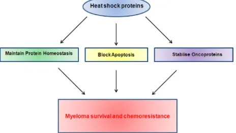
Being molecular chaperones responsible for protein
folding, the heat shock proteins play a key role in all of
the protein homeostasis pathways and thus the handling of
immunoglobulin folding in myeloma. In addition to their
chaperoning functions, heat shock proteins are found to be
involved in many other signalling pathways important for
myeloma growth and survival, making them particularly
attractive targets (Figure 3).
Numerous studies have shown that Hsp90 inhibition
in myeloma cells induces apoptosis and depletes multiple
client proteins such as AKT, STAT3, IL-6Rα, thereby
simultaneously disrupting multiple pathways known to
contribute to cell survival, including the JAK/STAT, PI3K,
NF-κβ, and RAS/ERK pathways [169]. Cytoplasmic
Hsp90 has also been shown to be a modulator of the
UPR by associating with and stabilising IRE1 and PERK,
two major transactivators of the UPR responsible for
the downstream upregulation of stress response genes
and translational repression [170]. It is also shown that
Hsp90 inhibition induces UPR in myeloma, and that
Hsp90 inhibitors induce myeloma cell death at least in
part via the
UPR
death pathway [11]. Given the support
of myeloma cell growth and survival by the bone marrow
microenvironment, the ability of HSP90 inhibition
to overcome exogenous IL-4-induced chemotherapy
resistance highlights the potential efficacy of HSP90
inhibitors
in vivo
[171].
Preclinical studies demonstrate that the inhibition
of Hsp90 is effective in myeloma
in vitro
and
in vivo
[image:7.612.59.291.555.686.2][11, 14, 172]. However, treatment of myeloma cells with
Figure 3: Heat shock proteins contribute to myeloma
compounds such as Hsp90 inhibitors (17-AAG,
NVP-AUY922), bortezomib and dexamethasone is usually
accompanied by the upregulation of other heat shock
proteins such as Hsp70 and Hsp27, protecting cells
from apoptosis and contributing to drug resistance [14,
173, 174]. For instance, it has been shown that Hsp27
is overexpressed in myeloma and inhibits the release of
Smac, an activator of caspases from the mitochondria,
thereby confering dexamethasone resistance [175].
Blocking the Hsp27 upregulation associated with
bortezomib treatment overcomes bortezomib resistance
[176], and inhibiting p38 MAPK, an upstream regulator
of Hsp27, sensitises myeloma cells to bortezomib induced
cell death by downregulating Hsp27 [177]. Evidence
for HSP90 inhibition having a cytostatic effect on colon
adenocarcinoma cells, rather than inducing cell death,
suggests that HSP90 inhibitors used in combination with
other agents can enhance tumour cell kill [178]. Further
studies in myeloma models will be needed to explore
possible drug combinations with HSP90 inhibitors.
The cytoplasmic Hsp70s, inducible Hsp72
and constitutively expressed Hsc70 are frequently
overexpressed in myeloma [179]. Inhibition of Hsp70 is
also effective in inducing myeloma cell death both
in vitro
and
in vivo
[179-181]. As they function as co-chaperones
of Hsp90, inhibition leads to Hsp90 client protein
depletion as well as cell death [179, 180]. Inhibition of
Hsp90 typically leads to a compensatory upregulation
of Hsp72, and inhibiting both Hsp70 and Hsp90 results
in a more effective anti-tumour response than inhibiting
Hsp90 alone [179, 180, 182]. It has also been shown that
Hsc70 and Hsp72, sharing 85% sequence homology, have
compensatory yet distinctive roles in immunoglobulin
folding and survival of myeloma [180, 182].
As Hsf1 is a major transcription factor responsible
for the rapid induction of heat shock proteins during stress,
recent studies have also explored its role as a potential
therapeutic target. Hsf1 regulates gene expression of
heat shock proteins such as Hsp90, Hsp72 and Hsp27,
as well as non-chaperone genes potentially utilized by
cancers such as the tumour necrosis factor (TNF) receptor
[104]. High level of Hsf1 is associated with cancer
malignancy and poor prognosis clinically, and there is
considerable evidence for the direct involvement of Hsf1
in tumourigenesis in cancers including myeloma [183,
184]. Early studies also suggest that inhibition of Hsf1
induces apoptosis in myeloma cells
in vitro
and reduces
tumour growth
in vivo
, and this is associated with lowered
expression of multiple downstream heat shock proteins.
Development of heat shock protein inhibitors for
the treatment of multiple myeloma
The activity of various inhibitors of the heat shock
response is currently being explored, however to date only
inhibitors of Hsp90 have reached the advanced stages of
clinical development.
Hsp90 inhibitors
The development of Hsp90 inhibitors was initially
based on the natural product geldanamycin, which has
potent anti-tumour activity in a wide range of tumour cell
lines. Geldanamycin binds to the N-terminal domain of
Hsp90, blocking the site of ATP binding and hydrolysis
[185]. A number of geldanamycin derivatives have since
been developed with improved solubility, stability and
toxicology [186]. The geldanamycin based tanespimycin
(17-AAG) was the first to enter the clinic as it showed
single agent activity
in vitro
on myeloma cell lines [12,
13], and combination treatment with bortezomib led to an
increased accumulation of ubiquitinated proteins compared
to single agent exposure [187]. Despite encouraging initial
results, the development of tanespimycin has since been
discontinued. Retaspimycin (IPI-504)
is a derivative of
17-AAG thought to be more potent and less toxic to the
liver than 17-AAG. A phase I trial showed that it is well
tolerated in myeloma patients [188]
, with similar synergy
when combined with bortezomib [189].
In addition to geldanamycin based compounds,
a number of novel Hsp90 inhibitors have recently been
developed and are undergoing preclinical and clinical
studies. NVP-AUY922 (VER52296) efficiently induced
apoptosis in myeloma cells at nanomolar concentrations
and triggered changes in the molecular signature of
cells characteristic of Hsp90 inhibition [14]. Phase I/II
studies of AUY922 with and without bortezomib, with
or without dexamethasone are currently being performed
in patients with relapsed or refractory multiple myeloma
(NCT00708292). KW-2478 is another promising
novel compound discovered through a unique lead
optimization strategy including microbial screening, X-ray
crystallography, cell-based screening and
in vivo
models
[15]. A study on myeloma cell lines showed that
KW-2478, a novel non-purine analogue antagonist, induced
growth inhibition and apoptosis associated with Hsp90
client protein depletion [15], and combination with
bortezomib exhibited synergistic activity
in vitro
and
in
vivo
[190]. Phase I/II Study of KW-2478 in combination
with bortezomib in multiple myeloma is ongoing
(NCT01063907).
being considered [193].
PU-H71, is an emerging purine scaffold HSP90
inhibitor that can not only bind a larger scope of
HSP90 conformations compared to its
geldanamycin-derived predecessors, but is also unaffected by HSP90
phosphosphorylation. PU-H71 exhibits potent
anti-myeloma
activity in cell lines by inhibiting both the
cytoplasmic and ER resident Hsp90 (Grp94) resulting in
the activation of the UPR and caspase dependent apoptosis
in myeloma cell lines [194] [195].
Hsp70 inhibitors
The consistent upregulation of Hsp70s following
Hsp90 and proteasome inhibition, and their proven
anti-apoptotic roles contributing to drug resistance leads to a
growing interest in the development of Hsp70 inhibitors to
be used as single anti-cancer agents or in combination with
conventional or targeted chemotherapies. However, to date
few Hsp70 specific inhibitors have been identified. Two
Hsp70 specific compounds, Ver-155008 and MAL3-101,
have been tested on myeloma in the preclinical setting.
Ver-155008 is an ATP-analogue capable of inducing
caspase dependent apoptosis in a panel of myeloma cell
lines via the modulation of multiple oncogenic pathways
and enhancing Hsp90 inhibition induced cell death [179,
180]. In contrast to Ver-155008, MAL3-101 inhibits the
ability of Hsp40 cochaperone to stimulate Hsp70 ATPase
activity, thereby blocking Hsp70 functions in cells
[196]. MAL3-101 exhibited promising anti-myeloma
properties on myeloma cell lines
in vitro
and
in vivo
,
and demonstrated synergy with proteasome and Hsp90
inhibitors [181]. Although these compounds have limited
potency, they may form the basis for the development of
future derivatives suitable for the clinical setting [197].
Hsf1 inhibitors
As an alternative to targeting individual heat shock
proteins, there has been an interest in the development
of inhibitors against Hsf1, the ‘master regulator’ of heat
shock response. Since the inhibition of a single heat shock
protein such as Hsp90 inevitably leads to the compensatory
upregulation of other heat shock proteins such as Hsp70
and Hsp27, targeting Hsf1 instead of the individual
chaperones separately is potentially more therapeutically
effective, as inhibition of Hsf1 could in theory abolish the
ability of a cancer cell to activate the whole heat shock
response during cellular stress. The increased sensitivity
of hepatocellular carcinoma and melanoma cell lines
to HSP90 inhibition with HSF1 knocked down
in vitro
,
illustrates the therapeutic potential of an HSF1 inhibitor
in combination with HSP90 inhibition [198]. While
several small molecular compounds can interfere with
the transcriptional activation of Hsf1 or the downstream
translational mechanisms, the precise mechanisms of how
these compounds work remains unclear and hence, they
are not yet valid for clinical investigation [199]. As the
development of inhibitors against transcriptional factors
lacking obvious druggable sites is challenging, a better
understanding of the molecular mechanism controlling
Hsf1 activation and function will aid the development of
specific inhibitors against this transcription factor [200].
CONCLUSION
It is becoming increasingly apparent that targeting
individual cellular stress pathways or components
may not be sufficient for killing myeloma cells as
other compensatory pathways or components can be
upregulated. Therefore, targeting multiple oncogenic and
signalling pathways simultaneously may be the future of
myeloma treatment, and cancer treatment in general.
The fact that cancers such as myeloma rely on
the protein handling pathway for survival creates a
‘therapeutic window’ for heat shock protein inhibition.
Evidence suggests that the inhibition of heat shock
proteins affect cancer cells more than normal cells [180,
182], making them attractive as potential therapeutic
targets in cancer and encouraging results are observed in
the early clinical trials on Hsp90 inhibitors. As individual
protein families, heat shock proteins are capable of
supporting multiple pathways critical to myeloma survival
and progression and inhibiting individual heat shock
proteins lead to myeloma cell death. The cell death effect
can also be significantly enhanced by combining heat
shock protein inhibition with inhibitors of other protein
handling pathways, such as proteasome and HDAC
inhibitors. Targeting multiple heat shock proteins at the
same time can also be a good strategy, exemplified by the
enhanced cell killing following dual inhibition of Hsp90
and Hsp70.
Challenges however remain in the effective
targeting of these proteins in myeloma. Firstly, the
molecular mechanisms of heat shock proteins are still
not fully understood, with multiple isoforms of the same
heat shock protein playing distinct or compensatory
roles. This is exemplified by the consistent upregulation
inhibition of other pathways such as the proteasome, but
the best combination treatment strategies are yet to be
established.
In conclusion, targeting the heat shock pathway is
a promising therapeutic strategy in myeloma as well as in
other cancers. Much work is currently ongoing in this area
and the results are eagerly awaited.
REFERENCES
1. Craig EA. Chaperones: helpers along the pathways to protein folding. Science. 1993; 260(5116):1902-1903. 2. Hartl FU. Molecular chaperones in cellular protein folding.
Nature. 1996; 381(6583):571-579.
3. Hartl FU and Hayer-Hartl M. Molecular chaperones in the cytosol: from nascent chain to folded protein. Science. 2002; 295(5561):1852-1858.
4. Muralidharan S and Mandrekar P. Cellular stress response and innate immune signaling: integrating pathways in host
defense and inflammation. J Leukoc Biol. 2013.
5. Udono H, Ichiyanagi T, Mizukami S and Imai T. Heat
shock proteins in antigen trafficking--implications on
antigen presentation to T cells. Int J Hyperthermia. 2009; 25(8):617-625.
6. Afzal E, Ebrahimi M, Najafi SM, Daryadel A and
Baharvand H. Potential role of heat shock proteins in neural differentiation of murine embryonal carcinoma stem cells (P19). Cell Biol Int. 2011; 35(7):713-720.
7. Wiesgigl M and Clos J. Heat shock protein 90 homeostasis controls stage differentiation in Leishmania donovani. Mol Biol Cell. 2001; 12(11):3307-3316.
8. Christians ES, Zhou Q, Renard J and Benjamin IJ. Heat shock proteins in mammalian development. Semin Cell Dev Biol. 2003; 14(5):283-290.
9. Ritossa F. A new puffing pattern induced by temperature
shock and DNP in drosophila. Experientia. 1962; 18(12):571-573.
10. Palumbo A and Anderson K. Multiple myeloma. N Engl J Med. 2011; 364(11):1046-1060.
11. Davenport EL, Moore HE, Dunlop AS, Sharp SY, Workman P, Morgan GJ and Davies FE. Heat shock protein inhibition is associated with activation of the unfolded protein response pathway in myeloma plasma cells. Blood. 2007; 110(7):2641-2649.
12. Richardson PG, Chanan-Khan AA, Alsina M, Albitar M, Berman D, Messina M, Mitsiades CS and Anderson KC. Tanespimycin monotherapy in relapsed multiple myeloma: results of a phase 1 dose-escalation study. Br J Haematol. 2010; 150(4):438-445.
13. Richardson PG, Chanan-Khan AA, Lonial S, Krishnan AY, Carroll MP, Alsina M, Albitar M, Berman D, Messina M and Anderson KC. Tanespimycin and bortezomib combination treatment in patients with relapsed or relapsed and refractory multiple myeloma: results of a phase 1/2
study. Br J Haematol. 2011; 153(6):729-740.
14. Stuhmer T, Zollinger A, Siegmund D, Chatterjee M, Grella E, Knop S, Kortum M, Unzicker C, Jensen MR, Quadt C, Chene P, Schoepfer J, Garcia-Echeverria C, Einsele H,
Wajant H and Bargou RC. Signalling profile and antitumour
activity of the novel Hsp90 inhibitor NVP-AUY922 in multiple myeloma. Leukemia. 2008; 22(8):1604-1612. 15. Nakashima T, Ishii T, Tagaya H, Seike T, Nakagawa
H, Kanda Y, Akinaga S, Soga S and Shiotsu Y. New molecular and biological mechanism of antitumor activities of KW-2478, a novel nonansamycin heat shock protein 90 inhibitor, in multiple myeloma cells. Clin Cancer Res. 2010; 16(10):2792-2802.
16. Borkovich KA, Farrelly FW, Finkelstein DB, Taulien J and Lindquist S. hsp82 is an essential protein that is required in higher concentrations for growth of cells at higher temperatures. Mol Cell Biol. 1989; 9(9):3919-3930. 17. Zhao R, Davey M, Hsu YC, Kaplanek P, Tong A, Parsons
AB, Krogan N, Cagney G, Mai D, Greenblatt J, Boone C, Emili A and Houry WA. Navigating the chaperone network: an integrative map of physical and genetic interactions mediated by the hsp90 chaperone. Cell. 2005; 120(5):715-727.
18. Aligue R, Akhavan-Niak H and Russell P. A role for Hsp90 in cell cycle control: Wee1 tyrosine kinase activity requires interaction with Hsp90. EMBO J. 1994; 13(24):6099-6106. 19. Chen CF, Chen Y, Dai K, Chen PL, Riley DJ and Lee WH.
A new member of the hsp90 family of molecular chaperones interacts with the retinoblastoma protein during mitosis and after heat shock. Mol Cell Biol. 1996; 16(9):4691-4699. 20. Cutforth T and Rubin GM. Mutations in Hsp83 and cdc37
impair signaling by the sevenless receptor tyrosine kinase in Drosophila. Cell. 1994; 77(7):1027-1036.
21. Joab I, Radanyi C, Renoir M, Buchou T, Catelli MG, Binart N, Mester J and Baulieu EE. Common non-hormone binding component in non-transformed chick oviduct receptors of four steroid hormones. Nature. 1984; 308(5962):850-853.
22. Oppermann H, Levinson W and Bishop JM. A cellular protein that associates with the transforming protein of Rous sarcoma virus is also a heat-shock protein. Proc Natl Acad Sci U S A. 1981; 78(2):1067-1071.
23. Sepehrnia B, Paz IB, Dasgupta G and Momand J. Heat shock protein 84 forms a complex with mutant p53 protein predominantly within a cytoplasmic compartment of the cell. J Biol Chem. 1996; 271(25):15084-15090.
24. Stancato LF, Chow YH, Hutchison KA, Perdew GH, Jove R and Pratt WB. Raf exists in a native heterocomplex with hsp90 and p50 that can be reconstituted in a cell-free system. J Biol Chem. 1993; 268(29):21711-21716. 25. Pearl LH and Prodromou C. Structure and mechanism of
the Hsp90 molecular chaperone machinery. Annu Rev Biochem. 2006; 75:271-294.
NP and Hartl FU. In vivo function of Hsp90 is dependent on ATP binding and ATP hydrolysis. J Cell Biol. 1998; 143(4):901-910.
27. Panaretou B, Prodromou C, Roe SM, O’Brien R, Ladbury JE, Piper PW and Pearl LH. ATP binding and hydrolysis are essential to the function of the Hsp90 molecular chaperone in vivo. EMBO J. 1998; 17(16):4829-4836.
28. Csermely P, Kajtar J, Hollosi M, Jalsovszky G, Holly S, Kahn CR, Gergely P, Jr., Soti C, Mihaly K and Somogyi J. ATP induces a conformational change of the 90-kDa heat shock protein (hsp90). J Biol Chem. 1993; 268(3):1901-1907.
29. Ali MM, Roe SM, Vaughan CK, Meyer P, Panaretou B, Piper PW, Prodromou C and Pearl LH. Crystal structure of an Hsp90-nucleotide-p23/Sba1 closed chaperone complex. Nature. 2006; 440(7087):1013-1017.
30. Hessling M, Richter K and Buchner J. Dissection of the ATP-induced conformational cycle of the molecular chaperone Hsp90. Nat Struct Mol Biol. 2009; 16(3):287-293.
31. Sato S, Fujita N and Tsuruo T. Modulation of Akt kinase activity by binding to Hsp90. Proc Natl Acad Sci U S A. 2000; 97(20):10832-10837.
32. Chen S, Sullivan WP, Toft DO and Smith DF. Differential interactions of p23 and the TPR-containing proteins Hop, Cyp40, FKBP52 and FKBP51 with Hsp90 mutants. Cell Stress Chaperones. 1998; 3(2):118-129.
33. Young JC, Obermann WM and Hartl FU. Specific binding
of tetratricopeptide repeat proteins to the C-terminal 12-kDa domain of hsp90. J Biol Chem. 1998; 273(29):18007-18010.
34. Marcu MG, Chadli A, Bouhouche I, Catelli M and Neckers LM. The heat shock protein 90 antagonist novobiocin interacts with a previously unrecognized ATP-binding domain in the carboxyl terminus of the chaperone. J Biol Chem. 2000; 275(47):37181-37186.
35. Soti C, Racz A and Csermely P. A Nucleotide-dependent molecular switch controls ATP binding at the C-terminal domain of Hsp90. N-terminal nucleotide binding unmasks a C-terminal binding pocket. J Biol Chem. 2002; 277(9):7066-7075.
36. Pratt WB, Galigniana MD, Harrell JM and DeFranco DB. Role of hsp90 and the hsp90-binding immunophilins in signalling protein movement. Cell Signal. 2004; 16(8):857-872.
37. Sharp S and Workman P. Inhibitors of the HSP90 molecular chaperone: current status. Adv Cancer Res. 2006; 95:323-348.
38. Whitesell L and Lindquist SL. HSP90 and the chaperoning of cancer. Nat Rev Cancer. 2005; 5(10):761-772.
39. Dittmar KD and Pratt WB. Folding of the glucocorticoid receptor by the reconstituted Hsp90-based chaperone machinery. The initial hsp90.p60.hsp70-dependent step is
sufficient for creating the steroid binding conformation. J
Biol Chem. 1997; 272(20):13047-13054.
40. Morishima Y, Murphy PJ, Li DP, Sanchez ER and Pratt WB. Stepwise assembly of a glucocorticoid receptor. hsp90 heterocomplex resolves two sequential
ATP-dependent events involving first hsp70 and then hsp90 in
opening of the steroid binding pocket. J Biol Chem. 2000; 275(24):18054-18060.
41. Sullivan W, Stensgard B, Caucutt G, Bartha B, McMahon N, Alnemri ES, Litwack G and Toft D. Nucleotides and two functional states of hsp90. J Biol Chem. 1997; 272(12):8007-8012.
42. Altieri DC, Stein GS, Lian JB and Languino LR. TRAP-1, the mitochondrial Hsp90. Biochim Biophys Acta. 2011; 1823(3):767-773.
43. Sciacovelli M, Guzzo G, Morello V, Frezza C, Zheng L, Nannini N, Calabrese F, Laudiero G, Esposito F,
Landriscina M, Defilippi P, Bernardi P and Rasola A. The
mitochondrial chaperone TRAP1 promotes neoplastic growth by inhibiting succinate dehydrogenase. Cell Metab. 2013; 17(6):988-999.
44. Melnick J, Dul JL and Argon Y. Sequential interaction of the chaperones BiP and GRP94 with immunoglobulin chains in the endoplasmic reticulum. Nature. 1994; 370(6488):373-375.
45. Christianson JC, Shaler TA, Tyler RE and Kopito RR. OS-9 and GRP94 deliver mutant alpha1-antitrypsin to the Hrd1-SEL1L ubiquitin ligase complex for ERAD. Nat Cell Biol. 2008; 10(3):272-282.
46. Eletto D, Dersh D and Argon Y. GRP94 in ER quality control and stress responses. Semin Cell Dev Biol. 2010; 21(5):479-485.
47. Mayer MP, Schroder H, Rudiger S, Paal K, Laufen T and Bukau B. Multistep mechanism of substrate binding determines chaperone activity of Hsp70. Nat Struct Biol. 2000; 7(7):586-593.
48. Liberek K, Marszalek J, Ang D, Georgopoulos C and Zylicz M. Escherichia coli DnaJ and GrpE heat shock proteins jointly stimulate ATPase activity of DnaK. Proc Natl Acad Sci U S A. 1991; 88(7):2874-2878.
49. Beckmann RP, Mizzen LE and Welch WJ. Interaction of Hsp 70 with newly synthesized proteins: implications for protein folding and assembly. Science. 1990; 248(4957):850-854.
50. Teter SA, Houry WA, Ang D, Tradler T, Rockabrand D, Fischer G, Blum P, Georgopoulos C and Hartl FU.
Polypeptide flux through bacterial Hsp70: DnaK cooperates
with trigger factor in chaperoning nascent chains. Cell. 1999; 97(6):755-765.
51. Thulasiraman V, Yang CF and Frydman J. In vivo newly translated polypeptides are sequestered in a protected folding environment. EMBO J. 1999; 18(1):85-95.
53. Mayer MP and Bukau B. Hsp70 chaperones: cellular functions and molecular mechanism. Cell Mol Life Sci. 2005; 62(6):670-684.
54. Mayer MP, Rudiger S and Bukau B. Molecular basis for interactions of the DnaK chaperone with substrates. Biol Chem. 2000; 381(9-10):877-885.
55. Pierpaoli EV, Sandmeier E, Baici A, Schonfeld HJ, Gisler S and Christen P. The power stroke of the DnaK/DnaJ/GrpE molecular chaperone system. J Mol Biol. 1997; 269(5):757-768.
56. Slepenkov SV and Witt SN. The unfolding story of the Escherichia coli Hsp70 DnaK: is DnaK a holdase or an unfoldase? Mol Microbiol. 2002; 45(5):1197-1206. 57. De Los Rios P, Ben-Zvi A, Slutsky O, Azem A and
Goloubinoff P. Hsp70 chaperones accelerate protein translocation and the unfolding of stable protein aggregates by entropic pulling. Proc Natl Acad Sci U S A. 2006; 103(16):6166-6171.
58. Qian SB, McDonough H, Boellmann F, Cyr DM and Patterson C. CHIP-mediated stress recovery by sequential ubiquitination of substrates and Hsp70. Nature. 2006; 440(7083):551-555.
59. Majeski AE and Dice JF. Mechanisms of chaperone-mediated autophagy. Int J Biochem Cell Biol. 2004; 36(12):2435-2444.
60. Pratt WB. The role of the hsp90-based chaperone system in signal transduction by nuclear receptors and receptors signaling via MAP kinase. Annu Rev Pharmacol Toxicol. 1997; 37:297-326.
61. Hendershot L, Wei J, Gaut J, Melnick J, Aviel S and Argon Y. Inhibition of immunoglobulin folding and secretion by dominant negative BiP ATPase mutants. Proc Natl Acad Sci U S A. 1996; 93(11):5269-5274.
62. Ni M and Lee AS. ER chaperones in mammalian development and human diseases. FEBS Lett. 2007; 581(19):3641-3651.
63. Shen Y, Meunier L and Hendershot LM. Identification and
characterization of a novel endoplasmic reticulum (ER) DnaJ homologue, which stimulates ATPase activity of BiP in vitro and is induced by ER stress. J Biol Chem. 2002; 277(18):15947-15956.
64. Delpino A and Castelli M. The 78 kDa glucose-regulated protein (GRP78/BIP) is expressed on the cell membrane, is released into cell culture medium and is also present in human peripheral circulation. Biosci Rep. 2002; 22(3-4):407-420.
65. Gray PC and Vale W. Cripto/GRP78 modulation of the TGF-beta pathway in development and oncogenesis. FEBS Lett. 2012; 586(14):1836-1845.
66. Misra UK, Payne S and Pizzo SV. The monomeric receptor binding domain of tetrameric alpha2-macroglobulin binds to cell surface GRP78 triggering equivalent activation of signaling cascades. Biochemistry. 2013; 52(23):4014-4025. 67. Neupert W and Brunner M. The protein import motor of
mitochondria. Nat Rev Mol Cell Biol. 2002; 3(8):555-565. 68. Brodsky JL, and Schekman, R. (1994). The Biology of Heat
Shock Proteins and Molecular Chaperones Cold Spring Harbor Laboratory Press, Plainview, NY ).
69. Corsi AK and Schekman R. Mechanism of polypeptide translocation into the endoplasmic reticulum. J Biol Chem. 1996; 271(48):30299-30302.
70. Park SH, Bolender N, Eisele F, Kostova Z, Takeuchi
J, Coffino P and Wolf DH. The cytoplasmic Hsp70
chaperone machinery subjects misfolded and endoplasmic reticulum import-incompetent proteins to degradation via the ubiquitin-proteasome system. Mol Biol Cell. 2007; 18(1):153-165.
71. Agarraberes FA, Terlecky SR and Dice JF. An intralysosomal hsp70 is required for a selective pathway of lysosomal protein degradation. J Cell Biol. 1997; 137(4):825-834.
72. Chiang HL, Terlecky SR, Plant CP and Dice JF. A role for a 70-kilodalton heat shock protein in lysosomal degradation of intracellular proteins. Science. 1989; 246(4928):382-385. 73. Evans CG, Chang L and Gestwicki JE. Heat shock protein
70 (hsp70) as an emerging drug target. J Med Chem. 2010; 53(12):4585-4602.
74. Sabirzhanov B, Stoica BA, Hanscom M, Piao CS and Faden AI. Over-expression of HSP70 attenuates caspase-dependent and caspase-incaspase-dependent pathways and inhibits neuronal apoptosis. J Neurochem. 2012; 123(4):542-554. 75. Benesch JL, Ayoub M, Robinson CV and Aquilina
JA. Small heat shock protein activity is regulated by variable oligomeric substructure. J Biol Chem. 2008; 283(42):28513-28517.
76. Garrido C, Paul C, Seigneuric R and Kampinga HH. The small heat shock proteins family: the long forgotten chaperones. Int J Biochem Cell Biol. 2012; 44(10):1588-1592.
77. Bruey JM, Ducasse C, Bonniaud P, Ravagnan L, Susin SA, Diaz-Latoud C, Gurbuxani S, Arrigo AP, Kroemer G, Solary E and Garrido C. Hsp27 negatively regulates cell death by interacting with cytochrome c. Nat Cell Biol. 2000; 2(9):645-652.
78. Charette SJ, Lavoie JN, Lambert H and Landry J. Inhibition of Daxx-mediated apoptosis by heat shock protein 27. Mol Cell Biol. 2000; 20(20):7602-7612.
79. Parcellier A, Schmitt E, Gurbuxani S, Seigneurin-Berny D, Pance A, Chantome A, Plenchette S, Khochbin S, Solary E and Garrido C. HSP27 is a ubiquitin-binding protein involved in I-kappaBalpha proteasomal degradation. Mol Cell Biol. 2003; 23(16):5790-5802.
conserved mitochondrial protein is structurally related to the protein encoded by the Escherichia coli groEL gene. Mol Cell Biol. 1988; 8(1):371-380.
82. Ostermann J, Horwich AL, Neupert W and Hartl FU. Protein folding in mitochondria requires complex formation with hsp60 and ATP hydrolysis. Nature. 1989; 341(6238):125-130.
83. Donald LJ, Stokell DJ, Holliday NJ, Ens W, Standing KG and Duckworth HW. Multiple equilibria of the Escherichia coli chaperonin GroES revealed by mass spectrometry. Protein Sci. 2005; 14(5):1375-1379.
84. Xu Z, Horwich AL and Sigler PB. The crystal structure of the asymmetric GroEL-GroES-(ADP)7 chaperonin complex. Nature. 1997; 388(6644):741-750.
85. Rye HS, Roseman AM, Chen S, Furtak K, Fenton WA, Saibil HR and Horwich AL. GroEL-GroES cycling: ATP and nonnative polypeptide direct alternation of folding-active rings. Cell. 1999; 97(3):325-338.
86. Czarnecka AM, Campanella C, Zummo G and Cappello F. Mitochondrial chaperones in cancer: from molecular biology to clinical diagnostics. Cancer Biol Ther. 2006; 5(7):714-720.
87. Martin J. Molecular chaperones and mitochondrial protein folding. J Bioenerg Biomembr. 1997; 29(1):35-43. 88. Deocaris CC, Kaul SC and Wadhwa R. On the brotherhood
of the mitochondrial chaperones mortalin and heat shock protein 60. Cell Stress Chaperones. 2006; 11(2):116-128. 89. Ghosh JC, Dohi T, Kang BH and Altieri DC. Hsp60
regulation of tumor cell apoptosis. J Biol Chem. 2008; 283(8):5188-5194.
90. Wadhwa R, Takano S, Kaur K, Aida S, Yaguchi T, Kaul
Z, Hirano T, Taira K and Kaul SC. Identification and
characterization of molecular interactions between mortalin/ mtHsp70 and HSP60. Biochem J. 2005; 391(Pt 2):185-190. 91. Chandra D, Choy G and Tang DG. Cytosolic accumulation
of HSP60 during apoptosis with or without apparent mitochondrial release: evidence that its pro-apoptotic or pro-survival functions involve differential interactions with caspase-3. J Biol Chem. 2007; 282(43):31289-31301. 92. Feng H, Zeng Y, Graner MW and Katsanis E. Stressed
apoptotic tumor cells stimulate dendritic cells and induce
specific cytotoxic T cells. Blood. 2002; 100(12):4108-4115.
93. Osterloh A, Meier-Stiegen F, Veit A, Fleischer B, von Bonin A and Breloer M. Lipopolysaccharide-free heat shock protein 60 activates T cells. J Biol Chem. 2004; 279(46):47906-47911.
94. Anraku I, Rajasuriar R, Dobbin C, Brown R, Lewin SR and Suhrbier A. Circulating heat shock protein 60 levels are elevated in HIV patients and are reduced by anti-retroviral therapy. PLoS One. 2012; 7(9):e45291.
95. Lewthwaite J, Owen N, Coates A, Henderson B and Steptoe A. Circulating human heat shock protein 60 in the plasma of British civil servants: relationship to physiological and psychosocial stress. Circulation. 2002; 106(2):196-201.
96. Dragovic Z, Broadley SA, Shomura Y, Bracher A and Hartl FU. Molecular chaperones of the Hsp110 family act as nucleotide exchange factors of Hsp70s. EMBO J. 2006; 25(11):2519-2528.
97. Ishihara K, Yamagishi N, Saito Y, Adachi H, Kobayashi Y, Sobue G, Ohtsuka K and Hatayama T. Hsp105alpha suppresses the aggregation of truncated androgen receptor with expanded CAG repeats and cell toxicity. J Biol Chem. 2003; 278(27):25143-25150.
98. Oh HJ, Chen X and Subjeck JR. Hsp110 protects heat-denatured proteins and confers cellular thermoresistance. J Biol Chem. 1997; 272(50):31636-31640.
99. Xu X, Sarbeng EB, Vorvis C, Kumar DP, Zhou L and Liu Q. Unique peptide substrate binding properties of 110-kDa heat-shock protein (Hsp110) determine its distinct chaperone activity. J Biol Chem. 2011; 287(8):5661-5672. 100. Baler R, Dahl G and Voellmy R. Activation of human
heat shock genes is accompanied by oligomerization,
modification, and rapid translocation of heat shock
transcription factor HSF1. Mol Cell Biol. 1993; 13(4):2486-2496.
101. Sarge KD, Murphy SP and Morimoto RI. Activation of heat shock gene transcription by heat shock factor 1 involves oligomerization, acquisition of DNA-binding activity, and nuclear localization and can occur in the absence of stress. Mol Cell Biol. 1993; 13(3):1392-1407.
102. Hahn JS, Hu Z, Thiele DJ and Iyer VR. Genome-wide analysis of the biology of stress responses through heat shock transcription factor. Mol Cell Biol. 2004; 24(12):5249-5256.
103. Page TJ, Sikder D, Yang L, Pluta L, Wolfinger RD,
Kodadek T and Thomas RS. Genome-wide analysis of human HSF1 signaling reveals a transcriptional program linked to cellular adaptation and survival. Mol Biosyst. 2006; 2(12):627-639.
104. Trinklein ND, Murray JI, Hartman SJ, Botstein D and Myers RM. The role of heat shock transcription factor 1 in the genome-wide regulation of the mammalian heat shock response. Mol Biol Cell. 2004; 15(3):1254-1261.
105. Biaoxue R, Xiling J, Shuanying Y, Wei Z, Xiguang C, Jinsui W and Min Z. Upregulation of Hsp90-beta and annexin A1 correlates with poor survival and lymphatic metastasis in lung cancer patients. J Exp Clin Cancer Res. 2012; 31:70.
106. Chant ID, Rose PE and Morris AG. Analysis of heat-shock
protein expression in myeloid leukaemia cells by flow
cytometry. Br J Haematol. 1995; 90(1):163-168.
107. McCarthy MM, Pick E, Kluger Y, Gould-Rothberg B, Lazova R, Camp RL, Rimm DL and Kluger HM. HSP90 as a marker of progression in melanoma. Ann Oncol. 2008; 19(3):590-594.
Gu X, Bailey C, Joseph M, et al. Antimyeloma activity of heat shock protein-90 inhibition. Blood. 2006; 107(3):1092-1100.
109. Pick E, Kluger Y, Giltnane JM, Moeder C, Camp RL, Rimm DL and Kluger HM. High HSP90 expression is associated with decreased survival in breast cancer. Cancer Res. 2007; 67(7):2932-2937.
110. Hanahan D and Weinberg RA. The hallmarks of cancer. Cell. 2000; 100(1):57-70.
111. Hanahan D and Weinberg RA. Hallmarks of cancer: the next generation. Cell. 2011; 144(5):646-674.
112. Wang J, Cui S, Zhang X, Wu Y and Tang H. High expression of heat shock protein 90 is associated with tumor aggressiveness and poor prognosis in patients with advanced gastric cancer. PLoS One. 2013; 8(4):e62876. 113. Zackova M, Mouckova D, Lopotova T, Ondrackova
Z, Klamova H and Moravcova J. Hsp90 - a potential prognostic marker in CML. Blood Cells Mol Dis. 2012; 50(3):184-189.
114. Ciocca DR and Calderwood SK. Heat shock proteins in cancer: diagnostic, prognostic, predictive, and treatment implications. Cell Stress Chaperones. 2005; 10(2):86-103. 115. Pocaly M, Lagarde V, Etienne G, Ribeil JA, Claverol S,
Bonneu M, Moreau-Gaudry F, Guyonnet-Duperat V, Hermine O, Melo JV, Dupouy M, Turcq B, Mahon FX and Pasquet JM. Overexpression of the heat-shock protein 70 is associated to imatinib resistance in chronic myeloid leukemia. Leukemia. 2007; 21(1):93-101.
116. Yang X, Wang J, Zhou Y, Wang Y, Wang S and Zhang W. Hsp70 promotes chemoresistance by blocking Bax mitochondrial translocation in ovarian cancer cells. Cancer Lett. 2012; 321(2):137-143.
117. Murphy ME. The HSP70 family and cancer. Carcinogenesis. 2013; 34(6):1181-1188.
118. Yang J, Hong Y, Wang W, Wu W, Chi Y, Zong H, Kong X, Wei Y, Yun X, Cheng C, Chen K and Gu J. HSP70 protects BCL2L12 and BCL2L12A from N-terminal ubiquitination-mediated proteasomal degradation. FEBS Lett. 2009; 583(9):1409-1414.
119. Stankiewicz AR, Lachapelle G, Foo CP, Radicioni SM and Mosser DD. Hsp70 inhibits heat-induced apoptosis upstream of mitochondria by preventing Bax translocation. J Biol Chem. 2005; 280(46):38729-38739.
120. Saleh A, Srinivasula SM, Balkir L, Robbins PD and Alnemri ES. Negative regulation of the Apaf-1 apoptosome by Hsp70. Nat Cell Biol. 2000; 2(8):476-483.
121. Beere HM, Wolf BB, Cain K, Mosser DD, Mahboubi A, Kuwana T, Tailor P, Morimoto RI, Cohen GM and Green DR. Heat-shock protein 70 inhibits apoptosis by preventing recruitment of procaspase-9 to the Apaf-1 apoptosome. Nat Cell Biol. 2000; 2(8):469-475.
122. Ravagnan L, Gurbuxani S, Susin SA, Maisse C, Daugas E, Zamzami N, Mak T, Jaattela M, Penninger JM, Garrido C and Kroemer G. Heat-shock protein 70 antagonizes
apoptosis-inducing factor. Nat Cell Biol. 2001; 3(9):839-843.
123. Ruchalski K, Mao H, Li Z, Wang Z, Gillers S, Wang Y, Mosser DD, Gabai V, Schwartz JH and Borkan SC. Distinct hsp70 domains mediate apoptosis-inducing factor release and nuclear accumulation. J Biol Chem. 2006; 281(12):7873-7880.
124. Mosser DD, Caron AW, Bourget L, Meriin AB, Sherman MY, Morimoto RI and Massie B. The chaperone function of hsp70 is required for protection against stress-induced apoptosis. Mol Cell Biol. 2000; 20(19):7146-7159. 125. Volloch V, Gabai VL, Rits S and Sherman MY. ATPase
activity of the heat shock protein hsp72 is dispensable for its effects on dephosphorylation of stress kinase JNK and on heat-induced apoptosis. FEBS Lett. 1999; 461(1-2):73-76. 126. Yaglom JA, Gabai VL, Meriin AB, Mosser DD and
Sherman MY. The function of HSP72 in suppression of c-Jun N-terminal kinase activation can be dissociated from its role in prevention of protein damage. J Biol Chem. 1999; 274(29):20223-20228.
127. Gabai VL, Yaglom JA, Waldman T and Sherman MY. Heat shock protein Hsp72 controls oncogene-induced senescence pathways in cancer cells. Mol Cell Biol. 2009; 29(2):559-569.
128. Meng L, Gabai VL and Sherman MY. Heat-shock transcription factor HSF1 has a critical role in human epidermal growth factor receptor-2-induced cellular transformation and tumorigenesis. Oncogene. 2010; 29(37):5204-5213.
129. Meng L, Hunt C, Yaglom JA, Gabai VL and Sherman MY. Heat shock protein Hsp72 plays an essential role in Her2-induced mammary tumorigenesis. Oncogene. 2011; 30(25):2836-2845.
130. Nylandsted J, Gyrd-Hansen M, Danielewicz A, Fehrenbacher N, Lademann U, Hoyer-Hansen M, Weber E, Multhoff G, Rohde M and Jaattela M. Heat shock protein 70 promotes cell survival by inhibiting lysosomal membrane permeabilization. J Exp Med. 2004; 200(4):425-435. 131. Ryhanen T, Hyttinen JM, Kopitz J, Rilla K, Kuusisto E,
Mannermaa E, Viiri J, Holmberg CI, Immonen I, Meri S, Parkkinen J, Eskelinen EL, Uusitalo H, Salminen A and Kaarniranta K. Crosstalk between Hsp70 molecular chaperone, lysosomes and proteasomes in autophagy-mediated proteolysis in human retinal pigment epithelial cells. J Cell Mol Med. 2009; 13(9B):3616-3631.
132. Cuervo AM and Dice JF. A receptor for the selective uptake and degradation of proteins by lysosomes. Science. 1996; 273(5274):501-503.
133. Yu Z, Zhi J, Peng X, Zhong X and Xu A. Clinical
significance of HSP27 expression in colorectal cancer. Mol
Med Rep. 2011; 3(6):953-958.