Copyright © 2002, American Society for Microbiology. All Rights Reserved.
Analysis of the Genetic Variability of Genes Encoding the RNA
III-Activating Components Agr and TRAP in a Population of
Staphylococcus aureus
Strains Isolated from
Cows with Mastitis
Philippe Gilot,
1* Ge´rard Lina,
2Thierry Cochard,
1and Bernard Poutrel
1Laboratoire de Pathologie Infectieuse et Immunologie, Institut National de la Recherche Agronomique, F-37380
Nouzilly,1and Centre National de Re´fe´rence des Toxe´mies Staphylococciques, EA1655, Faculte´ de Me´decine
Laennec, 69372 Lyon Cedex 08,2France
Received 7 March 2002/Returned for modification 19 June 2002/Accepted 25 July 2002
The expression of Staphylococcus aureus virulence proteins is under the control of RNA III, a central pleiotropic regulator transcribed from the agr locus. RNA III is activated by at least two two-component systems, one encoded by theagrlocus (AgrC-AgrA) and another encoded outside of this locus (TRAP-RAP). In this work, we developed new typing methods based on genes encoding these two systems, which we used to characterize a nonclonal population of S. aureusbovine mastitis isolates. Twelve agrrestriction types were identified in this population, but the majority of strains (56.3%) were grouped in the R III-A1 type. No strain isolated from humans, whoseagrsequence is available from GenBank, was found to belong to this major type. Restriction maps constructed for all of thoseagrvariants allowed the linking of all types in an evolution scheme and their grouping in one of the fouragrinterference groups. This analysis indicates that groups 2, 3, and 4 probably evolved from the more frequently encountered type, which belongs to group 1.agrgroup 1 was also found to be the most prevalent (69.0% of the strains) and the most polymorphic interference group. By developing anagrgroup-specific multiplex PCR, we confirmed the above classification of strains in theagr
interference groups. Four allelic variants of trap were also identified, indicating that this two-component system is also polymorphic. The majority of strains was grouped in the trap 1 type (71.8%). Whereas no relationships betweenagrgroup andtraptypes were found, strains of similaragrrestriction type were also of similartraptype (with the exception of strains belonging to theagrR IV-A5 and R VI-A8 types). Our analysis indicates thatS. aureusisolated from cows has predominantly a clonal structure and that the highly prevalent
agrR III-A1,trap1 type (56.3% of the strains) probably possesses a genetic background which endows it with superior ability to infect the bovine mammary gland.
Staphylococcus aureusis a gram-positive bacterium
respon-sible for various major diseases in both humans and domestic animals. In dairy animals,S. aureusis one of the major causes of intramammary infections (mastitis) of lactating females, from whose milk it is frequently isolated. The presence of this bacterium in raw milk represents a risk for human health and causes serious economic losses to milk producers around the world (17, 27).
The pathogenesis ofS. aureusis complex and involves both surface-associated proteins implicated in the adhesion of the bacterium to host tissues and the secretion of toxins that not only cause disease but also contribute to the bacterial spread (22). The expression of the virulence proteins is under the control of RNA III, a central pleiotropic regulator transcribed from the accessory gene regulator (agr) locus. RNA III is activated by at least two two-component systems, one (AgrC-AgrA) encoded byagrand another (target of RNA III-activat-ing protein [TRAP]-RNA III-activatIII-activat-ing protein [RAP]) en-coded outside of this locus (3, 22, 23).
Theagrsystem is a quorum-sensing system that, during the
transition from the exponential to the stationary phase of growth, down-regulates the transcription of genes encoding some surface proteins and up-regulates the transcription of certain extracellular toxins (14, 22). Theagr locus comprises two divergent transcriptional units, under the control of pro-moters P2 (RNA II) and P3 (RNA III). The P2 operon en-codes a two-component signal transduction system (AgrC, transmembrane receptor-histidine kinase; AgrA, cytoplasmic regulator), a propeptide (AgrD), and an integral membrane protein (AgrB) that is probably involved in the processing and/or secretion of the peptide (Fig. 1). The resulting mature autoinducing peptide (AIP) accumulates in the extracellular environment during bacterial growth, reaches a threshold con-centration (quorum sensing), and activates the two-component system by phosphorylation. The phosphorylated AgrA sensor then up-regulates the transcription from promoter P2, ampli-fying the response, and initiates transcription from promoter P3. The P3 transcript, RNA III, mediates up-regulation of secreted virulence factors as well as down-regulation of surface proteins (22). Interestingly, Ji et al. also described that AIP produced by a given strain of S. aureusactivates its ownagr
locus but may inhibit the expression ofagrin other strains. This phenomenon—due to polymorphism in a variable region of the
agrlocus comprising nucleotide sequences encoding AgrD, the C-terminal two-thirds of AgrB, and a portion of the N-terminal * Corresponding author. Mailing address: Laboratoire de
Patholo-gie Infectieuse et ImmunoloPatholo-gie, Institut National de la Recherche Agronomique (INRA), 37380 Nouzilly, France. Phone: 33-2-47 42 78 78. Fax: 33-2-47 42 77 79. E-mail: [email protected].
4060
on May 15, 2020 by guest
http://jcm.asm.org/
half of AgrC—actually led to the classification of S. aureus
isolates in four different interference groups (12, 15). This type of bacterial interference could be implicated in the struggle for the colonization of infected sites (15).
On the other hand, RAP-TRAP, a second two-component system, was also shown to be able to activate transcription of RNA III (3). The proposed model taking into account this second system suggests that autoinduction of virulence occurs in a two-step process. At the beginning of the bacterial growth, the autoinducer RAP accumulates and induces the phosphor-ylation of the surface-associated protein TRAP. This results in up-regulation of theagrlocus to produce RNA II. Onceagris activated (in the mid-exponential phase of growth), AIP and AgrC are produced. AIP accumulates in the environment and induces phosphorylation of AgrC, leading to phosphorylation of AgrA, up-regulation of RNA III synthesis, and down-regu-lation of TRAP phosphorydown-regu-lation (3, 32). Actually, it is not known iftrapinterstrain variation, similar to what was found foragr, does exist.
In this work, we developed new typing methods based on genes encoding the above-cited two-component systems, which we used to characterize the still poorly studied population ofS.
aureusstrains isolated from cows with mastitis.
MATERIALS AND METHODS
Bacterial strains.A total of 71S. aureusstrains isolated from milk of cows with mastitis were analyzed. All strains were identified asS. aureususing standard microbiological techniques (10). These strains were isolated from different loca-tions and at different times and were chosen to be epidemiologically unrelated. Most of the strains (n⫽65) were isolated from different regions of France by the Laboratory of Infectious Pathology and Immunology at the National Institute for Agronomic Research, but six strains were also isolated from different parts of the world: three strains were isolated in the United States (comprising strain 305 of Prasad and Newbould [ATCC 29470] [26]), one strain was isolated in the United Kingdom (the m strain of Neave and Oliver [ATCC 27543] [21]), and two strains were isolated in Japan (strains 125 and 130 of Takeuchi et al. [28]).
One strain was isolated in the 1950s, 15 strains were isolated in the 1960s, 16 strains were isolated in the 1970s, 12 strains were isolated in the 1980s, and 27 strains were isolated in the 1990s.
S. aureus agrreference strains RN6390 (agrgroup 1), RN6923 (agrgroup 2), RN8462 (agrgroup 3), and A880740 (agrgroup 4) (12, 15) and strains whose genomes are sequenced—N315 and Mu50 (18), COL (http://www.tigr.org/tdb /mdb/mbdinprogress.html), NCTC 8325 (http://www.genome.ou.edu/ staph.html), and MRSA-252 and MSSA-476 (http://www.sanger.ac.uk/Projects/ S_aureus/)—all from human origin, were used as controls.
Nucleic acid purification.For nucleic acid purification, strains were grown overnight on brain heart infusion agar plates at 37°C for 24 h. Ten to 15 colonies were then scraped from plates, and spheroplasts were prepared as described by van Leeuwen et al. (29). Nucleic acids were then purified from the spheroplasts as described by Boom et al. (4). Briefly, guanidine thiocyanate (catalog no. 50990; Fluka Biochemika) was added for cell lysis and the nucleic acids were purified by affinity chromatography with diatoms (high-purity, analytical grade Celite [catalog no. 16,743-6]; Aldrich). The nucleic acids were finally eluted from the diatom particles with 100l of a 10⫺4M EDTA–10⫺2M Tris䡠Cl (pH 8.0) solution and stored at⫺20°C until use.
PCR amplification of the variable region of theagroperon.PCR amplification of the 1,070-bp variable region of theagroperon was performed with primers B1 (5⬘-TAT GCT CCT GCA GCA ACT AA-3⬘) and C2 (5⬘-CTT GCG CAT TTC GTT GTT GA-3⬘) described by van Leeuwen et al. (Fig. 1) (29). The variableagr
region was amplified from 2l of the purified nucleic acid solution in a 100-l reaction mixture containing 2.5 U ofTaqDNA polymerase (TaqDNA polymer-ase in storage buffer A [Promega]), 200M deoxynucleotide triphosphates (dNTPs) (Promega), 0.5M primer B1, 0.5M primer C2, 2 mM MgCl2, 50 mM KCl, 0.1% Triton X-100, and 10 mM Tris䡠Cl (pH 9.0). Amplifications were carried out in a Perkin-Elmer thermocycler (GeneAmp PCR system 9600) through the following temperature program: 1 cycle of 4 min at 94°C; 40 cycles of 1 min at 94°C, 1 min at 50°C, and 2 min at 74°C; and finally 1 cycle at 74°C for 3 min. For some samples that were not amplified with the above protocol, the concentration of MgCl2in the PCR mixture was increased to 2.5 mM, and the annealing and elongation times were increased to 2 and 3 min, respectively. After precipitation with ethanol and centrifugation, the pellet was dissolved in 70l of a 10⫺4M EDTA–10⫺2M Tris䡠Cl (pH 9.0) solution. All samples were stored at ⫺20°C before restriction.
Agr group-specific multiplex PCR.Theagrsequences were amplified from 2l of the purified nucleic acid solutions in a 25-l reaction mixture containing 1.25 U ofTaqDNA polymerase (TaqDNA polymerase in storage buffer A [Pro-mega]), 200M dNTPs (Promega), 5 mM MgCl2, 50 mM KCl, 0.1% Triton X-100, 10 mM Tris䡠Cl (pH 9.0), and a 0.3M concentration of each of the following primers: Pan (5⬘-ATG CAC ATG GTG CAC ATG C-3⬘), agr1 (5⬘ -GTC ACA AGT ACT ATA AGC TGC GAT-3⬘), agr2 (5⬘-TAT TAC TAA TTG AAA AGT GGC CAT AGC-3⬘), agr3 (5⬘-GTA ATG TAA TAG CTT GTA TAA TAA TAC CCA G-3⬘), and agr4 (5⬘-CGA TAA TGC CGT AAT ACC CG-3⬘). These primers allow the amplification of a 441-bp DNA fragment of the
agrgroup 1 strains, of a 575-bp DNA fragment of theagrgroup 2 strains, of a 323-bp DNA fragment of theagrgroup 3 strains, and of a 659-bp DNA fragment of theagrgroup 4 strains. Amplifications were carried out in an MJ Research thermocycler (PTC-100) through the following temperature program: 1 cycle of 5 min at 94°C; 26 cycles of 30 s at 94°C, 30 s at 55°C, and 60 s at 72°C; and finally 1 cycle of 72°C for 10 min. Amplification products were electrophoresed in a 1.5% agarose gel containing ethidium bromide and visualized by transillumina-tion under UV.
PCR amplification of thetrapgene.The entire 504-bp open reading frame of thetrapgene was amplified with sense primer 5⬘-ACA TAA GGG GGA CCT TTC AT-3⬘(ending 1 nucleotide before the start codon) and antisense primer 5⬘-ACC AAT GGA AGT TTT CTT CG-3⬘(ending 4 nucleotides after the stop codon). Thetrapopen reading frame was amplified from 2l of the purified nucleic acid solutions in a 100-l reaction mixture containing 1.25 U ofTaqDNA polymerase (Taq DNA polymerase in storage buffer A [Promega]), 200M dNTPs (Promega), 1M sense primer, 1M antisense primer, 1 mM MgCl2, 50 mM KCl, 0.1% Triton X-100, and 10 mM Tris䡠Cl (pH 9.0). Amplifications were carried out in a Perkin-Elmer thermocycler (GeneAmp PCR system 9600) through the following temperature program: 1 cycle of 45 s at 95°C; 35 cycles of 45 s at 95°C, 60 s at 52°C, and 60 s at 72°C; and finally 1 cycle at 72°C for 10 min. All samples were stored at⫺20°C before restriction.
Restrictions of the PCR products.Thetrapamplicons were restricted with
MseI (New England Biolabs), whereas theagramplicons were restricted with
[image:2.603.49.282.71.181.2]RsaI (Roche Molecular Biochemicals) andAluI (Roche Molecular Biochemi-cals), according to manufacturer’s instructions. The restriction fragments were then separated by electrophoresis on a 3% agarose gel (SeaKel HGT agarose; FMC, Rockland, Maine) containing ethidium bromide and visualized by transil-lumination under UV.
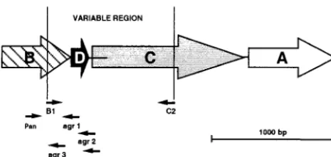
FIG. 1. Schematic map of theS. aureus agroperon (P2). The four members of the operon are represented with the position of the prim-ers used in this work.agrAandagrCencode the response regulator and receptor-histidine protein kinase components of a two-component sig-nal transduction pathway, whereasagrBandagrDcombine to generate the activating ligand for the receptor. Large open arrows indicate the direction of transcription for each gene. Horizontal lines indicate in-tergenic regions (the usual length of theagrD-agrCintergenic region is 25 bp, but lengths up to 160 bp are also described). Thin arrows indicate the position of the primers used to amplify part of the operon. Primer lengths are not shown to scale, but their 5⬘ends are correctly positioned.
on May 15, 2020 by guest
http://jcm.asm.org/
RESULTS
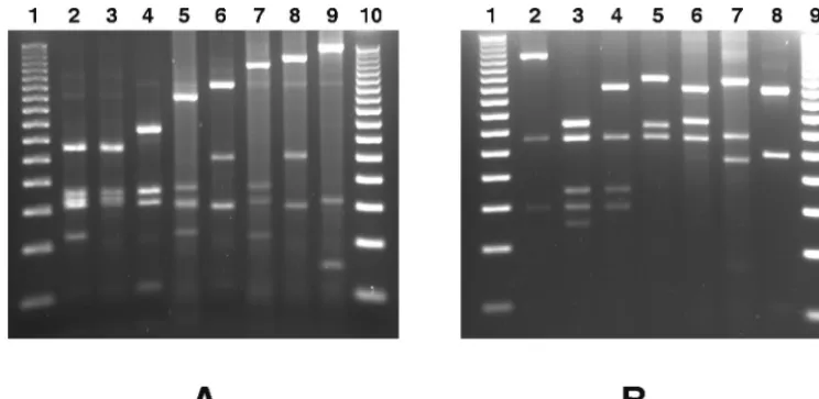
agr restriction fragment length polymorphism. The poly-morphism of theagroperon was analyzed by restriction endo-nuclease PCR in a population of 71 epidemiologically unre-lated S. aureus bovine mastitis isolates. To this end, the 1,070-bp variable region of the agr operon was amplified by PCR with primers B1 and C2 (Fig. 1). Sixty-eight of the tested strains gave an amplicon of the expected molecular weight, and one strain gave an amplicon of around 740 bp, whereas no amplicon could be amplified from two strains (results not shown). The PCR products were then restricted withRsaI or
AluI, giving 10 different profiles for each enzyme (Table 1; Fig. 2). The combination of these two restriction patterns allows the definition of 12 uniqueagrrestriction types (Table 1). Most of the strains belong to the R III-A1 type (56.3%), the R IV-A5 type (12.7%), and the R IV-A7 type (8.4%). The other nine restriction types are shared by only one to three strains (Table 1). The numerical index of the discriminatory ability of theagrtyping, calculated as described by Hunter and Gaston (11), indicates that if two strains were sampled randomly from the analyzed populations, then on 66.2% of occasions they would fall into different restriction types.
Relationship between restriction types of the agr operon.
Restriction maps were constructed to know the relationship between the unique restriction types of theagrlocus identified in the collection of strains isolated from cows with mastitis. To this end, partial hydrolysis was also conducted to correctly assign the position of some restriction fragments (results not shown). The maps were then compared with those correspond-ing to the same region of the agr operon sequenced from strains isolated from humans and available from nucleic acid databases (Fig. 3). Theagrinterference group reference strains isolated from humans were found to be type R I⬘-A1 for group 1 (GenBank accession no. X52543), type R IV-A9 for group 2 (GenBank accession no. AF001782), type R VI-A10 for group 3 (GenBank accession no. AF001783), and type R I-A6 for
group 4 (GenBank accession no. AF288215). Strain CMRSA-1 (GenBank accession no. AF210055) classified by Papakyriacou et al. (25) as being a variant of the agrinterference group 1 reference strain (Ia) was identified to be type R III-A2. It is worth noting that no type R VI-A10 and R IV-A9 were iden-tified among the strains isolated from cows with mastitis. Sim-ilarly, after amplification and restriction of theagroperon, the restriction maps of six strains of human origin whose genomes are sequenced were also constructed. Strains N315 and Mu50 are type R IV-A9, strains NCTC 8325 and COL are type R I⬘-A1, strain MRSA-252 is type R VI-A10, and strain MSSA-476 is type R VI-A8.
All restriction types were classified in one of the fouragr
groups on the basis of the presence or absence of a combina-tion of restriccombina-tion sites characteristic of each of the interfer-ence group referinterfer-ence sequinterfer-ences (Fig. 3 [types in grey]). All types were then linked one to the other inside each group, trying to produce the least gain or loss of restriction sites when evolving from one type to another (Fig. 3). This analysis allows the classification of allagrrestriction types into the four pre-viously knownagrgroups (Table 1). Nevertheless, it is of in-terest that the 3⬘ extremity of the variable region of the agr
locus (plus or minus two-thirds of the molecules) of two strains isolated from cows in Japan, the strain 125 (type R IX-A11) and the strain 130 (type R VIII-A12), possess the characteristic restriction sites of the agr group 1 strains, whereas the 5⬘
extremity of this region (plus or minus one-third of the mole-cule) contains restriction sites characteristic of theagrgroup 2 and of theagrgroup 3 strains, respectively (Fig. 3). As theagr
loci of both strains were sequenced (GenBank accession no. AB043555 and AB043554), this property was verified by align-ing the nucleotide sequences of their variableagrregion with those of the agr group 1 to 3 reference strains (results not shown).
The entireagrvariable region of strains 125 and 130 being nevertheless more similar to the agr group 1 reference se-TABLE 1. Characteristics of different restriction types of theagroperon identified among the analyzedS. aureusstrains isolated from cows
with mastitis
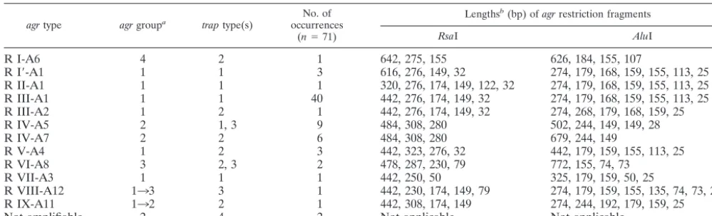
agrtype agrgroupa traptype(s) occurrencesNo. of
(n⫽71)
Lengthsb(bp) ofagrrestriction fragments
RsaI AluI
R I-A6 4 2 1 642, 275, 155 626, 184, 155, 107
R I⬘-A1 1 1 3 616, 276, 149, 32 274, 179, 168, 159, 155, 113, 25
R II-A1 1 1 1 320, 276, 174, 149, 122, 32 274, 179, 168, 159, 155, 113, 25
R III-A1 1 1 40 442, 276, 174, 149, 32 274, 179, 168, 159, 155, 113, 25
R III-A2 1 2 1 442, 276, 174, 149, 32 274, 268, 179, 168, 159, 25
R IV-A5 2 1, 3 9 484, 308, 280 502, 244, 149, 149, 28
R IV-A7 2 2 6 484, 308, 280 679, 244, 149
R V-A4 1 2 3 442, 323, 276, 32 442, 179, 159, 155, 113, 25
R VI-A8 3 2, 3 2 478, 287, 230, 79 772, 155, 74, 73
R VII-A3 1 1 1 442, 250, 50 325, 179, 159, 50, 25
R VIII-A12 133 3 1 442, 230, 174, 149, 79 274, 179, 159, 155, 135, 74, 73, 25
R IX-A11 132 2 1 442, 308, 174, 149 274, 244, 192, 179, 159, 25
Not amplifiable 2 4 2 Not applicable Not applicable
aStrains were classified in one of the fouragrgroups byagrgroup-specific PCR andagrrestriction map analysis. The putative evolving stage between two groups
is indicated by an arrow.
bLengths of restriction fragments were estimated by electrophoresis in the presence of molecular weight markers but, for some types, were also (R I-A6, GenBank
accession no. AF288215; R I⬘-A1, GenBank accession no. X52543 [strain COL at The Institute for Genomic Research data bank and strain NCTC 8325 at the University of Oklahoma’s ACGT data bank]; R III-A2, GenBank accession no. AF210055; R VI-A8, strain MSSA-476 at the Sanger data bank) or only (R VIII-A12, GenBank accession no. AB043555; R IX-A11, GenBank accession no. AB043554) calculated from computer-generated restrictions ofagrnucleic acid sequences deposited in data banks.
on May 15, 2020 by guest
http://jcm.asm.org/
[image:3.603.42.542.91.243.2]quence (88.2 and 91.1% of nucleotides identical, respectively) than to theagrreference sequences of group 2 (69.7 and 59.0% of nucleotides identical, respectively) or group 3 (64.9 and 76.0% of nucleotides identical, respectively), we tentatively classified these two strains in agr group 1, at the junction between groups 2 and 3, respectively (Fig. 3). Our analysis also showed that the isolate giving an agr amplicon of reduced length is anagrgroup 1 strain (type R VII-A3 [Fig. 3]) with a deletion of a 330-bp region comprising the entireagrDgene.
The restriction map analysis indicates that group 1 is the most diverse agr group. This group contains 8 out of the 14 restriction types identified, whereas group 2, group 3, and group 4 contain only 3 types, 2 types, and 1 type, respectively. Finally, we found that the sequenced human strains COL and NCTC 8325 belong toagrgroup 1, that strains N315 and Mu50 belong toagrgroup 2, and that strains MRSA-252 and MSSA-476 belong toagrgroup 3 (Fig. 3).
Development of anagrgroup-specific multiplex PCR.The analysis of the restriction maps of the variable region of theagr
operon (Fig. 3) suggests thatagrgroup-specific primers could be found in this region and used in anagrgroup-specific mul-tiplex PCR. We thus analyzed in detail the nucleotide se-quences of each of the four different agr operons obtained from GenBank (accession no. X52543, AF001782, AF001783, and AF288215) by using the Gene Jockey software (Biosoft, Cambridge, United Kingdom). One consensus forward primer (Pan) and four group-specific reverse primers (agr1, agr2, agr3, and agr4), which would allow the identification of theagrgroup on the basis of the molecular weight of its PCR product, were identified (Fig. 1). These primers were then tested experimen-tally in a multiplex PCR and shown to correctly identify theagr
group of theagrreference strains RN6390 (group 1), RN6923 (group 2), RN8462 (group 3), and A880740 (group 4) (Fig. 4). We then used the above multiplex PCR to analyze the popu-lation of strains isolated from cows with mastitis. All of the 71
strains but 1 gave an amplifiable product. The latter strain is the one giving an agr amplicon of reduced length and was shown by restriction analysis to have a deletion of the sequence corresponding to theagrgroup 1- and group 3-specific primers (Fig. 3). As the 3⬘ extremity of this amplicon possesses the characteristic restriction sites of group 1 strains and not those of group 3 strains, we definitely classify this strain in theagr
group 1.
Theagrgroup-specific multiplex PCR confirms the aboveagr
group classification of strains made by restriction analysis. Nev-ertheless, strains 125 and 130 isolated in Japan, tentatively classified by restriction analysis in group 1, are now classified by multiplex PCR in groups 2 and 3, respectively. This is due to the fact that primers agr2 and agr3 are not localized in the 3⬘
extremity of theagrvariable region, which for these two strains is characteristic of group 1. These two strains are probably in a process of evolution from group 1 to groups 2 and 3, respec-tively. The classification of all six human strains used as con-trols into agr groups was also confirmed. The majority of strains isolated from cows with mastitis belong to agrgroup1 (69.0%) and toagrgroup 2 (23.9%). Groups 3 and 4 contain only 2.8 and 1.4% of the analyzed strains, respectively. As discussed above, the classification of two strains (strains 125 and 130) is uncertain.
trap restriction fragment length polymorphism.As TRAP has been proved to be a component of a membrane-associated sensor able to induce the synthesis of RNA III via a signal transduction pathway other thanagr, we analyzed whethertrap
interstrain variation, similar to that which was found for agr, does exist. To this end, we first aligned thetrapsequences of six strains whose genomes are sequenced. Theoretical restrictions withMseI (a frequently cutting enzyme intrap) show that the
[image:4.603.107.479.69.250.2]agrgroup 1 or 2 strains COL, NCTC 8325, N315, and Mu50 have a similarMseI restriction profile and that this profile is different from those of theagrgroup 3 strains MRSA-252 and FIG. 2. Restriction polymorphism in theagrvariable region ofS. aureusstrains isolated from cows with mastitis. The amplified variable region of theagroperon was digested withAluI (A) orRsaI (B) and electrophoresed on a 3% agarose gel. Examples of the different restriction types obtained are shown: type A1 (lane A2), type A2 (lane A3), type A3 (lane A4), type A4 (lane A5), type A5 (lane A6), type A6 (lane A7), type A7 (lane A8), type A8 (lane A9), type R I (lane B2), type R II (lane B3), type R III (lane B4), type R IV (lane B5), type R V (lane B6), type R VI (lane B7), and type R VII (lane B8). Type R I⬘, being difficult to differentiate from type R I in a 3% agarose gel, is not shown. Types R VIII, R IX, A11, and A12 were identified by computer analysis of sequences with GenBank accession no. AB043554 and AB043555. Molecular weight markers (50-bp DNA ladder; Promega) are shown in lanes A1, A10, B1, and B9.
on May 15, 2020 by guest
http://jcm.asm.org/
MSSA-476 (results not shown). These preliminary results in-dicate that trap is polymorphic and suggest that a relation between trap types and agr group could perhaps exist. This prompted us to analyze the entireS. aureuspopulation isolated from cows with mastitis fortrappolymorphism.
trapgenes were amplified from all strains by PCR and re-stricted withMseI. This allowed the identification of four dif-ferent restriction types among the analyzed population (Fig. 5). The vast majority of strains isolated from cows belong to
traptype 1 (71.8%). Types 2, 3, and 4 account for 18.3, 7.0, and 2.8% of the analyzed strains, respectively (Table 1). It is worth noting that we were previously unable to amplify the variable region of theagr locus in the only two strains oftraptype 4 identified (Table 1). The human sequenced strains COL, NCTC 8325, Mu50, and N315 belong totraptype 1, whereas strains MRSA-252 and MSSA-476 belong totraptypes 2 and 3, respectively.
No particular relationship between unique agr group and
traptype could be displayed. Indeed, our experiments showed that theagrgroups 2, 1, and 3 contain strains oftraptypes 1 to 4, strains oftraptypes 1 to 3, and strains oftraptypes 2 and 3, respectively. Theagrgroup 4 contains a unique strain oftrap
type 2 (Table 1). Whereas no relationship was found between
agrgroups andtraptypes, strains possessing a similaragr re-striction type were also found to possess a similar traptype. Agr types R IV-A5 (containing strains oftraptypes 1 and 3) and R VI-A8 (containing strains of trap types 2 and 3) are nevertheless an exception to this rule (Table 1).
DISCUSSION
Polymorphism in theagrlocus was first described by Ji et al. (15). This led to the classification ofS. aureusisolates into four different interference groups (12, 15). Later, sequence varia-FIG. 3. Relationship between distinct restriction types of theagroperon identified amongS. aureusstrains isolated from cows with mastitis. The amplified variable region of theagroperon was digested withAluI andRsaI, and maps corresponding to the distinct restriction types identified in the population were constructed (3⬘end ofagrB[striped bars],agrD[solid bars], intergenic region [open bars], 5⬘end ofagrC[shaded bars]). These maps were compared with those corresponding to the same region of theagrgroup 1 to 4 reference sequences (type names in grey) obtained from GenBank (accession no. X52543 for group 1, accession no. AF001782 for group 2, accession no. AF001783 for group 3, and accession no. AF288215 for group 4). As the sequences of theagroperon deposited in GenBank do not always show any highly obvious translational start, the drafted length of the intergenic region represents the most frequently described length (25 bp). Based on the presence or absence of characteristic restriction sites, all restrictions types were classified in one of the fouragrgroups. The arrows indicate the appearance (⫹) or disappearance (⫺) of a restriction site with respect to the other types of the sameagrgroup. The dashed line indicates the position of a deletion in the operon.
on May 15, 2020 by guest
http://jcm.asm.org/
[image:5.603.48.533.70.420.2]tions within groups were also found (20, 25, 28, 29). In this work, we identified 14 differentagrrestriction types among the analyzed strains. Nevertheless, only 12 of them were present in our collection of bovine mastitis isolates. On the basis of the restriction maps of theagrvariable region, we classified allagr
types in one of the four interference groups (Fig. 3). This classification was confirmed by theagrgroup-specific PCR also developed in this work. Whereas strains belonging to each of the four agrgroups were found, most of them (69.0%) were assigned to group 1. This repartition of strains among inter-ference groups is quite similar to what was described by Moore and Lindsay for methicillin-sensitive hospital strains (19). The restriction sites used as markers to discriminate agr alleles indicate that theagrDsequences are stable within each inter-ference group. Most mutations within groups appear to arise in
agrC, the gene encoding the receptor of the AIP. Those mu-tations are probably not in sequences coding for amino acids interacting with the inducing peptide. The position of theAluI andRsaI sites inagrDseems to be sufficient to assign a strain to a particularagrgroup, whereas restriction sites characteris-tic of each group can also be found in other genes of this region (Fig. 3). This adds to previous reports showing that genes of the agr locus are submitted to a coevolutionary pressure, al-lowing the binding of a modified AIP to the receptor (15, 22). Our classification indicates that type R VII-A12 and type R IX-A11 are particular with respect to the coevolution of the propeptide and its receptor. Indeed, the receptor-encoding genes of type R VII-A12 and R IX-A11 are highly similar to those of the agr group 1 strains, whereas their propeptide-encoding genes are highly similar to those of groups 3 and 2, respectively. The fact that the propeptide and its receptor belong to different interference groups suggests that strains of type R VII-A12 and type R IX-A11 are impaired in the acti-vation of RNA III by theagrsystem. We postulate that these two types are in a process of evolution from group 1 to groups 2 and 3, respectively. We also identified a strain (type R VII-A3) which has a deletion of the completeagrDgene and which should thus also be impaired in the activation of RNA III by theagrsystem. Strains of types R VII-A12, R IX-A11, and R VII-A3 are nevertheless virulent, because they were all iso-lated from the milk of cows with mastitis. In connection with this, Wesson et al. showed that a strain mutated in agr was internalized by cultured bovine mammary epithelial cells at a level greater than the wild-type strain but contrary to the wild type failed to induce apoptosis (31). Others have also isolated virulentS. aureusstrains with an inactivatedagr system (30). These strains show increased adherence and biofilm formation, and these properties were considered important for the devel-opment of chronic infection (24).
We were also interested to know if the RAP-TRAP system is polymorphic and ubiquitously associated with S. aureus
strains. We identified thetrapgene in all strains analyzed, and we proved that at least four different alleles exist. We also tried to learn if different alleles of the RAP-encoding gene exist. As the nucleotide sequence of RAP is unknown, we made BlastN searches with the available RAP NH2-terminal sequence (IKKYKPITN). Curiously, homologies were only found with the well-conserved L2 ribosomal protein ofS. aureus (results not shown). The identification of the TRAP activator thus needs further clarification, as it is difficult to understand how a conserved ribosomal protein is able to activate the two-com-ponent system. RNA III-inhibiting peptide (RIP), a peptide of sequence YSPXTNF, isolated from culture supernatants of a coagulase-negativeStaphylococcusspecies that is believe to be
S. xylosus, was found to compete with RAP on inducing TRAP
[image:6.603.58.268.67.204.2]phosphorylation. This leads to inhibition of RNA III synthesis and to diminution of the virulence phenotype (2, 3, 9). These results need now to be extended to strains belonging to each of the fourtraptypes identified in this work. With the exception of the Newbould 305 and the NCTC 8325 strains that we classified as trap type 1, nothing is known about the allelic variation oftrapin the strains previously tested for inhibition by RIP. As we have shown that most of theS. aureus strains (71.8%) aretraptype 1, it can be speculated that most, if not all, of the strains tested for inhibition by RIP are alsotraptype FIG. 4. agr group-specific multiplex PCR. S. aureus DNA from
each of the four agr reference strains (RN6390 [group 1, lane 1], RN6923 [group 2, lane 2], RN8462 [group 3, lane 3], A880740 [group 4, lane 4]) were amplified with multiplex primers Pan, agr1, agr2, agr3, and agr4. PCR products were separated on a 1.5% agarose gel and visualized under UV.
FIG. 5. Restriction polymorphism in thetrapgene. Thetrapgene was amplified by PCR, digested withMseI, and electrophoresed on a 3% agarose gel. Examples of the different restriction types obtained are shown: type 1 (lane 2), type 2 (lane 3), type 3 (lane 4), and type 4 (lane 5). Molecular weight markers (50-bp DNA ladder; Promega) are shown in lanes 1 and 6.
on May 15, 2020 by guest
http://jcm.asm.org/
[image:6.603.49.276.477.668.2]1. It is thus still possible that RIP is not able to inhibit RNA synthesis in the other threetraptypes. Furthermore, it is also unknown if RAP purified from strains belonging to each of the fourtraptypes are, as AIPs isolated from each of the fouragr
types, able to activate RNA III synthesis in strains of the same type and to inhibit this synthesis in strains of different types.
Most of the strains (56.3%) isolated from cows with mastitis belong to theagrR III-A1,trap1 type. Our data indicate that strains belonging to this type have been able to infect cows from at least the end of the 1950s to date. TheagrR III-A1,
trap1 type is also the type of the Newbould 305 strain (ATCC 29470), a strain isolated in the United States and widely used for experimental mastitis. This indicates that this type is not linked to a particular geographical location (under the circum-stances of this work, France). The presence at a high preva-lence of typeagrR III-A1,trap1 in the population of strains isolated from cows with mastitis suggests that this type has unique characteristics which, in contrast to the other rare types, endow it with superior ability to infect the bovine mam-mary gland. TheagrR III-A1,trap1 type is thus probably anS.
aureuslineage that expands in the bovine population due to its
possession of a unique combination of virulence genes. As no strain isolated from humans, whoseagrsequence is available from GenBank, was found to belong to the R III-A1 type, it is tempting to think that on the contrary this S. aureustype is rarely isolated in the human population and that otheragrand
trap types are predominantly associated with human disease. This hypothesis will now be tested by analyzing the polymor-phism of agr and trap in a population of S. aureus strains isolated from humans. Previous works using other methods also indicated thatS. aureusisolated from humans and from cows has a predominantly clonal structure and that, whereas numerous types could be identified, only few of them are predominantly associated with a particular host and disease (1, 5–8, 13, 16, 33). The identification and characterization of a disease-dominant lineage(s) are very important for the devel-opment of vaccines and diagnostic tests. It could be expected that such works will lead in the future to the discovery of genetic determinants responsible for the tropism of differentS.
aureuslineages for specific hosts and tissues and to the
devel-opment of new prophylactic and diagnostic tools.
ADDENDUM
The sequence of the whole genome of MW2, a strain of community-acquired methicillin-resistant S. aureus, became available after this paper was submitted (GenBank accession no. AP004822 to AP004832). This strain falls into the classifi-cationsagrgroup 3 (type R VI-A8) andtraptype 3.
ACKNOWLEDGMENTS
We thank Mark Enright (University of Bath, Bath, United King-dom) for the gift of strains MRSA-252 and MSSA-476, John Iandolo (University of Oklahoma, Oklahoma City) for the gift of strain NCTC 8325, Keiichi Hiramatsu (Juntendo University, Tokyo, Japan) for the gift of strains N315 and Mu50, Philippe Moreillon (Centre Hospitalier Universitaire Vaudois, Lausanne, Switzerland) for the gift of strain COL, and Shotaro Takeuchi (Fukui Prefectural University, Fukui, Japan) for the gift of strains 125 and 130. The release of preliminary sequence data by theS. aureusNCTC 8325 Genome Sequencing Team at the University of Oklahoma Health Sciences Center, by the S.
aureusMRSA-252 and MSSA-476 Sequencing Group at the Sanger
Institute, and by theS. aureusCOL Sequencing Team at The Institute for Genomic Research is acknowledged. We are grateful to Martine Braibant for careful reading of the manuscript.
This work was supported by a grant (AIP P00060, P00223) from the French association Bureau des Ressources Ge´ne´tiques (BRG).
REFERENCES
1. Akineden, O., C. Annemu¨ller, A. Hassan, C. La¨mmler, W. Wolter, and M. Zscho¨ck.2001. Toxin genes and other characteristics ofStaphylococcus au-reusisolates from milk of cows with mastitis. Clin. Diagn. Lab. Immunol. 8:959–964.
2. Balaban, N., L. V. Collins, J. S. Cullor, E. B. Hume, E. Medina-Acosta, O. Vieira da Motta, R. O’Callaghan, P. V. Rossitto, M. E. Shirtliff, L. Serafim da Silveira, A. Tarkowski, and J. V. Torres.2000. Prevention of diseases caused byStaphylococcus aureususing the peptide RIP. Peptides21:1301– 1311.
3. Balaban, N., T. Goldkorn, Y. Gov, M. Hirshberg, N. Koyfman, H. R. Mat-thews, R. T. Nhan, B. Singh, and O. Uziel.2001. Regulation of Staphylococ-cus aureuspathogenesis via target of RNAIII-activating protein (TRAP). J. Biol. Chem.276:2658–2667.
4. Boom, R., C. J. Sol, M. M. Salimans, C. L. Jansen, P. M. Wertheim-van Dillen, and J. van der Noordaa.1990. Rapid and simple method for purifi-cation of nucleic acids. J. Clin. Microbiol.28:495–503.
5. Booth, M. C., L. M. Pence, P. Mahasreshti, M. C. Callegan, and M. S. Gilmore.2001. Clonal associations amongStaphylococcus aureusisolates from various sites of infection. Infect. Immun.69:345–352.
6. Day, N. P., C. E. Moore, M. C. Enright, A. R. Berendt, J. M. Smith, M. F. Murphy, S. J. Peacock, B. G. Spratt, and E. J. Feil.2001. A link between virulence and ecological abundance in natural populations ofStaphylococcus aureus. Science292:114–116.
7. Enright, M. C., N. P. Day, C. E. Davies, S. J. Peacock, and B. G. Spratt.2000. Multilocus sequence typing for characterization of methicillin-resistant and methicillin-susceptible clones ofStaphylococcus aureus. J. Clin. Microbiol. 38:1008–1015.
8. Fitzgerald, J., W. Meaney, P. Hartigan, C. Smyth, and V. Kapur.1997. Fine-structure molecular epidemiological analysis ofStaphylococcus aureus
recovered from cows. Epidemiol. Infect.119:261–269.
9. Gov, Y., A. Bitler, G. Dell’Acqua, J. V. Torres, and N. Balaban.2001. RNA III inhibiting peptide (RIP), a global inhibitor ofStaphylococcus aureus
pathogenesis: structure and function analysis. Peptides22:1609–1620. 10. Hogan, J., R. Gonzalez, R. Harmon, S. Nickerson, S. Oliver, J. Pankey, and
K. Smith.1999. Laboratory handbook on bovine mastitis. National Mastitis Council, Inc., Madison, Wis.
11. Hunter, P. R., and M. A. Gaston.1988. Numerical index of the discrimina-tory ability of typing systems: an application of Simpson’s index of diversity. J. Clin. Microbiol.26:2465–2466.
12. Jarraud, S., G. Lyon, A. Figueiredo, G. Lina, F. Vandenesch, J. Etienne, T. Muir, and R. Novick.2000. Exfoliatin-producing strains define a fourthagr
specificity group inStaphylococcus aureus. J. Bacteriol.182:6517–6522. 13. Jarraud, S., C. Mougel, J. Thioulouse, G. Lina, H. Meugnier, F. Forey, X.
Nesme, J. Etienne, and F. Vandenesch.2002. Relationships between Staph-ylococcus aureusgenetic background, virulence factors,agrgroups (alleles), and human disease. Infect. Immun.70:631–641.
14. Ji, G., R. Beavis, and R. Novick.1995. Cell density control of staphylococcal virulence mediated by an octapeptide pheromone. Proc. Natl. Acad. Sci. USA92:12055–12059.
15. Ji, G., R. Beavis, and R. P. Novick.1997. Bacterial interference caused by autoinducing peptide variants. Science276:2027–2030.
16. Kapur, V., W. M. Sischo, R. S. Greer, T. S. Whittam, and J. M. Musser.1995. Molecular population genetic analysis ofStaphylococcus aureusrecovered from cows. J. Clin. Microbiol.33:376–380.
17. Kluytmans, J., A. van Belkum, and H. Verbrugh.1997. Nasal carriage of
Staphylococcus aureus: epidemiology, underlying mechanisms, and associ-ated risks. Clin. Microbiol. Rev.10:505–520.
18. Kuroda, M., T. Ohta, I. Uchiyama, et al.2001. Whole genome sequencing of methicillin-resistantStaphylococcus aureus. Lancet357:1225–1240. 19. Moore, P. C., and J. A. Lindsay.2001. Genetic variation among hospital
isolates of methicillin-sensitiveStaphylococcus aureus: evidence for horizon-tal transfer of virulence genes. J. Clin. Microbiol.39:2760–2767.
20. Mullarky, I. K., C. Su, N. Frieze, Y. H. Park, and L. M. Sordillo.2001.
Staphylococcus aureus agrgenotypes with enterotoxin production capabilities can resist neutrophil bactericidal activity. Infect. Immun.69:45–51. 21. Neave, F., and J. Oliver.1962. The relationship between the number of
mastitis pathogens placed on the teats of dry cows, their survival, and the amount of intramammary infection caused. J. Dairy Res.29:79–93. 22. Novick, R.2000. Pathogenicity factors ofStaphylococcus aureusand their
regulation, p. 392–407.InV. Fischetti (ed.), Gram-positive pathogens. ASM Press, Washington, D.C.
23. Novick, R. P., H. F. Ross, S. J. Projan, J. Kornblum, B. Kreiswirth, and S. Moghazeh.1993. Synthesis of staphylococcal virulence factors is controlled by a regulatory RNA molecule. EMBO J.12:3967–3975.
on May 15, 2020 by guest
http://jcm.asm.org/
24. Otto, M.2001.Staphylococcus aureusandStaphylococcus epidermidispeptide pheromones produced by the accessory gene regulatoragrsystem. Peptides 22:1603–1608.
25. Papakyriacou, H., D. Vaz, A. Simor, M. Louie, and M. McGavin.2000. Molecular analysis of the accessory gene regulator (agr) locus and balance of virulence factor expression in epidemic methicillin-resistantStaphylococcus aureus. J. Infect. Dis.181:990–1000.
26. Prasad, L., and F. Newbould.1968. Inoculation of the bovine teat duct with
Staphylococcus aureus: the relationship of teat duct length, milk yield and milking rate to development of intramammary infection. Can. Vet. J.9:107– 115.
27. Sutra, L., and B. Poutrel.1994. Virulence factors involved in the pathogen-esis of bovine intramammary infections due to Staphylococcus aureus. J. Med. Microbiol.40:79–89.
28. Takeuchi, S., T. Maeda, N. Hashimoto, K. Imaizumi, T. Kaidoh, and Y. Hayakawa.2001. Variation of theagrlocus inStaphylococcus aureusisolates from cows with mastitis. Vet. Microbiol.79:267–274.
29. van Leeuwen, W., W. van Nieuwenhuizen, C. Gijzen, H. Verbrugh, and A. van
Belkum. 2000. Population studies of methicillin-resistant and -sensitive
Staphylococcus aureusstrains reveal a lack of variability in theagrDgene, encoding a staphylococcal autoinducer peptide. J. Bacteriol.182:5721–5729. 30. Vuong, C., H. L. Saenz, F. Gotz, and M. Otto.2000. Impact of theagr
quorum-sensing system on adherence to polystyrene inStaphylococcus au-reus. J. Infect. Dis.182:1688–1693.
31. Wesson, C. A., L. E. Liou, K. M. Todd, G. A. Bohach, W. R. Trumble, and K. W. Bayles.1998.Staphylococcus aureusAgr and Sar global regulators influence internalization and induction of apoptosis. Infect. Immun.66: 5238–5243.
32. West, A., and A. Stock. 2001. Histidine kinases and response regulator proteins in two-component signaling systems. Trends Biochem. Sci.26:369– 376.
33. Zadoks, R., W. van Leeuwen, H. Barkema, O. Sampimon, H. Verbrugh, Y. H. Schukken, and A. van Belkum.2000. Application of pulsed-field gel electro-phoresis and binary typing as tools in veterinary clinical microbiology and molecular epidemiologic analysis of bovine and humanStaphylococcus au-reusisolates. J. Clin. Microbiol.38:1931–1939.

![FIG. 4. agrRN6923 [group 2, lane 2], RN8462 [group 3, lane 3], A880740 [group4, lane 4]) were amplified with multiplex primers Pan, agr1, agr2, agr3,and agr4](https://thumb-us.123doks.com/thumbv2/123dok_us/8238945.829763/6.603.49.276.477.668/fig-agrrn-group-group-group-amplied-multiplex-primers.webp)
