R E S E A R C H A R T I C L E
Open Access
The effect of mobilization with movement
on pain and function in patients with knee
osteoarthritis: a randomized double-blind
controlled trial
Hani A. Alkhawajah
1*and Ali M. Alshami
2Abstract
Background:Few studies have investigated the effects of mobilization with movement (MWM) in patients with knee osteoarthritis (OA) compared to other procedures. Sham procedures are generally more appropriate control than using no or usual treatments. Moreover, studies investigating the widespread hypoalgesic effects of MWM in patients with knee OA are lacking. The aim was to investigate the effect of MWM on function and pain in patients with knee OA compared to sham MWM.
Methods:This is a randomized double-blind (patients and assessor) controlled trial. Forty adult patients with knee OA of grade II and above were recruited to receive either MWM treatment or sham MWM for the knee. The outcome measures included the following: a visual analogue scale (VAS) for pain, the pressure pain threshold (PPT) test, the Western Ontario and McMaster Universities Osteoarthritis (WOMAC) Index, the timed up and go (TUG) test, knee strength and knee range of motion (ROM). The measurements were taken at baseline, immediately after intervention and 2 days later.
Results:Compared with sham MWM, MWM resulted in greater immediate improvement in pain [mean difference (95% CI):−2.2 (−2.8,−1.6)], PPT at both the knee [176 (97, 254)] and shoulder [212 (136, 288)], TUG time [−1.6 (− 2.1,−1.1)], knee flexor strength [2.0 (1.3, 2.7)] and extensor strength [5.7 (4.1, 7.2)] and knee flexion ROM [12.8 (9.6, 15.9)] (all,p< 0.001) but not knee extension ROM [−0.8 (−1.6, 0.1)] (p= 0.067). After 2 days of intervention, patients who received MWM also demonstrated a greater improvement in pain [−1.0 (−1.8,−0.1)], PPT at the shoulder [107 (40, 175)], TUG time [−0.9 (−1.4,−0.4)], knee flexor strength [0.9 (0.2, 1.7)] and extensor strength [2.9 (2.1, 3.9)] and knee flexion ROM [8.3 (4.7, 11.9)] (all,p≤0.026). However, WOMAC scores and knee extension ROM showed no evidence of change at any stage after intervention (p≥0.067).
Conclusions:MWM provided superior benefits over sham MWM in terms of local and widespread pain, physical function (walking), knee flexion and extension muscle strength and knee flexion ROM for at least 2 days in patients with knee OA.
Trial registration:ClinicalTrials.gov (NCT02865252), registered on August 12, 2016.
Keywords:Hypoalgesia, Manual therapy, Pressure pain threshold, Quantitative sensory testing
© The Author(s). 2019Open AccessThis article is distributed under the terms of the Creative Commons Attribution 4.0 International License (http://creativecommons.org/licenses/by/4.0/), which permits unrestricted use, distribution, and reproduction in any medium, provided you give appropriate credit to the original author(s) and the source, provide a link to the Creative Commons license, and indicate if changes were made. The Creative Commons Public Domain Dedication waiver (http://creativecommons.org/publicdomain/zero/1.0/) applies to the data made available in this article, unless otherwise stated. * Correspondence:hkhawajah@iau.edu.sa
1Department of Physiotherapy, King Fahd Hospital of the University, Imam
Abdulrahman Bin Faisal University, P.O Box 40244, Khobar 31952, Saudi Arabia
Background
Osteoarthritis (OA) is the most prevalent form of joint arth-ritis [1]. Knee OA accounts for pain and functional disability in 19.2–27.8% of people aged > 45 [2, 3]. Approximately 37% of people aged ≥60 had knee OA on radiograph [4]. Data on the prevalence of OA in Arabic countries is scarce [5]. However, in Saudi Arabia, a cross-sectional study found that of 300 patients, 53.3% of men and 60.9% of women demonstrated radiographic features of knee OA. Eighty per cent of these patients reported knee pain [6].
There is no known cure for OA [7]. The management of knee OA aims to control pain while improving func-tion and quality of life [8]. The most common medical interventions include pharmacological agents and joint replacement surgery. However, the latter is high risk, es-pecially in older patients [9, 10]. In contrast, other less invasive treatments, such as targeted manual therapy and exercise, are cost-effective and can be safely admin-istered to older patients with OA [7]. Although clinical guidelines report that the efficacy of manual therapy and electrotherapeutic modalities is unclear in patients with knee OA [11], recent high-quality studies [12–14] have found that manual therapy decreases pain, increases range of motion (ROM) and improves physical function.
Mobilization with movement (MWM), which is a type of manual therapy with hypoalgesic effects, increases joint ROM, enhances muscle function and treats specific pathologies [15]. MWM is effective in the management of patients with tennis elbow [16, 17], ankle sprains [18, 19], shoulder impingement [20] and hip OA [21]. Other types of manual therapy, namely antero-posterior glide of the tibia on the femur, produce both local and widespread hypoalgesic effects in patients with knee OA [22].
To our knowledge, three studies have attempted to investigate the effects of MWM in patients with knee OA. These studies were either case series [23] or ran-domized controlled trials (RCTs) [24, 25] that used other treatment procedures in addition to MWM. Sham procedures more clearly distinguish the efficacy of a new procedure beyond the placebo response [26]. In addition, studies that particularly investigate the widespread hypoalgesic effects of MWM in patients with knee OA are lacking. Therefore, the aim of this study is to investigate the immediate and short-term effects of MWM on function and local and distant pain in patients with knee OA compared to sham MWM. This study will serve as the basis for long-term RCTs in the future. The current study is part of a larger study of a master’s thesis that has two phases. Phase one is presented in the current study, and phase two aims at evaluating the effect of MWM in a group of patients who demonstrate features of central sensitization.
Materials and methods
Study design and setting
This double-blind randomized controlled trial was con-ducted in the Department of Physiotherapy at King Fahd Hospital of the University (KFHU). The study was retro-spectively registered withClinicalTrials.gov(NCT02865252) and approved by the Institutional Review Board (IRB) (IRB-2014-04-323) at Imam Abdulrahman Bin Faisal University. The participants provided their written informed consent to undergo the treatment and to have their data used in the study. The study was carried out with CONSORT reporting guidelines [27] in mind.
Sample size determination and participants
Sample size calculation was performed using statistical software (G*Power 3.1) with the following combination: analysis of variance, repeated measures, within-between interaction, medium effect size (f) of 0.25, alpha level of 0.05, power (1-β) of 80%, correlation (r) of 0.5, with 2 groups and 3 measurements (time points) and non-sphericity correction (Є) of 1. The estimated desired sam-ple size was 28. A minimum of 18 patients per group was needed taking into consideration a 20% attrition rate.
Patients with knee OA who attended KFHU were re-cruited. Patients were diagnosed at the orthopaedic clinic and referred to the Department of Physiotherapy. Patients who were willing to participate in the study were screened for eligibility. The patients were included in the study if they were men or women aged ≥40, had unilateral or bilateral knee OA with a Kellgren and Lawrence (K&L) grade≥2 [28], fulfilled the classification criteria of the American College of Rheumatology for knee OA [29], reported peak knee pain of > 3 on a visual analogue scale (VAS) over the previous 24 h and were able to walk ≥6 m. Patients were excluded if they had knee or lower limb surgery, had received an intra-articular corticosteroid or hyaluronic acid injection within the past 6 months, reported current or past (within 4 weeks) oral corticosteroid use, had inflamma-tory or neurological disorders, had altered sensation (to cold, heat, or pressure) around their knee, exhibited cog-nitive difficulties, had low back-related leg pain or had any contraindication to manual therapy.
therapist (principal researcher) about patients’ allocation after the baseline measurements were taken. Patients were asked to attend on two occasions. The first visit took ap-proximately 2 h, during which measurements were taken at baseline, the intervention was delivered and immediate post-intervention measurements were taken. The second visit occurred 2 days later and lasted 30–45 min for meas-urement only (short-term effect). The testing procedures were identical for each patient, except that patients in the sham group received sham MWM.
Intervention
A physiotherapist (principal researcher), blind to the measurements until data analysis, who has 10 years of clinical experience and who is a certified Mulligan prac-titioner trained in the use of MWM administered treat-ments to all patients. MWM techniques were performed using a sustained medial, lateral, anterior, posterior or rotation glide of the tibia during active knee flexion and extension. The details of these techniques have been de-scribed previously [30]. The glides were tested in all pos-sible directions while the patient was in the supine position using the following order: frontal plane (medial/ lateral), sagittal plane (anterior/posterior) and then rota-tion. The glide direction that relieved pain to the lowest level and improved knee range most was selected as the glide for treatment. If the movement was not painful, overpressure was added at the end range. The glide dir-ection was examined in weight-bearing if there was no pain in the supine position. If several glide directions showed similar effects in the supine position, these tests were performed in a weight-bearing position to deter-mine the most effective glide direction [23].
In the treatment group, the therapist applied the glide force on the tibia with the knee in mid-range. Then this force was maintained while the patient was flexing and extending the knee to full range. Overpressure was per-formed at the end range. The MWM treatment tech-nique was repeated 10 times for three sets [23].
In the sham group, the patients were handled similarly to those in the treatment group, but they did not take the glide of direction. Alternatively, the therapist’s hands were lightly touching the knee skin without pressure, one hand on the tibia and one on the femur. Active knee flexion and extension movements, however, were per-formed 10 times for three sets.
Outcome measures
An independent experienced physiotherapist (assessor) from the Department of Physiotherapy (with > 5 years of clinical experience) who was blinded to the allocation of the patients collected the demographic data and baseline measurements of all outcome measurements. Then, the assessor left the room to remain blind to conditions
while the principal researcher applied either MWM treatment or sham MWM interventions according to the patient’s allocated group. After that, the principal re-searcher left the treatment area and the assessor per-formed the outcome measurements immediately after the intervention in a similar manner to the baseline measurements. Patients were asked not to discuss their treatment experience with the assessor. Two days later, the assessor performed the outcome measurements again to assess short-term effects [31].
Primary outcomes
Visual analogue scale (VAS)
Current pain intensity was measured using a 10-cm VAS with end points marked ‘no pain’ and ‘worst pain im-aginable’. The VAS is a valid and reliable measure of pain intensity [32–35].
Pressure pain threshold (PPT)
A digital pressure algometer (Somedic AB, Farsta, Sweden) was used to quantify pain intensity in accord-ance with similar clinical studies [16,22]. This measure has demonstrated high reliability with an intraclass cor-relation coefficient [ICC (2,3)] of 0.97 [36]. PPT is the lowest stimulus intensity at which a person feels mech-anical pain. Increased values of PPT may indicate hypoalgesia or decreased response to mechanical pain stimuli [37].
The most tender point on the medial aspect of the participant’s affected knee was palpated, marked and photographed to ensure standardization between mea-surements. With the participant in a side-lying position, a 1 cm2 algometer probe was used to apply pressure at 90oof knee flexion perpendicular to the skin at a rate of 40 kPa/s. Participants were asked to press a button when the sensation of non-painful pressure turned to become painful. The PPT value was recorded at this point. PPT was also examined on the middle deltoid, 10 cm away from the acromion of the ipsilateral shoulder, to investi-gate any widespread changes in sensitivity at a distant site. Three measurements were performed in each area (knee and shoulder), and the mean value was recorded for analysis. A rest period of 20 s was given after each measurement.
Western Ontario and McMaster universities osteoarthritis (WOMAC) index
and validity to test pain, stiffness and function, especially in patients with hip or knee OA [38–40].
Timed up and go (TUG)
This common test used to assess walking ability has been described in detail previously [41]. TUG has showed high inter- and intra-rater reliability [ICC (2, 1) = 0.96–0.97] in an arthritic population [42]. One prac-tice trial was performed prior to testing. Three measure-ments were performed, and the mean value was recorded for analysis. A rest period of 15 s was given after each measurement.
Secondary outcomes
Hand-held dynamometer
A digital dynamometer (Commander Power Track II, JTECH Medical Industries, Midvale, USA) was used to examine muscle strength and force development. It has good-to-excellent intra- and inter-rater reliability [ICC (2,1)≥0.70] and moderate-to-excellent validity to test muscle strength [43, 44]. The strength of knee flexors and extensors in kilograms was measured while sitting with the knee at 90o flexion. To provide resistance throughout the range, the hand-held dynamometer was placed on the distal tibia anteriorly when examining knee extensors and placed on the posterior ankle when examining knee flexors. Three repetitions were per-formed in each direction, and the mean value was used for analysis. A rest period of 15 s was given after each repetition.
Standard goniometer
A standard goniometer (EZ Read Jammar, Sammons Preston, Warrenville, USA) was used to measure active knee flexion and extension ROM in the supine position. The test was performed three times for each direction, and the mean value was used for analysis. Goniometer measurement demonstrated moderate to high inter-rater reliability (ICCs = 0.59–0.90) [45].
Statistical analysis
Data were analysed using IBM SPSS for Windows (version 24.0). Descriptive analysis included means, standard devia-tions, medians and interquartile ranges. Q-Q plot and Shapiro-Wilk test of standardized residuals were performed for checking the normality of residuals. All continuous vari-ables were approximately normally distributed, except for knee extension ROM. For this variable, the assumption was not met even after transformation, but the model residuals were acceptable. Homoscedasticity was tested for WOMAC by plotting a scatterplot of the standardized residuals against the predicted values. Linearity assumption was assessed by plotting a scatterplot of outcome values at follow-ups against baseline values in each treatment group. The scatterplots
did not indicate major departure from these assumptions. The primary analysis was performed on an intention-to-treat basis, and all randomised participants were included. For continuous outcomes, the least square means (LS means) and their 95% confidence intervals (CIs) were esti-mated using a linear mixed model (LMM) for repeated mea-sures with participant as a random effect, baseline score as a covariate [46,47] and outcomes at two follow-up visits as a dependent variable. This model contained the treatment group, time, baseline-by-time interaction and group-by-time interaction as fixed-effects with an unstructured covariance matrix among time points. For the WOMAC, which was measured with a single follow-up time (2 days), analysis of covariance (ANCOVA) with baseline value as a covariate was used. Mean changes for each group at each time point and mean between-group differences were estimated using appropriate contrasts in the models. All data were regarded as significant atp< 0.05 (two-sided).
Results
Forty-four patients were screened for eligibility. Forty patients satisfied the criteria. Of the 40 patients, four were excluded because of tibial osteotomy, two because of altered sensation around their knees and one because they were unable to walk a 6-m distance with or without an aid. Figure1shows the enrolment and randomization process. Table 1shows the demographic data of the pa-tients. Table 2 demonstrates the direction of glide ap-plied for the MWM intervention. The medial glide of the tibia over the femur was the most common technique.
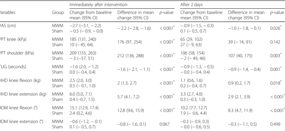
and a greater increase in knee flexion ROM (all, p< 0.001) but not in extension ROM (p= 0.067). Two days after intervention, patients who received MWM demon-strated a greater decrease in pain, a greater increase in PPT at the shoulder, a greater decrease in TUG time, a greater increase in knee flexor and extensor strength and a greater increase in knee flexion ROM compared to those who received sham MWM (all, p≤0.026). How-ever, no significant differences were found between the treatment and sham groups in PPT at the knee (p= 0.142) or knee extension ROM (p= 0.499) (Table3). The ANCOVA revealed no significant differences between the two groups in the total score or any sub-scale of WOMAC (p≥0.392) (Table4).
Discussion
This study investigated the immediate and short-term effects of MWM compared to sham MWM on function and local and widespread pain in patients with knee OA. MWM resulted in an immediate reduction in pain as
measured by VAS. The mean difference in VAS scores was 2.7 cm and 0.9 cm immediately post-intervention and after 2 days, respectively, more than the ‘minimal clinically relevant’difference of 0.84 cm [48]. This effect was similar to previously recorded results for ankle sprains [49], de Quervain’s tenosynovitis [50], lateral epi-condylalgia [51] and hip OA [21]. This reduction of pain lasted for 2 days. A similar result was found in patients with knee OA where MWM was applied in a case series [23] or in RCT’s where MWM was used in combination with other treatments [24,25].
A reduction of mechanical pain, as measured by an alg-ometer, was also observed following MWM, as demon-strated by increased PPTs in the knee. This result is similar to the findings of studies of spinal mobilization [31, 52] and peripheral joint mobilization of the elbow [53] and knee OA [22]. Interestingly, in this study, an im-provement in PPT was seen in the distant area (i.e. shoul-der) in the treatment group but not in the sham group. The increase in PPT was > 15% immediately post-intervention (for knee and shoulder) and after 2 days (for the shoulder), which is considered to reflect a clinically significant effect [22]. Previous studies revealed that
[image:5.595.64.540.86.293.2]Fig. 1Consort diagram of patients enrolment and randomization
Table 1Characteristics of patients in both groups at baseline
MWM (n= 20) Mean ± SD
Sham (n= 20) Mean ± SD
Age (years) 56.5 ± 7.6 56.6 ± 8.5
BMI (kg/m2) 32.6 ± 7.8 33.3 ± 6.1
Duration of symptoms (months)a 51 (46) 48 (42)
VAS (10 cm) 6.5 ± 1.9 5.7 ± 2.0
Gender (male/ female) 13 / 7 12 / 8
Affected knee side (right/left) 6 / 14 10 / 10
K&L knee OA grade (2 / 3 / 4) 14 / 4 / 2 13 / 3 / 4
MWMMobilization with movement,SDStandard deviation,BMIBody mass index,VASVisual analogue scale,K&LKellgren and Lawrence (1957) grading system,OAosteoarthritis
a
Values are expressed as median (interquartile range)
Table 2Direction of glide chosen for the MWM intervention
Direction of glide MWM
(n= 20)
Medial glide 7
Medial + internal rotation glide 2
Lateral glide 3
Lateral + external rotation glide 1
Internal rotation glide 5
Anterior glide 2
[image:5.595.57.291.577.698.2] [image:5.595.306.539.618.724.2]mobilization of the cervical spine decreases hyperalgesia in the upper limbs [31,54] and that knee mobilization in-duces hypoalgesic responses down to the heel [22].
Research has shown that joint mobilization not only initiates local physiological mechanisms but also involves central mechanisms such as facilitation of inhibitory pathways in the spinal cord or descending inhibitory pathways from higher levels in the brainstem [22]. Skyba et al. [55] reported that serotonergic and noradrenergic receptors in the spinal cord mediate analgesia produced by knee joint mobilization.
Knee flexion ROM improved significantly immediately after intervention with MWM in this study. This result corresponds to previous studies of the knee and hip. A case series [23] and RCT [24] reported improvement of knee flexion ROM following MWM in patients with knee OA. Beselga et al. [21] reported immediate improvement
of hip flexion and internal rotation ROM following a sin-gle treatment of MWM in patients with hip OA.
The present study demonstrated an immediate and short-term effect of knee MWM on motor activity, as in-dicated by significant improvements in knee flexor and extensor muscle strength. These improvements may be due to the reversal of reflex pain inhibition [56]. Alter-ation in motor activity may also be an indicAlter-ation of a response that is mediated at the level of the central ner-vous system [56]. MWM improved quadriceps muscle strength significantly in patients with knee OA up to 1-year follow-up [24]. Mobilization of the cervical spine improved the function of deep neck flexor in patients with neck pain [52] and increased pain-free grip strength in patients with lateral epicondylalgia [16,31].
[image:6.595.59.538.109.329.2]In this study, MWM improved TUG time. The de-crease in time needed to walk 6 m was 1.6 s immediately
Table 3Comparison of pain, pressure pain threshold, timed‘up and go’, muscle strength, and range of motion between both groups
Immediately after intervention After 2 days
Variables Group Change from baseline mean (95% CI)
Difference in mean change (95% CI)
p-value Change from baseline mean (95% CI)
Difference in mean change (95% CI)
p-value
VAS (cm) MWM
Sham
−2.7 (−3.1,−2.2)
−0.5 (−0.9,−0.0) −2.2 (−2.8,−1.6) < 0.001
* −0.9 (−1.5,−0.3)
0.1 (−0.5, 0.7) −1.0 (−1.8,−0.1) 0.026
*
PPT knee (kPa) MWM Sham
185 (131, 240)
10 (−45, 64) 176 (97, 254) < 0.001
* 65 (29, 102)
27 (−9, 63) 39 (−14, 91) 0.142
PPT shoulder (kPa) MWM Sham
209 (155, 263)
−3 (−57, 51) 212 (136, 288) < 0.001
* 106 (58, 154)
−2 (−49, 46) 107 (40, 175) 0.003
*
TUG (seconds) MWM Sham −
1.6 (2.0,−1.2)
0.0 (−0.4, 0.4) −1.6 (−2.1,−1.1) < 0.001
* −0.9 (−1.3,−0.5)
−0.0 (−0.4, 0.4) −0.9 (−1.4,−0.4) 0.001
*
HHD knee flexion (kg) MWM Sham
2.5 (2.0, 3.0)
0.5 (−0.1, 1.0) 2 (1.3, 2.7) < 0.001
* 1.1 (0.6, 1.6)
0.2 (−0.4, 0.7) 0.9 (0.2, 1.7) 0.018
*
HHD knee extension (kg) MWM Sham
6.0 (5.0, 7.1)
0.4 (−0.7, 1.5) 5.7 (4.1, 7.2) < 0.001
* 3.3 (2.7, 4.0)
0.3 (−0.3, 1.0) 2.9 (2.1, 3.9) < 0.001
*
ROM knee flexion (°) MWM Sham
15.1 (12.9, 17.4)
2.4 (0.2, 4.6) 12.8 (9.6, 15.9) < 0.001
* 10.2 (7.7, 12.7)
1.9 (−0.6, 4.4) 8.3 (4.7, 11.9) < 0.001
*
ROM knee extension (°) MWM Sham −
0.6 (−1.2,−0.1)
0.1 (−0.5, 0.7) −0.8 (−1.6, 0.1) 0.067 −
0.3 (−0.9, 0.3)
−0.0 (−0.6, 0.5) −0.3 (−1.1, 0.5) 0.499 CIConfidence interval,HHDHand-held dynamometer,MWMMobilization with movement,ROMRange of motion,PPTPressure pain threshold,TUGTimed“Up and Go”, VAS Visual analogue scale
*
Significance difference (p< 0.05)
Table 4Comparison of the Western Ontario and McMaster Universities Osteoarthritis Index between both groups
Variables Group Change from baseline
mean (95% CI)
Difference in mean
change (95% CI) p
-value
Pain scale MWM
Sham
- 0.2 (− 1.1, 0.9) - 0.1 (− 0.9, 0.7)
- 0.1 (− 1.3, 1.0) 0.813
Stiffness scale MWM
Sham
0.0 (−0.4, 0.5) - 0.1 (− 0.5, 0.3)
0.1 (− 0.5, 0.7) 0.700
Function scale MWM
Sham
0.3 (− 1.9, 2.4) 1.6 (− 0.6, 3.7)
- 1.3 (− 4.4, 1.8) 0.392
Total score MWM
Sham
- 0.2 (− 3.1, 2.7) 1.6 (− 1.3, 4.4)
- 1.8 (− 5.9, 2.4) 0.396
[image:6.595.56.540.606.725.2]after the intervention, which is considered to reflect a clinically significant effect [42]. Our finding is consistent with the study by Altmış et al. [25]. In patients with hip OA, Beselga et al. [21] found that MWM reduced the time needed to walk 6 m in this functional test. How-ever, another manual therapy technique, namely antero-posterior glide, had no effect on this test in patients with knee OA [22]. This disagreement may be attributed to the different mobilization techniques used, test proce-dures and/or the characteristics of the patients in the two studies. These contradictory findings emphasize the need of further research in this area. In this study, sev-eral patients received MWM in weight-bearing positions. Thus, patients simultaneously received self-feedback from their painless joint movement.
While the reduction of pain and the improvement of physical function were achieved by MWM, the WOMAC Index scores did not change. This may be because the grade of OA (on the K&L scale) was relatively low, which may represent a non-major limitation of functional activ-ity. Moreover, 2 days might not be sufficient for a per-ceived improvement in daily activities. Moss et al. [22] reported no improvement in WOMAC Index scores after the initial effect of antero-posterior glide in patients with knee OA. However, longer sessions of MWM or other manual therapy techniques in combination with exercise produced significant improvements in WOMAC Index scores in other studies [24,57,58].
A strength of this study is that a sham treatment was used, which is considered more appropriate than no or usual treatment as a control. A limitation of is the short-term design, which may suggest that the immediate changes of any outcome cannot be extrapolated to long-term changes. However, significant improvements in pain, function, ROM and muscle strength were noted in this study, as in previous studies [21–23].
Conclusion
The current study suggests that MWM but not sham MWM for patients with knee OA provides a local and widespread hypoalgesic effect, increases knee flexion ROM, increases knee flexor and extensor strength and improves physical function. Although this study demon-strated immediate and short-term effects that persisted for 2 days after the intervention, more research is needed to determine the long-term efficacy of this approach.
Abbreviations
ANCOVA:Analysis of covariance; CI: Confidence interval; ES: Effect size; ICCs: Intraclass correlation coefficients; IRB: Institutional review board; K&L: Kellgren and Lawrence; KFHU: King Fahd Hospital of the University; LMM: Linear mixed model; MWM: Mobilization with movement;
OA: Osteoarthritis; PPT: Pressure pain threshold; RCTs: Randomized controlled trials; ROM: Range of motion; TUG: Timed“Up and Go”; VAS: Visual analogue scale; WOMAC: Western Ontario and McMaster Universities
Acknowledgements
The authors in this study would like to thank physiotherapists Mr. Ahmed Aldandan and Ms. Ranya Alsaif for their help in the process of data collection, and Mr. Melbin John for his assistance with data analysis.
Authors’contributions
This research was a part of a thesis submitted as partial fulfilment of the requirements for the degree of Master of Science. HA is a former post-graduate student who made data collection and wrote all the sections of this research under close supervision of AA who analyzed and interpreted the patient’s data. All authors read and approved the final manuscript.
Funding
The authors in this study declare that this research did not receive any specific grant from funding agencies in the public, commercial, or not-for-profit sectors.
Availability of data and materials
The datasets used and/or analyzed during the current study are available from the corresponding authors on reasonable request.
Ethics approval and consent to participate
The ethical approval was obtained from the Institutional Review Board (IRB) (IRB-2014-04-323) at Imam Abdulrahman Bin Faisal University. The participants provided their written informed consent to undergo the treatment and to have their data used in the study.
Consent for publication
The participants in this study provided their written informed consent to undergo the treatment and to have their data used in the study.
Competing interests
The authors declare that they have no competing interests.
Author details
1
Department of Physiotherapy, King Fahd Hospital of the University, Imam Abdulrahman Bin Faisal University, P.O Box 40244, Khobar 31952, Saudi Arabia.2Department of Physical Therapy, College of Applied Medical Sciences, Imam Abdulrahman Bin Faisal University, P.O. Box 2435, Dammam 31441, Saudi Arabia.
Received: 19 October 2018 Accepted: 20 September 2019
References
1. Creamer P, Hochberg MC. Osteoarthritis Lancet. 1997;350(9076):503–8. 2. Felson DT, Naimark A, Anderson J, Kazis L, Castelli W, Meenan RF. The
prevalence of knee osteoarthritis in the elderly. The Framingham Osteoarthritis Study. Arthritis Rheum. 1987;30(8):914–8.
3. Jordan JM, Helmick CG, Renner JB, Luta G, Dragomir AD, Woodard J, Fang F, Schwartz TA, Abbate LM, Callahan LF, et al. Prevalence of knee symptoms and radiographic and symptomatic knee osteoarthritis in African Americans and Caucasians: the Johnston County osteoarthritis project. J Rheumatol. 2007;34(1):172–80.
4. Lawrence RC, Felson DT, Helmick CG, Arnold LM, Choi H, Deyo RA, Gabriel S, Hirsch R, Hochberg MC, Hunder GG, et al. Estimates of the prevalence of arthritis and other rheumatic conditions in the United States. Part II Arthritis Rheum. 2008;58(1):26–35.
5. Alghamdi MA, Olney S, Costigan P. Exercise treatment for osteoarthritis disability. Ann Saudi Med. 2004;24(5):326–31.
6. Al-Arfaj A, Al-Boukai AA. Prevalence of radiographic knee osteoarthritis in Saudi Arabia. Clin Rheumatol. 2002;21(2):142–5.
7. Gross KD, Hillstrom H. Knee osteoarthritis: primary care using noninvasive devices and biomechanical principles. Med Clin North Am. 2009;93(1):179–200. 8. Mease PJ, Hanna S, Frakes EP, Altman RD. Pain mechanisms in osteoarthritis:
understanding the role of central pain and current approaches to its treatment. J Rheumatol. 2011;38(8):1546–51.
10. Fajardo M, Di Cesare PE. Disease-modifying therapies for osteoarthritis: current status. Drugs Aging. 2005;22(2):141–61.
11. Jevsevar DS. Treatment of osteoarthritis of the knee: evidence-based guideline, 2nd edition. J Am Acad Orthop Surg. 2013;21(9):571–6. 12. Abbott JH, Robertson MC, Chapple C, Pinto D, Wright AA. Leon de la Barra
S, Baxter GD, Theis JC, Campbell AJ. Manual therapy, exercise therapy, or both, in addition to usual care, for osteoarthritis of the hip or knee: a randomized controlled trial. 1: clinical effectiveness. Osteoarthr Cartil. 2013; 21(4):525–34.
13. Kappetijn O, van Trijffel E, Lucas C. Efficacy of passive extension mobilization in addition to exercise in the osteoarthritic knee: an observational parallel-group study. Knee. 2014;21(3):703–9.
14. Ali SS, Ahmed SI, Khan M, Soomro RR. Comparing the effects of manual therapy versus electrophysical agents in the management of knee osteoarthritis. Pak J Pharm Sci. 2014;27(4 Suppl):1103–6.
15. Bisset L, Hing W, Vicenzino B. A systematic review of the efficacy of MWM. In: Vicenzino B, Hing W, Rivett D, Hall T, editors. Mobilization with movement: the art and the science. Chatswood: Churchill Livingstone; 2011. p. 26–63. 16. Vicenzino B, Paungmali A, Buratowski S, Wright A. Specific manipulative
therapy treatment for chronic lateral epicondylalgia produces uniquely characteristic hypoalgesia. Man Ther. 2001;6(4):205–12.
17. Paungmali A, O’Leary S, Souvlis T, Vicenzino B. Naloxone fails to antagonize initial hypoalgesic effect of a manual therapy treatment for lateral epicondylalgia. J Manip Physiol Ther. 2004;27(3):180–5.
18. Collins N, Teys P, Vicenzino B. The initial effects of a Mulligan’s mobilization with movement technique on dorsiflexion and pain in subacute ankle sprains. Man Ther. 2004;9(2):77–82.
19. Vicenzino B, Branjerdporn M, Teys P, Jordan K. Initial changes in posterior talar glide and dorsiflexion of the ankle after mobilization with movement in individuals with recurrent ankle sprain. J Orthop Sports Phys Ther. 2006;36(7):464–71.
20. Delgado-Gil JA, Prado-Robles E, Rodrigues-de-Souza DP, Cleland JA, Fernandez-de-las-Penas C, Alburquerque-Sendin F. Effects of mobilization with movement on pain and range of motion in patients with unilateral shoulder impingement syndrome: a randomized controlled trial. J Manip Physiol Ther. 2015;38(4):245–52.
21. Beselga C, Neto F, Alburquerque-Sendin F, Hall T, Oliveira-Campelo N. Immediate effects of hip mobilization with movement in patients with hip osteoarthritis: a randomised controlled trial. Man Ther. 2015.
22. Moss P, Sluka K, Wright A. The initial effects of knee joint mobilization on osteoarthritic hyperalgesia. Man Ther. 2007;12(2):109–18.
23. Takasaki H, Hall T, Jull G. Immediate and short-term effects of Mulligan’s mobilization with movement on knee pain and disability associated with knee osteoarthritis: a prospective case series. Physiother Theory Pract. 2013;29(2):87–95. 24. Kaya Mutlu E, Ercin E, Razak Ozdincler A, Ones N. A comparison of two
manual physical therapy approaches and electrotherapy modalities for patients with knee osteoarthritis: a randomized three arm clinical trial. Physiother Theory Pract. 2018;34(8):600–12.
25. Altmis H, Oskay D, Elbasan B, Duzgun I, Tuna Z. Mobilization with movement and kinesio taping in knee arthritis-evaluation and outcomes. Int Orthop. 2018;42(12):2807–15.
26. Brim RL, Miller FG. The potential benefit of the placebo effect in sham-controlled trials: implications for risk-benefit assessments and informed consent. J Med Ethics. 2013;39(11):703–7.
27. Rennie D. CONSORT revised--improving the reporting of randomized trials. JAMA. 2001;285(15):2006–7.
28. Kellgren JH, Lawrence JS. Radiological assessment of osteo-arthrosis. Ann Rheum Dis. 1957;16(4):494–502.
29. Altman RD. The classification of osteoarthritis. J Rheumatol Suppl. 1995;43:42–3. 30. Mulligan BR. Manual therapy: NAGS, SNAGS, MWMS etc. 6th ed. Wellington:
Plane View Services Ltd; 2010.
31. Vicenzino B, Collins D, Benson H, Wright A. An investigation of the interrelationship between manipulative therapy-induced hypoalgesia and sympathoexcitation. J Manip Physiol Ther. 1998;21(7):448–53.
32. Sindhu BS, Shechtman O, Tuckey L. Validity, reliability, and responsiveness of a digital version of the visual analog scale. J Hand Ther. 2011;24(4):356–64. 33. Hawker GA, Mian S, Kendzerska T, French M. Measures of adult pain: visual analog scale for pain (VAS pain), numeric rating scale for pain (NRS pain), McGill pain questionnaire (MPQ), short-form McGill pain questionnaire MPQ), chronic pain grade scale (CPGS), short Form-36 bodily pain scale (SF-36 BPS), and measure of intermittent and constant osteoarthritis pain (ICOAP). Arthritis Care Res (Hoboken). 2011;63(Suppl 11):S240–52.
34. Wessel J. The reliability and validity of pain threshold measurements in osteoarthritis of the knee. Scand J Rheumatol. 1995;24(4):238–42. 35. Grafton KV, Foster NE, Wright CC. Test-retest reliability of the short-form
McGill pain questionnaire: assessment of intraclass correlation coefficients and limits of agreement in patients with osteoarthritis. Clin J Pain. 2005;21(1):73–82.
36. Mutlu EK, Ozdincler AR. Reliability and responsiveness of algometry for measuring pressure pain threshold in patients with knee osteoarthritis. J Phys Ther Sci. 2015;27(6):1961–5.
37. Vanderweeen L, Oostendorp RA, Vaes P, Duquet W. Pressure algometry in manual therapy. Man Ther. 1996;1(5):258–65.
38. Bellamy N, Buchanan WW, Goldsmith CH, Campbell J, Stitt LW. Validation study of WOMAC: a health status instrument for measuring clinically important patient relevant outcomes to antirheumatic drug therapy in patients with osteoarthritis of the hip or knee. J Rheumatol. 1988;15(12):1833–40.
39. Angst F, Aeschlimann A, Steiner W, Stucki G. Responsiveness of the WOMAC osteoarthritis index as compared with the SF-36 in patients with
osteoarthritis of the legs undergoing a comprehensive rehabilitation intervention. Ann Rheum Dis. 2001;60(9):834–40.
40. Parent E, Moffet H. Comparative responsiveness of locomotor tests and questionnaires used to follow early recovery after total knee arthroplasty. Arch Phys Med Rehabil. 2002;83(1):70–80.
41. Podsiadlo D, Richardson S. The timed“up & go”: a test of basic functional mobility for frail elderly persons. J Am Geriatr Soc. 1991;39(2):142–8. 42. Alghadir A, Anwer S, Brismée J-M. The reliability and minimal detectable
change of Timed Up and Go test in individuals with grade 1–3 knee osteoarthritis. BMC Musculoskelet Disord. 2015;16(1):174.
43. Mentiplay BF, Perraton LG, Bower KJ, Adair B, Pua YH, Williams GP, McGaw R, Clark RA. Assessment of lower limb muscle strength and power using hand-held and fixed dynamometry: a reliability and validity study. PLoS One. 2015;10(10):e0140822.
44. Maffiuletti NA, Bizzini M, Desbrosses K, Babault N, Munzinger U. Reliability of knee extension and flexion measurements using the con-Trex isokinetic dynamometer. Clin Physiol Funct Imaging. 2007;27(6):346–53.
45. van Trijffel E, van de Pol RJ, Oostendorp RA, Lucas C. Inter-rater reliability for measurement of passive physiological movements in lower extremity joints is generally low: a systematic review. J Physiother. 2010;56(4):223–35. 46. Davis S. Mixed Models for Repeated Measures using Categorical time Effects
(MMRM). In, editors. Chichester, UK: Wiley; 2014. p. 130–184. 47. Kenward MG, White IR, Carpenter JR. Should baseline be a covariate or
dependent variable in analyses of change from baseline in clinical trials? By G. F. Liu, K. Lu, R. Mogg, M. Mallick and D. V. Mehrotra, statistics in medicine 2009; 28:2509-2530. Stat Med. 2010;29(13):1455–6 author reply 1457. 48. Eberle E, Ottillinger B. Clinically relevant change and clinically relevant
difference in knee osteoarthritis. Osteoarthr Cartil. 1999;7(5):502–3. 49. O’Brien T, Vicenzino B. A study of the effects of Mulligan’s mobilization with
movement treatment of lateral ankle pain using a case study design. Man Ther. 1998;3(2):78–84.
50. Backstrom KM. Mobilization with movement as an adjunct intervention in a patient with complicated de Quervain's tenosynovitis: a case report. J Orthop Sports Phys Ther. 2002;32(3):86–94 discussion 94-87. 51. Paungmali A, Vicenzino B, Smith M. Hypoalgesia induced by elbow
manipulation in lateral epicondylalgia does not exhibit tolerance. J Pain. 2003;4(8):448–54.
52. Sterling M, Jull G, Wright A. Cervical mobilisation: concurrent effects on pain, sympathetic nervous system activity and motor activity. Man Ther. 2001;6(2):72–81.
53. Paungmali A, O’Leary S, Souvlis T, Vicenzino B. Hypoalgesic and sympathoexcitatory effects of mobilization with movement for lateral epicondylalgia. Phys Ther. 2003;83(4):374–83.
54. Vicenzino B, Collins D, Wright A. The initial effects of a cervical spine manipulative physiotherapy treatment on the pain and dysfunction of lateral epicondylalgia. Pain. 1996;68(1):69–74.
55. Skyba DA, Radhakrishnan R, Rohlwing JJ, Wright A, Sluka KA. Joint manipulation reduces hyperalgesia by activation of monoamine receptors but not opioid or GABA receptors in the spinal cord. Pain. 2003;106(1–2):159–68.
57. Deyle GD, Henderson NE, Matekel RL, Ryder MG, Garber MB, Allison SC. Effectiveness of manual physical therapy and exercise in osteoarthritis of the knee. A randomized, controlled trial. Ann Intern Med. 2000;132(3):173–81. 58. Deyle GD, Allison SC, Matekel RL, Ryder MG, Stang JM, Gohdes DD, Hutton
JP, Henderson NE, Garber MB. Physical therapy treatment effectiveness for osteoarthritis of the knee: a randomized comparison of supervised clinical exercise and manual therapy procedures versus a home exercise program. Phys Ther. 2005;85(12):1301–17.
Publisher’s Note