INTRODUCTION
Magnesium is the most abundant intracellular bivalent cation. It plays a major role in overall cell functions, including DNA and protein synthesis, glucose and fat metabolism, oxidative
phosphorylation, neuromuscular excitability, and enzyme ac-tivity.1 Magnesium is also well known for its important roles in maintaining mineral bone metabolism and regulating vascular tone. Several studies have reported that magnesium deficiency is associated with the development of atherosclerosis and vas-cular diseases.2,3 Other studies in animals and humans have demonstrated that magnesium supplementation can limit ath-erosclerosis and decrease the intima-media thickness.4,5 Ath-erosclerosis can be attributed to endothelial dysfunction, which was demonstrated by several studies that endothelial dysfunc-tion is present in the preclinical stage of atherosclerosis.6
Most of magnesium contents in human body is deposited in intracellular compartment (bone, 85%, soft tissue and liver, 14%), with only 1% present in the extracellular compartment. A trace amount of magnesium is absorbed from the intestine. However, this absorbed magnesium is mostly excreted through
The Relationship between Magnesium and
Endothelial Function in End-Stage Renal Disease
Patients on Hemodialysis
Shina Lee, Jung-Hwa Ryu, Seung-Jung Kim, Dong-Ryeol Ryu, Duk-Hee Kang, and Kyu Bok Choi
Department of Internal Medicine, School of Medicine, Ewha Womans University, Seoul, Korea.Purpose: Chronic kidney disease (CKD) patients tend to have higher serum magnesium values than healthy population due to their positive balance of magnesium in kidney. Recent studies found that magnesium level is positively correlated with endothelial function. Therefore, this study was conducted to define the relationship between magnesium level and endothelial dysfunction in end stage renal disease (ESRD) patients on hemodialysis (HD).
Materials and Methods: A total of 27 patients were included in this cross-sectional study. Iontophoresis with laser-Doppler flowm-etry, flow mediated dilation (FMD), and carotid intima-media thickness were measured. Patients’ average serum magnesium lev-els were measured over previous three months, including the examination month. Pearson’s correlation coefficient analysis and multivariate regression model were used to define the association between magnesium and endothelial function.
Results: In the univariate analysis, higher magnesium levels were associated with better endothelium-dependent vasodilation (EDV) of the FMD in ESRD patients on HD (r=0.516, p=0.007). When the participants were divided into two groups according to the median magnesium level (3.47 mg/dL), there was a significant difference in EDV of FMD (less than 3.47 mg/dL, 2.8±1.7%; more than 3.47 mg/dL, 5.1±2.0%, p=0.004). In multivariate analysis, magnesium and albumin were identified as independent factors for FMD (β=1.794, p=0.030 for serum magnesium; β=3.642, p=0.012 for albumin).
Conclusion: This study demonstrated that higher serum magnesium level may be associated with better endothelial function in ESRD patients on HD. In the future, a large, prospective study is needed to elucidate optimal range of serum magnesium levels in ESRD on HD patients.
Key Words: Magnesium, hemodialysis, microcirculation, endothelium
Yonsei Med J 2016 Nov;57(6):1446-1453
http://dx.doi.org/10.3349/ymj.2016.57.6.1446 pISSN: 0513-5796 · eISSN: 1976-2437
Received: August 27, 2015 Revised: January 6, 2016
Accepted: March 12, 2016
Corresponding author: Dr. Kyu Bok Choi, Department of Internal Medicine, School of Medicine, Ewha Womans University, 1071 Anyangcheon-ro, Yangcheon-gu, Seoul 07985, Korea.
Tel: 82-2-2650-5132, Fax: 82-2-2650-2505, E-mail: kbchoi@ewha.ac.kr
•The authors have no financial conflicts of interest.
© Copyright: Yonsei University College of Medicine 2016
the kidney or/and intestine itself in order to maintain its prop-er magnesium balance. Excreted magnesium accounts for less than 1% of the total stores.7 For chronic kidney disease (CKD), urinary magnesium excretion may be normal or even increased until the glomerular filtration rate falls to ≤30 mL/ min. As CKD progresses (<30 mL/min), urinary magnesium excretion is inadequate to balance intestinal magnesium ab-sorption, at which dietary magnesium intake becomes a ma-jor determinant of serum and total body magnesium levels.8 In the case of patients receiving hemodialysis (HD), adminis-tration of magnesium-containing medication and high mag-nesium content of the dialysate are largely contributable for magnesium balance.9 Consequently, they are more likely to have elevated serum magnesium concentration and to be at risk for magnesium overload than are patients with function-ing kidneys.8
To our knowledge, there are few prior studies that addressed the relationship between the serum magnesium level and en-dothelial dysfunction in patients with end-stage renal disease (ESRD) undergoing HD. Therefore, the aim of this study was to define this relationship using flow mediated dilation (FMD) and iontophoresis with laser-Doppler flowmetry (LDF), and carotid intima-media thickness (cIMT).
MATERIALS AND METHODS
Subjects and study design
A total of 27 HD patients from Ewha Womans University Mok-dong Hospital Dialysis Center between February 2011 and Sep-tember 2012 were enrolled. The participants who were under-going HD for at least three months were between 18 and 65 years of age. They were medically stable (without any acute ill-nesses, significant infections, inflammation, or malignancies) and anuric with urine output of <100 mL/day, which can be seen as loss of residual renal function. HD was maintained us-ing bicarbonate dialysate with the followus-ing electrolyte con-centration: sodium 140 mEq/L, potassium 2.0 mEq/L, chloride 120 mEq/L, calcium 3.0 mEq/L, phosphate 0 mEq/L, magne-sium 1.0 mEq/L (1.2 mg/dL), and bicarbonate 30 mEq/L. Data were collected from the medical record, including enrollment information and previous medical history. To evaluate endo-thelial function and atherosclerosis, FMD, iontophoresis with LDF, and cIMT were measured prior to starting a dialysis ses-sion after the patients had fasted for at least 8 hours. Each subject avoided food, drugs, tobacco, alcohol, coffee, or tea 10 hours prior to the test and had 20 minutes of acclimation in supine position before the test.
This protocol was approved by the hospital’s ethics commit-tee. All patients gave written informed consent before partici-pating.
Assessment of endothelial function
Flow-mediated dilation (FMD)
Measurements were made by a single observer using a SONOS 5500 ultrasound system (Philips North America Corporation, Andover, MA, USA) with a 11-MHz probe. Brachial FMD was assessed using methods described previously by Celermajer, et al.10 Briefly, after 20 minutes of resting in the supine position in a temperature controlled room (22–24°C), the patient’s study arm was extended and comfortably immobilized throughout the measurement. Changes in the luminal diameter of the bra-chial were measured during the reactive hyperemia phase in order to assess endothelium-dependent vasodilation. After the subjects had been lying down for 15 minutes, endothelium-in-dependent vasodilation was measured based on the change in the luminal diameter of the brachial artery in response to 0.6 mg nitroglycerine. The lag times from the baseline to the initial reaction and peak reaction were defined as the initial reaction time and peak reaction time, respectively.
Iontophoresis with laser-Doppler flowmetry (LDF)
Iontophoresis employs electrically repulsive forces to deliver lo-cally applied drug across the skin for therapeutic and diagnostic purposes. It is a non-invasive and proper tool to determine en-dothelial dysfunction.11 LDF measures cutaneous blood perfu-sion using the principle of the Doppler shift for lasers; it pro-vides a linear relationship with the velocity of red blood cells.12
defined as the flow rate reached after the last drug delivery. In order to eliminate the baseline variability, the blood flow re-sponses to locally delivered Ach and SNP were expressed as ratios of response PU to baseline PU.11
Intima-media thickness of the common carotid artery (cIMT) Carotid ultrasonography was performed using a 10-MHz scan-ning frequency in B mode. The carotid IMT was analyzed using a computer-based software called Intimascope® (Media Cross
Co. Ltd., Tokyo, Japan).15 With the subject in the supine position, one skillful observer scanned the vessel in transverse planes. Images were obtained 20 mm proximal to the origin of the bulb at the far wall of the right common carotid artery. The comput-er-based IMT was evaluated by three methods, including 3-point, maximal, and average evaluations. Three-point evalu-ation refers to the average value of 3-point IMT, including two end points and the middle point in the >2 cm region. Maximal evaluation was obtained by the IMT value at the region’s max-imal point. Average IMT was the average value of 250 comput-er-based points in the region. In this study, average IMT was used because the computer-automated average IMT evalua-tions was thought to be more reliable indices for atherosclero-sis rather than 3-point evaluation.15
Biochemical analyses
Venous blood was sampled just before the start of a dialysis ses-sion after 8 hour of fasting. Routine laboratory methods were used to measure serum albumin, total calcium, phosphate, to-tal cholesterol, triglycerides, high-density lipoprotein, low-density lipoprotein and intact parathyroid hormone (PTH) in the blood. Serum magnesium was measured using the Xylene blue method (Clinimate MG kit, Sekisui Medical Co. Ltd., To-kyo, Japan). In healthy subjects, serum magnesium ranges be-tween 1.9 and 3.1 mg/dL (0.79–1.29 mmol/L). The laboratory data (except magnesium) was taken from those at the begin-ning of the month, on which the endothelial function tests
were carried out.
Statistical analyses
All data are expressed as means±SDs. Student’s t-tests were used to assess differences in the means between two groups. Pear-son’s rank correlation was used to determine correlations be-tween paired variables. Stepwise multivariate regression analy-sis was used to assess the predictors for FMD. p values<0.05 were considered statistically significant. All calculations were performed using SPSS 18.0 (SPSS Inc., Chicago, IL, USA).
RESULTS
Baseline characteristics
[image:3.595.300.540.425.729.2]A total of 27 patients between 33 and 64 years old were in-cluded (12 men, 15 women). The baseline demographic and clinical characteristics are shown in Table 1. The etiology of CKD in this study included diabetes mellitus, hypertension, glomerulonephritis, and trauma, in order of decreasing frequ-ency. All patients with cardiovascular disease were being treat-ed for diabetes mellitus. One patient with cerebrovascular dis-ease was not diagnosed with diabetes mellitus. There were no patients with peripheral vascular disease. Two patients (on male and one female) were current smokers.
Table 1. Baseline Characteristics
Variables n=27
Age, yrs (range) 52 (33–64)
Sex, M/F 12/15
Etiology of chronic kidney disease, number of patients
Diabetes mellitus 11
Hypertension 5
Chronic glomerulonephritis 3
Trauma 1
Unknown 7
Current smoker 2
History, number of patients
Cardiovascular disease 3
Cerebrovascular disease 3
Peripheral vascular disease 0
Table 2. Biochemical and Vascular Assessment
Variables Mean±
standard deviation (n)* Laboratory finding
Magnesium (mg/dL) 3.43±0.46 (27) Hemoglobin (g/dL) 10.27±0.61 (27) Blood urea nitrogen (mg/dL) 71.96±15.14 (27) Creatinine (mg/dL) 10.82±2.82 (27) Albumin (g/dL) 3.81±0.30 (27) Total cholesterol (mg/dL) 139.93±30.27 (27) Triglycerides (mg/dL) 112.81±69.23 (27) Low density cholesterol (mg/dL) 78.09±22.27 (11) Total calcium (mg/dL) 8.51±0.81 (27) Phosphorus (mg/dL) 5.31±1.00 (27) Uric acid (mg/dL) 7.77±0.93 (27) Intact parathyroid hormone (pg/mL) 154.86±148.32 (27) Vascular assessment
Flow-mediated dilation (%) 3.9±2.2 (26) Nitroglycerine-mediated dilation (%) 11.7±6.7 (26) Acetylcholine-induced iontophoresis
(ratio of response to baseline) 8.5±4.3 (27) Nitropurusside-induced iontophoresis
[image:3.595.43.284.554.731.2]Biochemical and vascular assessments
The biochemical and vascular assessments are presented in Table 2. The average serum magnesium concentration (3.43±
0.46 mg/dL, range 2.47–4.5 mg/dL) was higher than the normal limit (1.9–3.1 mg/dL). Phosphorus, uric acid, and intact PTH were also higher than the references values. In the endothelial dysfunction test, FMD (3.9±2.2%) was lower than the nitroglyc-erin-mediated dilation (NMD, 11.7±6.7%). The response to Ach-induced iontophoresis (8.5±4.3), however, was greater than that of SNP-induced iontophoresis (7.5±4.5). The average cIMT of 18 subjects was <1 mm (0.80±0.10 mm).
The relationship between the serum magnesium concentra-tion and vascular parameters was evaluated (Figs. 1, 2, and 3). There was a strong positive relationship between FMD and the serum magnesium concentration (r=0.561, p=0.003) (Fig. 2A), in contrast to NMD (r=-0.057, p=0.783) (Fig. 2B). Both Ach-induced and SNP-Ach-induced ratio of response to baseline from iontophoresis with LDF had weak correlation with statistical insignificance (r=-0.022, p=0.914) (Fig. 1A) (r=0.017, p=0.932) (Fig. 1B).
The cIMT decreased with increasing serum magnesium concentration, which was not statistically significant (p=0.221).
25.0 20.0 15.0 10.0 5.0 0.0
Serum magnesium (mg/dL)
2.00 2.50 3.00 3.50 4.00 4.50 r=-0.022, p=0.914
Ach-induced iontophoresis (ratio of response to baseline)
A
25.0 20.0 15.0 10.0 5.0 0.0
Serum magnesium (mg/dL)
2.00 2.50 3.00 3.50 4.00 4.50 r=0.017, p=0.932
SNP-induced iontophoresis (ratio of response to baseline)
[image:4.595.86.517.66.225.2]B
Fig. 1. Scatter plot of the relationships between serum magnesium concentration and endothelial function using iontophoresis with laser-Doppler flowmetry. Neither endothelium-dependent vascular dilation (A) nor endothelium-independent vascular dilation (B) was significantly related to serum magnesium. Ach, acetylcholine; SNP, sodium nitroprusside.
6.0
4.0
2.0
0.0
Serum magnesium (mg/dL)
2.00 2.50 3.00 3.50 4.00 4.50 r=0.561, p=0.003
Flow-mediated dilation (%)
A
25.0 20.0 15.0 10.0 5.0 0.0
Serum magnesium (mg/dL)
2.00 2.50 3.00 3.50 4.00 4.50 r=-0.057, p=0.783
Nitroglycerin-mediated dilation (%)
[image:4.595.90.520.270.431.2]B
Fig. 2. Scatter plot of the relationships between serum magnesium concentration and endothelial function, measured by flow mediated dilation. En-dothelium-dependent vasodilation (A) is significantly associated with serum magnesium. There is no significant trend in endothelium-independent vasodilatation (B).
Fig. 3. Negative relationship between serum magnesium concentration and carotid intima-media thickness (p=0.221).
1.10
1.00
0.90
0.80
0.70
0.60
Serum magnesium (mg/dL)
2.00 2.50 3.00 3.50 4.00 4.50 r=-0.303, p=0.221
[image:4.595.56.297.476.662.2]Measurements comparisons based on the median magnesium concentration
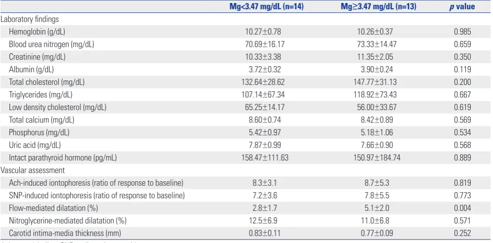
We compared the laboratory parameters and vascular assess-ments of patients with magnesium levels above and below the median value (3.47 mg/dL), shown in Table 3. One group (n=14 patients) had serum magnesium levels below the median val-ue, and other group (with 13 patients) had the levels above the median value. The FMD was significantly different be-tween the two groups (2.8±1.7% below vs. 5.1±2.0% above, p=0.004). However, there were inconsistent values between the two groups with regard to the other endothelial
dysfunc-tion, measured by iontophoresis with LDF and cIMT.
Factors associated with flow-mediated dilation
[image:5.595.42.541.86.332.2]There was a significant correlation between FMD and serum magnesium levels. Multivariate analysis was performed in or-der to determine the factors associated with FMD. Variables that were significant in univariate analysis were included in the multivariate regression analysis. These included age, gen-der, diabetes mellitus, history of cerebrovascular disease, al-bumin, calcium, phosphorus, and intact PTH. Serum magne-sium and albumin were found to be independent factors for FMD (β=1.794, p=0.030 for serum magnesium; β=3.642, p= Table 3. Measurement Comparisons Based on the Median Magnesium Value
Mg<3.47 mg/dL (n=14) Mg≥3.47 mg/dL (n=13) p value Laboratory findings
Hemoglobin (g/dL) 10.27±0.78 10.26±0.37 0.985
Blood urea nitrogen (mg/dL) 70.69±16.17 73.33±14.47 0.659
Creatinine (mg/dL) 10.33±3.38 11.35±2.05 0.350
Albumin (g/dL) 3.72±0.32 3.90±0.24 0.119
Total cholesterol (mg/dL) 132.64±28.62 147.77±31.13 0.200
Triglycerides (mg/dL) 107.14±67.34 118.92±73.43 0.667
Low density cholesterol (mg/dL) 65.25±14.17 56.00±33.67 0.619
Total calcium (mg/dL) 8.60±0.74 8.42±0.89 0.569
Phosphorus (mg/dL) 5.42±0.97 5.18±1.06 0.534
Uric acid (mg/dL) 7.87±0.99 7.66±0.90 0.568
Intact parathyroid hormone (pg/mL) 158.47±111.63 150.97±184.74 0.889 Vascular assessment
Ach-induced iontophoresis (ratio of response to baseline) 8.3±3.1 8.7±5.3 0.819 SNP-induced iontophoresis (ratio of response to baseline) 7.2±3.6 7.8±5.5 0.773
Flow-mediated dilatation (%) 2.8±1.7 5.1±2.0 0.004
[image:5.595.299.536.355.550.2]Nitroglycerine-mediated dilatation (%) 12.5±6.9 11.0±6.8 0.571 Carotid intima-media thickness (mm) 0.83±0.11 0.77±0.09 0.252 Ach, acetylcholine; SNP, sodium nitroprusside.
Table 4. Univariate and Multivariate Analysis of the Factors Associated with FMD
Univariate* Multivariate
r p value β p value
Age NS
-Female 0.468 0.016 0.801 0.292
Diabetes mellitus NS
-Cerebrovascular disease -0.390 0.049 -0.918 0.284 Albumin (g/dL) 0.550 0.004 3.642 0.012
Total calcium (mg/dL) NS
-Phosphorus (mg/dL) NS
-Intact PTH (pg/mL) NS
-Magnesium (mg/dL) 0.549 0.004 1.794 0.030 FMD, flow-mediated dilation; PTH, parathyroid hormone.
*Statistically significant (p<0.05) p values as assessed by Pearson’s correla-tion, as well as β estimates and p values from multivariate regression models. The r2 of the multivariate model was 0.638. Variables known to influence FMD
(age, sex, diabetes mellitus, history of cerebrovascular disease, albumin, cal-cium, phosphorus, intact PTH) were included in the multivariate analyses.
Fig. 4. Positive relationship between serum magnesium concentration and iPTH (p=0.207). iPTH, intact parathyroid hormone.
600
400
200
0
Serum magnesium (mg/dL)
2.00 2.50 3.00 3.50 4.00 4.50 r=0.251, p=0.207
[image:5.595.43.282.382.527.2]0.012 for albumin) (Table 4).
The relationship between intact PTH and magnesium
As shown in Fig. 4, the univariate analysis showed positive cor-relation (r=0.251) between intact PTH and magnesium, altho-ugh it was not statistically significant (p=0.207).
DISCUSSION
Systemic endothelial dysfunction is an early triggering event in the pathogenesis of atherosclerosis, hypertension and cardio-vascular disease.16 A loss of endothelium-dependent vasodila-tion precedes structural atherosclerotic lesions.6,17 Endothelial dysfunction was identified in a child with known risk factors for later cardiovascular disease.18,19 Therefore, evaluating the func-tion of the vascular endothelium allows detecfunc-tion and preven-tion of cardiovascular diseases.
Hypomagnesemia is believed to play a significant role in the development of cardiovascular disease, hypertension, and th-rombosis in the general population.20,21 Magnesium is well known to modulate vascular tone and response, and act as a cofactor for Ach-induced endothelium-dependent relaxation.1 Magnesium deficiency promotes oxidative stress in various cell types, including endothelial cells.22,23 In addition, oral mag-nesium therapy is associated with significant improvement in endothelial function in patients with coronary artery disease.24 Baseline serum magnesium levels are higher in patients with CKD than those in healthy population due to their positive bal-ance of magnesium in kidney.25 Thus, it is important to define the relationship between serum magnesium levels and endo-thelial dysfunction in CKD patients on HD because cardiovas-cular complications are the most common cause of death am-ong dialysis patients.
Our present data demonstrated that macrovascular endothe-lial function is significantly correlated with serum magnesium levels in patients on HD, whereas microvascular endothelial function (presented as iontophoresis with LDF) was not cantly related to serum magnesium values. Although not signifi-cant, there was also positive correlation between cIMT and se-rum magnesium level.
FMD has been established as valid method of assessing mac-rovascular endothelial function in patients at risk for atheroscle-rosis and other cardiovascular diseases. We found that FMD was positively correlated with serum magnesium. This suggests that higher magnesium values may actually improve macro-vascular endothelial function in patients undergoing HD. The reactive hyperemia during FMD measurement produces a shear stress stimulus that induces the endothelium to release nitric oxide (NO), a vasodilator.26 Endothelial dysfunction refers to a worsening in endothelium-dependent relaxation. Endo-thelial dysfunction is normally mediated by compounds in-cluding NO, prostacyclin, and endothelium-derived
hyperpo-larizing factors (EDHFs), which directly facilitate vascular sm-ooth muscle cell relaxation.27 Magnesium is thought to modify the vascular tone not only by regulating the endothelial and smooth muscle cell functions, but also by playing a role in the classical pathway of NO release. One animal experiment dem-onstrated that magnesium increases the production of prosta-cyclin and NO, which ultimately promote endothelium-inde-pendent and endothelium-deendothelium-inde-pendent vasodilation, respec-tively.28 Similarly, our study confirmed this positive relationship between serum magnesium and FMD, especially in patients on HD. In addition, serum magnesium levels may actually be protective against macrovascular disease.
Unlike FMD of the brachial artery, there are no standardized assessments of endothelial function in the microvasculature. Iontophoresis with LDF is one of non-invasive methods to as-sess microvascular endothelial function. This technology uses transdermal delivery of selective endothelium-dependent va-sodilators, such as Ach in this study. The non-NO dependent vasodilatory pathway is likely to play a more critical role in the peripheral microcirculation than it does in the macrocircula-tion. In other words, the contributions of recovered EDHF, which was suppressed by NO, or compensatorily enhanced prostanoid-dependent vasodilatory response is the determin-ing factor for the preservation of endothelial function of mi-crovessels.29 EDHF is a non-characterized endothelial-factor that plays a role in non-NO, non-prostaglandin-mediated en-dothelium-dependent vasodilation. It ultimately causes hy-perpolarization and relaxation of vascular smooth muscle cells.29 The EDHF contribution is inversely correlated with vessel size. The predominant EDHF activity occurs in resis-tance vessels. In smaller vessels, there is a compensatory up-regulation of EDHF under conditions of reduced NO bioavail-ability.29 In this study, exogenous transdermal Ach induced peripheral vasodilation through a non-NO dependent path-way. Ach has been shown to mediate cutaneous vasodilation through both prostanoid and non-NO, non-prostanoid-de-pendent pathways. This phenomenon suggests that there is no direct role for NO in vasodilation.30 These findings are sup-ported by an in vitro study, which demonstrated that Ach re-laxation is mainly dependent on a non-NO, non-prostanoid endothelium-dependent hyperpolarization.31 Studies that have used iontophoresis application of Ach have also demonstrat-ed that NO plays a limitdemonstrat-ed role in the cutaneous response to Ach.32,33 Other researchers have suggested that prostaglandins act in the late phase of Ach-induced iontophoresis.34 Ultimate-ly, it appears that the NO contribution in the peripheral mi-crocirculation is smaller than in the mami-crocirculation. In ad-dition, cutaneous Ach-mediated iontophoresis is unresponsive regardless of the serum magnesium level. Consequently, the irrelevant response to Ach-mediated iontophoresis in the present study is expected.
serum magnesium levels were associated with decreased cIMT, although the correlation was not statistically significant. Previ-ously, HD patients were studied with regard to the relationship between magnesium level (intra- and extracellular) and ath-erosclerosis (as measured using the cIMT).36 HD patients were found to have significantly higher mean common cIMTs than controls patients, and their serum magnesium and intracellu-lar magnesium were negatively associated with the common cIMT. This suggests that magnesium may play a protective role in the development and acceleration of atherosclerosis in patients with chronic renal insufficiency.
The interplay between intact PTH and magnesium is com-plicated. Several studies have reported that hypermagnesemia play a role for inhibition of PTH secretion, presenting a signifi-cant linear inverse correlation between PTH and magnesium in patients on peritoneal dialysis as well as HD.37 However, a linear correlation was statistically insignificant. PTH may be regulated by phosphorus and calcium rather than magnesium, and these factors are tied up for sustaining homeostasis. Barada-ran and Nasri38 reported that magnesium was correlated with serum 25-OH Vit D level rather than PTH, concluding that fac-tors such as serum 25-OH Vit D rather than serum magnesium might be more important for regulation of PTH secretion. Ad-ditionally, when we analyzed the relationship between PTH and endothelial function, there was no obvious correlation be-tween them (not shown). Therefore, future studies are required to ascertain the relationship between magnesium and PTH.
This study has several limitations. Given its cross-sectional design, the case and effect relationships cannot be determined. Assessments were performed only one time and were not based on a time series. In addition, because the control group was not settled, these data cannot be generalized to all dialysis pa-tients. Another limitation is that there is no objective reference to define endothelial dysfunction as assessed by iontophore-sis with LDF. In order to describe the blood volume change, we used a ratio of response to baseline PU. However, there are no reference values of iontophoresis in HD patients. Finally, al-though each magnesium level ranged either above or within the upper limit of normal reference, the participants did not show any symptoms from hypermagnesemia. Fur-thermore, because there were no signs of toxicity, we could not propose the upper limit of magnesium concentration, which may pos-itively affect the endothelial function. Regardless of despite these limitations, our study is the first to suggest that serum magnesium prevents endothelial dysfunction in HD patients.
In conclusion, higher serum magnesium levels in hemodi-laysis patients may have a beneficial effect on endothelial dys-function. In the future, a large prospective study of HD patients is needed to clarify the relationship between serum magnesium and endothelial function. Furthermore, it is important to de-fine the optimal magnesium reference range in patients with end stage renal disease undergoing HD.
REFERENCES
1. Altura BM, Altura BT. New perspectives on the role of magnesium in the pathophysiology of the cardiovascular system. I. Clinical aspects. Magnesium 1985;4:226-44.
2. Reffelmann T, Ittermann T, Dörr M, Völzke H, Reinthaler M, Peters-mann A, et al. Low serum magnesium concentrations predict car-diovascular and all-cause mortality. Atherosclerosis 2011;219:280-4. 3. Amighi J, Sabeti S, Schlager O, Mlekusch W, Exner M, Lalouschek
W, et al. Low serum magnesium predicts neurological events in patients with advanced atherosclerosis. Stroke 2004;35:22-7. 4. Ouchi Y, Tabata RE, Stergiopoulos K, Sato F, Hattori A, Orimo H.
Effect of dietary magnesium on development of atherosclerosis in cholesterol-fed rabbits. Arteriosclerosis 1990;10:732-7. 5. Turgut F, Kanbay M, Metin MR, Uz E, Akcay A, Covic A. Magnesium
supplementation helps to improve carotid intima media thickness in patients on hemodialysis. Int Urol Nephrol 2008;40:1075-82. 6. McLenachan JM, Williams JK, Fish RD, Ganz P, Selwyn AP. Loss of
flow-mediated endothelium-dependent dilation occurs early in the development of atherosclerosis. Circulation 1991;84:1273-8. 7. Floege J, Johnson RJ, Feehally J. Comprehensive clinical
nephrol-ogy, 4th ed. St. Louis: Elsevier Health Sciences; 2010.
8. Navarro-González JF, Mora-Fernández C, García-Pérez J. Clinical implications of disordered magnesium homeostasis in chronic renal failure and dialysis. Semin Dial 2009;22:37-44.
9. Truttmann AC, Faraone R, Von Vigier RO, Nuoffer JM, Pfister R, Bianchetti MG. Maintenance hemodialysis and circulating ion-ized magnesium. Nephron 2002;92:616-21.
10. Celermajer DS, Sorensen K, Ryalls M, Robinson J, Thomas O, Leonard JV, et al. Impaired endothelial function occurs in the sys-temic arteries of children with homozygous homocystinuria but not in their heterozygous parents. J Am Coll Cardiol 1993;22:854-8. 11. Cupisti A, Rossi M, Placidi S, Caprioli R, Morelli E, Vagheggini G, et
al. Responses of the skin microcirculation to acetylcholine and to sodium nitroprusside in chronic uremic patients. Int J Clin Lab Res 2000;30:157-62.
12. Niwayama J, Sanaka T. Development of a new method for monitor-ing blood purification: the blood flow analysis of the head and foot by laser Doppler blood flowmeter during hemodialysis. Hemodial Int 2005;9:56-62.
13. Morris SJ, Shore AC, Tooke JE. Responses of the skin microcircu-lation to acetylcholine and sodium nitroprusside in patients with NIDDM. Diabetologia 1995;38:1337-44.
14. Davis KR, Ponnampalam J, Hayman R, Baker PN, Arulkumaran S, Donnelly R. Microvascular vasodilator response to acetylcholine is increased in women with pre-eclampsia. BJOG 2001;108:610-4. 15. Yanase T, Nasu S, Mukuta Y, Shimizu Y, Nishihara T, Okabe T, et al.
Evaluation of a new carotid intima-media thickness measurement by B-mode ultrasonography using an innovative measurement software, intimascope. Am J Hypertens 2006;19:1206-12.
16. Verma S, Anderson TJ. Fundamentals of endothelial function for the clinical cardiologist. Circulation 2002;105:546-9.
17. Celermajer DS, Sorensen KE, Bull C, Robinson J, Deanfield JE. En-dothelium-dependent dilation in the systemic arteries of asymp-tomatic subjects relates to coronary risk factors and their interac-tion. J Am Coll Cardiol 1994;24:1468-74.
18. Celermajer DS, Sorensen KE, Gooch VM, Spiegelhalter DJ, Miller OI, Sullivan ID, et al. Non-invasive detection of endothelial dys-function in children and adults at risk of atherosclerosis. Lancet 1992;340:1111-5.
20. Ma J, Folsom AR, Melnick SL, Eckfeldt JH, Sharrett AR, Nabulsi AA, et al. Associations of serum and dietary magnesium with cardio-vascular disease, hypertension, diabetes, insulin, and carotid arte-rial wall thickness: the ARIC study. Atherosclerosis Risk in Com-munities Study. J Clin Epidemiol 1995;48:927-40.
21. Liao F, Folsom AR, Brancati FL. Is low magnesium concentration a risk factor for coronary heart disease? The Atherosclerosis Risk in Communities (ARIC) Study. Am Heart J 1998;136:480-90.
22. Wiles ME, Wagner TL, Weglicki WB. Effect of acute magnesium deficiency (MgD) on aortic endothelial cell (EC) oxidant produc-tion. Life Sci 1997;60:221-36.
23. Dickens BF, Weglicki WB, Li YS, Mak IT. Magnesium deficiency in vitro enhances free radical-induced intracellular oxidation and cy-totoxicity in endothelial cells. FEBS Lett 1992;311:187-91.
24. Shechter M, Sharir M, Labrador MJ, Forrester J, Silver B, Bairey Merz CN. Oral magnesium therapy improves endothelial function in pa-tients with coronary artery disease. Circulation 2000;102:2353-8. 25. Mordes JP, Wacker WE. Excess magnesium. Pharmacol Rev 1977;
29:273-300.
26. Ras RT, Streppel MT, Draijer R, Zock PL. Flow-mediated dilation and cardiovascular risk prediction: a systematic review with meta-analysis. Int J Cardiol 2013;168:344-51.
27. Cracowski JL, Minson CT, Salvat-Melis M, Halliwill JR. Method-ological issues in the assessment of skin microvascular endotheli-al function in humans. Trends Pharmacol Sci 2006;27:503-8. 28. Northcott CA, Watts SW. Low [Mg2+]e enhances arterial
spontane-ous tone via phosphatidylinositol 3-kinase in DOCA-salt hyperten-sion. Hypertension 2004;43:125-9.
29. Ozkor MA, Quyyumi AA. Endothelium-derived hyperpolarizing factor and vascular function. Cardiol Res Pract 2011;2011:156146. 30. Holowatz LA, Thompson CS, Minson CT, Kenney WL. Mechanisms
of acetylcholine-mediated vasodilatation in young and aged hu-man skin. J Physiol 2005;563(Pt 3):965-73.
31. Buus NH, Simonsen U, Pilegaard HK, Mulvany MJ. Nitric oxide, prostanoid and non-NO, non-prostanoid involvement in acetyl-choline relaxation of isolated human small arteries. Br J Pharmacol 2000;129:184-92.
32. Noon JP, Walker BR, Hand MF, Webb DJ. Studies with iontophoretic administration of drugs to human dermal vessels in vivo: choliner-gic vasodilatation is mediated by dilator prostanoids rather than nitric oxide. Br J Clin Pharmacol 1998;45:545-50.
33. Khan F, Davidson NC, Littleford RC, Litchfield SJ, Struthers AD, Belch JJ. Cutaneous vascular responses to acetylcholine are medi-ated by a prostanoid-dependent mechanism in man. Vasc Med 1997;2:82-6.
34. Durand S, Tartas M, Bouyé P, Koïtka A, Saumet JL, Abraham P. Prostaglandins participate in the late phase of the vascular re-sponse to acetylcholine iontophoresis in humans. J Physiol 2004; 561(Pt 3):811-9.
35. Pignoli P, Tremoli E, Poli A, Oreste P, Paoletti R. Intimal plus me-dial thickness of the arterial wall: a direct measurement with ul-trasound imaging. Circulation 1986;74:1399-406.
36. Tzanakis I, Virvidakis K, Tsomi A, Mantakas E, Girousis N, Karefyl-lakis N, et al. Intra- and extracellular magnesium levels and ather-omatosis in haemodialysis patients. Magnes Res 2004;17:102-8. 37. Navarro JF, Macía ML, Gallego E, Méndez ML, Chahín J,
García-Nieto V, et al. Serum magnesium concentration and PTH levels. Is long-term chronic hypermagnesemia a risk factor for adynamic bone disease? Scand J Urol Nephrol 1997;31:275-80.