INTRODUCTION
With the rapid development of diagnostic and therapeutic procedures performed outside the operating room (OR), pa-tient needs for sedation or monitored anesthesia care have been increasing. Sedation relaxes anxiety, discomfort, and pain during a procedure. This makes the patient comfortable and allows children or uncooperative adults to undergo proce-dures without body movement.1 In an analysis of 63000 diag-nostic or therapeutic procedures performed under sedation or monitored anesthesia care, 41% of sedations was performed by non-anesthesiologists.2 The most common procedures per-formed under non-anesthesia sedation are gastrointestinal endoscopy (64%) and cardiovascular procedures (30.5%).2
The depth of sedation depends on the type and purpose of the procedure, and possible complications are closely associat-ed with the depth of sassociat-edation. According to a report by the
Pe-diatric Sedation Research Consortium in 2009, pulmonary com-plications, such as apnea, aspiration, or desaturation, occurred 235 times per 10000 sedation/anesthesia administrations out-side the OR.3 Many non-anesthesiologists practice anesthesia and sedation in their field, and while some of them are anxious when performing these, others perform these without any awareness of the dangers. Moreover, despite the recommen-dation of a standard anesthesia setup, including an anesthesia machine, standard monitoring, anesthesia cart, and suction apparatus at the endoscopy location, great variations are ob-served in the arrangement of equipment in each clinical field.1,4,5 This review serves as a general guide focusing on sedation procedures outside the OR and describes pre-procedure pa-tient evaluation, intra-procedure monitoring, and administra-tion strategies for sedatives and analgesics that are needed to provide safe and satisfactory sedation outside the OR.
PRE-PROCEDURE EVALUATION AND
PREPARATION
Clinicians should be aware of the following: 1) reviewing pre-vious medical records and interviewing the patients or care-giver for knowing underlying medical conditions (e.g., abnor-malities of major organ systems, allergies); 2) previous experience or adverse events with sedation and anesthesia, sensitivity to sedatives or analgesics, and pain tolerance; and 3) current medical history and exposure to psychotropic drug.1 A
Sedation Strategies for Procedures
Outside the Operating Room
Youn Yi Jo and Hyun Jeong Kwak
Department of Anesthesiology and Pain Medicine, Gil Medical Center, Gachon University College of Medicine, Incheon, Korea.
With the rapid development of diagnostic and therapeutic procedures performed outside the operating room (OR), the need for appropriate sedation care has emerged in importance to ensure the safety and comfort of patients and clinicians. The preparation and administration of sedatives and sedation care outside the OR require careful attention, proper monitoring systems, and clini-cally useful sedation guidelines. This literature review addresses proper monitoring and selection of sedatives for diagnostic and interventional procedures outside the OR. As the depth of sedation increases, respiratory depression and cardiovascular suppres-sion become serious, necessitating careful surveillance using appropriate monitoring equipment.
Key Words: Sedation, procedure, monitoring, capnography, dexmedetomidine, remifentanil
pISSN: 0513-5796 · eISSN: 1976-2437
Received: February 25, 2019 Revised: April 3, 2019
Accepted: April 9, 2019
Corresponding author: Hyun Jeong Kwak, MD, PhD, Department of Anesthesiol-ogy and Pain Medicine, Gil Medical Center, Gachon University College of Medi-cine, 21 Namdong-daero 774beon-gil, Namdong-gu, Incheon 21565, Korea. Tel: 82-32-460-3637, Fax: 82-32-469-6319, E-mail: hyun615@gilhospital.com
•The authors have no potential conflicts of interest to disclose. © Copyright: Yonsei University College of Medicine 2019
This is an Open Access article distributed under the terms of the Creative Com-mons Attribution Non-Commercial License (https://creativecomCom-mons.org/licenses/ by-nc/4.0) which permits unrestricted non-commercial use, distribution, and repro-duction in any medium, provided the original work is properly cited.
physical examination, including patient demographics (weight and height), vital signs (blood pressure, heart rate, re-spiratory rate, temperature, and oxygen saturation), evaluation of the airway (mouth opening, short neck, neck mobility, facial anomaly, neck mass, and tracheal deviation), state of the teeth (protruding, damaged, or shaken teeth), and lung and heart sounds (mummer, irregular beats, and abnormal breathing sound), should be performed before starting sedation.1,6
According to the American Society of Anesthesiologists (ASA) pre-procedure fasting guidelines, the minimal fasting time is 2 h for clear liquids, 4 h for breast milk, 6 h for infant formula, 6 h for nonhuman milk, 6 h for a light meal, and 8 h or more for fried foods, fatty foods, or meat for gastric emptying.1 However, in a previous analysis of 400 patients who underwent propofol se-dation, although 70% of the enrolled patients had a shorter fasting time than recommended, no differences in respiratory events regardless of the fasting status were observed.7 In addi-tion, other researchers reported no significant differences in aspiration or emesis according to fasting times in the emer-gency department.8,9 Fasting time is determined by the required level of sedation, type and site of procedures, and necessity of airway manipulation.1,9 Recently, drinking clear liquids (water, apple juice, orange juice without pulp, etc.) until 2–3 h prior to anesthesia has been allowed because avoiding hypoglycemia improves patient comfort.10 Fasting time, which is applied flex-ibly, is based on the premise that immediate treatment should be possible when a problem occurs, and it is generally recom-mended to fast for 6 h for light meals before sedation.
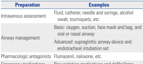
Pre-procedure laboratory testing is guided by underlying medical conditions and the predictable affected result of seda-tion.1,6 Sedatives or analgesics can cause cardiopulmonary compromise, and hence, emergency equipment and drugs should be prepared (Table 1).1
PATIENT MONITORING
An analysis of the ASA Closed Claims database demonstrated that respiratory depression caused by an overdose of seda-tives or opioids was responsible for 21% of monitored anes-thesia care-related claims, and about half of these claims were judged as preventable with better monitoring with vigilance
and an alarm system.11
Respiratory monitoring
Respiratory depression is the most frequent adverse event during sedation,12 and hence, pulse oximetry is widely applied for detecting hypoxia or a desaturation event.13 However, pulse oximetry tends to delay the detection of respiratory suppres-sion. A previous study reported that pulse oximetry can detect only 38% of apnea or hypoventilation events during colonos-copy with sedation, whereas capnography is more reliable than pulse oximetry for early detection of hypoventilation.14 A clinical study concluded that monitoring respiratory activity by using capnography improved patient safety related to respira-tory adverse events during sedation with a combination of benzodiazepines and opioids for endoscopic retrograde chol-angiopancreatography (ERCP) and endoscopic ultrasonogra-phy (EUS).15 Capnographic monitoring of respiratory activity during sedation can lead to rapid interventions, such as pa-tient stimulation, withholding medication and/or oxygen sup-plementation, thus reducing the frequency of hypoxemia, se-vere hypoxemia, and apnea.15
The ASA guidelines recommend monitoring of pulse oxim-etry with appropriate alarms and exhaled carbon dioxide via capnography and continuous observation of qualitative clini-cal signs.1
Hemodynamic monitoring
For hemodynamic monitoring, the Standards of Practice Com-mittee of the American Society for Gastrointestinal Endoscopy recommended the monitoring of blood pressure and heart rate during sedation.16 In addition, other organizations of an-esthesiologists have suggested that procedural sedation should require hemodynamic monitoring for the assessment of blood pressure, heart rate, and electrocardiography.11,17-20 Unless monitoring interferes with procedures, such as magnetic reso-nance imaging, it is recommended to check blood pressure be-fore sedation, and then continuously monitor blood pressure (e.g., at 5 min intervals), heart rate, and electrocardiography during moderate sedation, especially in patients with signifi-cant cardiovascular disease or dysrhythmias.1
Monitoring the depth of sedation
Sedation levels can be evaluated by the clinician. Evaluating the depth of sedation is very important because the greater the depth of sedation, the greater the impact on cardiopulmo-nary function.
[image:2.595.41.279.622.729.2]The depth of sedation is classified as follows: minimal seda-tion (anxiolysis), normal response to verbal commands and unaffected cardiopulmonary function; moderate sedation/ analgesia (conscious sedation), purposeful response to verbal commands and intact airway and cardiopulmonary functions; deep sedation/analgesia, response to painful stimulation and requirement of assistance for proper ventilation and airway Table 1. Emergency Preparedness during Sedation
Preparation Examples
Intravenous assessment Fluid, catheter, needle and syringe, alcohol swab, tourniquets, etc.
Airway management
Basic: oxygen, suction, face mask and bag, and oral or nasal airway
Advanced: supraglottic airway device and endotracheal intubation set
Pharmacologic antagonists Flumazenil, naloxone, etc.
patency; and general anesthesia.1,21 Clinicians sometimes pre-fer the digitalized form because it is more convenient than clin-ical observations.
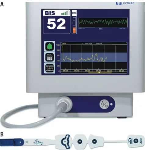
The bispectral index (BIS) monitor (BIS vista monitor revi-sion 3.0; Aspect Medical Systems, Norwood, MA, USA) is the most widely used monitoring instrument and is based on the interpretation of electroencephalograms (Fig. 1). It can be ap-plied simply by attaching a single patch on the forehead to the temporal region of the head. BIS presents values between 90 and 100 for ‘awaken’, between 70 and 90 for ‘light to moderate sedation’, between 60 and 70 for ‘superficial anesthesia’, and between 45 and 60 for ‘general anesthesia’. Previous clinical studies did not provide satisfactory results for applying the BIS for short procedural sedation.22-24 In a previous clinical study, the Spearman correlation between the BIS and the observer’s assessment of alertness/sedation was 0.59 [95% confidence interval (CI), 0.44–0.74] and that between the BIS and the con-tinuum of depth of sedation was 0.53 (95% CI, 0.36–0.70).22 The correlations were not strong enough, and no clinical rele-vance was observed in the sedation complications regardless of the BIS.22 Moreover, in a comparative study between the BIS and conventional clinical assessment during short proce-dures, no significant differences were observed in propofol dosage, oxygen desaturation, and requirement of hemody-namic and respiratory support between groups of patients undergoing bronchoscopy under propofol sedation.23 Fur-thermore, no clinical benefit regarding awareness was
ob-served during the procedures between the study groups.23 In contrast, during long procedures that required moderate seda-tion, BIS monitoring provided some clinical benefits.25,26 In a comparison between the BIS and invisible groups during deep sedation for ERCP, BIS monitoring led to a reduction in the re-quired propofol dose.25 Another study on ERCP also reported an improvement in recovery time, but did not report a reduc-tion in cardiopulmonary complicareduc-tions.26
Designated individual for patient monitoring
ASA guidelines emphasize the presence of a designated indi-vidual other than the practitioner or procedural team to mon-itor the patient throughout the procedure. The designated in-dividual should be trained to recognize apnea and airway obstruction and to check the level of sedation and vital signs.1
SEDATIVES AND ANALGESICS
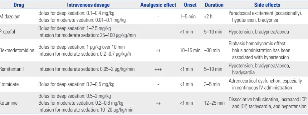
For procedures outside the OR, the use of inhalation agents is limited, and hence, most institutes prefer the use of intrave-nous agents. The dosage and side effects of individual sedative or analgesic agents commonly used are listed in Table 2.27-35
Midazolam
Midazolam is the most frequently used benzodiazepine be-cause of the rapid onset of and short duration for procedural sedation. It provides proper anxiolysis and antegrade amne-sia.29,32 It enables respiratory depression and obtuse responses to carbon dioxide retention via central respiratory depression.36 In particular, rapid intravenous administration might increase respiratory depression.32,36 The dose requirements decrease with increasing age, which results in prolonged and profound drug responses in older adults.32 Because it is a central ner-vous system depressant, geriatric patients and those with se-vere illness and compromised cardiopulmonary reserves have to be closely monitored.32 Because midazolam has no analge-sic effect, it is often used in combination with opioids, such as fentanyl; however, the combined use thereof can increase the risk of respiratory depression and severe hypotension.32
[image:3.595.57.295.421.668.2]The administration of midazolam sometimes induces para-doxical reactions (disinhibitory reactions), including uncon-trolled aggressiveness, agitation, or hallucinations. Paradoxical reactions are manifested within 5 min of intravenous midazol-am administration and are preceded by transient sedation before sudden agitation.37 The paradoxical reactions are related to genetic factors, alcohol abuse, or psychological disturbance, and are assumed to be due to the loss of cortical resistance caused by the inhibitory reaction of midazolam and reduced serotonin control.38 Flumazenil, an antidote to benzodiaze-pines, and haloperidol are helpful to attenuate paradoxical re-actions after midazolam administration.38-40
Fig. 1. BIS monitor and a sensor. The BIS is a processed electroencepha-logram monitor that measures the hypnotic effects of anesthetics and sedatives. The BIS monitor (A) reports a single number from 0 to 100 that represents an integrated measure of cerebral electrical activity. A sensor (B) is placed usually placed at the forehead. BIS, bispectral index. A
Propofol
Propofol is a white-colored formula with benefits of rapid onset of anesthesia and a short recovery time. It provides smoother recovery than do other intravenous sedatives, and enables quicker recovery of psychomotor performance and lower in-cidence of postoperative nausea and vomiting than do other regimens.41 Because propofol has no analgesic effect, it can be combined with opioids. Owing to its fast onset and recovery profiles, it is also used for sedation in pediatric patients under-going MRI.42,43 When combined with ketamine, it has lower side effects.44 The authors prefer to co-administer propofol (a bolus of 1 mg/kg) and ketamine (a bolus of 0.5–1 mg/kg) for shorter sedation (<20 min) in children. This is because ket-amine can compensate for the cardiovascular and respiratory depressive effects caused by propofol due to its sympathomi-metic effects and can reduce propofol-injection pain.
Propofol sedation has been shown to cause euphoria in over 40% of patients undergoing gastroenteroscopy45 and is associated with a risk of drug addiction or abuse. Since propo-fol addiction was first reported in 1992, many people have be-come aware of the danger of addiction: the biggest event was the death of popstar Michael Jackson in 2009 due to propofol misuse. Propofol was designated as a controlled substance in Korea in February, 2011 (the first in the world), as there is a potential risk of abuse and propofol abusers are increasing. Because injection pain is the most frequent side effect of pro-pofol, the concomitant use of lidocaine is recommended.46,47 Propofol induces respiratory depression and exerts a greater effect on cardiovascular depression with profound hypoten-sion than do other intravenous agents.48,49 Rapid injection of the sedative formula, old age, and poor physical status results in the debilitation of patients, especially those vulnerable to catastrophic cardiorespiratory effects.48,49 Because propofol is a lipid-based formula, rapid bacterial contamination might
eas-ily develop and induce life-threatening sepsis,48-50 and hence, sterile and aseptic handling is important. Although very rare, propofol infusion syndrome, which involves severe metabolic acidosis, renal failure, rhabdomyolysis, and cardiac failure, may develop in cases of single administration of propofol.51
Dexmedetomidine
Dexmedetomidine is a selective α2-receptor agonist and pro-vides anxiolytic, sedative, and analgesic effects.52,53 Dexme-detomidine reduces norepinephrine release and inhibits sym-pathetic outflow in the central nervous system; therefore, it can cause profound bradycardia, especially in young patients with a high vagal tone.52,54 If transient hypertension occurs dur-ing the infusion of loaddur-ing dose, a reduction of infusion rate should be considered.52 Meanwhile, hypotension may also oc-cur, especially in geriatric patients or patients with diabetes mellitus or chronic hypertension.52,53
A feature of dexmedetomidine is that it has analgesic prop-erties in addition to its role as a hypnotic, while being opioid sparing; thus, it is not associated with significant respiratory depression. Dexmedetomidine is most often used in the inten-sive care unit for light to moderate sedation. An earlier study suggested that using dexmedetomidine for sedation in me-chanically ventilated adults may reduce the time to extubation and intensive care unit stay.55 It should not be administered over 24 h,52 because it induces potential withdrawal respons-es, such as agitation and an abrupt increase in blood pressure.
[image:4.595.44.538.85.269.2]Patients on dexmedetomidine can be cooperative, which are beneficial in some procedures, such as blepharoplasty. Previous clinical studies demonstrated that dexmedetomi-dine provides less respiratory depression with better analgesic efficacy and deeper sedation level than does midazolam for double-balloon enteroscopy56 and ablation for atrial fibrilla-tion.57 It can be used in combination with other sedatives, like Table 2. Summary of Sedation Drugs Commonly Used
Drug Intravenous dosage Analgesic effect Onset Duration Side effects
Midazolam Bolus for deep sedation: 0.1Bolus for moderate sedation: 0.01–0.4 mg/kg–0.1 mg/kg - 1–5 min <2 h Paradoxical excitement (occasionally), hypotension, bradypnea
Propofol Bolus for deep sedation: 1–2.5 mg/kg
Infusion for moderate sedation: 25–100 μg/kg/min - <1 min 5–10 min Hypotension, bradypnea/apnea
Dexmedetomidine Bolus for deep sedation: 1 μg/kg over 10 minInfusion for moderate sedation: 0.2–0.7 μg/kg/h ++ 10–15 min ~30 min Biphasic hemodynamic effect: bolus administration has been associated with hypertension
Remifentanil Infusion for moderate sedation: 0.05–2 μg/kg/min +++ <1 min 5–10 min Hypotension, bradypnea/apnea, bradycardia
Etomidate Bolus for deep sedation: 0.2–0.5 mg/kg - <1 min 3–5 min Adrenocortical dysfunction, especially in continuous IV administration
Ketamine Bolus for deep sedation: 0.5 –2 mg/kg Bolus for moderate sedation: 0.2–0.8 mg/kg Infusion for moderate sedation: 10–20 μg/kg/min
++ <1 min 12–25 min Dissociative hallucination, increased ICP and IOP, tachycardia, and hypertension
ICP, intracranial pressure; IOP, intraocular pressure; IV, intravenous.
propofol, opioids, and benzodiazepines, to enhance sedation and to help maintain hemodynamic stability by decreasing the requirement for other sedatives.58,59 Because dexmedeto-midine has a late onset of 10–15 min, combined administra-tion of small doses of midazolam (1.5–2 mg) for rapid hypno-sis or fentanyl (25–50 µg) for rapid analgesia when starting sedation with infusion of dexmedetomidine at a rate of 0.5± 0.3 µg/kg/min is generally favored. Dexmedetomidine is also used for procedural sedation in children.60 However, it should be noted that the use of dexmedetomidine for procedural se-dation in pediatric patients has not been well evaluated and its use is not currently approved in children in any country.
Opioids
Some clinicians prefer to use additional opioids with hypnot-ics. An addition of opioids effectively reduces the hypnotic requirements and controls procedure-induced discomfort. However, it should be noted that respiratory depression and hemodynamic suppression might be possible even when low doses of sedatives are used with opioids; therefore, special at-tention should be paid.
Intravenous fentanyl has an onset of 5 min and a duration of 30–60 min. A previous study demonstrated that the com-bined use of fentanyl could reduce propofol requirements for procedural sedation without any delay in recovery time for patients undergoing elective EUS.61
Remifentanil, an ultra-short-acting opioid is preferred for use in combination with sedatives because of its rapid recov-ery. Remifentanil has been reportedly used as a component of conscious sedation in patients undergoing painful medical procedures.62 Remifentanil infusion at a rate of 0.5±0.3 μg/kg/ min provided sufficient analgesia, but was accompanied by a high incidence of respiratory depression at subtherapeutic lev-els.62 Because of its significant respiratory depression, careful monitoring of capnography during remifentanil infusion is recommended.
Morphine and meperidine might induce bronchospasm re-lated with histamine release. Rapid administration of opioids, especially fentanyl, alfentanil, sufentanil, and remifentanil, might induce chest wall rigidity, which can disturb proper ven-tilation.28
Etomidate
Etomidate has unique characteristics, including an easy dos-ing profile, limited suppression of ventilation, lack of hista-mine liberation, and protection from myocardial and cerebral ischemia.63 It is frequently used for procedural sedation64 and as an induction agent for rapid sequence intubation63 in the emergency department. In addition, etomidate is a good induc-tion agent for hemodynamically unstable patients.65 Etomidate is also used in patients with traumatic brain injury, because it is one of the only anesthetic agents able to decrease intracranial pressure and maintain a normal arterial pressure.
Despite its numerous cardiovascular and respiratory ad-vantages, etomidate has a notable side effect of adrenocortical suppression. It is possible even in single administration, and sometimes, exogenous glucocorticoid supply is required during the postoperative period.66 Moreover, etomidate has disad-vantages, such as pain at the injection site, myoclonus, and fre-quent nausea, which have led to its decreased usage as an an-esthetic, and it not being recommend for elective sedation.27,67
Ketamine
Unlike most sedatives, including midazolam and propofol, that potentiate the inhibitory action of γ-aminobutyric acid, ketamine is an antagonist of N-methyl-D-aspartate receptor.27 The unique characteristic of ketamine is dissociative anesthe-sia, which is a status in which the patients appear conscious with eye opening but have catatonia that prevent them from responding to external stimuli.27 Ketamine induces psychomi-metic effects, such as hallucinations or dysphoria.27 Unlike other sedatives, ketamine has a central sympathomimetic effect and can transiently increase blood pressure and heart rate.68 However, when catecholamines are depleted, ketamine exhib-its negative cardiovascular responses.27,69 Ketamine preserves the airway reflex and respiratory drive, but increases oral secre-tion, which might increase the incidence of laryngospasms.68 Because of the above-mentioned characteristics of ketamine, even sub-anesthetic ketamine administration is contraindi-cated in cases of high-risk coronary disease, uncontrolled hy-pertension, increased intracranial pressure, increased intra-ocular pressure, psychosis, and hepatic dysfunction.70
SPECIAL CONSIDERATIONS FOR
INDIVIDUAL PROCEDURES
Gastrointestinal procedures
of midazolam to the regimen of continuous propofol and remi-fentanil infusion may be helpful in overcoming this problem. A retrospective review of sedation for endoscopic submucosal dissection also reported that complete resection rates were significantly higher and that procedure times were signifi-cantly shorter with continuous infusion of propofol with opi-oid by an anesthesiologist than with intermittent propofol/ midazolam injection by an endoscopist.74 However, aspiration pneumonia was more frequent in patients receiving continu-ous propofol and opioid infusion than in those receiving the intermittent injection.74 A combined administration of propo-fol and opioid may have difficulties for non-anesthesiologists to adequately titrate the dosages of these drugs, because these co-administration enhances their side effects of respiratory depression, hypotension, and bradycardia.
ERCP is more complex than other endoscopic procedures. It often requires precise intervention and complete immobili-zation without gagging or squirming to ensure the safety and success of the procedure. Moreover, many patients who require ERCP are vulnerable. In a recent clinical study of conscious sedation for ERCP, the combined use of dexmedetomidine (a loading dose of 1 μg/kg over 10 min) resulted in significantly better patient satisfaction scores and lower desaturation rates than did the combined use of midazolam (0.05 mg/kg) during remifentanil infusion (a loading dose of 1 μg/kg and an infu-sion rate of 0.05–0.2 μg/kg/min).75 In addition, dexmedetomi-dine has been reported to be safe and to decrease the total dose of other hypnotics in very old patients undergoing ERCP.76
Sedation for MRI or CT
For ensuring patient satisfaction and acquiring good-quality MRI and CT images, immobilization of the patient is important during these imaging procedures. However, staying alone for long periods in a dark, noisy environment is not easy for chil-dren and adults with claustrophobia. In a review focused on sedation for pediatric MRI, dexmedetomidine was found to have a greater sedative effect than did chloral hydrate, pento-barbital, and midazolam; in addition, preterm or small children should preferably be given general anesthesia for the safety and success of the diagnostic test.77 A randomized controlled study compared pharmacodynamic responses to a combina-tion of dexmedetomidine (a loading dose of 1 μg/kg and an infusion rate of 0.5 μg/kg/h) and midazolam (0.1 mg/kg) vs. propofol (250–300 μg/kg/min) in children anesthetized using sevoflurane for MRI, and demonstrated that dexmedetomi-dine-midazolam provided adequate anesthesia, although it had a more prolonged recovery time than did propofol.78 Neu-rodevelopmental disorders might change the sedative require-ments.79 In an animal study, autistic rats showed increased re-quirements of propofol and dexmedetomidine than did the control rats.79
Neurologic interventions
A recent matched-cohort study comparing conscious sedation and general anesthesia for patients undergoing flow diverter placement for aneurysms demonstrated that conscious seda-tion could be successfully applied for short and simple neuro-logic procedures.80 When selecting sedatives for neurologic procedures, ketamine should be avoided because of its char-acteristics of increasing intracranial pressure81 and inducing psychomimetic activity,82 which may affect the validity of the neurologic examination.
Cardiologic procedures
Cardiologic procedures that require sedation include cardio-version, ablation, transesophageal echocardiography, device implantation, and percutaneous transcatheter valve proce-dures. Propofol administration by nursing staff might be ap-propriate for some cardiologic procedures that require mod-erate sedation. However, proper training is essential for using capnography to detect respiratory depression, and using a tar-get-controlled infusion pump is recommended for propofol administration.83 A study comparing dexmedetomidine (a loading dose of 1 μg/kg over 10 min and a maintenance infu-sion rate of 0.2 μg/kg/h) and thiamylal (a bolus of 1.25 mg/kg and the same bolus dose every 15 min) for sedation during ab-lation of atrial fibrilab-lation showed that both sleep-disordered breathing events and the number of body movements were significantly lower in the dexmedetomidine group than in the thiamylal group.84 Therefore, they suggested that dexmedeto-midine was a safe and proper sedative for cardiologic proce-dural sedation.84
CONCLUSION
The need for sedation and anesthesia outside the OR is in-creasing because of the increased use of diagnostic tools and procedural treatment methods. It is important to understand the characteristics and side effects of sedatives and analgesics when selecting them, because the degree or depth of sedation required to improve the patient’s stability and to ensure the success of the procedure may vary. Clinicians should remem-ber that as the depth of sedation increases, the risks of respira-tory depression and cardiovascular suppression become seri-ous, and hence, precautions should be taken using appropriate surveillance systems.
AUTHOR CONTRIBUTIONS
ORCID iDs
Youn Yi Jo https://orcid.org/0000-0002-9214-7039 Hyun Jeong Kwak https://orcid.org/0000-0003-4432-8510
REFERENCES
1. Practice Guidelines for Moderate Procedural Sedation and Anal-gesia 2018: a report by the American Society of Anesthesiologists Task Force on Moderate Procedural Sedation and Analgesia, the American Association of Oral and Maxillofacial Surgeons, can College of Radiology, American Dental Association, Ameri-can Society of Dentist Anesthesiologists, and Society of Interven-tional Radiology. Anesthesiology 2018;128:437-79.
2. Pino RM. The nature of anesthesia and procedural sedation out-side of the operating room. Curr Opin Anaesthesiol 2007;20:347-51. 3. Cravero JP, Beach ML, Blike GT, Gallagher SM, Hertzog JH; Pedi-atric Sedation Research Consortium. The incidence and nature of adverse events during pediatric sedation/anesthesia with propo-fol for procedures outside the operating room: a report from the Pediatric Sedation Research Consortium. Anesth Analg 2009;108: 795-804.
4. Michel Foehn ER. Adult and pediatric anesthesia/sedation for gastrointestinal procedures outside of the operating room. Curr Opin Anaesthesiol 2015;28:469-77.
5. Youn AM, Ko YK, Kim YH. Anesthesia and sedation outside of the operating room. Korean J Anesthesiol 2015;68:323-31.
6. Gozal D, Gozal Y. Pediatric sedation/anesthesia outside the oper-ating room. Curr Opin Anaesthesiol 2008;21:494-8.
7. Bell A, Treston G, McNabb C, Monypenny K, Cardwell R. Profil-ing adverse respiratory events and vomitProfil-ing when usProfil-ing propofol for emergency department procedural sedation. Emerg Med Australas 2007;19:405-10.
8. Agrawal D, Manzi SF, Gupta R, Krauss B. Preprocedural fasting state and adverse events in children undergoing procedural seda-tion and analgesia in a pediatric emergency department. Ann Emerg Med 2003;42:636-46.
9. Godwin SA, Burton JH, Gerardo CJ, Hatten BW, Mace SE, Silvers SM, et al. Clinical policy: procedural sedation and analgesia in the emergency department. Ann Emerg Med 2014;63:247-58. 10. Van De Velde M, Kuypers M, Teunkens A, Devroe S. Risk and
safety of anesthesia outside the operating room. Minerva Aneste-siol 2009;75:345-8.
11. Bhananker SM, Posner KL, Cheney FW, Caplan RA, Lee LA, Dom-ino KB. Injury and liability associated with monitored anesthesia care: a closed claims analysis. Anesthesiology 2006;104:228-34. 12. Choi JW, Kim DK, Cho CK, Park SJ, Son YH. Trends in medical
dis-putes involving anesthesia during July 2009-June 2018: an analysis of the Korean Society of Anesthesiologists database. Korean J An-esthesiol 2019;72:156-63.
13. Metzner J, Domino KB. Risks of anesthesia or sedation outside the operating room: the role of the anesthesia care provider. Curr Opin Anaesthesiol 2010;23:523-31.
14. Cacho G, Pérez-Calle JL, Barbado A, Lledó JL, Ojea R, Fernández-Rodríguez CM. Capnography is superior to pulse oximetry for the detection of respiratory depression during colonoscopy. Rev Esp Enferm Dig 2010;102:86-9.
15. Qadeer MA, Vargo JJ, Dumot JA, Lopez R, Trolli PA, Stevens T, et al. Capnographic monitoring of respiratory activity improves safety of sedation for endoscopic cholangiopancreatography and ultrasonography. Gastroenterology 2009;136:1568-76.
16. Waring JP, Baron TH, Hirota WK, Goldstein JL, Jacobson BC,
Leighton JA, et al.; American Society for Gastrointestinal Endos-copy, Standards of Practice Committee. Guidelines for conscious sedation and monitoring during gastrointestinal endoscopy. Gas-trointest Endosc 2003;58:317-22.
17. ASA Committee on Standards and Practice Parameters. Stan-dards for basic anesthetic monitoring [accessed on 2019 January 31]. Available at: https://www.asahq.org/~/media/Sites/ASAHQ/ Files/Public/Resources/standards-guidelines/standards-for-ba-sic-anesthetic-monitoring.pdf.
18. Academy of Medical Royal Colleges. Safe sedation practice for healthcare procedures: standards and guidance [accessed on 2019 January 31]. Available at: https://www.rcoa.ac.uk/system/files/ PUB-SafeSedPrac2013.pdf.
19. Hinkelbein J, Lamperti M, Akeson J, Santos J, Costa J, De Robertis E, et al. European Society of Anaesthesiology and European Board of Anaesthesiology guidelines for procedural sedation and analge-sia in adults. Eur J Anaesthesiol 2018;35:6-24.
20. Australian and New Zealand College of Anaesthetists. Guidelines on sedation and/or analgesia for diagnostic and interventional medical, dental or surgical procedures [accessed on 2019 January 31]. Available at: http://www.anzca.edu.au/documents/ps09-2014-guidelines-on-sedation-and-or-analgesia.
21. American Society of Anesthesiologists. Continuum of depth of se-dation: definition of general anesthesia and levels of sedation/an-algesia [accessed on 2019 January 31]. Available at: https://www. asahq.org/standards-and-guidelines/continuum-of-depth-of-se- dation-definition-of-general-anesthesia-and-levels-of-sedation-analgesia.
22. Weaver CS, Hauter WH, Duncan CE, Brizendine EJ, Cordell WH. An assessment of the association of bispectral index with 2 clini-cal sedation sclini-cales for monitoring depth of procedural sedation. Am J Emerg Med 2007;25:918-24.
23. Fruchter O, Tirosh M, Carmi U, Rosengarten D, Kramer MR. Pro-spective randomized trial of bispectral index monitoring of seda-tion depth during flexible bronchoscopy. Respiraseda-tion 2014;87:388-93.
24. Yang KS, Habib AS, Lu M, Branch MS, Muir H, Manberg P, et al. A prospective evaluation of the incidence of adverse events in nurse-administered moderate sedation guided by sedation scores or Bispectral Index. Anesth Analg 2014;119:43-8.
25. Paspatis GA, Chainaki I, Manolaraki MM, Vardas E, Theodoropou-lou A, Tribonias G, et al. Efficacy of bispectral index monitoring as an adjunct to propofol deep sedation for ERCP: a randomized controlled trial. Endoscopy 2009;41:1046-51.
26. von Delius S, Salletmaier H, Meining A, Wagenpfeil S, Saur D, Ba-jbouj M, et al. Bispectral index monitoring of midazolam and propofol sedation during endoscopic retrograde cholangiopan-creatography: a randomized clinical trial (the EndoBIS study). En-doscopy 2012;44:258-64.
27. Analgesic agents. In: Butterworth JF, Mackey DC, Wasnick JD, edi-tors. Clinical anesthesiology. 6th ed. New York: McGraw-Hill Edu-cation; 2018. p. 187-97.
28. Intravenous anesthetics. In: Butterworth JF, Mackey DC, Wasnick JD, editors. Clinical anesthesiology. 6th ed. New York: McGraw-Hill Education; 2018. p. 171-85.
29. Intravenous anesthetics. In: Morgan JF, Mackey DC, Wasnick JD, editors. Morgan & Mikhail’s clinical anesthesiology, 6th ed. New York: McGraw-Hill Education; 2018. p. 324-62.
30. Glaxo Wellcome. Ultiva (remifentanil hydrochloride) product in-formation [accessed on 2019 January 31]. Available at: https:// au.gsk.com/media/216414/pi_ultiva.pdf.
sedation. Gastroenterology 2007;133:675-701.
32. Pfizer, Inc. Product monograph - midazolam injection USP (pre-servative-free) [accessed on 2019 January 31]. Available at: https://www.pfizer.ca/pm/en/midazolam_pres_free.pdf. 33. Hospira, Inc. Etomidate injection, solution [accessed on 2019
Jan-uary 31]. Available at: https://www.pfizer.com/sites/default/files/ products/material_safety_data/Amidate_(Etomidate)_(hospi-ra)060214.pdf.
34. Hospira, Inc. Precedex package insert [accessed on 2019 January 31]. Available at: https://www.accessdata.fda.gov/drugsatfda_ docs/label/2013/021038s021lbl.pdf.
35. Ketamine injection [accessed on 2019 January 31]. Available at: https://www.drugs.com/pro/ketamine-injection.html#s-34068-7. 36. Forster A, Gardaz JP, Suter PM, Gemperle M. Respiratory depres-sion by midazolam and diazepam. Anesthesiology 1980;53:494-7. 37. Golparvar M, Saghaei M, Sajedi P, Razavi SS. Paradoxical reaction
following intravenous midazolam premedication in pediatric pa-tients - a randomized placebo controlled trial of ketamine for rap-id tranquilization. Paediatr Anaesth 2004;14:924-30.
38. Mancuso CE, Tanzi MG, Gabay M. Paradoxical reactions to benzo-diazepines: literature review and treatment options. Pharmaco-therapy 2004;24:1177-85.
39. Khan LC, Lustik SJ. Treatment of a paradoxical reaction to mid-azolam with haloperidol. Anesth Analg 1997;85:213-5.
40. Jackson BF, Beck LA, Losek JD. Successful flumazenil reversal of paradoxical reaction to midazolam in a child. J Emerg Med 2015; 48:e67-72.
41. Bryson HM, Fulton BR, Faulds D. Propofol. An update of its use in anaesthesia and conscious sedation. Drugs 1995;50:513-59. 42. Machata AM, Willschke H, Kabon B, Kettner SC, Marhofer P.
Pro-pofol-based sedation regimen for infants and children undergo-ing ambulatory magnetic resonance imagundergo-ing. Br J Anaesth 2008; 101:239-43.
43. Na SH, Song Y, Kim SY, Byon HJ, Jung HH, Han DW. A simulation study of propofol effect-site concentration for appropriate seda-tion in pediatric patients undergoing brain MRI: pharmacody-namic analysis. Yonsei Med J 2017;58:1216-21.
44. Yan JW, McLeod SL, Iansavitchene A. ketamine-propofol versus propofol alone for procedural sedation in the emergency depart-ment: a systematic review and meta-analysis. Acad Emerg Med 2015;22:1003-13.
45. Brechmann T, Maier C, Kaisler M, Vollert J, Schmiegel W, Pak S, et al. Propofol sedation during gastrointestinal endoscopy arouses euphoria in a large subset of patients. United European Gastroen-terol J 2018;6:536-46.
46. Mamiya H, Noma T, Fukuda K, Kasahara M, Ichinohe T, Kaneko Y. Pain following intravenous administration of sedative agents: a comparison of propofol with three benzodiazepines. Anesth Prog 1998;45:18-21.
47. Youn AM, Hsu TM. Heated carrier fluids in decreasing propofol injection pain: a randomized, controlled trial. Korean J Anesthesi-ol 2017;70:33-8.
48. Fresenius Kabi, INC. –Diprivan (propofol) injectable emulsion, USP [accessed on 2019 January 31]. Available at: https://www.ac-cessdata.fda.gov/drugsatfda_docs/label/2013/021038s021lbl.pdf. 49. APP Pharmaceuticals. Diprivan (propofol) injectable emulsion
for IV administration prescribing information [accessed on 2019 January 31]. Available at: https://www.accessdata.fda.gov/drug-satfda_docs/label/2008/019627s046lbl.pdf.
50. Bennett SN, McNeil MM, Bland LA, Arduino MJ, Villarino ME, Per-rotta DM, et al. Postoperative infections traced to contamination of an intravenous anesthetic, propofol. N Engl J Med 1995;333:147-54. 51. Michel-Macías C, Morales-Barquet DA, Reyes-Palomino AM,
Ma-chuca-Vaca JA, Orozco-Guillén A. Single dose of propofol causing propofol infusion syndrome in a newborn. Oxf Med Case Reports 2018;2018:omy023.
52. Abbott Laboratories. Precedex (dexmedetomidine) injection pre-scribing information [accessed on 2019 January 31]. Available at: https://www.accessdata.fda.gov/drugsatfda_docs/label/1999/21038lbl. pdf.
53. Gertler R, Brown HC, Mitchell DH, Silvius EN. Dexmedetomi-dine: a novel sedative-analgesic agent. Proc (Bayl Univ Med Cent) 2001;14:13-21.
54. Kang D, Lim C, Shim DJ, Kim H, Kim JW, Chung HJ, et al. The cor-relation of heart rate between natural sleep and dexmedetomidine sedation. Korean J Anesthesiol 2019;72:164-8.
55. Chen K, Lu Z, Xin YC, Cai Y, Chen Y, Pan SM. Alpha-2 agonists for long-term sedation during mechanical ventilation in critically ill patients. Cochrane Database Syst Rev 2015;1:CD010269. 56. Oshima H, Nakamura M, Watanabe O, Yamamura T, Funasaka K,
Ohno E, et al. Dexmedetomidine provides less body motion and respiratory depression during sedation in double-balloon enter-oscopy than midazolam. SAGE Open Med 2017;5:20503121177 29920.
57. Cho JS, Shim JK, Na S, Park I, Kwak YL. Improved sedation with dexmedetomidine-remifentanil compared with midazolam-remi-fentanil during catheter ablation of atrial fibrillation: a random-ized, controlled trial. Europace 2014;16:1000-6.
58. Paris A, Tonner PH. Dexmedetomidine in anaesthesia. Curr Opin Anaesthesiol 2005;18:412-8.
59. Giovannitti JA Jr, Thoms SM, Crawford JJ. Alpha-2 adrenergic re-ceptor agonists: a review of current clinical applications. Anesth Prog 2015;62:31-9.
60. Ahmed SS, Unland T, Slaven JE, Nitu ME, Rigby MR. Successful use of intravenous dexmedetomidine for magnetic resonance im-aging sedation in autistic children. South Med J 2014;107:559-64. 61. Singh SA, Prakash K, Sharma S, Dhakate G, Bhatia V. Comparison
of propofol alone and in combination with ketamine or fentanyl for sedation in endoscopic ultrasonography. Korean J Anesthesiol 2018;71:43-7.
62. Litman RS. Conscious sedation with remifentanil during painful medical procedures. J Pain Symptom Manage 2000;19:468-71. 63. Hohl CM, Kelly-Smith CH, Yeung TC, Sweet DD, Doyle-Waters
MM, Schulzer M. The effect of a bolus dose of etomidate on corti-sol levels, mortality, and health services utilization: a systematic review. Ann Emerg Med 2010;56:105-13.
64. Vinson DR, Bradbury DR. Etomidate for procedural sedation in emergency medicine. Ann Emerg Med 2002;39:592-8.
65. Sivilotti ML, Filbin MR, Murray HE, Slasor P, Walls RM; NEAR In-vestigators. Does the sedative agent facilitate emergency rapid se-quence intubation? Acad Emerg Med 2003;10:612-20.
66. Wagner RL, White PF. Etomidate inhibits adrenocortical function in surgical patients. Anesthesiology 1984;61:647-51.
67. Owen H, Spence AA. Etomidate. Br J Anaesth 1984;56:555-7. 68. Craven R. Ketamine. Anaesthesia 2007;62 Suppl 1:48-53.
69. Christ G, Mundigler G, Merhaut C, Zehetgruber M, Kratochwill C, Heinz G, et al. Adverse cardiovascular effects of ketamine infu-sion in patients with catecholamine-dependent heart failure. An-aesth Intensive Care 1997;25:255-9.
70. Gorlin AW, Rosenfeld DM, Ramakrishna H. Intravenous sub-an-esthetic ketamine for perioperative analgesia. J Anaesthesiol Clin Pharmacol 2016;32:160-7.
72. Park CH, Shin S, Lee SK, Lee H, Lee YC, Park JC, et al. Assessing the stability and safety of procedure during endoscopic submu-cosal dissection according to sedation methods: a randomized trial. PLoS One 2015;10:e0120529.
73. Thomson A, Andrew G, Jones DB. Optimal sedation for gastroin-testinal endoscopy: review and recommendations. J Gastroenter-ol HepatGastroenter-ol 2010;25:469-78.
74. Park CH, Min JH, Yoo YC, Kim H, Joh DH, Jo JH, et al. Sedation methods can determine performance of endoscopic submucosal dissection in patients with gastric neoplasia. Surg Endosc 2013; 27:2760-7.
75. Lu Z, Li W, Chen H, Qian Y. Efficacy of a dexmedetomidine-remi-fentanil combination compared with a midazolam-remidexmedetomidine-remi-fentanil combination for conscious sedation during therapeutic endoscop-ic retrograde cholangio-pancreatography: a prospective, random-ized, single-blinded preliminary trial. Dig Dis Sci 2018;63:1633-40. 76. Inatomi O, Imai T, Fujimoto T, Takahashi K, Yokota Y, Yamashita
N, et al. Dexmedetomidine is safe and reduces the additional dose of midazolam for sedation during endoscopic retrograde cholangiopancreatography in very elderly patients. BMC Gastro-enterol 2018;18:166.
77. Schulte-Uentrop L, Goepfert MS. Anaesthesia or sedation for MRI in children. Curr Opin Anaesthesiol 2010;23:513-7.
78. Heard C, Burrows F, Johnson K, Joshi P, Houck J, Lerman J. A comparison of dexmedetomidine-midazolam with propofol for maintenance of anesthesia in children undergoing magnetic res-onance imaging. Anesth Analg 2008;107:1832-9.
79. Elmorsy SA, Soliman GF, Rashed LA, Elgendy H. Dexmedetomi-dine and propofol sedation requirements in an autistic rat model. Korean J Anesthesiol 2019;72:169-77.
80. Griessenauer CJ, Shallwani H, Adeeb N, Gupta R, Rangel-Castilla L, Siddiqui AH, et al. Conscious sedation versus general anesthe-sia for the treatment of cerebral aneurysms with flow diversion: a matched cohort study. World Neurosurg 2017;102:1-5.
81. Takeshita H, Okuda Y, Sari A. The effects of ketamine on cerebral circulation and metabolism in man. Anesthesiology 1972;36:69-75. 82. Laskowski K, Stirling A, McKay WP, Lim HJ. A systematic review of
intravenous ketamine for postoperative analgesia. Can J Anaesth 2011;58:911-23.
83. Furniss SS, Sneyd JR. Safe sedation in modern cardiological prac-tice. Heart 2015;101:1526-30.