Epidemiology and Risk Factors for Bacteremia in 144
Consecutive Living-Donor Liver Transplant Recipients
Sang Il Kim,1 Youn Jeong Kim,1 Yoon Hee Jun,1 Seong Heon Wie,1 Yang Ree Kim,1 Jong Young Choi,1 Seung Kyu Yoon,1 In Sung Moon,2 Dong Goo Kim,2 Myung Duk Lee,2 and Moon Won Kang1
Departments of 1Internal Medicine and 2Surgery, Kangnam St. Mary’s Hospital, College of Medicine, The Catholic University of Korea, Seoul, Korea.
Received June 2, 2008 Accepted October 22, 2008
This study was supported by a grant of the Korea Health 21 R&D Project, Ministry of Health & Welfare, Republic of Korea (Project No.: A040004).
Reprint address: requests to Dr. Dong Goo Kim, Department of Surgery, Kangnam St. Mary's Hospital, College of Medicine, The Catholic University of Korea, 505 Banpo-dong, Soecho-gu, Seoul 137-701, Korea. Tel: 82-2-590-2724, Fax: 82-2-595-2992, E-mail: kimdg@catholic.ac.kr
Purpose: Bacteremia is a major infectious complication associated with mortality in liver transplant recipients. The causative organisms and clinical courses differ between medical centers due to variations in regional bacterial epidemiology and posttransplant care. Further, living donors in Korea con-tribute to 83% of liver transplants, and individualized data are required to improve survival rates. Patients and Methods: We retrospectively analyzed 104 subjects who had undergone living-donor liver transplant from 2005 to 2007. Results: Among the 144 consecutive living-donor liver transplant recipients, 24% (34/144) developed bacteremia, 32% (46/144) developed non-bacteremic infections, and 44% (64/144) did not develop any infectious complications. Forty episodes of bacteremia occurred in 34 recipients. The major sources of bacteremia were intravascular catheter (30%; 12/40), biliary tract (30%; 12/40), and abdomen (22.5%; 9/40). Gram-positive cocci were more common (57.5%; 23/40) than Gram-negative rods (32.5 %; 13/40) and fungi (10%; 4/40). The data revealed that the following factors were significantly different between the bacteremia, non-bacteremic infection, and no infection groups: age (p= 0.024), posttransplant hemodialysis (p= 0.002), ICU stay (p= 0.012), posttransplant hospitalization (p< 0.0001), and duration of catheterization (p< 0.0001). The risk factors for bacteremia were older than 55 years (odds ratio, 6.1; p= 0.003), catheterization for more than 22 days (odds ratio, 4.0; p= 0.009), UNOS class IIA (odds ratio, 6.6; p= 0.039), and posttransplant hemodialysis (odds ratio, 23.1; p= 0.001). One- year survival rates in the bacteremic, non-bacteremic infection, and no infection groups were 73.2%, 91.3%, and 93.5%, respectively. Conclusion: Early catheter removal and
preserva-tion of renal funcpreserva-tion should focus for improving survival after transplant.
Key Words: Living-donor liver transplant, bacteremia, risk factor
INTRODUCTION
Bacteremia has been reported to be the main cause of mortality in liver transplant recipients.1-3
The mortality in bacteremic liver transplant reci-pients has been found to range between 24% and 36%.4,5 In a previous study, it was found that the proportion of all infections due to bacteremias increased significantly over time. Furthermore, of other major infections, a trend of fungal infection and cytomegalovirus infection to decrease was noted.6 Most bacterial infections in liver transplant
recipients occur within the first month after trans-plantation, with the incidence of bacteremia ranging between 21% and 33%.7,8
The proportions of types bacterial infections have changed since the early 1990s, and differ-ences have also been noted in the proportions among hospitals. Several centers have reported that the infections due to Gram-negative bacteria constitute 65% of all types of bacterial infections, resulting from intra-abdominal or biliary sources.7-9
In contrast, some centers have reported that infections due to Gram-positive bacteria, such as staphylococci and enterococci, outnumber those due to Gram-negative bacteria.10,11 Diabetes and
serum albumin levels are significant clinical pre-dictors for bacteremia in liver transplant recipients.12
envir-onment, antibacterial prophylaxis, regional bacte-rial epidemiology, and posttransplant managements differ among centers. Further, living donors in Korea contribute to more than 80% of liver trans-plants.13-15 Therefore, clinical predictors need to be
reassessed to improve the survival.
PATIENTS AND METHODS
Patients
We analyzed 144 patients who had undergone living-donor liver transplant from January 2005 to September 2007 at Kangnam St. Mary's hospital, Seoul, Korea-a 1,000-bed tertiary-care university hospital.
Antimicrobial prophylaxis
The perioperative prophylaxis consisted of cefo-perazone/sulbactam (2 g/day, IV) and ampicillin (8g/day, IV) for 5 days. Nystatin (800,000 units/ day, oral) was administered for 1 month for fungal prophylaxis. Pneumocystis pneumonia prophylaxis consisted of trimethoprim/sulfamethoxazole (80 mg/400 mg/day, oral). Routine antiviral prophyl-axis was not administered.
Immunosuppression
All patients received tacrolimus or cyclosporine, mycophenolate mofetil, and low-dose prednisone as routine immunosuppressive agents. Rejections were treated with a high-dose steroid along with or without change in the immunosuppressive agents to tacrolimus or OKT3.
Definition of bacteremia and infection
We used a previously reported diagnostic de-finition of infection in transplant recipients.16 Bacteremia was considered to be present when Staphylococcus aureus, Candida species, or Gram- negative rods were isolated from at least 1 blood culture. The other pathogens were considered positive when they were isolated from 2 blood cultures from the site considered as the infection site. Primary bacteremia was defined as bacteremia
with no physical, radiological, or pathological evidence of a definite infection source. Catheter- related bacteremia was defined when more than 15 colony-forming units of bacteria were cultured from the catheter tip, irrespective of whether the same organism was isolated from the blood culture. Intra-abdominal infection was defined as presence of fever, abdominal pain, tenderness, or elevation of liver function indices with evidence of cholangitis, liver abscess, or infected biloma through radiological examination. These definitions were also stated in the Center for Disease Control criteria.17
Statistical analysis
Student's t-test was used to analyze the continu-ous variables, and chi-square test was used to analyze the categorical variables. We analyzed the risk factors associated with bacteremia by univari-ate and multivariunivari-ate logistic regression analyses. Statistical analysis was performed using SPSS for Windows, version 12.0 software package (SPSS Inc, Chicago, IL, USA) and p values less than 0.05 was considered to be statistically significant.
RESULTS
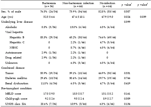
Table 1. Demographic and Clinical Characteristics of Living-Donor Liver Transplant Recipients*
Bacteremia(n = 34) Non-bacteremic infection(n = 46) No-infection(n = 64) p value† p value‡
Sex, % of male 76.5% (26/34) 73.9% (34/46) 82.8% (53/64) 0.507
Age (yrs) 52.5 ± 6.4 47.4 ± 10.1 47.9 ± 9.0 0.024 0.039
Underlying liver disease 0.399
Alcoholic 8.8% (3/34) 13.0% (6/46) 6.3% (4/64)
Viral hepatitis
Hepatitis B 85.3% (29/34) 65.2% (30/46) 76.6% (49/64)
Hepatitis C 0 2.2% (1/46) 4.7% (3/64)
NBNC 0 8.7% (4/46) 6.3% (4/64)
Autoimmune 2.9% (1/34) 2.2% (1/46) 0
Drug related 2.9% (1/34) 2.2% (1/46) 0
Unknown 0 6.5% (3/46) 6.3% (4/64)
Combined disease
Tumor 55.9% (19/34) 39.1% (18/46) 46.9% (30/64) 0.331
Diabetes mellitus 29.4% (10/34) 30.4% (14/46) 29.7% (19/64) 0.766
Renal dysfunction 11.8% (4/34) 6.5% (3/46) 4.7% (3/64) 0.419
Pre-transplant condition
MELD score 17.0 ± 9.5 18.3 ± 8.7 15.1 ± 8.2 0.161
Child-pugh score 9.2 ± 2.6 9.5 ± 2.4 8.9 ± 2.7 0.509
UNOS class IIA 20.6% (7/34) 10.9% (5/46) 6.3% (4/64) 0.134
NBNC, non-B non-C; MELD, model for end-stage liver disease; UNOS, united network of organ sharing. *Data are presented as percent (no.) or mean ± SD.
†Chi-square test was used to analyze categorical variables between three groups and ANOVA for continuous variables. ‡Student's t-test was used between bacteremia group and non-bacteremic infection group.
differ between the 3 groups. Pre-transplant para-meters such as the Model for End-Stage Liver Disease (MELD) scores, Child-Pugh scores, and United Network of Organ Sharing (UNOS) classes did not significantly differ between the 3 groups (Table 1).
Sources and causes of bacteremia
Of the 144 patients, 34 had 40 episodes of bacte-remia, and 17% (6/34) of these patients experienced an additional bacteremia episode during the study period. None of these patients showed relapse or recurrence of bacteremia, and each episode was considered a different case. Among 40 episodes of bacteremia, 22.5% (9/40) occurred within 14 days
of operation and 77.5% (31/40) occurred later than 14 days after transplantation.
The 3 most common sources of bacteremia were intravascular catheter (30%; 12 of 40 episodes), biliary tract (30%; 12 of 40 episodes), and abdomen (22.5%; 9 of 40 episodes). The sources and patho-gens in bacteremic patients are listed in Table 2. Catheter-related bacteremia in the majority of the cases was caused by staphylococci (75.0%; 9 of 12 episodes). Of them, 88.9% (8 of 9 organisms) were methicillin-resistant. Two patients died due to catheter-related bacteremia caused by Candida albicans and Acinetobacter lwoffii.
Table 2. Sources and Pathogens Associated with Bacteremia in Study Patients
Source
(No. of episodes) Pathogen No. of episodes
Sensitivity to major antibiotics (No. of episodes) Catheter related (n = 12) Coagulase-negative Staphylococci 7
Staphylococcus epidermidis 4 MR (4)
Staphylococcus hominis 2 MR (1), MS (1)
Staphylococcus haemolyticus 1 MR (1)
Staphylococcus aureus 2 MR (2)
Acinetobacter lwoffii 1 CS (1)
Candida albicans 1 FS (1)
Candida parapsilosis 1 FR (1)
Biliary (n = 12) Enterococcus faecium 3 VR (2), VS (1)
Enterococcus casseliflavus 1 VR (1)
Klebsiella pneumoniae 3 ESBL (1), non-ESBL (2)
Escherichia coli 2 non-ESBL (2)
Pseudomonas aeruginosa 1 non-ESBL (1)
Enterobacter cloacae 1 ESBL (1)
Candida species* 1
Abdomen (n = 9) Enterococcus faecium 5 VR (3), VS (2)
Escherichia coli 2 non-ESBL (2)
Acinetobacter baumannii 1 CS (1)
Candida glabrata 1 FR (1)
Pneumonia (n = 2) Streptococcus pneumoniae 1 CS (1)
Chryseobacterium meningosepticum 1 CR (1)
Wound (n = 1) Staphylococcus aureus 1 MR (1)
Urinary tract (n = 1) Enterococcus faecium 1 VS (1) Unknown (n = 3) Staphylococcus epidermidis† 2 MR (1), MS (1)
Empedobacter brevis 1 CR (1)
MR, methicillin resistant; MS, methicillin sensitive; CS, cephalosporin sensitive; CR, cephalosporin resistant; FS, fluconazole sensitive; FR, fluconazole resistant; VR, vancomycin resistant; VS, vancomycin sensitive; ESBL, extended-spectrum beta lactamase positive; non-ESBL, extended-spectrum beta lactamase negative.
*Species were not demonstrated at the point of detected.
†Both 2 episodes fulfilled the definition for primary bacteremia.
Among the Gram-positive cocci, methicillin-resis-tant coagulase-negative staphylococci (30.4%; 7/23) were the most common, followed by vancomycin- resistant enterococci (26.1%; 6/23), vancomycin- sensitive enterococci (17.4%; 4/23), methicillin- resistant S. aureus (13%; 3/23), methicillin-sensitive coagulase-negative staphylococci (8.7%; 2/23), and
penicillin-resistant pneumococci (4.3%; 1/23). Over-all, 26.1% (6 of 23 Gram-positives) of the Gram- positive cocci were resistant to vancomycin. None of the enterococci were resistant to linezolid.
Table 3. Pre-operative Variables with Bacteremia Compared to Non-Bacteremic Infections and No-Infection*
Bacteremia
(n = 34)
Non-bacteremic infection (n = 46)
No-infection
(n = 64) p value
†
Donor relationship 0.912
Offspring 47.1% (16/34) 43.5% (20/46) 43.8% (28/64)
Sibling 20.6% (7/34) 21.7% (10/46) 15.6% (10/64)
Parent 0 2.2% (1/46) 1.6% (1/64)
Distant family 0 4.3% (2/46) 6.3% (4/64)
Conjugal 11.8% (4/34) 8.7% (4/46) 15.6% (10/64)
Unrelated 20.6% (7/34) 19.6% (9/46) 17.2% (11/64)
Positive lymphocyte crossmatch 6.9% (2/34) 11.8% (4/46) 3.4% (2/64) 0.300 History of Previous operation 11.8% (4/34) 13.0% (6/46) 9.4% (6/64) 0.825
Pre-operational infectious disease
Fever of unproven source 11.8% (4/34) 17.4% (8/46) 7.8% (5/64) 0.307
Infectious disease 0.309
SBP 17.6% (6/34) 15.2% (7/46) 4.7% (3/64)
Pneumonia 0 0 3.1% (2/64)
Sinusitis 5.9% (2/34) 4.3% (2/46) 7.8% (5/64)
Cholecystitis 2.9% (1/34) 0 0
Tuberculosis 0 2.2% (1/46) 1.6% (1/64)
Others 5.9% (2/34) 6.5% (3/46) 1.6% (1/64)
None 67.6% (23/34) 71.7% (33/46) 81.3% (52/64)
Pre-operational non-infectious variables
Acute hepatic failure 23.5% (8/34) 26.1% (12/46) 18.8% (12/64) 0.645
Portal vein thrombosis 14.7% (5/34) 8.7% (4/46) 20.3% (13/64) 0.246
Small graft size 0 6.5% (3/46) 7.8% (5/64) 0.259
Laboratory findings
White blood cell count (/mm3) 4,264 ± 3,181 4,089 ± 2,894 3,649 ± 2,498 0.529
Creatinine (mg/dL) 0.96 ± 0.74 0.81 ± 0.38 1.03 ± 1.32 0.502 Total bilirubin (mg/dL) 11.6 ± 13.4 10.9 ± 11.9 7.2 ± 10.3 0.116
Albumin (mg/dL) 3.2 ± 0.5 3.1 ± 0.5 3.2 ± 0.5 0.987
SGPT (IU/L) 50 ± 41 57 ± 77 48 ± 51 0.709
INR 1.6 ± 0.4 1.9 ± 1.0 1.6 ± 0.5 0.067
SBP, spontaneous bacterial peritonitis; SGPT, serum glutamate pyruvate transaminase; INR, international normalized ratio. *Data are presented as percent(no.) or mean ± SD.
†Chi-square test was used to analyze categorical variables between three groups and ANOVA for continuous variables.
(15.4%; 2/13). Among the Gram-negatives, 46.2% (6/13) demonstrated resistance to third-generation cephalosporins such as ceftazidime, ceftriaxone,
Table 4. Intra-Operative and Post-Operative Variables of Bacteremic Group Compared to Non-Bacteremic Infection and No Infection*
Bacteremia
(n = 34)
Non-bacteremic infection (n = 46)
No-infection
(n = 64) p value
† p value‡
Intraoperative variables Operative duration (min, min ± SD)
640 ± 94 645 ± 103 613 ± 72 0.150
RBC transfusion (packs, min ± SD)
15.3 ± 8.4 15.2 ± 10.4 12.4 ± 6.7 0.143
Post-operative variables
Re-operation 8.8% (3/34) 15.2% (7/46) 6.3% (4/64) 0.288 Hemodyalisys 26.5% (9/34) 10.9% (5/46) 3.1% (2/64) 0.002
ICU stay (days) 15.6 ± 23.6 10.0 ± 7.1 7.6 ± 2.0 0.012 0.009 Catheter days (days) 29.6 ± 22.6 17.5 ± 6.2 16.6 ± 6.4 < 0.0001 < 0.0001
Biliary complication 26.5% (9/34) 37.0% (17/46) 18.8% (12/64) 0.102 Duration of hospitalization
Post-operation 38.9 ± 22.1 35.7 ± 21.4 25.3 ± 6.6 < 0.0001 < 0.005 ICU, intensive care unit.
*Data are presented as percent (no.) or mean ± SD.
†Chi-square test was used to analyze categorical variables between three groups and ANOVA for continuous variables. ‡Student's t-test was used between bacteremia group and non-bacteremic infection group.
tazobactam (data not shown). Fungi caused 10% (4 of 40 episodes) of the episodes, and 50% of these fungi were resistant to fluconazole.
Predictors of bacteremia
The perioperative clinical and laboratory vari-ables predictive of post-transplant bacteremia were compared between bacteremia, non-bactere-mic infection, and no infection groups (Table 3). No difference was noted between the groups with regard to preoperative variables such as donor relationship, positive lymphocyte cross-match, history of previous surgery, presence of preope-rative infectious disease, and laboratory findings. The intraoperative and postoperative variables of the recipients are listed in Table 4. Surgery dura-tion and the amount of transfusion did not differ between groups. However, bacteremic patients were significantly more likely to have undergone posttransplant hemodialysis (p= 0.002), and to have longer duration of ICU stay (p= 0.012) and longer period of intravascular catheterization (p< 0.0001).
Postoperative hospitalization period was longer in bacteremic patients than in patients with non- bacteremic infection (p= 0.005).
Risk factors for bacteremia and mortality
Univariate analysis revealed that age above 55 years (p= 0.014), intravascular catheterization for more than 8 days (p= 0.003), UNOS class IIA (p= 0.044), and hemodialysis (p= 0.001) were significant risk factors for bacteremia (Table 5). Multivariate analysis revealed an association between bacteremia and age (odds ratio, 6.05; 95% confidence interval, 1.86 to 19.66; p= 0.003), catheterization for more than 22 days (odds ratio, 3.97; 95% confidence interval, 1.4 to 11.22; p= 0.009), UNOS class IIA (odds ratio, 6.59; 95% confidence interval, 1.1 to 39.37; p= 0.039), and posttransplant hemodialysis (odds ratio, 23.12; 95% confidence interval, 3.78 to 141.55; p= 0.001).
infec-Table 5. Logistic Regression Analysis of Risk Factors for Bacteremia after Living-Donor Liver Transplant
Risk factors Percentage of bacteremia with risk factor (No.*)
p value (univariate)
p value
(multivariate) OR (95% CI) Preoperational variables
Age (yrs) ≥55
< 55
36.2% (17/47)
17.5% (17/97) 0.014 0.003
6.05 (1.86-19.66) Diabetes Yes
No
21.9% (7/32)
24.1% (27/112) 0.793
UNOS class IIA
IIB or III
43.8% (7/16)
21.1% (27/128) 0.044 0.039
6.59
(1.10-39.37)
Child class C
A or B
22.1% (17/77)
25.4% (17/67) 0.642
MELD score ≥25
< 25
29.0% (9/31)
22.1% (25/113) 0.422
Tumor Yes
No
28.4% (19/67)
19.5% (15/77) 0.211
History of previous surgery Yes
No
25.0% (4/16)
23.4% (30/128)
0.890
Renal dysfunction Yes
No
40.0% (4/10)
22.4% (30/134) 0.206
Portal vein thrombosis Yes
No
22.7% (5/22)
23.8% (29/122)
0.916
Intra- and post-operational variables Duration of operation (h) ≥10 < 10
22.4% (19/85)
25.4% (15/59) 0.670
Re-operation Yes
No
21.4% (3/14)
23.8% (31/130) 0.840
Posttransplant hemodialysis Yes
No
56.3% (9/16)
19.5% (25/128) 0.001 0.001
23.12 (3.78 - 141.55) Bilary complication Yes
No
23.7% (9/38)
23.6% (25/106) 0.990
ICU stay(d) ≥8
< 8
22.6% (14/62)
24.4% (20/82) 0.800
Catheter days ≥22
< 22
39.1% (18/46)
16.3% (16/98) 0.003 0.009
3.97 (1.40 - 11.22)
OR, odd ratio; CI, confidence interval; UNOS, united network of organ sharing; MELD, model for end-stage liver disease; ICU, intensive care unit.
*No. of bacteremia/No. of patients with risk factors.
tions, and 23.5% (4/17) did not have any infec-tious complication. The mortality rates were 26.5% (9/34) in bacteremia group, 8.7% (4/46) in non- bacteremic infection group, and 6.3% (4/64) in no
[image:7.595.65.531.124.634.2]Fig. 1. Survival curves of study groups; the survival rate at 1 month: bacteremia group, 94.1%; non-bacteremic infection group, 95.7%; and no infection group, 95.3%, the survival rate at 12 months: bacteremia group, 73.2%; non-bacteremic infection group, 91.3%; and no infection group, 93.5%. It was statistically different between the groups as follows: bacteremia vs. no infection, p= 0.006; bacteremia vs. non-bacteremic infection, p= 0.044. No difference was noted between the no infection and non-bacteremic infection groups (p= 0.65).
candidemias. One patient developed Candida and enterococci bacteremia simultaneously. Of the 46 patients in the non-bacteremic infection group, 4 died: 1 due to multi-drug resistant Acinetobacter pneumonia, 1 due to Aspergillus pneumonia, 1 due to refractory shock with pneumonia and infected hematoma, and 1 due to pneumonia with ischemic heart disease. Of 64 patients in the no infection group, 4 died: 1 due to primary graft failure, 2 due to varix bleeding, and 1 due to cardiac ischemia.
The survival rate at 1 month was as follows: bacteremia group, 94.1%; non-bacteremic infection group, 95.7%; and no infection group, 95.3% (Fig. 1). However, the survival rate at 12 months was as follows: bacteremia group, 73.2%; non-bactere-mic infection group, 91.3%; and no infection group, 93.5%. It was statistically different between the groups as follows: bacteremia vs. no infection, p= 0.006; bacteremia vs. non-bacteremic infection, p= 0.044. No difference was noted between the no infec-tion and non-bacteremic infecinfec-tion groups (p= 0.65).
DISCUSSION
In this study, patients who developed bacteremia
after liver transplantation had a decreased 1-year survival rate compared to patients who developed non-bacteremic infection (p= 0.044) or who did not develop any infectious complications (p= 0.006). No difference was noted in the survival rate between the patients who developed non-bacteremic infec-tion and those who did not develop any infecinfec-tion (p= 0.650). Even though all the cases of mortality did not occur because of bacteremia, major causes of death were infectious complications with bac-teremia rather than rejection, surgical complica-tions, and non-bacteremic infections. Despite im-provements in surgical technique, prolonged graft function, and progress in therapeutic options, the rate of major infectious complications remains high.2 Technical complexity due to the high rate of bile leakage has been the main factor respon-sible for infectious complications.18 Our data revealed that the biliary tract was one of the most common sources of bacteremia (Table 2). In the present study, biliary complications were not sig-nificantly different between the groups, however, reduction of biliary complications is thought to be important in reducing bacteremia, especially in living-donor liver transplantation, because biliary trees are one of the most common entries of bacteria and surgical technique is complex.
Liver transplant recipients are generally immu-nocompromised.1 Furthermore, the pretransplant
could have led to these differences.
In our study, patients who developed bacteremia had undergone posttransplant hemodialysis more frequently than other patients and had longer duration of ICU stay and longer period of intra-vascular catheterization. These results are not very different from those observed in a larger study.9,12
The independent risk factors for bacteremia were age above 55 years, catheterization for more than 22 days, UNOS class IIA, and posttransplant hemodialysis. However, in a previous study, only diabetes mellitus and serum albumin level were found to be the risk factors for bacteremia.12 This
indicates that our medical center performs cathe-terization for a longer period and needs to improve posttransplant care. In a previous study, age above 65 years, donor age above 50 years, male gender recipient, re-transplant, and pretransplant MELD score greater than 25 were associated with poor patient and graft survival.19 Furthermore, a high MELD score was found to be indicative of poor outcome and showed maximal impact during the first year posttransplant. Our data also revealed that bacteremic patients tended to have a higher MELD score than other patients, nevertheless it was not a significant risk factor. Therefore, we analyzed 4 variables of the MELD score separately to determine which factors were directly related to bacteremia. In the above mentioned study,19
recipients above the age of 65 years showed signi-ficantly lower survival rates, whereas age above 55 years was found to be a significant risk factor for bacteremia in our study. However, old age is a well-known risk factor for bacteremia even in non-transplant patients.20 Combined tumor, largely
hepatoma, was more common in bacteremic patients than in other patients, however, was not significant.
Catheter-related infection was the main cause of bacteremia; this has also been reported in a previous study.12 However, the common etiologic
organisms such as methicillin-resistant S. aureus and coagulase-negative staphylococci differ between centers.12,21,22 At our center, coagulase-negative staphylococci were found to be more common than S. aureus. Clinically, it is of importance to note that 88% of the staphylococci were methi-cillin-resistant. Therefore, it can be suggested that, whenever a patient develops fever, glycopeptide
should be added empirically during the pending blood culture in addition to removing the catheter. Furthermore, 26.1% of the Gram-positive cocci were vancomycin-resistant enterococci, therefore, linezolid can be considered when a patient is febrile 48 to 72 hours after glycopeptide admini-stration even when no growth is detected in the blood culture.
One-year survival rate showed a significant difference between the 3 groups, whereas 1-month survival rate did not differ between the 3 groups: bacteremia group 94.1%; non-bacteremic infection group, 95.7%; and no infection group, 95.3% (Fig. 1). Majority of the deaths occurred between 1 and 6 months. Posttransplant infection within 1 month occurs primarily due to surgical and technical complexity, wound infection, urinary tract infec-tion, catheter-related infecinfec-tion, and pneumonia.1,2 Our data revealed that a large percentage of events related to bacteremia occurred within 1 month, although death occurred between 1 and 6 months after transplant. Furthermore, long hospital stay along with hemodialysis or catheterization could be another factors responsible for bacteremia that contribute to the decreased survival rate in liver transplant recipients.23 The limitations of our data are that, since this is a retrospective analysis, we could not identify the precise operational break-age during the transplant that could have an effect on the posttransplant clinical course and also could not evaluate biliary variation in donors, which could be an important factor. A large prospective study in the future can provide more informative data.
In conclusion, posttransplant bacteremia de-creases the 1-year survival rate in liver transplant recipients. To reduce the occurrence of bacteremia, recipients with older than 55 years of age and UNOS class IIA need to be carefully monitored for bacteremia, and antibacterial agents should be changed or added for resistant organisms based on the epidemiologic data. Early catheter removal and preservation of renal function could improve posttransplant survival.
REFERENCES
recipients. N Engl J Med 1998;338:1741-51.
2. Snydman DR. Infection in solid organ transplantation. Transpl Infect Dis 1999;1:21-8.
3. Kwak EJ, Kusne S. Risks and epidemiology of infec-tions after liver transplantation. In: Bowden RA, Ljungman P, Paya CV, editors. Transplant infection. 2nd ed. Philadelphia: Lippincott Williams & Wilkins; 2003. p.120.
4. Wade JJ, Rolando N, Hayllar K, Philpott-Howard J, Casewell MW, Williams R. Bacterial and fungal infec-tions after liver transplantation: an analysis of 284 patients. Hepatology 1995;21:1328-36.
5. Singh N, Gayowski T, Wagener MM, Marino IR. Pre-dictors and outcome of early-versus late-onset major bacterial infections in liver transplant recipients re-ceiving tacrolimus (FK506) as primary immunosuppres-sion. Eur J Clin Microbiol Infect Dis 1997;16:821-6. 6. Singh N, Wagener MM, Obman A, Cacciarelli TV, de
Vera ME, Gayowski T. Bacteremias in liver transplant recipients: shift toward gram-negative bacteria as pre-dominant pathogens. Liver Transpl 2004;10:844-9. 7. George DL, Arnow PM, Fox AS, Baker AL, Thistlethwaite
JR, Emond JC, et al. Bacterial infection as a complica-tion of liver transplantacomplica-tion: epidemiology and risk factors. Rev Infect Dis 1991;13:387-96.
8. Kusne S, Dummer JS, Singh N, Iwatsuki S, Makowka L, Esquivel C, et al. Infections after liver transplantation. An analysis of 101 consecutive cases. Medicine (Baltimore) 1988;67:132-43.
9. Wagener MM, Yu VL. Bacteremia in transplant reci-pients: a prospective study of demographics, etiologic agents, risk factors, and outcomes. Am J Infect Control 1992;20:239-47.
10. Paya CV, Hermans PE, Washington JA 2nd, Smith TF, Anhalt JP, Wiesner RH, et al. Incidence, distribution, and outcome of episodes of infection in 100 orthotopic liver transplantations. Mayo Clin Proc 1989;64:555-64. 11. Kawecki D, Chmura A, Pacholczyk M, Lagiewska B,
Adadynski L, Wasiak D, et al. Etiological agents of bac-teremia in the early period after liver transplantation. Transplant Proc 2007;39:2816-21.
12. Singh N, Paterson DL, Gayowski T, Wagener MM, Marino IR. Predicting bacteremia and bacteremic mor-tality in liver transplant recipients. Liver Transpl 2000; 6:54-61.
13. Korean Network for Organ Sharing. Annual Report 2007. Available from: URL: http://www.konos.go.kr accessed 1 June 2008.
14. Cho WH, Kim YS. Landmarks in clinical transplantation in Korea. Yonsei Med J 2004;45:963-7.
15. Moon DB, Lee SG. Adult-to-adult living donor liver transplantation at the Asan medical center. Yonsei Med J 2004;45:1162-8.
16. Chang FY, Singh N, Gayowski T, Wagener MM, Marino IR. Fever in liver transplant recipients: changing spec-trum of etiologic agents. Clin Infect Dis 1998;26:59-65. 17. Garner JS, Jarvis WR, Emori TG, Horan TC, Hughes JM.
CDC definitions for nosocomial infections, 1988. Am J Infect Control 1988;16:128-40.
18. Simon DM, Levin S. Infectious complications of solid organ transplantations. Infect Dis Clin North Am 2001; 15:521-49.
19. Habib S, Berk B, Chang CC, Demetris AJ, Fontes P, Dvorchik I, et al. MELD and prediction of post-liver transplantation survival. Liver Transpl 2006;12:440-7. 20. Mellors JW, Horwitz RI, Harvey MR, Horwitz SM. A
simple index to identify occult bacterial infection in adults with acute unexplained fever. Arch Intern Med 1987;147:666-71.
21. Candel FJ, Grima E, Matesanz M, Cervera C, Soto G, Almela M, et al. Bacteremia and septic shock after solid- organ transplantation. Transplant Proc 2005;37:4097-9. 22. Bedini A, Codeluppi M, Cocchi S, Guaraldi G, Di
Benedetto F, Venturelli C, et al. Gram-positive blood-stream infections in liver transplant recipients: incidence, risk factors, and impact on survival. Transplant Proc 2007;39:1947-9.