Ionization–Time of Flight Mass Spectrometry System for Identification
of Aerobically Growing Gram-Positive Bacilli
E. Farfour,a,bJ. Leto,fM. Barritault,a,bC. Barberis,gJ. Meyer,aB. Dauphin,fA.-S. Le Guern,hA. Leflèche,iE. Badell,jN. Guiso,j A. Leclercq,kA. Le Monnier,kM. Lecuit,a,kV. Rodriguez-Nava,lE. Bergeron,lJ. Raymond,a,cS. Vimont,dE. Bille,a,bE. Carbonnelle,a,e H. Guet-Revillet,a,bH. Lécuyer,a,bJ.-L. Beretti,a,bC. Vay,gP. Berche,a,bA. Ferroni,a,bX. Nassif,a,band O. Join-Lamberta,b
Université Paris Descartes, Paris, Francea; Laboratoire de Microbiologie, Hôpital Necker-Enfants Malades,bLaboratoire de Bactériologie, Hôpital Cochin,cLaboratoire de Bactériologie, Hôpital Tenon,dand Laboratoire de Microbiologie, Hôpital Européen Georges Pompidou,eAssistance Publique-Hôpitaux de Paris, Paris, France; Andromas SAS, Pépinière Paris Santé Cochin, Paris, Francef; Laboratorio de Bacteriologia, Hospital de Clínicas, Facultad de Farmacia y Bioquímica, Universidad de Buenos Aires, Buenos Aires, Argentinag; Laboratoire du Centre Médical de l’Institut Pasteur,hCellule d’Intervention Biologique d’Urgence,iMolecular Prevention and Therapy of Human Diseases, National Centre of Reference of Corynebacteria of theDiphtheriaeComplex,jand Centre National de Référence desListeria, Institut Pasteur, Paris, Francek; and Research Group on Bacterial Opportunistic Pathogens and Environment, UMR5557-Observatoire Français des Nocardioses, Université Claude Bernard Lyon 1, Lyon, Francel
Matrix-associated laser desorption ionization–time of flight mass spectrometry (MALDI-TOF MS) is a rapid and simple
micro-bial identification method. Previous reports using the Biotyper system suggested that this technique requires a preliminary
ex-traction step to identify Gram-positive rods (GPRs), a technical issue that may limit the routine use of this technique to identify
pathogenic GPRs in the clinical setting. We tested the accuracy of the MALDI-TOF MS Andromas strategy to identify a set of 659
GPR isolates representing 16 bacterial genera and 72 species by the direct colony method. This bacterial collection included 40
C.
diphtheriae
, 13
C. pseudotuberculosis
, 19
C. ulcerans
, and 270 other
Corynebacterium
isolates, 32
L. monocytogenes
and 24 other
Listeria
isolates, 46
Nocardia
, 75
Actinomyces
, 18
Actinobaculum
, 11
Propionibacterium acnes
, 18
Propionibacterium avidum
, 30
Lactobacillus
, 21
Bacillus
, 2
Rhodococcus equi
, 2
Erysipelothrix rhusiopathiae
, and 38 other GPR isolates, all identified by
refer-ence techniques. Totals of 98.5% and 1.2% of non-
Listeria
GPR isolates were identified to the species or genus level, respectively.
Except for
L. grayi
isolates that were identified to the species level, all other
Listeria
isolates were identified to the genus level
because of highly similar spectra. These data demonstrate that rapid identification of pathogenic GPRs can be obtained without
an extraction step by MALDI-TOF mass spectrometry.
A
erobically growing Gram-positive rods (GPRs) form a very
heterogeneous and extensive group of bacterial species (
23
).
Some of them, such as
Corynebacterium diphtheriae
,
Listeria
monocy-togenes
, and
Bacillus anthracis
are highly pathogenic. These bacterial
species are associated with severe community-acquired infections
and may be associated with outbreaks, thus requiring rapid
identifi-cation for both therapeutic and infection control measures. Other
clinically important pathogenic GPR species include
Nocardia
spp.
and
Rhodococcus equi
, which can cause severe opportunistic
infec-tions.
Corynebacterium urealyticum
and
Corynebacterium jeikeium
are, respectively, associated with urinary tract infections and vascular
catheter infections, prosthetic endocarditis, and septicemia and can
be multidrug resistant (
31
,
49
). Finally, during the last two decades, a
number of species of low pathogenicity, such as
Propionibacterium
acnes
and new actinomycetes (
Actinobaculum schaalii
,
Actinomyces
neuii
,
Actinomyces turicensis
, and
Actinomyces radingae
), have been
associated with various infectious conditions (
5
,
25
,
37
). Rapid
iden-tification of GPRs is therefore increasingly required for therapeutic
and/or epidemiological concerns.
Systematic identification of GPRs is a challenge. Phenotypic
identification methods are accurate for identification of
patho-genic GPRs. However, it isn’t easy to identify most commensal
GPR isolates that are frequently cultured in microbiology
labora-tories by these techniques, due to weak or variable reactivity
among single species or because additional tests are required in up
to 50% of cases to obtain a 90% correct identification rate (
22
). In
addition, as most recently described GPR species have been
char-acterized using molecular techniques such as 16S rRNA gene
se-quencing, these species cannot be identified using phenotypic
methods in the absence of appropriate identification systems or
updated databases (
1
,
10
,
21
,
22
,
45
,
54
). In some cases, 16S
ribo-somal gene sequencing poorly discriminates closely related
spe-cies, and alternative molecular methods have been developed to
solve these issues (
28
,
36
,
38
). Matrix-assisted laser desorption
ionization–time of flight mass spectrometry (MALDI-TOF MS) is
a rapid bacterial identification technique that is increasingly used
in microbiology laboratories (
13
,
30
,
48
). MALDI-TOF MS
iden-tification systems are based on the comparison of the tested isolate
mass spectrum with reference databases. Several databases and
identification strategies have been developed, including the
Bio-typer (Bruker Daltonics, Bremen, Germany), the Saramis
(bio-Mérieux, Marcy l’Etoile, France), and the Andromas (Paris,
France) systems. Previous studies reported that the Bruker
MALDI-TOF MS system accurately identified GPR species such as
Listeria
spp.,
Nocardia
spp., and
Corynebacterium
species (
3
,
6
,
29
,
Received10 February 2012 Returned for modification19 March 2012 Accepted17 May 2012
Published ahead of print12 June 2012
Address correspondence to Olivier Join-Lambert, olivier.join-lambert@nck.aphp.fr.
Copyright © 2012, American Society for Microbiology. All Rights Reserved.
doi:10.1128/JCM.00368-12
on May 16, 2020 by guest
http://jcm.asm.org/
51
). In these studies, a preliminary extraction step was mandatory
to obtain satisfactory results, a procedure that is more
cumber-some and time-consuming than the direct colony method that can
be used to identify Gram-negative bacilli (
43
). Available
MALDI-TOF MS databases have also been evaluated in the routine use of
clinical microbiology laboratories, but few GPRs were included, and
in some cases, poor identification results were reported (
7
,
47
).
The Andromas identification strategy is based on a limited
number of species-specific profiles for each entry (
11
,
16
). A
par-ticularity of the Andromas database is that it was built without any
extraction step (
7
,
12
). Previous studies have shown that this
iden-tification strategy provides good ideniden-tification of bacteria,
myco-bacteria, yeasts, and
Aspergillus
spp. by the direct colony method
(
2
,
7
,
11
,
16
,
18
,
19
,
32
). We evaluated the accuracy of this method
to identify a large collection of GPR isolates that included both
pathogenic and commensal isolates.
MATERIALS AND METHODS
Bacterial isolates.A collection of 659 GPR isolates was set up for this study, originating from French clinical microbiology laboratories and ref-erence centers and from the clinical microbiology laboratory of the Uni-versity of Buenos Aires, Argentina. Altogether, this collection comprised 16 bacterial genera and 73 bacterial species that are listed inTable 1. Except forListeria,Nocardia,Corynebacterium diphtheriae, Corynebacte-rium pseudotuberculosis, andCorynebacterium ulceransisolates, we used 16S rRNA gene sequencing as a reference technique to identify clinical isolates. 16S rRNA gene sequences were compared with those of bacterial strains obtained from the GenBank or Bioinformatic Bacterial Identifica-tion database (17). Aⱖ99.0% similarity cutoff was required for definite identification.
Listeriaisolates were identified by the French National Reference Cen-tre forListeriawith the API Listeria identification system (bioMérieux, Marcy l’Etoile, France) (8,20,34) and additional phenotypic confirma-tion tests (41,46). AllNocardiaisolates were identified by the French Reference Center forNocardiausing a polyphasic approach, including morphological examination, biochemical profiling, antimicrobial suscep-tibility testing, and 16S rRNA gene sequencing using specific primers de-signed to identifyNocardiaspp. (42). Partial sequencing of thehsp65gene was done only for isolates identified asNocardia abscessus,Nocardia ar-thritidis, andNocardia beijingensisto confirm species identification (42). C. diphtheriae,C. ulcerans, andC. pseudotuberculosisisolates were identi-fied by the French National Reference Centre of Corynebacteria of the DiphtheriaeComplex using the API Coryne system (bioMérieux, Marcy l’Etoile, France), additional phenotypical assays, and specific molecular methods (a PCR targeting thedtxRgene forC. diphtheriaeand a multiplex PCR targeting the 16S rRNA,rpoB, andpldgenes forC. pseudotuberculosis andC. ulcerans) (36,38).
Instrumentation and data analysis.Bacterial isolates were grown on 5% horse blood Columbia agar plates and incubated for 24 h (48 h for slow-growth bacteria) at 37°C in a 10% CO2atmosphere. Whole-cell bacteria were deposited without an extraction step using a cotton swab on a MALDI-TOF MS target plate and allowed to dry at room temperature. Samples were then fixed by adding 1l of absolute ethanol to inactivate bacteria and allowed to dry at room temperature. Samples were then overlaid with 1l of saturated ␣-cyano-4-hydroxycinnamic acid and allowed to crystallize at room temper-ature. Samples were processed on the Microflex MALDI-TOF MS spectrom-eter with the FlexControl software (Bruker Daltonics, Bremen, Germany). Positive ions were extruded at an accelerating voltage of 20 kV in linear mode. Each spectrum was the sum of the ions obtained from 350 laser shots per-formed automatically on different regions of the same well. The spectra were analyzed in anm/zrange of 3,500 to 20,000.
Evaluation of the Andromas MALDI-TOF MS system for identifica-tion of GPRs.Bacterial isolates were identified using the Andromas soft-ware, while being unaware of the identification result of the reference
method. This software compares the mass spectrum of the tested isolate with the spectra included in the Andromas database, taking into account a possible MS peak variation of⫾10m/z(7). The Andromas database was created using both reference and clinical isolates routinely used in our laboratory. To date, this database encompasses more than 700 bacterial species, including 33 genera and 156 species of aerobically growing Gram-positive rods. Using this software, species identification is considered to be valid if the percentage of common peaks isⱖ68% of those of a species-specific profile of the database. A 10% difference between the first two species diagnostics having the best match in the database is also required to give species identification. If the latter condition is not fullfilled, the identification is considered to be correct at the genus or group level for closely related species if the first two matches belonged to the same genus or group of bacteria. In all other cases, results are considered to be incon-clusive.
In this study, a misidentification was defined as a discrepancy between the identification result given by MALDI-TOF MS and the result obtained by 16S rRNA sequencing or other reference identification methods de-scribed above. Identifications to the genus level and absence of identifica-tion were not considered misidentificaidentifica-tions.
Listeriaspecies discrimination.A similarity study ofListeriaisolates’ mass spectra was performed using the BioNumerics 5.1 software (Applied Maths, Belgium) for in-gel-view representation and calculation of den-drograms. Similarities between mass spectra were calculated using the Euclidean distance. Hierarchical clustering was performed using the un-weighted-pair group method using average linkages (UPGMA) aggrega-tion method.
RESULTS
Except for
Listeria
isolates, 98.5% (594/603) of strains were
iden-tified to the species level, and only 1.2% (7/603) of them could not
be identified beyond the genus level (
Table 1
). Correct
identifica-tions were obtained for the closely related pathogenic species
C.
diphtheriae
,
C. ulcerans
, and
C. pseudotuberculosis
, as well as for
frequently encountered nonlipophilic commensal
Corynebacte-rium
species that are difficult to identify using biochemical tests:
C. striatum
or
C. amycolatum
, which have been misidentified as
C.
xerosis
(
21
);
C. simulans
, which fits the biochemical profile of
C.
striatum
or
C. minutissimum
(
53
), and
C. aurimucosum
, which is
difficult to differentiate from
C. minutissimum
(
55
). MALDI-TOF
MS also allowed a rapid identification of bacterial species that
would have required molecular methods for definitive
identifica-tion as
Nocardia
spp.,
Rhodococcus equi
,
Erysipelothrix
rhusio-pathiae
,
Actinomyces
spp., and
Bacillus
and
Lactobacillus
spp. (
9
).
In most cases, MALDI-TOF MS performed as well as extended
phenotypic and specific molecular methods to identify GPR
iso-lates.
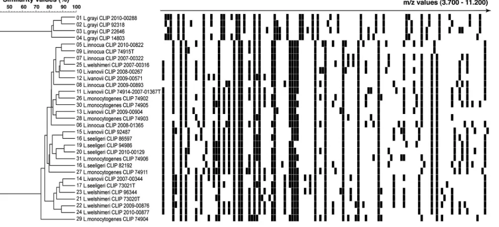
Conversely, except for
Listeria grayi
isolates that were
identi-fied to the species level, all other
Listeria
species isolates could not
be identified beyond the genus level. Indeed, the mass spectrum of
these isolates always matched more than one
Listeria
species-spe-cific profile of the database with a high similarity value (ⱖ68% of
common peaks). Moreover, the 10% difference between the two
best matchs that is required by the Andromas algorithm to
pro-vide species identification was not reached. Therefore,
identifica-tion was only given at the genus level. The similarity analysis of the
mass spectra of
Listeria
isolates accounts for these results, showing
that
Listeria grayi
and non
-grayi
isolates form two distinct clusters
with high intrinsic similarity values (
ⱖ
70%), but these two
clus-ters only share a 40% similarity value (
Fig. 1
). These findings
cor-roborate molecular 16S and 23S rRNA phylogenetic studies that
on May 16, 2020 by guest
http://jcm.asm.org/
showed that two clusters can be identified within this genus: one
for
L. grayi
and a second for other
Listeria
species (
14
,
44
).
Only 7 other GPRs were identified to the genus level, including
4
Nocardia
and 2
C. aurimucosum
isolates and 1
A. viscosus
isolate.
The 4
Nocardia
isolates, which belonged to 4 different species,
were not identified to the species level because of poor mass
spec-tra, a technical issue that has been previously reported for
Nocar-dia
spp. without an extraction step (
51
). The two
C. aurimucosum
isolates and the
A. viscosus
isolate had satisfactory mass spectra but
matched another species-specific profile of the same genus with a
less than 10% difference.
Finally, 1
C. aurimucosum
isolate was misidentified as
C.
stria-tum
, and 1
C. diphtheriae
isolate could not be identified. Analysis
of the mass spectrum of the
C. aurimucosum
isolate showed that it
corresponded to a new specific
C. aurimucosum
profile. The
C.
diphtheriae
isolate was not identified because its mass spectrum
[image:3.585.51.453.71.682.2] [image:3.585.273.536.81.571.2]comprised only 5 peaks, whereas species-specific profiles of the
TABLE 1Identification of 659 Gram-positive rods using the MALDI-TOF MS Andromas systema
Genus or species
No. of isolates tested
Identification result (no. of isolates)
Species level
Genus level
Not
identified Error
Actinobaculumspp.b 18 18 0 0 0
A. schaalii 14 14 0 0 0
A. massiliense 4 4 0 0 0
Actinomycesspp.b 76 75 1 0 0
A. europaeus 1 1 0 0 0
A. funkei 4 4 0 0 0
A. naeslundii 1 1 0 0 0
A. neuii 33 33 0 0
A. odontolyticus 5 5 0 0 0
A. radingae 6 6 0 0 0
A. turicensis 23 23 0 0 0
A. urogenitalis 2 2 0 0 0
A. viscosus 1 0 1 0 0
Bacillusspp.b 21 21 0 0 0
B. cereus/B. thuringiensis 10 10 0 0 0
B. licheniformis 2 2 0 0 0
B. pumilus/B. safensis 4 4 0 0 0
B. subtilis 3 3 0 0 0
B. simplex 2 2 0 0 0
Corynebacteriumspp.b 342 338 2 1 1
C. amycolatum 97 97 0 0 0
C. aurimucosum 30 27 2 0 1
C. coyleae 3 3 0 0 0
C. diphtheriae 40 39 0 1 0
C. glucuronolyticum 10 10 0 0 0
C. imitans 2 2 0 0 0
C. jeikeium 16 16 0 0 0
C. massiliensis 1 1 0 0 0
C. pseudodiphtheriticum 10 10 0 0 0
C. pseudotuberculosis 13 13 0 0 0
C. simulans 25 25 0 0 0
C. singulare 1 1 0 0 0
C. striatum 44 44 0 0 0
C. tuberculostearicum 18 18 0 0 0
C. ulcerans 19 19 0 0 0
C. urealyticum 10 10 0 0 0
Lactobacillusspp.b 30 30 0 0 0
L. casei 1 1 0 0
L. crispatus 8 8 0 0 0
L. delbrueckii 1 1 0 0 0
L. fermentum 2 2 0 0 0
L. gasseri 7 7 0 0 0
L. jensenii 6 6 0 0 0
L. rhamnosus 5 5 0 0 0
Listeriaspp.b 56 4 52 0 0
L. grayi 4 4 0 0
L. innocua 5 0 5 0 0
L. ivanovii 5 0 5 0 0
L. monocytogenes 32 0 32 0 0
L. seeligeri 5 0 5 0 0
L. welshimeri 5 0 5 0 0
TABLE 1(Continued)
Genus or species
No. of isolates tested
Identification result (no. of isolates)
Species level
Genus level
Not
identified Error
Nocardiaspp.b 46 42 4 0 0
N. abscessus 4 4 0 0 0
N. asteroides 2 2 0 0 0
N. arthritidis 1 0 1 0 0
N. beijingensis 3 3 0 0 0
N. brasiliensis 7 6 1 0 0
N. cyriacigeorgica 7 6 1 0 0
N. farcinica 11 10 1 0 0
N. nova 5 5 0 0
N. mexicana 1 1 0 0 0
N. otitidiscavium 2 2 0 0 0
N. paucivorans 1 1 0 0 0
N. veterana 2 2 0 0 0
Propionibacteriumspp.b 29 29 0 0 0
P. acnes 11 11 0 0 0
P. avidum 18 18 0 0 0
Other GPR speciesc 41 41 0 0 0
Arthrobacter cumminsii 1 1 0 0 0
Brevibacterium casei 3 3 0 0 0
Brevibacterium paucivorans
1 1 0 0 0
Dermabacter hominis 12 12 0 0 0
Erysipelothrix rhusiopathiae
2 2 0 0 0
Gardnerella vaginalis 3 3 0 0 0
Paenibacillus motobuensis
1 1 0 0 0
Rhodococcus equi 2 2 0 0 0
Turicella otitidis 16 16 0 0 0
aThe mass spectra of the isolates studied were compared with those of the whole
Andromas database, which encompasses more than 700 bacterial species, including 33 genera and 156 species of aerobically growing Gram-positive rods (GPRs). b
For these genera, the total numbers of taxa included in the Andromas database are 18
Actinomyces, 3Actinobaculum, 13Bacillus, 32Corynebacterium, 24Lactobacillus, 6
Listeria, 25Nocardia, and 4Propionibacteriumspecies.
cFor these genera, the total numbers of taxa included in the Andromas database are 5
Arthrobacter, 6Brevibacterium, 1Dermabacter, 1Erysipelothrix, 1Gardnerella, and 3
Paenibacillusspecies, 1Rhodococcusspecies, and 1Turicellaspecies.
on May 16, 2020 by guest
http://jcm.asm.org/
closely related species
C. diphtheriae
,
C. pseudotuberculosis
, and
C.
ulcerans
comprise a minimum of 23 peaks. In that case, a formic
acid protein extraction step slightly improved the mass spectrum
profile (13 peaks), but species identification could not be obtained
because the two best matches in the database were too close (
C.
diphtheriae
at 63% and
C. pseudotuberculosis
at 60%). This
non-toxigenic isolate was recovered from a patient with bacteremia. It
grew well on agar plates and was correctly identified by phenotypic
and molecular assays.
All
Bacillus cereus
isolates were identified as
Bacillus cereus
/
B.
thurigiensis
group since these two species display similar
species-specific spectra in the Andromas database. Four additional
Bacil-lus
isolates were identified as
B. pumilus
/
B. safensis
for the same
reason and because they cannot be identified to the species level
using 16S rRNA gene sequencing.
DISCUSSION
This work is the first MALDI-TOF MS study that addressed the
issue of the accuracy of this new bacterial identification method
against a fairly extensive collection of Gram-positive rods,
includ-ing both pathogenic and commensal species. The most important
result of our study is that the vast majority of pathogenic GPRs
could be identified to the species level. In addition, we also
accu-rately identified nonpathogenic or low-pathogenicity species. As
most of these strains were obtained from clinical microbiology
laboratories and represent the GPRs that are most frequently
re-covered in this setting, our data suggest that this identification
strategy can also be used to rule out pathogenic pathogens and to
describe new GPR-associated infectious conditions.
In this study, MALDI-TOF MS identification of bacterial
iso-lates were obtained by the direct colony method—i.e., under
workflow conditions that can be routinely used in all
microbiol-ogy laboratories. Similar results were only obtained after a
prelim-inary extraction step using the Bruker Biotyper system (
3
,
6
,
29
,
51
). Each MALDI-TOF MS identification system has its own
spec-ificities, including the hardware itself (the mass spectrometer), the
way the database was built, and the algorithms used to compare
the mass spectrum of a sample with those of the database.
There-fore, it is not an easy task to identify the reason why better results
were obtained with the Andromas system by the direct colony
method. It is well established that the quality of mass spectra varies
according to bacterial genera. For instance, better MALDI-TOF
MS acquisitions (i.e., higher number of peaks and/or higher
rela-tive intensity of peaks) are usually obtained for Gram-negarela-tive
bacteria (except for mucoid colonies) compared to enterococci
and Gram-positive rods (
4
). Consequently, using a MALDI-TOF
database that was built with an extraction step, it is likely that
identification errors will occur more frequently for Gram-positive
bacteria than for Gram-negative bacteria. The Andromas database
was built without any extraction step, a technical issue that
prably had an important impact on the quality of results we
ob-tained. To the best of our knowledge, the way the bioMérieux and
Bruker databases were engineered has not been previously
re-ported or published.
Our data suggest that species identification of
Listeria
spp. by
the direct colony method can only be achieved at the genus level,
except for
L. grayi
. It has been reported that species identification
and typing of
Listeria
spp. can be obtained with the Biotyper
sys-tem (
6
). Nevertheless, a complex extraction step and several
cen-trifugations that may not be routinely implemented in clinical
microbiology laboratories were required to obtain these results.
As
L. monocytogenes
causes almost all cases of human listeriosis,
rapid identification of a GPR isolate as
Listeria
sp. is significant
progress. For a limited number of GPRs belonging to other
gen-era, no identification was obtained at the species level, because the
mass spectrum of the tested isolate matched two species-specific
profiles of the same genus, because of an atypical mass spectrum,
or due to poor mass spectrum acquisition. Altogether, these
ob-FIG 1MALDI-TOF MS discrimination ofListeriaspecies. MALDI-TOF MS profiles ofListeriaisolates, as obtained by the direct colony method, are visualized in a gel-view representation. Spectra were clustered using the UPGMA clustering algorithm. The scale represents the percentage of matching peak mass signals between individual spectra.
on May 16, 2020 by guest
http://jcm.asm.org/
[image:4.585.45.541.71.297.2]servations illustrate the limitations, albeit limited, of the
Andro-mas MALDI-TOF MS strategy for identification of GPRs. By the
direct colony method, we obtained species identification rates of
Corynebacterium
and
Nocardia
isolates similar to those reported
by the extraction method using the Biotyper system, suggesting
that a simpler approach can be used to identify GPRs.
Rapid identification of GPRs should have an important clinical
impact on the diagnosis of GPR-associated infectious diseases.
Rarely encountered diseases, such as granulomatous
lymphadeni-tis due to
C. pseudotuberculosis
or cutaneous manifestations of
C.
ulcerans
or
C. diphtheriae
, may be easier to diagnose (
27
,
33
,
52
).
In that case, the main advantage of this direct colony method is to
give rapid identification of colonies that cannot be distinguished
from those of other nonlipophilic
Corynebacterium
species.
MALDI-TOF MS should also improve the diagnosis of fastidious
or recently described GPR species, like new
Actinomyces
species
(
26
,
35
,
39
,
45
). In addition, rapid identification of GPRs should
improve the probabilistic antimicrobial treatment of patients.
In-deed, some GPRs have specific and atypical susceptibility patterns,
such as resistance to broad-spectrum cephalosporins for
L.
mono-cytogenes
and resistance to glycopeptides for
E. rhusiopathiae
,
some
Nocardia
spp., and some
Lactobacillus
spp. (
15
,
24
,
50
).
Some GPRs, such as
C. jeikeium
, and, to a lesser extent,
C.
urea-lyticum
and
C. amycolatum
, can be multidrug resistant (
40
).
To conclude, our data suggest that the Andromas strategy is an
accurate method for identification and/or screening of pathogenic
GPRs. As taxonomic changes and characterization of new species
or genomospecies of GPRs are frequent, continuous enrichment
of the MALDI-TOF MS databases will have to be performed as a
dynamic process.
ACKNOWLEDGMENTS
We thank Gilles Quesnes for technical assistance.
This work was supported by grants from the Programme Hospitalier de Recherche Clinique (PHRC; grant no. BOS07001 and AOM08181).
Julie Leto and Brunhilde Dauphin are employees of Andromas. Xavier Nassif is a shareholder of Andromas.
REFERENCES
1.Adderson EE, et al.2008. Identification of clinical coryneform bacterial isolates: comparison of biochemical methods and sequence analysis of 16S rRNA andrpoBgenes. J. Clin. Microbiol.46:921–927.
2.Alanio A, et al.2011. Matrix-assisted laser desorption ionization time-of-flight mass spectrometry for fast and accurate identification of clini-cally relevant Aspergillus species. Clin. Microbiol. Infect.17:750 –755. 3.Alatoom AA, Cazanave CJ, Cunningham SA, Ihde SM, Patel R.2012.
Identification of non-diphtheriae Corynebacteriumby use of matrix-assisted laser desorption ionization–time of flight mass spectrometry. J. Clin. Microbiol.50:160 –163.
4.Alatoom AA, Cunningham SA, Ihde SM, Mandrekar J, Patel R.2011. Comparison of direct colony method versus extraction method for iden-tification of Gram-positive cocci by use of Bruker Biotyper matrix-assisted laser desorption ionization–time of flight mass spectrometry. J. Clin. Mi-crobiol.49:2868 –2873.
5.Bank S, Jensen A, Hansen TM, Soby KM, Prag J.2010. Actinobaculum schaalii, a common uropathogen in elderly patients, Denmark. Emerg. Infect. Dis.16:76 – 80.
6.Barbuddhe SB, et al.2008. Rapid identification and typing ofListeria species by matrix-assisted laser desorption ionization–time of flight mass spectrometry. Appl. Environ. Microbiol.74:5402–5407.
7.Bille E, et al.27 September 2011. MALDI-TOF MS Andromas strategy for the routine identification of bacteria, mycobacteria, yeasts, Aspergillus spp. and positive blood cultures. Clin. Microbiol. Infect. [Epub ahead of print.] doi:10.1111/j.1469-0691.2011.03688.x.
8.Bille J, et al.1992. API Listeria, a new and promising one-day system to identifyListeriaisolates. Appl. Environ. Microbiol.58:1857–1860. 9.Bizzini A, et al.2011. Matrix-assisted laser desorption ionization-time of
flight mass spectrometry as an alternative to 16S rRNA gene sequencing for identification of difficult-to-identify bacterial strains. J. Clin. Micro-biol.49:693– 696.
10. Boyd MA, Antonio MA, Hillier SL.2005. Comparison of API 50 CH strips to whole-chromosomal DNA probes for identification of Lactoba-cillusspecies. J. Clin. Microbiol.43:5309 –5311.
11. Carbonnelle E, et al.2007. Rapid identification of staphylococci isolated in clinical microbiology laboratories by matrix-assisted laser desorption ionization–time of flight mass spectrometry. J. Clin. Microbiol.45:2156 – 2161.
12. Carbonnelle E, et al.2012. Robustness of two MALDI-TOF mass spec-trometry systems for bacterial identification. J. Microbiol. Methods89: 133–136.
13. Carbonnelle E, et al.2011. MALDI-TOF mass spectrometry tools for bacterial identification in clinical microbiology laboratory. Clin. Biochem. 44:104 –109.
14. Collins MD, et al.1991. Phylogenetic analysis of the genusListeriabased on reverse transcriptase sequencing of 16S rRNA. Int. J. Syst. Bacteriol. 41:240 –246.
15. Danielsen M, Wind A.2003. Susceptibility of Lactobacillus spp. to anti-microbial agents. Int. J. Food Microbiol.82:1–11.
16. Degand N, et al.2008. Matrix-assisted laser desorption ionization-time of flight mass spectrometry for identification of nonfermenting Gram-negative bacilli isolated from cystic fibrosis patients. J. Clin. Microbiol. 46:3361–3367.
17. Devulder G, Perriere G, Baty F, Flandrois JP.2003. BIBI, a bioinfor-matics bacterial identification tool. J. Clin. Microbiol.41:1785–1787. 18. Dupont C, et al.2010. Identification of clinical coagulase-negative
staph-ylococci, isolated in microbiology laboratories, by matrix-assisted laser desorption/ionization-time of flight mass spectrometry and two auto-mated systems. Clin. Microbiol. Infect.16:998 –1004.
19. Ferroni A, et al.2010. Real-time identification of bacteria andCandida species in positive blood culture broths by matrix-assisted laser desorption ionization–time of flight mass spectrometry. J. Clin. Microbiol.48:1542– 1548.
20. Fujisawa T, Mori M.1994. Evaluation of media for determining hemo-lytic activity and that of API Listeria system for identifying strains of Lis-teria monocytogenes. J. Clin. Microbiol.32:1127–1129.
21. Funke G, Lawson PA, Bernard KA, Collins MD.1996. Most Corynebac-terium xerosisstrains identified in the routine clinical laboratory corre-spond toCorynebacterium amycolatum. J. Clin. Microbiol.34:1124 –1128. 22. Funke G, Renaud FN, Freney J, Riegel P.1997. Multicenter evaluation of the updated and extended API (RAPID) Coryne database 2.0. J. Clin. Microbiol.35:3122–3126.
23. Funke G, von Graevenitz A, Clarridge JE III, Bernard KA.1997. Clinical microbiology of coryneform bacteria. Clin. Microbiol. Rev.10:125–159. 24. Gorby GL, Peacock JE, Jr.1988. Erysipelothrix rhusiopathiae
endocar-ditis: microbiologic, epidemiologic, and clinical features of an occupa-tional disease. Rev. Infect. Dis.10:317–325.
25. Hall V.2008. Actinomyces— gathering evidence of human colonization and infection. Anaerobe14:1–7.
26. Hwang SS, et al.2011. Actinomyces graevenitzii bacteremia in a patient with alcoholic liver cirrhosis. Anaerobe17:87– 89.
27. Join-Lambert OF, et al.2006. Corynebacterium pseudotuberculosis ne-crotizing lymphadenitis in a twelve-year-old patient. Pediatr. Infect. Dis. J. 25:848 – 851.
28. Khamis A, Raoult D, La Scola B.2005. Comparison betweenrpoBand 16S rRNA gene sequencing for molecular identification of 168 clinical isolates ofCorynebacterium. J. Clin. Microbiol.43:1934 –1936.
29. Konrad R, et al.2010. Matrix-assisted laser desorption/ionisation time-of-flight (MALDI-TOF) mass spectrometry as a tool for rapid diagnosis of potentially toxigenic Corynebacterium species in the laboratory manage-ment of diphtheria-associated bacteria. Euro Surveill.15:19699.http: //www.eurosurveillance.org/ViewArticle.aspx?ArticleId⫽19699 30. La Scola B.2011. Intact cell MALDI-TOF mass spectrometry-based
ap-proaches for the diagnosis of bloodstream infections. Expert Rev. Mol. Diagn.11:287–298.
31. Lavollay M, et al.2009. The beta-lactam-sensitive D,D-carboxypeptidase activity of Pbp4 controls the L,D and D,D transpeptidation pathways in Corynebacterium jeikeium. Mol. Microbiol.74:650 – 661.
on May 16, 2020 by guest
http://jcm.asm.org/
32. Lotz A, et al.2010. Rapid identification of mycobacterial whole cells in solid and liquid culture media by matrix-assisted laser desorption ioniza-tion–time of flight mass spectrometry. J. Clin. Microbiol.48:4481– 4486. 33. Lowe CF, Bernard KA, Romney MG.2011. Cutaneous diphtheria in the urban poor population of Vancouver, British Columbia, Canada: a 10-year review. J. Clin. Microbiol.49:2664 –2666.
34. McLauchlin J.1997. The identification of Listeria species. Int. J. Food Microbiol.38:77– 81.
35. Nielsen HL, Soby KM, Christensen JJ, Prag J. 2010. Actinobaculum schaalii: a common cause of urinary tract infection in the elderly popula-tion. Bacteriological and clinical characteristics. Scand. J. Infect. Dis.42: 43– 47.
36. Pacheco LG, et al.2007. Multiplex PCR assay for identification of Co-rynebacterium pseudotuberculosis from pure cultures and for rapid de-tection of this pathogen in clinical samples. J. Med. Microbiol.56:480 – 486.
37. Perry AL, Lambert PA.2006. Propionibacterium acnes. Lett. Appl. Mi-crobiol.42:185–188.
38. Pimenta FP, et al.2008. A PCR for dtxR gene: application to diagnosis of non-toxigenic and toxigenic Corynebacterium diphtheriae. Mol. Cell. Probes22:189 –192.
39. Reinhard M, et al.2005. Ten cases ofActinobaculum schaaliiinfection: clinical relevance, bacterial identification, and antibiotic susceptibility. J. Clin. Microbiol.43:5305–5308.
40. Riegel P, Ruimy R, Christen R, Monteil H.1996. Species identities and antimicrobial susceptibilities of corynebacteria isolated from various clin-ical sources. Eur. J. Clin. Microbiol. Infect. Dis.15:657– 662.
41. Rocourt J, Buchrieser C.2007. The genus Listeria and Listeria monocy-togenes: phylogenetic position, taxonomy, and identification, p 1–20.In Ryser ET, Marth EH (ed), Listeria, listeriosis, and food safety, 3rd ed, vol 1. CRC Press, Boca Raton, FL.
42. Rodriguez-Nava V, et al.2006. Use of PCR-restriction enzyme pattern analysis and sequencing database forhsp65gene-based identification of Nocardiaspecies. J. Clin. Microbiol.44:536 –546.
43. Saffert RT, et al.2011. Comparison of Bruker Biotyper matrix-assisted laser desorption ionization–time of flight mass spectrometer to BD Phoe-nix automated microbiology system for identification of Gram-negative bacilli. J. Clin. Microbiol.49:887– 892.
44. Sallen B, Rajoharison A, Desvarenne S, Quinn F, Mabilat C. 1996. Comparative analysis of 16S and 23S rRNA sequences ofListeriaspecies. Int. J. Syst. Bacteriol.46:669 – 674.
45. Santala AM, et al.2004. Evaluation of four commercial test systems for identification ofActinomycesand some closely related species. J. Clin. Mi-crobiol.42:418 – 420.
46. Seeliger HPR, Jones D.1986. GenusListeria, p 1235–1245.InHolt JG, Krieg NR, Sneath PH (ed), Bergey’s manual of systematic bacteriology, vol 2. Williams & Wilkins, Baltimore, MD.
47. Seng P, et al.2009. Ongoing revolution in bacteriology: routine identifi-cation of bacteria by matrix-assisted laser desorption ionization time-of-flight mass spectrometry. Clin. Infect. Dis.49:543–551.
48. Seng P, et al. 2010. MALDI-TOF-mass spectrometry applications in clinical microbiology. Future Microbiol.5:1733–1754.
49. Tauch A, et al.2008. The lifestyle of Corynebacterium urealyticum de-rived from its complete genome sequence established by pyrosequencing. J. Biotechnol.136:11–21.
50. Troxler R, von Graevenitz A, Funke G, Wiedemann B, Stock I.2000. Natural antibiotic susceptibility of Listeria species: L. grayi, L. innocua, L. ivanovii, L. monocytogenes, L. seeligeri and L. welshimeri strains. Clin. Microbiol. Infect.6:525–535.
51. Verroken A, et al.2010. Evaluation of matrix-assisted laser desorption ionization–time of flight mass spectrometry for identification ofNocardia species. J. Clin. Microbiol.48:4015– 4021.
52. Wagner J, et al.2001. Infection of the skin caused by Corynebacterium ulcerans and mimicking classical cutaneous diphtheria. Clin. Infect. Dis. 33:1598 –1600.
53. Wattiau P, Janssens M, Wauters G.2000. Corynebacterium simulans sp. nov., a non-lipophilic, fermentative Corynebacterium. Int. J. Syst. Evol. Microbiol.50:347–353.
54. Woo PC, Lau SK, Teng JL, Tse H, Yuen KY.2008. Then and now: use of 16S rDNA gene sequencing for bacterial identification and discovery of novel bacteria in clinical microbiology laboratories. Clin. Microbiol. In-fect.14:908 –934.
55. Yassin AF, Steiner U, Ludwig W.2002. Corynebacterium aurimucosum sp. nov. and emended description of Corynebacterium minutissimum Collins and Jones (1983). Int. J. Syst. Evol. Microbiol.52:1001–1005.