www.impactjournals.com/oncotarget/ Oncotarget, Vol. 6, No.8
Clinical and biological significance of
de novo CD5
+diffuse large
B-cell lymphoma in Western countries
Zijun Y. Xu-Monette1,*, Meifeng Tu2,*, Kausar J. Jabbar1, Xin Cao1, Alexandar Tzankov3, Carlo Visco4, Qingqing Cai1, Santiago Montes-Moreno5, Yuji An1, Karen Dybkaer6, April Chiu7, Attilio Orazi8, Youli Zu9, Govind Bhagat10, Kristy L. Richards11, Eric D. Hsi12, William W.L. Choi13, J. Han van Krieken14, Jooryung Huh15, Maurilio Ponzoni16, Andrés J.M. Ferreri16, Xiaoying Zhao17, Michael B. Møller18, John P. Farnen19, Jane N. Winter20, Miguel A. Piris5, Roberto N. Miranda1, L. Jeffrey Medeiros1 and Ken H. Young1,21
1 Department of Hematopathology, The University of Texas MD Anderson Cancer Center, Houston, TX, USA 2 Peking University Cancer Hospital and Institute, Beijing, China
3 University Hospital, Basel, Switzerland 4 San Bortolo Hospital, Vicenza, Italy
5 Hospital Universitario Marques de Valdecilla, Santander, Spain 6 Aalborg University Hospital, Aalborg, Denmark
7 Memorial Sloan-Kettering Cancer Center, New York, NY, USA 8 Weill Medical College of Cornell University, New York, NY, USA 9 The Methodist Hospital, Houston, TX, USA
10 Columbia University Medical Center and New York Presbyterian Hospital, New York, NY, USA 11 University of North Carolina School of Medicine, Chapel Hill, NC, USA
12 Cleveland Clinic, Cleveland, OH, USA
13 University of Hong Kong Li Ka Shing Faculty of Medicine, Hong Kong, China 14 Radboud University Nijmegen Medical Centre, Nijmegen, Netherlands 15 Asan Medical Center, Ulsan University College of Medicine, Seoul, Korea 16 San Raffaele H. Scientific Institute, Milan, Italy
17 Zhejiang University School of Medicine, Second University Hospital, Hangzhou, China 18 Odense University Hospital, Odense, Denmark
19 Gundersen Lutheran Health System, La Crosse, WI, USA
20 Feinberg School of Medicine, Northwestern University, Chicago, IL, USA
21 The University of Texas School of Medicine, Graduate School of Biomedical Sciences, Houston, TX, USA * These authors made equal contributions to this work
Correspondence to: Ken H. Young, email: khyoung@mdanderson.org
Keywords: ABC, BCL2, CD5, diffuse large B-cell lymphoma, NF-κB
Received: December 29, 2014 Accepted: January 02, 2015 Published: March 08, 2015
This is an open-access article distributed under the terms of the Creative Commons Attribution License, which permits unrestricted use, distribution, and reproduction in any medium, provided the original author and source are credited.
AbstrAct
CD5 is a pan-T-cell surface marker and is rarely expressed in diffuse large B-cell lymphoma (DLBCL). Large-scale studies of de novo CD5+ DLBCL are lacking
in Western countries. In this study by the DLBCL Rituximab-CHOP Consortium, CD5 was expressed in 5.5% of 879 DLBCL patients from Western countries. CD5+
IntroductIon
CD5 is a cell surface glycoprotein typically expressed on normal and neoplastic T- cells, as well as on a subset of normal naïve B-cells and lymphoma cells, mainly in chronic lymphocytic leukemia/small lymphocytic lymphoma (CLL/SLL) and mantle cell lymphoma [1-4]. CD5 has an immunoreceptor tyrosine inhibitory motif and functionally inhibits the T-cell response [5,6] and B-cell receptor (BCR) signaling-mediated apoptosis, probably by recruiting the SH2 domain-containing protein tyrosine phosphatase-1 (SHP-1) after being phosphorylated by Lyn [7,8]. In lymphocytes, CD5 also inhibits signaling downstream of the BCR pathway, including the calcium response and interleukin-2 (IL2) production whereas augments BCR-mediated IL10 production, an
anti-inflammatory cytokine and a survival factor for B-cells
[9,10]. In CLL/SLL, CD5 governs the phosphorylation and nuclear translocation of STAT3 and nuclear factor of activated T cells 2 (NFAT2); activated STAT3/NFAT2 in turn leads to excess production of IL10 [11,12].
CD5 has also been found to be expressed, albeit rarely, in de novo diffuse large B-cell lymphoma (DLBCL). To date, large-scale studies of de novoCD5+DLBCL have been conducted only in Japan, with a reported frequency of 5 to 22% of all DLBCL [13-20]. Compared with patients with CD5– DLBCL, CD5+ DLBCL patients are reportedly more often elderly, female, and have >1 ECOG performance status, elevated serum lactate dehydrogenase (LDH) level, advanced stage disease, >1 extranodal sites, B-symptoms, and high International Prognostic Index (IPI) at diagnosis [13,14,19]. Pathologically, CD5+ DLBCL are associated with centroblastic morphology (rarely immunoblastic), Bcl-2 overexpression, and non-germinal center B-cell (non-GCB) subtype [16,19].
Most studies from Japan have shown that clinical outcomes of CD5+ DLBCL patients treated with standard CHOP (cyclophosphamide, doxorubicin, vincristine, and prednisone) chemotherapy with or without rituximab are poor, although the prognostic
significance of CD5 positivity may depend on associated
aggressive clinical parameters [13-15,17,19]. Bone marrow (BM) involvement (28%) and central nervous system (CNS) relapse (12.7%) are increased in CD5+
DLBCL patients [14,16,20]. However, in one study CD5 expression status did not correlate with prognosis by univariate or multivariate analysis, either in all patients or in rituximab-treated patients [18]. The effect of adding rituximab to CHOP on survival of CD5+ DLBCL patients also has been inconsistent in different studies [18-20]. In Western countries, a few cases of de novo CD5+ DLBCL have been reported [21,22], and a morphologic and immunophenotypic study of 13 cases of denovo CD5+ DLBCL showed heterogeneous features [23]. No large-scale study of CD5+ DLBCL in Western countries has been performed with attention focused on the clinicopathological features and clinical response to R-CHOP.
Biological study of CD5+ DLBCL can enhance understanding of the pathogenesis. Recent gene
expression profiling (GEP) analysis by two groups
yielded contradictory results. In one study comparison of 11 de novo CD5+ DLBCL and 9 CD5– DLBCL
cases showed upregulation of integrin-β 1 and/or CD36 adhesion molecules, which were confirmed by
immunohistochemistry (IHC) to be expressed in tumor cells and vascular endothelia, respectively [24]. In another study that compared 22 CD5+ and 26 CD5– DLBCL cases, CD5 positivity was associated with downregulation of extracellular matrix (ECM)-related genes [25].
The purpose of this study is to assess the frequency, clinicopathologic and biological features of de novo CD5+
DLBCL and to evaluate the prognostic significance of
CD5expression in DLBCL treated with rituximab-CHOP (R-CHOP) in Western countries.
results
Frequency of cd5 expression in dlbcl and associated clinicopathologic features
Figures 1A-B shows representative positive and negative CD5 IHC staining in DLBCL. We observed that thirty (5.6%) DLBCLs in the training set, and eighteen (5.3%) DLBCLs in the validation set were CD5 positive DLBCL. Expression of CD5 was noted on most tumor standard R-CHOP chemotherapy, CD5+ DLBCL patients had significantly worse
overall survival (median, 25.3 months vs. not reached, P< .0001) and progression-free survival (median, 21.3 vs. 85.8 months, P< .0001) than CD5– DLBCL patients, which was independent of Bcl-2, STAT3, NF-κB and the International Prognostic Index. Interestingly, SSBP2 expression abolished the prognostic significance of CD5 expression, suggesting a tumor-suppressor role of SSBP2 for CD5 signaling. Gene-expression profiling demonstrated that B-cell receptor signaling dysfunction and microenvironment alterations are the important mechanisms underlying the clinical impact of CD5 expression. This study shows the distinctive clinical and biological features of CD5+ DLBCL patients in Western countries and underscores important
cells of CD5+ DLBCL; 67% of CD5+ tumors had >80% of the tumor cells positive for CD5. Most (76.7%) CD5+ DLBCL patients were of activated B-cell–like (ABC) subtype (Figure 1C). CD5+ patients DLBCL had
significantly higher CD5 mRNA levels compared to CD5– DLBCL patients (P = .0019, Supplemental Figures 1A-B). Comparison of the clinical characteristics of CD5+
vs. CD5– DLBCL patients in the training set showed that CD5+ DLBCL patients were more frequently elderly (>60 years), and had B-symptoms, high performance status, an IPI score >2, and BM involvement (Table 1). None of the CD5+ patients, compared to eight (1.6%) CD5– DLBCL patients, showed CNS involvement at diagnosis. However, four (8.3%) CD5+ DLBCL patients had CNS relapse during follow-ups.
Pathological features of CD5+ vs. CD5– DLBCL patients were characterized by comparing their protein
expression profiles (Table 2 showed the results for most
but not all the biomarkers in 879 patients). CD5+ DLBCL, as compared with CD5– DLBCL, were more often positive for Bcl-2, FOXP1, pSTAT3, c-Rel and CXCR4, and less often expressed GCET, CD10, CD30, and SSBP2 (single-stranded DNA binding protein 2),or had MYC nonsilent mutations(Table 2, Supplemental Figures 1C-E). REL
amplification or BCL2 translocation, which was found in only one CD5+ GCB-DLBCL DLBCL, did not account for the increased c-Rel or Bcl-2 level. CD5+ DLBCL also had no association with BCL2 amplifications, unlike one
earlier study [26]. When comparison was restricted to the ABC subtype, CD5+ DLBCL were associated with
significantly higher frequencies of Bcl-2+ and pSTAT3+ and lower frequencies of MYC mutations, CD30+, SSBP2+,
and NF-κB1/p50+ (Table 2).
[image:3.612.76.543.264.661.2]CD5 expression is associated with significantly poorer survival in dlbcl
CD5+ DLBCL patients had significantly poorer overall survival (OS) (median OS: 25.3 months vs. not
reached, hazard ratio [HR]: 3.87, 95% confidence interval
[CI] of rate: 1.99-7.51, P < .0001) and progression-free survival (PFS) (median PFS: 21.3 vs. 85.8 months, HR: 4.31, 95% CI: 2.26-8.23, P < .0001) in the training set, regardless of cell-of-origin (COO) (Figures 1D-I). The 5-year OS rates for patients with CD5+vs. CD5– DLBCL were 35.5% vs. 64.8%, and the 5-year PFS rates for patients with CD5+vs. CD5– DLBCL were 29.6% vs. 59%, respectively. Between CD5+ patients with GCB- and
ABC-DLBCL there was no significant difference in OS or PFS
(P = .76 for OS, and P = .51 for PFS).
BM involvement significantly impacted OS (P = .0052) and PFS (P = .033) of CD5+ DLBCL patients
(Figures 1J-K), and CD5 expression appeared to
impact nodal DLBCL more than extranodal DLBCL (Supplemental Figures 1F-I) in the training set. CXCR4, a chemokine receptor involved in tumor cell homing to bone marrow and lymph node [27-29] was expressed at higher levels in CD5+ compared with CD5– DLBCL
(P= .05, Supplemental Figure 1E). However, only in
GCB-DLBCL was expression of CXCR4 significantly
upregulated (P = .044). Expression levels of CXCR4 in CD5+ were similar to that in CD5–ABC-DLBCL (P = .59),
although significantly higher than in CD5– patients with GCB-DLBCL (P = .028, Figure 1L).
effect on bcl-2 expression and prognostic independence of cd5 expression
Almost 75% of CD5+ DLBCL patients had
concurrent overexpression (≥70% of the tumor cells) of
antiapoptotic Bcl-2, an unfavorable biomarker [30,31].
This frequency was significantly higher than that in CD5– DLBCL patients (53%, P = .0039, Table 2). Moreover, the correlation of CD5+ and Bcl-2 overexpression was observed at both the mRNA and protein levels and
remained significant in the comparison of CD5+ with CD5– DLBCL within the ABC subtype, which commonly overexpressed Bcl-2 (Figures 2A-D).
[image:7.612.67.551.359.675.2]However, CD5 expression predicted unfavorable clinical outcomes independent of Bcl-2+ status in the overall or ABC-DLBCL (Figures 2E-L), and vice versa.
Effect on NF-κB and STAT3 activation and prognostic independence of cd5 expression
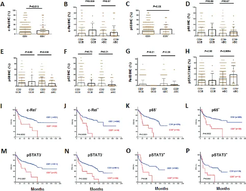
CD5 expression was associated with nuclear
expression of the NF-κB subunits c-Rel and p65, but not
mRNA levels of REL or RELA (Figures 3A-D, Table 2,
Supplemental Figures 1J-K). However, nuclear expression of p50, which is also indicative of canonical NF-κB pathway activation [32], was significantly decreased in
CD5+ ABC-DLBCL (Figure 3E, Table 2), which was not due to the NFKB1 downregulation at the mRNA level (Supplemental Figure 1L). There was no correlation between CD5+ and nuclear expression of p52 or RelB, two subunits involved in noncanonical/alternative
NF-κB pathway activation [32] (although showing trends of
downregulation in CD5+ DLBCL, Figures 3F-G). CD5+ was also associated significantly with nuclear expression of phosphorylated/activated STAT3 but not
upreglulated STAT3 mRNA in the ABC-DLBCL subtype (Figure 3H, Supplemental Figure 1O). The nuclear expression of pSTAT3 has been associated with poorer survival [33].
However, the adverse effect of CD5 expression on prognosis did not depend on c-Rel, p65 or STAT3 activation. In both c-Rel– and c-Rel+, p65– and p65+, and pSTAT3– and pSTAT3+ patients, CD5 expression
correlated with significantly poorer OS and PFS (Figures
3I-P, showing analysis in the combined training and validation sets due to the limited CD5+ cases).
Frequent loss of ssbP2 expression in cd5+
dlbcl
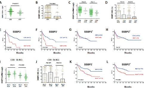
CD5 positivity in DLBCL was frequently associated with lack of SSBP2 expression, a tumor suppressor protein for lymphoma and leukemia (Figures 4A-D, Table 2)
[image:8.612.67.550.287.663.2]36]. Furthermore, the prognostic significance of CD5
expression was restricted to DLBCLs without or with very low SSBP2 expression. In SSBP2+ patients, CD5 expression in DLBCL was not associatedwith prognosis (Figures 4E-H).
In ABC-DLBCL, Bcl-2+ vs. Bcl-2– also had decreased SSBP2 mRNA and protein expression independent of CD5 expression (Figures 4I-J, Supplemental Figures 1P-Q). However, the prognostic
significance of Bcl-2 was independent of SSBP2 expression (Figures 4K-L).
Multivariate survival analysis
Multivariate analysis of clinical and pathological
factors including IPI (defined by age, stage, serum
LDH, performance status, and extranodal sites), sex, B-symptoms and tumor size, CD5+, Bcl-2+, and COO
confirmed that CD5 expression independently predicted significantly poorer OS (P = .005) and PFS (P = .014) in DLBCL, in addition to the known unfavorable prognostic factors IPI >2 and Bcl-2+ (Table 3). COO classification
did not reach statistical significance to be an independent
factor in this multivariate analysis, but did remain as an independent prognostic factor after removal of CD5 as
a factor in the survival analysis, suggesting that CD5
expression significantly contributed to the poor prognosis
of ABC-DLBCL.
The prognostic significance of these variables
(except COO) was further analyzed in the ABC-DLBCL subset. CD5 positivity remained as an independent prognostic factor predicting poorer OS (P = .032). However, the P value for PFS did not reach statistical
significance (P = .12). IPI>2 and Bcl-2+ remained independent prognostic factors for both OS and PFS (Table 3).
Prognostic and biological impact of cd5 expression in the validation set
In an independent validation set from multiple
medical centers, we validated the prognostic significance
(Figures 5A-B) as well biological impact of CD5+ discovered in the training set. In the validation set, CD5+
DLBCL were also associated with elevated Bcl-2 and decreased SSBP2 expression (Figures 5C-D), and its prognosis was independent of Bcl-2+ (Figures 5E-F) but depended on SSBP2– (Figures 5G-H).
[image:9.612.63.546.366.661.2]In both the training and validation sets, the
prognostic significance of CD5+ was independent of the IPI. Figures 5I-L shows analysis in the combined training and validation sets.
Effect of CD5 expression on BCR/TCR signaling
CD5+ DLBCL had significantly higher levels of
CARD11/CARM1 mRNA compared with CD5– DLBCL (Figure 6A). CARD11 is a scaffold protein downstream
of BCR signaling that activates NF-κB via interaction
with BCL10 [32,37,38]. A20, a negative regulator of
NF-κB signaling [37], and IL10, which mediates immune inhibition and cell survival downstream the CD5 signaling [10,11], did not show differential expression of mRNAs between CD5+ and CD5– DLBCL patients. (P= .30 and
P= .36). In addition, TCL1A (which modulates AKT
activation downstream of the TCR signaling [39]) and BCL11A (proto-oncogene with a critical role in lymphoid
development [40]), which was often co-amplified with
REL in lymphoid malignancies [41,42], were significantly
upregulated in CD5+ ABC-DLBC patients (Figures 6B-C). PTEN encoding a tumor suppressor antagonizing the
PI3K/AKT signaling was significantly downregulated in
CD5+ GCB-DLBCL (Figure 6D).
Differential expression of CARD11, TCL1A, and BCL11A between Bcl-2+ and Bcl-2– DLBCL patients resembled the effect of CD5 signaling (Figures 6E-G), suggesting that there are similar signaling pathways potentially underlying the association of CD5 and Bcl-2 expression. In contrast, regulation of other genes such as A20 (Figure 6H), PTEN, CD10, PIK3CA, MYC, CXCL12, PRDM1, STAT3 showed different patterns in Bcl-2+ (vs.
Bcl-2–) and CD5+ (vs. CD5–)patients.
cd5 expression signatures in de novo dlbcl
Gene expression profiles of CD5+ and CD5– DLBCL were compared to identify CD5 gene signatures in de novo DLBCL, which showed 86 differentially expressed genes (DEGs) by comparison within the overall cohort, and 39 DEGs by comparison within the ABC-DLBCL subcohort (Figures 5A-B, Table 4). Comparison within the GCB-DLBCL subcohort with limited CD5+ cases found 17 DEGs, most of which are related to cytoskeleton, microtubule or nervous system function (Figure 5C). Only a few genes were found expressed differentially between CD5+ GCB- and ABC-DLBCLs (Table 5).
CD5 expression signatures included both activators and inhibitors of TCR/BCR signaling. Activation of BCR/TCR was suggested by upregulation of CARD11, CLECL1 (encoding a T-cell costimulatory molecule), and IGHM. However, inhibition of BCR/TCR and increased threshold for activation in CD5+ DLBCL was also suggested by upregulation of PTPN2 and SIT1 (which negatively regulate TCR signaling), LYN (which has roles in inhibiting BCR), and SH3BP5 (which
inhibits BTK signaling), and downregulation of PDE4D (which hydrolyzes c-AMP, thereby removing the c-AMP constraint for TCR) in CD5+ DLBCL [43].
[image:12.612.67.549.451.660.2]DEGs downstream of TCR/BCR also suggested activated BCR signaling with negative feedback in CD5+ DLBCL. Activation of the NF-κB, MAPK, and Wnt pathways was indicated by upregulation of CXXC5 (activating NF-κB and MAPK) and GLYR1
Figure 6: mrnA expression of bcr signaling-related genes between cd5+ and cd5– dlbcl patients, or bcl-2+ and
bcl-2– dlbcl patients. (A-B) Association of CD5 expression with upregulated CARD11 and TCL1 mRNA levels; (C) CD5 expression
(activating MAPK) and downregulation of APCDD1 (negative regulator of Wnt signaling), PI15 (trypsin inhibitor), CAMK2N1 (CAMK2 inhibitor), PPM1L (dephosphorylating MAPKs), and PTPRR (sequester
and inhibitor of MAPKs). In turn, antiapoptotic BCL2 and TNFAIP8 downstream of the NF-κB pathway were significantly upregulated. On the other hand, inhibition
of the calcium-dependent signaling, which is required for the activation of proteins downstream BCR including
NF-κB and NFAT [44,45], was indicated by upregulation of
BSPRY (inhibiting calcium influx) and downregulation of
SGK1,which activates ion channels and calcium entry. CD5 expression signatures also included 18 transcription factors, suggesting distinct transcription programs in CD5+ DLBCL patients. Upregulated transcription factors included CREB3L2 (a transcription activator binding to the cAMP response element),
ETV6 (a transcriptional repressor), FOXP1 (an essential transcriptional repressor of B-cell development), IRF2BP2 (a transcription corepressor repressing the NFAT target genes including IL2/IL4), JARID2 (a transcriptional repressor), TCF4 (a transcription factor binding to the immunoglobulin enhancer), and ZNF589 (a transcriptional repressor). Downregulated transcription factors included LIM factors LMO2, LMO4, and LHX2 (which have roles in proliferation, differentiation and hematopoietic development), MAML3 (a transcriptional coactivator for NOTCH proteins), TFEC (which binds to the immunoglobulin heavy-chain/IGH gene enhancer), and MYBL1 (a strong transcriptional activator). Altogether it appears that transcription factors that repress TCR/ BCR signaling and proliferation outnumbered those that enhance TCR/BCR signaling.
under concurrent positive and negative regulations in CD5+ DLBCL. Three genes promoting cell cycle progression were upregulated, including CCND2 (a cyclin important for G1/S transition), CDK3 (a cyclin-dependent protein kinase involved in G0-G1 and G1-S transitions), and TLK1 (a kinaseinvolved in the regulation of chromatin assembly), whereas NEK6 (playing an important role in mitotic cell cycle progression) was downregulated. JARID2 (which negatively regulates cell proliferation signaling) was upregulated, and CLEC11A, PTK2, MYBL1, and SGK2 genes (which stimulate proliferation) were downregulated.
Supporting a previous study [25], another distinctive feature of the CD5 expression signatures were the downregulation of genes related to cell adhesion, ECM remodeling, and migration (CCBE1, COL5A1, COL6A3, ENPP3, FAM198B, FN1, ITGBL1, PCDH9, PTK2, RAPH1,and MYO7A). LTBP1 (functioning in the
assembly, secretion, and targeting of TGFβ1 to ECM),
INHBA (encoding a TGFβ family member), and PTPRB (involvedin blood vessel remodeling and angiogenesis) were downregulatedin CD5+ DLBCL.
Gene set enrichment analysis (GSEA) was performed to enrich the relevant pathways. Downregulated “ECM Receptor Interaction and upregulated “Nitrogen Metabolism” had nominal P-values < 1% ( .006 and .0039 respectively) although no gene sets were enriched by an FDR threshold of 25% (probably due to the small number of CD5+ cases, and the highly heterogeneous nature of CD5– DLBCLpatients). In addition, “Focal Adhesion” had a nominal P-value of .026 with an FDR of 30%. These results reinforced the notion that downregulation of genes involved in ECM and cell adhesion is a prominent feature of the CD5 expression signature (Figures 5D-F). Pathway
analysis by the Ingenuity Pathway Analysis (IPA) software showed CD5 expression signatures were associated with functional networks of Hematopoiesis, Nervous System Development and Function, Cellular Growth and Proliferation (Supplemental Figure 2).
dIscussIon
De novo CD5+ DLBCL is a unique subset of DLBCL [1] and has not been studied on a large scale in Western countries. The current study of 879 patients with de novo
DLBCL identified 48 (5.5%) CD5+ patients, associated with higher frequencies of >1 ECOG performance status, BM involvement, CNS relapse, ABC subtype, Bcl-2+, and STAT3 activation whereas with lower frequencies of CD30+, SSBP2+, and MYC mutations. CD5 signaling
appears to differentially regulate NF-κB subunits,
activating c-Rel and p65 but decreasing p50 activation. Other features associated with CD5+ DLBCL in this study, such as elderly age, B-symptoms, IPI, GCET, CD10, FOXP1 and CXCR4, were probably due to the predominance of ABC subtype of CD5+ DLBCL patients. Compared with previous studies conducted in Japan, CD5+ DLBCL in Western countries had lower prevalence, shared common features of performance status, ABC subtype, Bcl-2 overexpression, BM involvement, and development of CNS recurrence, but lacked features of female predominance, extranodal involvement, elevated serum LDH, and higher disease stage [13-15,17-19].
These differences may reflect ethnic and genetic variation,
the heterogeneity of CD5+ DLBCL, the larger number of CD5+ DLBCLs (n = 109) in the Japanese cohort and
cohort-specific features.
[image:14.612.89.536.75.275.2]We further assessed the prognostic impact of CD5
Figure 7: Gene expression profiling and gene set enrichment analysis of CD5+ dlbcl. (A-C) Gene expression profiling of
expression and found that CD5+ independently correlated with poorer survival in DLBCL with R-CHOP treatment. Moreover, the 48 CD5+ DLBCL patients treated by
R-CHOP in this study cohort did not show significant
improvement in OS (P= .66) or PFS (P= .81) compared to the 14 CD5+ DLBCL patients treated with CHOP from an independent CHOP-treated DLBCL cohort (results not shown). Furthermore, our attempt to understand the biology of CD5+ DLBCL suggested that molecular pathways downstream BCR signaling which promote cell proliferation and survival (such as Bcl-2 [but not Myc] overexpression, and activation of c-Rel, p65, and STAT3) were likely relevant for the pathogenesis of CD5+ DLBCL; however, the adverse impact of CD5 expression did not depend on any of these factors alone.
BM involvement also appeared to impact prognosis of CD5+ DLBCL significantly in the training set (Figures
1J-K, which however was not confirmed in the validation
set), and development of CNS relapse (0% at diagnosis, 8.3% after treatment) was remarkable for CD5+ DLBCL. A role of CXCR4/CXCL12 axis in BM involvement and CNS relapse of CD5+ DLBCL was suggested by the higher CXCR4 expression in the studied CD5+ DLBCL patients (Supplemental Figure 1E) [28,46]. However, restricting within ABC-DLBCL, CD5+ compared to CD5– patients had similar levels of CXCR4 but had a higher incidence of BM involvement (34.5% vs. 7.2%, P < .0001, Table 2),
suggesting that CXCR4/CXCL12 axis was not sufficient to
explain for the BM involvement. A previous study in CLL/ SLL also suggested that other factors in addition to the
CXCR4/CXCL12 axis may account for marrow infiltration
of neoplastic cells [46]. In this study, downregulation of genes involved in ECM and cell adhesion, which was a prominent feature of the CD5+ signature revealed by GEP analysis, likely contributed to the BM involvement and development of CNS relapse in CD5+ DLBCL.
Interestingly, CD30 and SSBP2 (a tumor-suppressor [34,36,47]) expression was frequently negative in CD5+ DLBCL patients. SSBP2 has a critical regulatory role in the transcriptional program of hematopoietic stem and progenitor cells in vivo, via modulating the abundance and function of multiple transcription cofactors including LIM domain-binding protein 1 (LDB1), LMO, and LHX. In mouse models, loss of SSBP2 resulted in hypoplastic hematopoietic tissues and impaired hematopoiesis and was associated with shortened lifespan and greater susceptibility to B-cell lymphomas [34,35,48]. Prognostic
significance of CD5 expression in SSBP2+ and SSBP2– DLBCL patients also suggests a tumor-suppressor function of SSBP2 for CD5 signaling.
GEP analysis suggested both positive and negative regulation of TCR/BCR in CD5+ DLBCL patients, and differential regulation of BCR downstream pathways
(activation of NF-κB, MAPK, and Wnt pathways and inhibition of calcium influx). This may suggest the
activated but reprogrammed BCR signaling in CD5+
DLBCL patients, and the role of CD5 expression in mitigating BCR signaling, and promoting tumor cell survival by previous studies [7-10]. Likewise, both positive and negative regulations of proliferation, growth, and cell cycle were suggested by CD5 expression signatures. Therefore it appears that CD5 signaling contributes to survival yet an anergic-like state of B-cells.
It is also possible that the unique characteristics of the CD5 expression signature in antiapoptosis,
proliferation, signaling, and transcription reflects the
COO and the differentiation stages of the lymphoma cells. For example, downregulated LMO2, LMO4, LHX2, and CLEC11A as well as upregulated FOXP1, ZNF589, LYN, and BCL2 are hematopoietic mediators and have distinct expression patterns during B-cell development. In addition, IGHM expressed in naïve B-cells and plasma cells was upregulated in CD5+ patients, whereas
MYC mutations, which may arise from an aberrant hypermutation process in DLBCL [49], were almost absent in CD5+ DLBCL patients (only one CD5+ GCB-DLBCL case had MYC mutations). Previous studies indicated that CD5+ DLBCL and CD5+ CLL had higher frequencies of germline vs. somatically hypermutated IGHV genes compared with CD5– DLBCL [50], suggesting the COO of CD5+ DLBCL might be distinct from that of CD5– DLBCL and yet similar to that of CD5+ CLL [51]. Our collective results of GEP, MYC mutations, and Blimp-1 expression (indicating commitment to plasma cell differentiation) [52-54] in CD5+ and CD5– DLBCL (Tables 2, 4) suggest that subsets of CD5+ DLBCL may originate from pre-GC, memory B-cells, or neoplasms differentiated into plasma cells yet never through GC reaction [60, 61].
In summary, in this study we show that de novo CD5+ DLBCL, which occurs at a low frequency (5.5%) in Western countries, was associated with unfavorable clinicopathologic variables and with inferior survival following R-CHOP treatment. Although heterogeneity still exists in this disease subset, dysregulated BCR signaling
is significantly implicated in lymphoma cell survival and
disease dissemination. Bcl-2 inhibitors, STAT3 inhibitors, and therapeutic strategies modulating BCR signaling, tumor microenvironment and cytokine/chemokine axes may help in the management of CD5+ DLBCL patients.
MAterIAls And Methods
Patients
primary mediastinal, cutaneous, or central nervous system
DLBCL, or human immunodeficiency virus infection
were excluded. All patients were reviewed by a group of hematopathologists and were diagnosed according to
the World Health Organization classification criteria. The
study was conducted in accordance with the Declaration of Helsinki, and the protocol was reviewed and approved by the Institutional Review Boards of each participating center, and the comprehensive collaborative study was approved by the Institutional Review Board at The University of Texas MD Anderson Cancer Center.
tissue microarray and biomarkers
Immunohistochemical analysis for CD5 expression
using a monoclonal CD5 antibody (Novocastra Labs, UK)
was performed on 879 DLBCL biopsy specimens using
formalin-fixed, paraffin-embedded tissue microarrays as
described previously [55,56]. CD5 expression was scored by three pathologists independently on 400 cells in each of
cases under a microscope at 40× magnification. Expression
of other markers such as CD10, GCET1, MUM1, FOXP1,
Bcl-6, Bcl-2, Myc, Ki-67, p53, MDM2, NF-κB subunits,
pSTAT3, CD30, CXCR4, and SSBP2 [31,33,55-59] was also assessed using respective antibodies. SSBP2 antibody was kindly provided by Dr. Lalitha Nagarajan, PhD from the Department of Genetics, MD Anderson Cancer Center [34,36]. Due to tissue exhaustion, IHC analysis for some markers other than CD5 was not successful in few cases.
Gene translocations and amplifications were detected
using the methods described previously [30,31,59]. MYC mutation was detected using the Sanger sequencing method.
Cell-of-origin classification
Cell-of-origin classification as either GCB or ABC
DLBCLs was determined by GEP for patients in the training set and by IHC according to the Visco-Young algorithm and/or Choi algorithms [55] for all the patients in the training and validation sets.
Gene expression profiling
GEP were achieved in 488 DLBCL (27 CD5+ and 461 CD5–) patients of the training set using total RNAs
extracted from each formalin-fixed, paraffin-embedded
tissue sample and Affymetrix GeneChips array as described previously [30,55,57,58]. The microarray data
were quantified and normalized, and the DEGs between
CD5+ and CD5– DLBCL patients at false discovery rate
of .01 were identified using multiple t-tests. Gene set enrichment analysis was performed on the KEGG pathway
gene sets. Pathway analysis for the DEGs was performed
using the Ingenuity Pathway Analysis software program (IPA, http://www.qiagen.com/ingenuity).
statistical analysis
Clinicopathologic differences between different DLBCL subgroups were assessed using Fisher’s exact test and the Spearman rank correlation. The mRNA expression levels of affected genes were also retrieved from the GEP data and compared between CD5+ and CD5– DLBCL patients using unpaired t tests. Overall survival was calculated from the time of diagnosis to death from any cause or last follow-up. Progression-free survival was calculated from the time of diagnosis to disease progression, relapse, or death from any cause. Patients who were alive and/or had no disease progression were censored at last follow-up. Survival
analysis was performed using the Kaplan–Meier method
with GraphPad Prism 6 (GraphPad Software, San Diego, CA), and differences were compared using the log-rank test. Multivariate survival analysis was performed using the Cox proportional hazards regression model with the SPSS statistics software program (version 19.0; IBM Corporation, Armonk, NY). All differences with P ≤ .05 were considered statistically significant.
AcknowledgeMents
This work is supported by The University of Texas MD Anderson Cancer Center Institutional Research Grant Award, an MD Anderson Lymphoma Specialized Programs of Research Excellence (SPORE) Research Development Program Award, an MD Anderson Myeloma SPORE Research Development Program Award, MD Anderson Collaborative Research Funds with High-Throughput Molecular Diagnostics, Gilead Pharmaceutical, Adaptive Biotechnology, and Roche Molecular Systems, and National Cancer Institute and National Institutes of Health grants (R01CA138688 and R01CA187415) to
K.H.Y. This work was also partially supported by National
Cancer Institute and National Institutes of Health grants (P50CA136411 and P50CA142509), and by the MD Anderson Cancer Center Support Grant CA016672. Dr. Xu-Monette, PhD is the recipient of the Harold C. and Mary L. Daily Endowment Fellowships and Shannon Timmins Fellowship for Leukemia Research Award. Dr.
Kausar J. Jabbar, MD is the recipient of the Pathology
Division Biomarker Fellowship Award.
Author contributions
WWLC, JHK, MP, AJMF, XZ, MAP, JPF, JNW, RNM, LJM, KHY; Collection and assembly of data under approved IRB and MTA: ZYXM, KJJ, XC, AZ, CV, SMM, KD, AC, AO, YZ, GB, KLR, EDH, WWLC, JHK, MP, AJMF, XZ, MAP, JPF, JNW, KHY; Data analysis and interpretation: ZYXM, KHY; Manuscript writing: ZYXM, MT, KHY; Final approval of manuscript: All authors.
editorial note
This paper has been accepted based in part on peer-review conducted by another journal and the authors’ response and revisions as well as expedited peer-review in Oncotarget.
conFlIcts oF Interest dIsclosure
The authors declare no conflicts of interest.
reFerences
1. Young KH, Medeiros LJ, Chan WC. Diffuse large B-cell lymphoma. In: Orazi A, Weiss LM, Foucar K, Knowles DM, eds. Neoplastic Hematopathology. Philadelphia, PA, USA. . Lippincott Willaims & Wilkins; 2014:502-565. 2. Stein H, Warnke RA, Chan WC, Jaffe ES, Chan JKC, Gatter
KC, Campo E, Swerdlow SH, Campo E. Diffuse large B-cell lymphoma, not otherwise specified. In: Swerdlow SH, Campo E, Harris NL, Jaffe ES, Pileri SA, Stein H, Thiele J, Vardiman JW, et al., eds. WHO classification of tumours of haematopoetic and lymphoid tissues in. 4th ed. Lyon, France. International Agency for Research on Cancer (IARC) 2008:233-261.
3. Jain P, Fayad LE, Rosenwald A, Young KH, O’Brien S. Recent advances in de novo CD5+ diffuse large B cell lymphoma. Am J Hematol 2013; 88:798-802.
4. Berland R, Wortis HH. Origins and functions of B-1 cells with notes on the role of CD5. Annu Rev Immunol 2002; 20:253-300.
5. Brossard C, Semichon M, Trautmann A, Bismuth G. CD5 inhibits signaling at the immunological synapse without impairing its formation. J Immunol 2003; 170:4623-9. 6. Bamberger M, Santos AM, Goncalves CM, Oliveira MI,
James JR, Moreira A, Lozano F, Davis SJ, Carmo AM. A new pathway of CD5 glycoprotein-mediated T cell inhibition dependent on inhibitory phosphorylation of Fyn kinase. J Biol Chem 2011; 286:30324-36.
7. Jevremovic D, Dronca RS, Morice WG, McPhail ED, Kurtin PJ, Zent CS, Hanson CA. CD5+ B-cell lymphoproliferative disorders: Beyond chronic lymphocytic leukemia and mantle cell lymphoma. Leuk Res 2010; 34:1235-8.
8. Tibaldi E, Brunati AM, Zonta F, Frezzato F, Gattazzo C, Zambello R, Gringeri E, Semenzato G, Pagano MA, Trentin L. Lyn-mediated SHP-1 recruitment to CD5 contributes
to resistance to apoptosis of B-cell chronic lymphocytic leukemia cells. Leukemia 2011; 25:1768-81.
9. Gary-Gouy H, Bruhns P, Schmitt C, Dalloul A, Daeron M, Bismuth G. The pseudo-immunoreceptor tyrosine-based activation motif of CD5 mediates its inhibitory action on B-cell receptor signaling. J Biol Chem 2000; 275:548-56. 10. Gary-Gouy H, Harriague J, Bismuth G, Platzer C, Schmitt
C, Dalloul AH. Human CD5 promotes B-cell survival through stimulation of autocrine IL-10 production. Blood 2002; 100:4537-43.
11. Garaud S, Morva A, Lemoine S, Hillion S, Bordron A, Pers JO, Berthou C, Mageed RA, Renaudineau Y, Youinou P. CD5 promotes IL-10 production in chronic lymphocytic leukemia B cells through STAT3 and NFAT2 activation. J Immunol 2011; 186:4835-44.
12. Challagundla P, Jorgensen JL, Kanagal-Shamanna R, Gurevich I, Pierson DM, Ferrajoli A, Reyes SR, Medeiros LJ, Miranda RN. Utility of quantitative flow cytometry immunophenotypic analysis of CD5 expression in small B-cell neoplasms. Arch Pathol Lab Med 2014; 138:903-9. 13. Yamaguchi M, Ohno T, Oka K, Taniguchi M, Ito M, Kita
K, Shiku H. De novo CD5-positive diffuse large B-cell lymphoma: clinical characteristics and therapeutic outcome. Br J Haematol 1999; 105:1133-9.
14. Yamaguchi M, Seto M, Okamoto M, Ichinohasama R, Nakamura N, Yoshino T, Suzumiya J, Murase T, Miura I, Akasaka T, Tamaru J, Suzuki R, Kagami Y, et al. De novo CD5+ diffuse large B-cell lymphoma: a clinicopathologic study of 109 patients. Blood 2002; 99:815-21.
15. Harada S, Suzuki R, Uehira K, Yatabe Y, Kagami Y, Ogura M, Suzuki H, Oyama A, Kodera Y, Ueda R, Morishima Y, Nakamura S, Seto M. Molecular and immunological dissection of diffuse large B cell lymphoma: CD5+, and CD5- with CD10+ groups may constitute clinically relevant subtypes. Leukemia 1999; 13:1441-7.
16. Yamaguchi M, Nakamura N, Suzuki R, Kagami Y, Okamoto M, Ichinohasama R, Yoshino T, Suzumiya J, Murase T, Miura I, Ohshima K, Nishikori M, Tamaru J, et al. De novo CD5+ diffuse large B-cell lymphoma: results of a detailed clinicopathological review in 120 patients. Haematologica 2008; 93:1195-202.
17. Ennishi D, Takeuchi K, Yokoyama M, Asai H, Mishima Y, Terui Y, Takahashi S, Komatsu H, Ikeda K, Yamaguchi M, Suzuki R, Tanimoto M, Hatake K. CD5 expression is potentially predictive of poor outcome among biomarkers in patients with diffuse large B-cell lymphoma receiving rituximab plus CHOP therapy. Ann Oncol 2008; 19:1921-6. 18. Hyo R, Tomita N, Takeuchi K, Aoshima T, Fujita A,
Kuwabara H, Hashimoto C, Takemura S, Taguchi J, Sakai R, Fujita H, Fujisawa S, Ogawa K, et al. The therapeutic effect of rituximab on CD5-positive and CD5-negative diffuse large B-cell lymphoma. Hematol Oncol 2010; 28:27-32.
N, Nakamura S, Ohshima K, Nakamine H, Hirano M. Clinicopathologic characteristics and treatment outcome of the addition of rituximab to chemotherapy for CD5-positive in comparison with CD5-negative diffuse large B-cell lymphoma. Ann Oncol 2010; 21:2069-74.
20. Miyazaki K, Yamaguchi M, Suzuki R, Kobayashi Y, Maeshima AM, Niitsu N, Ennishi D, Tamaru JI, Ishizawa K, Kashimura M, Kagami Y, Sunami K, Yamane H, et al. CD5-positive diffuse large B-cell lymphoma: a retrospective study in 337 patients treated by chemotherapy with or without rituximab. Ann Oncol 2011; 22:1601-7. 21. Davidson-Moncada JK, McDuffee E, Roschewski M. CD5+
diffuse large B-cell lymphoma with hemophagocytosis. J Clin Oncol 2013; 31:e76-9.
22. Westin J, McLaughlin P. De novo CD5+ diffuse large B-cell lymphoma: a distinct subset with adverse features, poor failure-free survival and outcome with conventional therapy. Leuk Lymphoma 2010; 51:161-3.
23. Kroft SH, Howard MS, Picker LJ, Ansari MQ, Aquino DB, McKenna RW. De novo CD5+ diffuse large B-cell lymphomas. A heterogeneous group containing an unusual form of splenic lymphoma. Am J Clin Pathol 2000; 114:523-33.
24. Kobayashi T, Yamaguchi M, Kim S, Morikawa J, Ogawa S, Ueno S, Suh E, Dougherty E, Shmulevich I, Shiku H, Zhang W. Microarray reveals differences in both tumors and vascular specific gene expression in de novo CD5+ and CD5- diffuse large B-cell lymphomas. Cancer Res 2003; 63:60-6.
25. Suguro M, Tagawa H, Kagami Y, Okamoto M, Ohshima K, Shiku H, Morishima Y, Nakamura S, Seto M. Expression profiling analysis of the CD5+ diffuse large B-cell lymphoma subgroup: development of a CD5 signature. Cancer Sci 2006; 97:868-74.
26. Yamamoto K, Okamura A, Yakushijin K, Hayashi Y, Matsuoka H, Minami H. Tandem triplication of the BCL2 gene in CD5-positive intravascular large B cell lymphoma with bone marrow involvement. Ann Hematol 2014; 93:1791-3.
27. Azab AK, Runnels JM, Pitsillides C, Moreau AS, Azab F, Leleu X, Jia X, Wright R, Ospina B, Carlson AL, Alt C, Burwick N, Roccaro AM, et al. CXCR4 inhibitor AMD3100 disrupts the interaction of multiple myeloma cells with the bone marrow microenvironment and enhances their sensitivity to therapy. Blood 2009; 113:4341-51. 28. Balkwill F. Cancer and the chemokine network. Nat Rev
Cancer 2004; 4:540-50.
29. Chang BY, Francesco M, De Rooij MF, Magadala P, Steggerda SM, Huang MM, Kuil A, Herman SE, Chang S, Pals ST, Wilson W, Wiestner A, Spaargaren M, et al. Egress of CD19(+)CD5(+) cells into peripheral blood following treatment with the Bruton tyrosine kinase inhibitor ibrutinib in mantle cell lymphoma patients. Blood 2013; 122:2412-24.
30. Visco C, Tzankov A, Xu-Monette ZY, Miranda RN, Tai YC, Li Y, Liu WM, d’Amore ES, Li Y, Montes-Moreno S, Dybkaer K, Chiu A, Orazi A, et al. Patients with diffuse large B-cell lymphoma of germinal center origin with BCL2 translocations have poor outcome, irrespective of MYC status: a report from an International DLBCL rituximab-CHOP Consortium Program Study. Haematologica 2013; 98:255-63.
31. Hu S, Xu-Monette ZY, Tzankov A, Green T, Wu L, Balasubramanyam A, Liu WM, Visco C, Li Y, Miranda RN, Montes-Moreno S, Dybkaer K, Chiu A, et al. MYC/BCL2 protein coexpression contributes to the inferior survival of activated B-cell subtype of diffuse large B-cell lymphoma and demonstrates high-risk gene expression signatures: a report from The International DLBCL Rituximab-CHOP Consortium Program. Blood 2013; 121:4021-31; quiz 4250. 32. Shaffer AL, 3rd, Young RM, Staudt LM. Pathogenesis
of human B cell lymphomas. Annu Rev Immunol 2012; 30:565-610.
33. Ok CY, Chen J, Xu-Monette ZY, Tzankov A, Manyam GC, Li L, Visco C, Montes-Moreno S, Dybkaer K, Chiu A, Orazi A, Zu Y, Bhagat G, et al. Clinical Implications of Phosphorylated STAT3 Expression in De Novo Diffuse Large B-cell Lymphoma. Clin Cancer Res 2014; 20:5113-23.
34. Wang Y, Klumpp S, Amin HM, Liang H, Li J, Estrov Z, Zweidler-McKay P, Brandt SJ, Agulnick A, Nagarajan L. SSBP2 is an in vivo tumor suppressor and regulator of LDB1 stability. Oncogene 2010; 29:3044-53.
35. Li J, Kurasawa Y, Wang Y, Clise-Dwyer K, Klumpp SA, Liang H, Tailor RC, Raymond AC, Estrov Z, Brandt SJ, Davis RE, Zweidler-McKay P, Amin HM, et al. Requirement for ssbp2 in hematopoietic stem cell maintenance and stress response. J Immunol 2014; 193:4654-62.
36. Liang H, Samanta S, Nagarajan L. SSBP2, a candidate tumor suppressor gene, induces growth arrest and differentiation of myeloid leukemia cells. Oncogene 2005; 24:2625-34.
37. Staudt LM. Oncogenic activation of NF-kappaB. Cold Spring Harb Perspect Biol 2010; 2:a000109.
38. Thome M, Charton JE, Pelzer C, Hailfinger S. Antigen receptor signaling to NF-kappaB via CARMA1, BCL10, and MALT1. Cold Spring Harb Perspect Biol 2010; 2:a003004.
39. Rodig SJ, Vergilio JA, Shahsafaei A, Dorfman DM. Characteristic expression patterns of TCL1, CD38, and CD44 identify aggressive lymphomas harboring a MYC translocation. Am J Surg Pathol 2008; 32:113-22.
41. Satterwhite E, Sonoki T, Willis TG, Harder L, Nowak R, Arriola EL, Liu H, Price HP, Gesk S, Steinemann D, Schlegelberger B, Oscier DG, Siebert R, et al. The BCL11 gene family: involvement of BCL11A in lymphoid malignancies. Blood 2001; 98:3413-20.
42. Deambrogi C, De Paoli L, Fangazio M, Cresta S, Rasi S, Spina V, Gattei V, Gaidano G, Rossi D. Analysis of the REL, BCL11A, and MYCN proto-oncogenes belonging to the 2p amplicon in chronic lymphocytic leukemia. Am J Hematol 2010; 85:541-4.
43. Peter D, Jin SL, Conti M, Hatzelmann A, Zitt C. Differential expression and function of phosphodiesterase 4 (PDE4) subtypes in human primary CD4+ T cells: predominant role of PDE4D. J Immunol 2007; 178:4820-31.
44. Woyach JA, Johnson AJ, Byrd JC. The B-cell receptor signaling pathway as a therapeutic target in CLL. Blood 2012; 120:1175-84.
45. Feske S. Calcium signalling in lymphocyte activation and disease. Nat Rev Immunol 2007; 7:690-702.
46. Barretina J, Junca J, Llano A, Gutierrez A, Flores A, Blanco J, Clotet B, Este JA. CXCR4 and SDF-1 expression in B-cell chronic lymphocytic leukemia and stage of the disease. Ann Hematol 2003; 82:500-5.
47. Fleisig HB, Orazio NI, Liang H, Tyler AF, Adams HP, Weitzman MD, Nagarajan L. Adenoviral E1B55K oncoprotein sequesters candidate leukemia suppressor sequence-specific single-stranded DNA-binding protein 2 into aggresomes. Oncogene 2007; 26:4797-805.
48. Xu Z, Meng X, Cai Y, Liang H, Nagarajan L, Brandt SJ. Single-stranded DNA-binding proteins regulate the abundance of LIM domain and LIM domain-binding proteins. Genes Dev 2007; 21:942-55.
49. Pasqualucci L, Neumeister P, Goossens T, Nanjangud G, Chaganti RS, Kuppers R, Dalla-Favera R. Hypermutation of multiple proto-oncogenes in B-cell diffuse large-cell lymphomas. Nature 2001; 412:341-6.
50. Nakamura N, Abe M. Histogenesis of CD5-positive and CD5-negative B-cell neoplasms on the aspect of somatic mutation of immunoglobulin heavy chain gene variable region. Fukushima J Med Sci 2003; 49:55-67.
51. Nakamura N, Kuze T, Hashimoto Y, Tasaki K, Hojo H, Sasaki Y, Sato M, Abe M. Analysis of the immunoglobulin heavy chain gene variable region of 101 cases with peripheral B cell neoplasms and B cell chronic lymphocytic leukemia in the japanese population. Pathol.Int. 1999; 49:595-600.
52. Angelin-Duclos C, Cattoretti G, Lin KI, Calame K. Commitment of B lymphocytes to a plasma cell fate is associated with Blimp-1 expression in vivo. J Immunol 2000; 165:5462-71.
53. Nutt SL, Fairfax KA, Kallies A. BLIMP1 guides the fate of effector B and T cells. Nat Rev Immunol 2007; 7:923-7. 54. Shaffer AL, Lin KI, Kuo TC, Yu X, Hurt EM, Rosenwald
A, Giltnane JM, Yang L, Zhao H, Calame K, Staudt
LM. Blimp-1 orchestrates plasma cell differentiation by extinguishing the mature B cell gene expression program. Immunity 2002; 17:51-62.
55. Visco C, Li Y, Xu-Monette ZY, Miranda RN, Green TM, Li Y, Tzankov A, Wen W, Liu WM, Kahl BS, d’Amore ES, Montes-Moreno S, Dybkaer K, et al. Comprehensive gene expression profiling and immunohistochemical studies support application of immunophenotypic algorithm for molecular subtype classification in diffuse large B-cell lymphoma: a report from the International DLBCL Rituximab-CHOP Consortium Program Study. Leukemia 2012; 26:2103-13.
56. Hu S, Xu-Monette ZY, Balasubramanyam A, Manyam GC, Visco C, Tzankov A, Liu WM, Miranda RN, Zhang L, Montes-Moreno S, Dybkaer K, Chiu A, Orazi A, et al. CD30 expression defines a novel subgroup of diffuse large B-cell lymphoma with favorable prognosis and distinct gene expression signature: a report from the International DLBCL Rituximab-CHOP Consortium Program Study. Blood 2013; 121:2715-24.
57. Xu-Monette ZY, Wu L, Visco C, Tai YC, Tzankov A, Liu WM, Montes-Moreno S, Dybkaer K, Chiu A, Orazi A, Zu Y, Bhagat G, Richards KL, et al. Mutational profile and prognostic significance of TP53 in diffuse large B-cell lymphoma patients treated with R-CHOP: report from an International DLBCL Rituximab-CHOP Consortium Program Study. Blood 2012; 120:3986-96.
58. Xu-Monette ZY, Moller MB, Tzankov A, Montes-Moreno S, Hu W, Manyam GC, Kristensen L, Fan L, Visco C, Dybkaer K, Chiu A, Tam W, Zu Y, et al. MDM2 phenotypic and genotypic profiling, respective to TP53 genetic status, in diffuse large B-cell lymphoma patients treated with rituximab-CHOP immunochemotherapy: a report from the International DLBCL Rituximab-CHOP Consortium Program. Blood 2013; 122:2630-40.
59. Tzankov A, Xu-Monette ZY, Gerhard M, Visco C, Dirnhofer S, Gisin N, Dybkaer K, Orazi A, Bhagat G, Richards KL, Hsi ED, Choi WW, van Krieken JH, et al. Rearrangements of MYC gene facilitate risk stratification in diffuse large B-cell lymphoma patients treated with rituximab-CHOP. Mod Pathol 2014; 27:958-71.
60. Dybkær K, Bøgsted M, Falgreen S, Bødker JS, Kjeldsen MK, Schmitz A, et al: A diffuse large B-cell lymphoma classification system that associates normal B-cell subset phenotypes with prognosis. J Clin Oncol 2015. In press. 61. Testoni M, Zucca E, Young KH, Bertoni F. Genetic lesions