Dr Silni Chandra et al JMSCR Volume 06 Issue 07 July 2018 Page 111
How to Minimize Corneal Astigmatism during Cataract Surgery
Authors
Dr Silni Chandra, Dr Smitha M.
Abstract
Aims: To study the corneal astigmatism in manual small incision cataract surgery following temporal incision, superior incision, superotemporal incision and superior incision with infinity suture.
Setting: Government Medical College, Kozhikode; Government Medical College, Palakkad
Design: Retrospective Study
Material and Method: This study included cases of cataract surgery done during 9 months period from January 2016 to September 2016 at Government Medical College Palakkad. A retrospective Chart review of 199 eyes of 199 patients was done to assess the post operative corneal astigmatism. Visual Acuity & Keratometry was recorded preoperatively and at 2 months post operatively. The results were tabulated into four groups: Superior incision with infinity suture, temporal incision, Superotemporal incision and Superior incision. Postoperative corneal astigmatism, induced corneal astigmatism and post operative visual acuity were analyzed among the four groups.
Statistics: SPSS analysis, Microsoft Excel
Result: 82 (41.2%) patients had undergone SICS with superior incision, 45 (22.6%) patients had SICS with superior incision with infinity suture, 42(21.1%) patients had undergone SICS with superotemporal incision and 30 (15%) patients had undergone SICS with temporal incision.
Post operatively the mean corneal astigmatism at two months was significantly higher in the superior with infinity suture group (1.6444 ±0.640) and superior section group (1.548 ± 0.767) and lowest in the temporal (0.4750 ± 0.401) followed by superotemporal section group (1.035 ± 0.656)
The induced corneal astigmatism was lowest in the in the Temporal section group(-0.158 ±0.3181 followed by 0.2917 ±0.6460 in the superotemporal section group and 0.5722 ±0.6563 in the Superior section with infinity suture group and highest in the superior section group ( 0.7622 ±0.7119). This difference in the four groups is found to be statistically significant (p=0.000).
Conclusion: Temporal incision induces the least corneal astigmatism followed by superotemporal incision. Highest corneal astigmatism is induced by Superior incision. Infinity suture will help in marginally reducing the corneal astigmatism.
Keywords: Cataract, Small Incision, Infinity suture, Superior, Temporal, Superotemporal, Astigmatism.
Introduction
Cataract is the leading cause of preventable blindness in the world. Presently surgery is the only option for treatment of this condition. At present Phacoemulsification and manual small
incision cataract surgery are the two best available options for its treatment. In the developing world with shortage of resources manual small incision cataract surgery remains the best option.
www.jmscr.igmpublication.org Impact Factor (SJIF): 6.379
Dr Silni Chandra et al JMSCR Volume 06 Issue 07 July 2018 Page 112 In the last few decades the patients and the
surgeons expectations from cataract surgery has increased manifold. Spectacle independence is increasingly being expected by all patients, both rich and poor. Therefore improving the unaided visual acuity has become the main aim of cataract surgery. For this to occur, astigmatism should be reduced to the minimal. Pre existing astigmatism is one of the main factors which affect postoperative astigmatism[3,7,9]. Presently there are many techniques which are available to reduce this pre-existing corneal astigmatism which include toric IOLs, limbal relaxing incision, surface ablation and placement of surgical section at the steep axis of astigmatism
Placing the incision on the steeper meridian to flatten it so as to reduce the post operative corneal astigmatism ultimately leading to better unaided visual acuity should be our ultimate aim.
In this study we aim to study the surgically induced corneal astigmatism following manual small incision with superior, superotemporal, temporal sections and the effect of placing infinity suture in superior incisions. Superior incisions induce the greatest amount of astigmatism followed by superotemporal, nasal, superonasal and least of all, temporal.[16,18]
Material and Method
Inclusion criteria- All patients operated from January 2016 to September 2016 according to standard operating protocol were included in the study.
Standard Operating protocol ensured that all surgeries done in a similar manner except for the difference in the site of the incision were selected. Characteristics of the incision included straight 6 mm scleral incision at either superior, temporal or superotemporal location without any sutures except Infinity sutures in superior incision..
Exclusion criteria- All patients with traumatic cataract, complicated cataract, lenticular subluxation, previous intraocular or corneal surgeries or glaucoma surgeries and corneal scarring or degeneration were excluded.
All those patients who did not have atleast a mean post operative follow up of two months were excluded from the study.
Any patient with intra operative complications like posterior capsular rent with our without vitreous prolapse which required a deviation from the standard operating protocol, or post operative complications like post operative endophthalmitis etc were excluded from the study.
Patients were divided into four groups based on their incision site: Superior, superior with infinity suture, supero-temporal and temporal group. The following variables were recorded from their files –demographic parameters such as age, sex, pre operative and post operative ocular findings such as visual acuity, intra ocular pressure, slit lamp findings, fundus and pupil dilatation. Astigmatism and keratometry measured using autorefractometer was also noted down. Ocular comobidities such as glaucoma, age related macular degeneration, diabetic retinopathy, pseudo exfoliation and presence of systemic diseases like diabetes mellitus, hypertension, cardiovascular diseases and dyslipidemia were also noted. The outcome variable of interest which included Post operative refraction, keratometry, uncorrected visual acuity at two months were also recorded were recorded from their files
Microsoft excel and SPSS statistical packages were used for data management and analysis. The one-way analysis of variance (ANOVA) was applied to compare the outcomes between the different groups in this study. A p value of less than 0.05 was considered significant.
Results
Dr Silni Chandra et al JMSCR Volume 06 Issue 07 July 2018 Page 113 groups of patients. The mean age was 62.78 years
and it ranged from 23 to 82 years. There were 83 (41.7%) males and 116 (58.2%) females. 115 (57.7%) patients had their right eye operated while 84(42.2%) patients had their left eye operated. Coexisting systemic conditions were seen as follows: Diabetes mellitus in 24(12%) patients, hypertension in 36(18%) patients, coronary artery disease in 9(0.04%) patients, chronic obstructive pulmonary disease in 6(0.03%)patients, parkinsonism in1 (0.005% patients) and dyslipidemia in 1(0.005%)patients. 122(61.3%) patients were nil systemically.
Coexisting ocular conditions were as follows: 5(0.02%) patients had pseudo exfoliation, 6(0.03%) patients were one eyed, and 1 (0.005%) patient had scleritis in the other eye. Pre operative visual acuity ranged between perception of light to 6/18. Cataract status ranged from nuclear sclerosis
grade 2 to grade 4, posterior subcapsular cataract and mature cataract. 82 (41.2%) patients had undergone SICS with 6mm superior straight incision, 45 (22.6%) patients had SICS with superior incision with infinity suture, 42(21.1%) patients had undergone SICS with superotemporal 6mm straight incision and 30 (15%)patients had undergone SICS with 6 mm temporal straight incision.
Astigmatism was calculated from the difference in the keratometry value in the steeper and flatter meridian. The mean corneal astigmatism preoperatively (KDIFFPRE) was against the rule astigmatism which ranged from 1.0778 ± 0.714 in the Superior section with infinity suture, 0.6333 ±0.4535 in the Temporal section group, 0.7440 ±0.4765 in the Superotemporal section and 0.8110 ± 0.559 in the Superior section groups (p=0.005) (Table 1)
Table 1 Paired Samples Statistics
SECTION Mean N Std. Deviation Std. Error Mean
SUP WITH INFINITY Pair 1 KDIFFPOST 1.6444 45 .64069 .09551
KDIFFPRE 1.0778 45 .71474 .10655
TEMP Pair 1 KDIFFPOST .4750 30 .40124 .07326
KDIFFPRE .6333 30 .45359 .08281
SUPEROTEMP Pair 1 KDIFFPOST 1.0357 42 .65698 .10137
KDIFFPRE .7440 42 .47655 .07353
SUP Pair 1 KDIFFPOST 1.5488 82 .76723 .08473
KDIFFPRE .8110 82 .55910 .06174
Post operatively the mean corneal astigmatism (KDIFFPOST) at two months was 1.6444 ±0.640, 0.4750 ±0.401, 1.035 ±0.656 and 1.548 ±0.767 in the four groups respectively. This difference in the four groups is found to be statistically significant.
(p=0.000)[14,19]
Postoperative corneal astigmatism is greater than preoperative corneal astigmatism in all three groups except the temporal section group which showed a decline in the astigmatism. (Table 3)
Table 2 Induced Astigmatism
Paired Samples Test
SECTION Paired Differences
Mean
Std. Deviatio
n
Std. Error Mean
95% Confidence Interval of the Difference
Lower Upper
SUP WITH INFINITY Pair 1 KDIFFPOST - KDIFFPRE .56667 .66657 .09937 .36641 .76693
TEMP Pair 1 KDIFFPOST - KDIFFPRE -.15833 .31815 .05809 -.27713 -.03953
SUPEROTEMP Pair 1 KDIFFPOST - KDIFFPRE .29167 .64609 .09969 .09033 .49300
SUP Pair 1 KDIFFPOST - KDIFFPRE .73780 .73483 .08115 .57635 .89926
Table 3 Paired Samples Correlations
SECTION N Correlation Sig.
SUP WITH INFINITY Pair 1 KDIFFPOST & KDIFFPRE 45 .521 .000
TEMP Pair 1 KDIFFPOST & KDIFFPRE 30 .729 .000
SUPEROTEMP Pair 1 KDIFFPOST & KDIFFPRE 42 .385 .012
Dr Silni Chandra et al JMSCR Volume 06 Issue 07 July 2018 Page 114 The superior incision induced 0.7378 D ± 0.7348
against the rule astigmatism while the temporal incision induced 0.1583 D ± 0.3181 of astigmatism. The induced corneal astigmatism was 0.5666 ±0.6665 in the Superior section with
infinity suture group and 0.2917 ±0.6460 in the superotemporal section group. This difference in the four groups is found to be statistically significant. (p=0.000)[11,12,16] (Table 3)
Figure 1 Figure 2
Figure 3 Figure 4
This is a simple scatter plot of preoperative and postoperative corneal astigmatism in the four groups. The coordinates in the Temporal group (Figure2) are more clustered towards the left, lower quadrant of the scatter plot indicating a low post op astigmatism while its scattered all over in the Superior incision group(Figure4). Most of the coordinates in the superotemporal group (Figure 3) are in the left lower quadrant but a few are scattered in the left upper quadrant too indicating higher post op astigmatism when the pre op astigmatism is high. Figure 1 shows mild reduction in post op astigmatism but the results are not consistently low for all cases in Superior section with infinity suture.
The post operative astigmatism in the Superior, Superior with infinity Suture and Superotemporal
group showed an increase in comparison to the pre operative astigmatism. In the temporal group the post op astigmatism (Figure 2) showed a definite decrease in majority of the cases. In Superotemporal group (Figure 3) the post op astigmatism is low in case the pre op astigmatism is low but in cases where the preop astigmatism is high the results are not so favourable.
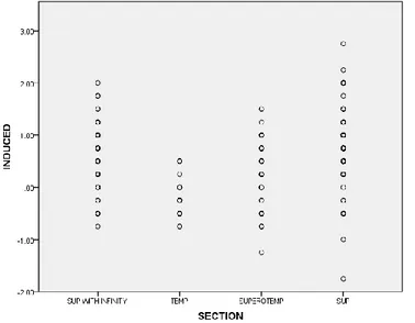
Dr Silni Chandra et al JMSCR Volume 06 Issue 07 July 2018 Page 115 Figure 5
Simple scatter plot of induced corneal astigmatism in the four groups showing highest induced
astigmatism in the superior section group and the least in the temporal group (figure 5)
Table 4 Descriptive Statistics- Post Op Visual acuity
SECTION N Minimum Maximum Mean Std. Deviation
SUP WITH INFINITY VAPOST 45 .1000 1.0000 .502667 .2365510
Valid N (listwise) 45
TEMP VAPOST 30 .0000 .5000 .207000 .1650737
Valid N (listwise) 30
SUPEROTEMP VAPOST 42 .0000 1.0000 .353333 .2203840
Valid N (listwise) 42
SUP VAPOST 82 .0000 1.1760 .513610 .2899039
Valid N (listwise) 82
ANOVA
VAPOST
Sum of Squares df Mean Square F Sig.
Between Groups 2.549 3 .850 13.750 .000
Within Groups 12.051 195 .062
Total 14.601 198
The mean Post Operative unaided visual acuity (VAPOST) at two months in the Superior section with Infinity suture was 0.50 ± 0.23(6/18). 59.5% patients had unaided visual acuity of 6/18 or better, 35.5% patients had visual acuity equal to or better than 6/12.(Table 4)
In the Temporal Section group the mean Post Operative unaided visual acuity was 0.20 ± 0.16(6/9). 100% of the patients had unaided visual acuity of 6 /18 or better at two months. 82.6% of the patients had unaided visual acuity of 6/12 or better.[20]
In the superotemporal group the mean Post Operative unaided visual acuity was 0.35 ± 0.22 (6/12). 78.5% patients had unaided visual acuity
of 6/18 or better.66.5% patients had unaided visual acuity of 6/12 or better.
In the superior section group the mean Post Operative unaided visual acuity was 0.51 ± 0.28(6/18). 56.4% patients had visual acuity of 6/18 or better. 33.4%patients had unaided visual acuity of 6/12 or better. This difference in the postoperative visual acuity among the four groups was found to be statistically significant (p=0.000)
Discussion
Dr Silni Chandra et al JMSCR Volume 06 Issue 07 July 2018 Page 116 astigmatism was found to be highest (1.6444
±0.640) in the superior section with infinity suture group while it was lowest in the temporal group (0.4750 ±0.401). But the preoperative astigmatism was highest in this group which resulted in a higher post operative corneal astigmatism. Preoperative corneal astigmatism ranged from 1.0778 ± 0.714 to 0.6333 ±0.4535 and was statistically and clinically significantly different between the incision types.
The amount of against the rule astigmatism induced post operatively was highest in the superior group (0.7622 D ± 0.7119), followed by superior section with infinity suture group (0.5722 ±0.6563) followed by superotemporal group (0.2917 ±0.6460) while it was lowest (0.158 D ± 0.3181) in the temporal section group [2,11,14,19]. This difference in the four groups is found to be statistically significant. (p=0.000)[11,12,16]. This was consistent with the findings of Gokhale et al (2005) who found SIA vector in superior group to be 1.28D, 0.37D in temporal group but it differed from the findings in the superotemporal group where the SIA was 0.2D which was less than in the temporal group but in our study the induced astigmatism in superotemporal group was higher than in the temporal group.
There was a decrease in the against the rule astigmatism in the temporal section group while it showed an increase in against the rule asigmatism in the rest of the three groups.[3,4] These were consistent with findings in the study done by Bhaskar Reddy et al16 who found that incisions placed temporally tend to decrease the against the rule astigmatism mostly prevalent in the adult population due to the absence of the lid tone in both phaco and Manual small incision cataract surgery.
There is a greater shift in corneal astigmatism towards against the rule astigmatism following superior incision SICS. This shift is reduced marginally when an infinity suture is placed on the superior incision. The shift is minimal if the incision is placed superotemporally. There is a decrease in against the rule astigmatism following
temporal incision. Thus, our results are consistent with the idea of increase in against the rule astigmatism following superior incision [2,11] The post operative unaided distance visual acuity was the best in the temporal section group. 100 percent of patients achieved a visual acuity of 6/18 or better in the temporal group[19] while it was lowest in the superior section group. Only 56.4% patients achieved unaided visual acuity of 6/18 or better. The infinity suture did not make any change in mean unaided visual acuity but marginally increased the percentage (59.5%) of patients achieving 6/18 or better vision.
This is a retrospective non comparative study with a limited number of patients. Despite these limitations it is believed that this study adequately throws light on the importance of Temporal and superotemporal incisions[10] as a means of controlling corneal astigmatism which ultimately reduces the overall surgically induced astigmatism and thus paves the way for achieving a better spectacle free vision.
Majority of Ophthalmic surgeons are trained in Superior approach in cataract surgery. But we all have realised that it has only resulted in poor unaided visual acuity. So it is time we switch over to Temporal approach. But those of us who have difficulty in switching over to this entirely new approach may try the Superotemporal approach with equally good results if not better. And those of us who have difficulty with both these techniques may try placing an appropriately tight infinity suture at the superior section to get better results.
References
1. Gogate PM. Small incision cataract surgery: Complications and mini-review. Indian J Ophthalmol 2009;57:45-9
Dr Silni Chandra et al JMSCR Volume 06 Issue 07 July 2018 Page 117 3. Xia WQ ,Chen W,Li JP et al .Astigmatism
changes after cataract surgery with different positions of 6 mm sutureless incision. Chinese Journal of Practical Ophthalmology, 2005:23(8): 804-809 4. Wang YS . The astigmatism study after
extracapsular cataract extraction. Journal of Clinical Ophthalmology 2009:17(6) :537-539
5. P. Mishra, S. Manavalan, M. Ramya, M. Jeevitha, R. Vinnarasi, Latha Priyangaa, V. Sridevi, R. Parth. “Manual Small Incision Cataract Surgery (MSICS)”. Journal of Evolution of Medical and Dental Sciences 2014; Vol. 3, Issue 46, September22; Page: 11249-11261,DOI:
10.14260/jemds/2014/3467
6. Haldipurkar SS, Shikari HT, Gokhale V. Wound construction in manual small incision cataract surgery. Indian J Ophthalmol 2009;57:9-13.
7. Investigating the impact of preoperative corneal astigmatism orientati on on the postoperative spherical equivalent refraction following intraocular lens implantation. McNeely RN, Moutari S, Pazo E, Moore JE.Eye Vis (Lond). 2018 Apr 25;5:7. doi: 10.1186/s40662-018-0103-4. eCollection 2018.
8. Influence of ocular features and incision width on surgically induced astigmatism after cataract surgery. Chang SW, Su TY, Chen YL. J Refract Surg. 2015 Feb;31(2):82-8. doi: 10.3928/1081597X-20150122-02.
9. Astigmatism following small incision cataract extraction through superotemporal incision.Guan C1, Xiao T Eye Sci. 2012 Jun;27(2):94-7. doi: 10.3969/j.issn.1000-4432.2012.02.009.
10.Gokhale NS, Sawhney S. Reduction in astigmatism in manual MSICS through change in astigmatism site. Indian J Ophthalmol 2005;53:201-3.
11.Evaluation and comparison of surgically induced astigmatism between phacoemul-sification and small incision cataract surgery...www.sjopthal.net/article.asp?issn =1858-540X;year=2013;volume=5;issue... y atil - 2013
12.Radwan AA (2011) Comparing Surgical-Induced Astigmatism through Change of Incision Site in Manual Small Incision Cataract Surgery (SICS). J Clinic Experiment Ophthalmol 2:161 doi:10.4172/2155-9570.1000161
13.Edmund Arthur et al. Postoperative Corneal and Surgically Induced Astigmatism following Superior Approach Manual Small Incision Cataract Surgery in Patients with Preoperative Against-the-Rule Astigmatism .J Ophthalmol. 2016; doi: 10.1155/2016/9489036
14.International J. of Healthcare and Biomedical Research, Volume: 06, Issue: 01, October 2017, 43-48 43 www.ijhbr.com ISSN: 2319-7072 Original article: Post-operative Corneal Astigmatism in Superior vs Temporal straight scleral incisions after Manual Small Incision Cataract Surgery (MSICS) Harshavardhan Reddy1, *Shubhangi Nigwekar2, Surekha V. Bangal3
15.Reddy B, Raj A, Singh VP. The sites of incision and corneal astigmatism in conventional SICS versus phacoemulsi cation. Ann Ophthal- mol (Skokie) 2007; 39(3):209-16.
16.Choosing the location of corneal incision based on preexisting astigmatism in phacoemulsification. Tejedor J1, Murube J .Am J Ophthalmol. 2005 May;139(5):767-76.
17.Zawar SV, Gogate P. Safety and efficacy of temporal manual small incision cataract surgery in India. Eur J Ophthalmol. 2011; 21(6):748–753.
Dr Silni Chandra et al JMSCR Volume 06 Issue 07 July 2018 Page 118 incision in manual small-incision cataract
surgery. Nepal J Ophthalmol. 2011 Jan-Jun.